In the published article, there were errors in “Figures 2, 3, 6–8” as published. Certain images were mixed between groups, resulting in the unintentional misplacement of the representative images in Figures 2B, 3A, 6A, 7A, B, 8A. The corrected Figures 2, 3, 6, 7, 8 and their captions appear below.
FIGURE 2

Myricetin inhibited the phosphorylation level of MAPK/ERK and PI3K/AKT signaling pathways. Analysis of the level of (A) p-ERK and ERK and (B) p-PI3K, PI3K, p-AKT, and AKT in OC cells (A2780 and HO8910) by western blotting. *p < 0.05; **p < 0.01; ***p < 0.001; ****p < 0.0001 were considered statistically significant, n = 3.
FIGURE 3

Myricetin promoted apoptosis of OC cell lines. (A) The apoptosis levels of A2780 and HO8910 cells were determined by Flow cytometry after myricetin treatment. (B) Analysis of the expression level of cleaved-caspase-3 and cleaved-PARP in OC cells (A2780 and HO8910) by western blotting. (C) Analysis of the expression level of Bax and Bcl-2 in OC cells (A2780 and HO8910) by western blotting.*p < 0.05; **p < 0.01; ***p < 0.001; ****p < 0.0001 were considered statistically significant, n = 3.
FIGURE 6
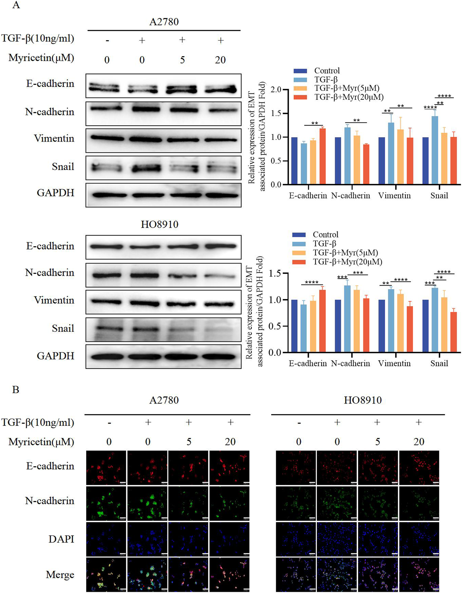
Myricetin reverses TGF-β-induced EMT to inhibit the metastasis and invasion of OC cells. (A) Analysis of the expression level of E-cadherin, N-cadherin, Vimentin, and Snail in OC cells by western blot. (B) The expression levels of E-cadherin and N-cadherin in A2780 and HO8910 cells were detected by immunofluorescence assay.*p < 0.05; **p < 0.01; ***p < 0.001; ****p < 0.0001 were considered statistically significant, n = 3.
FIGURE 7
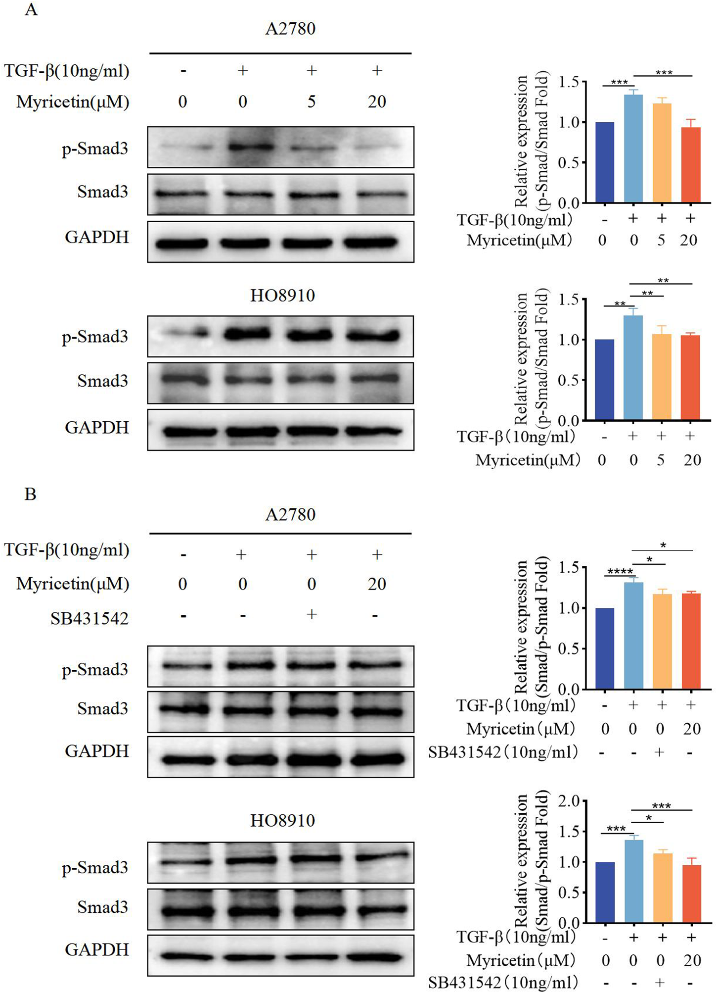
Myricetin inhibits TGF-β-induced EMT in OC cells through the classical Smad signaling pathway. (A) Analysis of the expression level of phosphorylated Smad3 and Smad3 in OC cells. (B) Analysis of the phosphorylated Smad3 and Smad3 in OC cells after different combination treatments of TGF-β, myricetin, or SB431542(10 ng/mL). *p < 0.05; **p < 0.01; ***p < 0.001; ****p < 0.0001 were considered statistically significant, n = 3.
FIGURE 8

Myricetin inhibits TGF-β-induced EMT in OC cells through ERK/MAPK and PI3K/AKT signaling pathways. (A) Analysis of the expression level of phosphorylated ERK and ERK in OC cells. (B) Analysis of the expression level of phosphorylated PI3K, PI3K, phosphorylated AKT, and AKT in OC cells. Results are presented as mean ± SD (n = 5). *p < 0.05; **p < 0.01; ***p < 0.001; ****p < 0.0001 were considered statistically significant, n = 3.
In the published article, there was an error in Figure 4 as published. The tumors in the control group exceeded the usual size in Figures 4A–C. The corrected Figure 4 and its caption appear below.
FIGURE 4

Suppressive influence of myricetin in the growth of OC cells in vivo. (A) Construction of subcutaneous heterograft tumor model in nude mice. (B, C) The change of tumor weight and size in model mice. (D) Analysis of the expression level of Bax and Bcl-2 in tumor by western blotting. (E) Analysis of liver and lung metastasis in model mice by HE staining. (F) Analysis of the level of EGFR, Ki-67, and MMP-9 in tumor tissues of nude mice by immunohistochemistry. *p < 0.05; **p < 0.01; ***p < 0.001; ****p < 0.0001 were considered statistically significant, n = 5.
The authors apologize for these errors and state that these do not change the scientific conclusions of the article in any way. The original article has been updated.
Statements
Publisher’s note
All claims expressed in this article are solely those of the authors and do not necessarily represent those of their affiliated organizations, or those of the publisher, the editors and the reviewers. Any product that may be evaluated in this article, or claim that may be made by its manufacturer, is not guaranteed or endorsed by the publisher.
Summary
Keywords
myricetin, epithelial-to-mesenchymal transition, TGF-β, ovarian cancer, PI3K/AKT, TGF-β/Smad
Citation
Yang H-W, Lan Y, Li A, Wu H, Song Z-W, Wan A-L, Wang Y, Li S-B, Ji S, Wang Z-C, Wu X-Y and Lan T (2024) Corrigendum: Myricetin suppresses TGF-β-induced epithelial-to-mesenchymal transition in ovarian cancer. Front. Pharmacol. 15:1447095. doi: 10.3389/fphar.2024.1447095
Received
11 June 2024
Accepted
10 October 2024
Published
14 November 2024
Volume
15 - 2024
Edited and reviewed by
Olivier Feron, Université Catholique de Louvain, Belgium
Updates
Copyright
© 2024 Yang, Lan, Li, Wu, Song, Wan, Wang, Li, Ji, Wang, Wu and Lan.
This is an open-access article distributed under the terms of the Creative Commons Attribution License (CC BY). The use, distribution or reproduction in other forums is permitted, provided the original author(s) and the copyright owner(s) are credited and that the original publication in this journal is cited, in accordance with accepted academic practice. No use, distribution or reproduction is permitted which does not comply with these terms.
*Correspondence: Ting Lan, tinglan@xzhmu.edu.cn; Xin-Yu Wu, xzfywxy@163.com; Zhong-Cheng Wang, wangzhongcheng@xzhmu.edu.cn
†These authors have contributed equally to this work and share first authorship
Disclaimer
All claims expressed in this article are solely those of the authors and do not necessarily represent those of their affiliated organizations, or those of the publisher, the editors and the reviewers. Any product that may be evaluated in this article or claim that may be made by its manufacturer is not guaranteed or endorsed by the publisher.