Abstract
Atherosclerosis (AS) constitutes a major cardiovascular disorder posing a severe threat to human health, with the rupture of vulnerable plaques marking a critical turning point in the progression of AS. This pathological event can trigger acute myocardial infarction and stroke, thereby exerting a profound adverse impact on patient prognosis. In contrast to normal arterial tissues, vulnerable plaques are characterized by an abundance of neovascularization, which is generated through angiogenic pathways. Although these neovessels serve to alleviate the hypoxic microenvironment within the plaque, they concurrently compromise plaque stability. Notably, angiogenesis engages in crosstalk with AS-associated phenotypic processes, including cellular apoptosis, extracellular matrix remodeling, inflammatory responses, and oxidative stress. This interplay forms a positive feedback loop that further exacerbates the destabilization of vulnerable plaques. The vascular endothelial growth factor (VEGF) pathway plays a central regulatory role in angiogenesis. Targeting the VEGF pathway to inhibit angiogenesis and enhance plaque stability has thus opened a novel therapeutic avenue for AS management. In comparison, this strategy has demonstrated promising efficacy in preclinical studies; however, a lack of safe and reliable pharmaceutical agents remains, hindering their translation into clinical practice for AS treatment. In this review, the authors summarize the underlying mechanisms governing angiogenesis and vulnerable plaque formation, and further explore the phenotypic crosstalk between these processes. The ultimate aim is to provide valuable insights to facilitate future breakthroughs in the development of therapeutic strategies for AS.
1 Introduction
Atherosclerosis (AS) is a lipid-driven inflammatory disease that primarily affects large and medium-sized arteries, characterized by lipid deposition in the arterial intima and the formation of atherosclerotic plaques (Monaco et al., 2025). AS serves as the most significant pathological basis for ischemic heart disease, stroke, and peripheral arterial disease, imposing a substantial disease burden worldwide (Herrington et al., 2016; So et al., 2020; Hou et al., 2025). Vulnerable plaques refer to those atherosclerotic plaques with an increased risk of rupture and thrombosis, representing a turning point for the acute exacerbation of AS (Björkegren and Lusis, 2022). Patients with AS exhibiting features of vulnerable plaques face a significantly elevated risk of adverse events such as acute myocardial infarction and cardiac death (Del et al., 2024). Promoting the evolution of atherosclerotic plaques towards a stable phenotype is a practical approach to improving the prognosis of AS (Räber et al., 2019).
Angiogenesis refers to the process of forming new capillaries from pre-existing vascular networks, which serves as a crucial mechanism in physiological and pathological processes, such as organismal growth and development, tissue repair, and tumorigenesis (Hutchings et al., 2021). As one of the significant characteristics of vulnerable plaques, dense newly formed capillaries have been detected in vulnerable plaque tissues (Su et al., 2017; Cai et al., 2021; Gössl et al., 2010). These neovessels, originating from the vasa vasorum in the adventitia of arterial walls, while meeting the demand for blood and oxygen supply in the expanding plaque tissues, can promote inflammatory cell infiltration, cholesterol deposition, and intraplaque hemorrhage, thereby impairing plaque stability (Cheng et al., 2013; Zorc-Pleskovič et al., 2018). The inhibition of intraplaque angiogenesis provides a novel strategy for stabilizing atherosclerotic vulnerable plaques (Ugusman et al., 2024). A comprehensive understanding of the mechanisms underlying angiogenesis and the vulnerable plaques is a prerequisite for implementing targeted interventions. This review summarizes the mechanisms of angiogenesis and vulnerable plaque formation, with a focus on the phenotypic interactions between them, and reveals potential targets for regulating angiogenesis to intervene in plaque stability. Additionally, the latest advancements in interventional strategies targeting angiogenesis in vulnerable plaques will be discussed. The data for this review were retrieved from PubMed and ClinicalTrials.gov, with keywords including “atherosclerosis”, “vulnerable plaque”, “angiogenesis”, “neovascularization”, and “vascular endothelial growth factor”. Studies published between 1980 and 2025 were included, with a particular focus on those released in the recent 5 years.
2 Mechanisms of neovascularization
2.1 Vasculogenesis, arteriogenesis, and angiogenesis
In physiological or pathological conditions, neovascularization occurs via vasculogenesis, arteriogenesis, and angiogenesis, each differing in initiation, process, and outcomes. Vasculogenesis refers to the formation of the primary vascular system from mesodermal progenitors during the early stages of embryonic development (Hutchings et al., 2021). Arteriogenesis, driven by hemodynamic forces, involves positive remodeling of pre-existing collateral arterioles—narrow, high-resistance vessels—into larger conductance arteries to compensate for occluded trunks (Helisch and Schaper, 2003; Moraes et al., 2013). Occurring in tissues with pre-existing collaterals, it generates structurally and functionally intact arteries, thereby restoring perfusion to ischemic regions (Wang et al., 2025). Angiogenesis, distinct from the above, involves the migration and proliferation of differentiated endothelial cells to form new capillary networks from existing vessels, encompassing sprouting and intussusceptive subtypes (Semenza, 2014). Sprouting—budding and branching from existing vessels—predominates in forming small vascular plexuses and plays a key role in neovascularization of atherosclerotic plaques (Mitsos et al., 2012). Mechanisms related to neovascularization are illustrated in Figure 1.
FIGURE 1
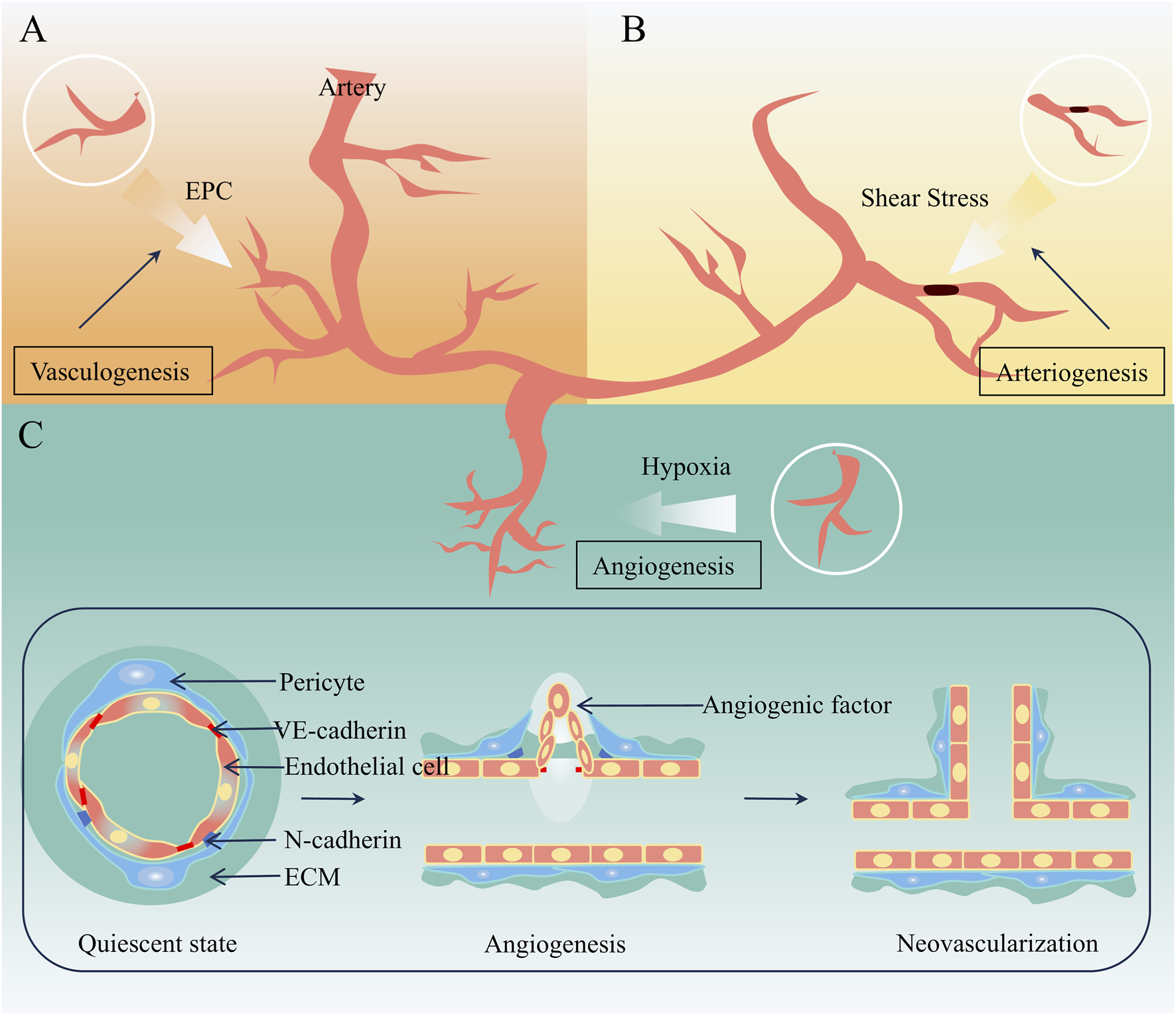
Mechanisms related to neovascularization. (A) Vasculogenesis (B) Arteriogenesis (C) Angiogenesis.
2.2 Mechanisms of angiogenesis
Mature capillaries consist of interconnected endothelial cells on the inner side and tightly arranged pericytes, along with an extracellular matrix (ECM), on the outer side, which collectively ensure normal blood circulation (Bergers and Song, 2005). Under physiological conditions, endothelial cells stay in a quiescent state, regulating blood flow to provide nutrients and oxygen for tissues. When triggers like hypoxia exist, the biosynthesis of pro-angiogenic signals increases, thereby activating angiogenesis. This process encompasses stages such as cell proliferation, migration, differentiation, maturation, and lumen formation, each featuring unique endothelial cell functions (Xiang et al., 2024). The basic process is as follows: A variety of angiogenic factors induce vascular dilation and enhance permeability by disrupting intercellular junctions and degrading the ECM. Then, endothelial cells (stalk cells and tip cells) migrate with the ECM serving as a scaffold, facilitating the formation of branches. Eventually, branch fusion, ECM deposition, and pericyte maturation result in the formation of functional new blood vessels. The molecular mechanism of angiogenesis is depicted in Figure 2. Angiogenesis is extensively involved in various physiological and pathological processes such as the AS, tumorigenesis, and tissue repair. Its purpose, regulatory mechanisms, and biological effects vary across different diseases. In brief, angiogenesis is a key driver of tumor progression and represents a pathological uncontrolled proliferation. It aims to provide nutrients and pathways for tumor growth and metastasis, with more complex regulatory mechanisms—tumor cells continuously secrete pro-angiogenic factors while inhibiting anti-angiogenic signals (Liu X. et al., 2025). Angiogenesis during wound healing is a physiological repair process with temporal specificity (Huang et al., 2025). Its goal is to restore blood supply to damaged tissues, support granulation tissue formation and epithelialization, and naturally terminate once wound healing is completed. In AS, angiogenesis is a pathological process primarily occurring within atherosclerotic plaques. While it intends to supply oxygen to hypoxic plaque tissues, it exacerbates plaque instability and rupture risk.
FIGURE 2
Mechanism of angiogenesis.
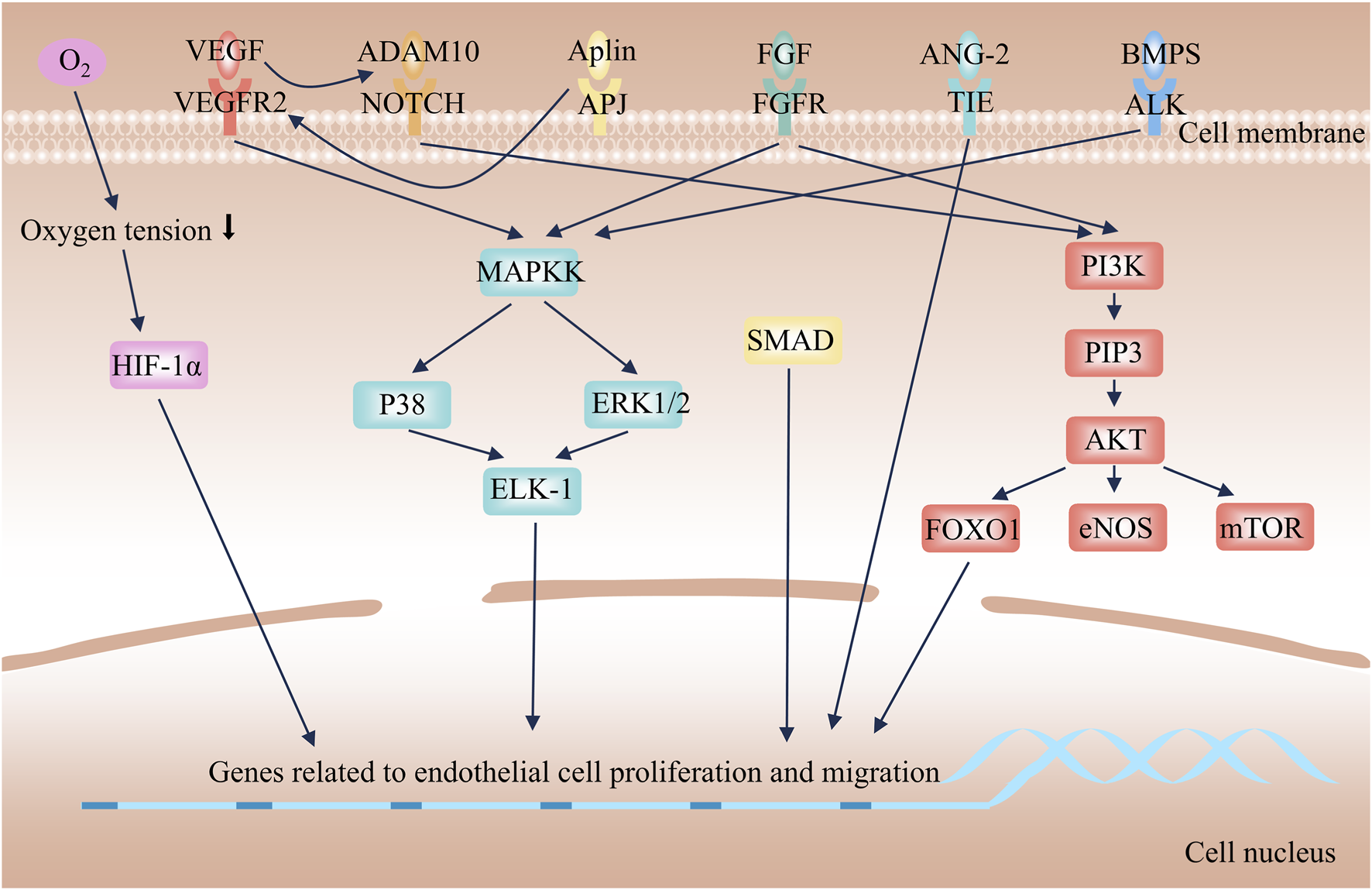
2.2.1 Angiogenic factors
Multiple molecules mediate the process of angiogenesis. Among them, the vascular endothelial growth factor (VEGF) and the heparin - binding fibroblast growth factor (FGF) family are the core angiogenic factors. VEGF is recognized as the most critical one. The VEGF family comprises six structurally related proteins, including VEGF-A, VEGF-B, VEGF-C, VEGF-D, VEGF-E, and placental growth factor (PlGF) (Lee et al., 2015). Angiogenesis primarily relies on the interaction between VEGF-A (often referred to simply as VEGF) and VEGF Receptor-2 (VEGFR2) (Ferrara, 2002). VEGF acts as a potent vascular protective cytokine, responsible for maintaining the homeostasis of the vascular network. Simultaneously, it serves as an indispensable key component for angiogenesis in various diseases (Lee et al., 2025). Researchers have demonstrated that VEGF can induce increased vascular permeability, vasodilation, and degradation of the ECM (Choi et al., 2025; Nagasaki et al., 2016). Similarly, FGF has received extensive attention. The FGF family consists of 22 members, which bind to seven types of receptors (Phan et al., 2021). FGF-1 is referred to as acidic FGF (aFGF), and FGF-2 is known as basic FGF (bFGF); both are potent mitogens for endothelial cells (Kharitonenkov, 2009). FGF can induce de novo angiogenesis and plays a crucial role in the growth of new blood vessels in various scenarios, such as wound healing and embryonic development (Itoh and Ornitz, 2011).
2.2.2 Angiogenic signaling pathway
Angiogenic factors are upregulated in response to stimuli such as hypoxia, activating downstream angiogenesis signaling pathways to promote endothelial cell sprouting, migration, and lumen formation. Hypoxia-inducible factors (HIFs) are a class of transcription factors activated under hypoxic conditions, playing a central role in hypoxic adaptation (Acuña-Pilarte and Koh, 2025). Tissue hypoxia inhibits the ubiquitin-proteasomal degradation of HIF-1α, allowing it to translocate into the nucleus, where it binds to the β subunit and recruits transcriptional coactivators. This complex then binds to hypoxia response elements on target genes, such as VEGF-A and FGF, inducing the expression of pro-angiogenic signaling molecules (Plastino et al., 2021; Basavaraju et al., 2021). Increased biosynthesis of pro-angiogenic signals induces matrix metalloproteinases (MMPs) to degrade ECM components. This process leads to the detachment of pericytes from the basement membrane, disruption of endothelial junctions, and increased vascular permeability, thereby creating conditions that are conducive to endothelial cell migration (Carmeliet and Jain, 2011). Individual endothelial cells, known as tip cells, undergo sprouting and migration in response to pro-angiogenic signaling molecules. The migration of tip cells relies on PI3K/Akt-eNOS-mTOR-FOXO1 axis-mediated cell proliferation and cytoskeletal remodeling (Ackah et al., 2005; Fukumura et al., 2001). Additionally, the FGF/FGFR and Apelin/APJ systems synergistically promote this process by regulating energy metabolism and directional polarity (Vimalraj, 2022; Wagenaar and Moll, 2025). Concurrently, BMP2 enhances sprouting through the activation of the PI3K/Akt and MEK/ERK pathways (Cai et al., 2012; Goumans et al., 2009; Chen et al., 2018). Notch signaling further activates the PI3K/Akt pathway to sustain the survival and proliferation of endothelial cells (Gude et al., 2008; Meurette et al., 2009). The aforementioned signaling pathways collectively synergize to promote the sprouting and migration of endothelial cells. Following the tip cells are endothelial cells called stalk cells, which are responsible for lumen elongation and pericyte recruitment (ávila-Gálvez et al., 2025). After branch fusion, endothelial cells in the new blood vessels revert to a quiescent state, reorganize cellular junctions, promote pericyte maturation, deposit the basement membrane, and maintain the balance between pro-angiogenic and anti-angiogenic factors (Liu et al., 2023).
2.2.3 Emerging detection technologies for angiogenesis
A variety of imaging technologies can achieve quantitative analysis of angiogenesis and neovascularization by virtue of different biomarkers. With the continuous emergence of emerging techniques such as single-cell RNA sequencing, spatial transcriptomics, and high-resolution imaging of vasa vasorum, more precise methods have been provided for angiogenesis detection. Most studies adopt immunohistochemical staining or immunofluorescence staining to evaluate neovascularization, and the biomarkers used include CD31, CD34, vWF, Ki67, and TER-119 (Parma et al., 2020; Tziakas et al., 2013; Kurdi et al., 2019; Qi et al., 2023). All the aforementioned methods are indirect detection approaches. Contrast-enhanced ultrasound and micro-ultrasound, two non-invasive techniques for neovascularization detection, enable dynamic imaging of blood flow status (Liu et al., 2017; Ding et al., 2019). The application of single-cell RNA sequencing facilitates the accurate identification of specific cell subsets and specific molecular expressions (Zhao et al., 2025). Spatial transcriptomics overcomes the limitation of traditional sequencing in losing spatial location information. It can resolve gene expression profiles while preserving the morphological characteristics of tissues (Liu S. et al., 2025). High-resolution imaging of vasa vasorum is a pivotal technique for the precise detection of angiogenesis from a morphological perspective. It can compensate for the limitations of traditional molecular detection and conventional imaging techniques, and intuitively visualize the morphological characteristics and distribution patterns of vasa vasorum (Cebral et al., 2025).
3 Mechanism of vulnerable plaque
With the progression of AS, the arterial intima may develop intimal thickening, fatty streaks, fibrolipid plaques, calcified plaques, or complex plaques. Plaque rupture, surface erosion, and calcified nodules are the primary pathological bases for acute thrombosis in AS, with plaque rupture playing a dominant role (Yahagi et al., 2016). Plaques with a high tendency to rupture are defined as vulnerable plaques, and the most common morphological type is the thin-cap fibroatheroma (TCFA) (Fleg et al., 2012). TCFA exhibits typical phenotypic characteristics: it contains a large necrotic core composed of lipids and cellular debris, covered by a thin fibrous cap (usually less than 65 μm in thickness) (Suzuki et al., 2024; Roleder-Dylewska et al., 2023). A large number of inflammatory cells infiltrate the fibrous cap, often presenting with punctate calcification and intraplaque hemorrhage. Additionally, the arterial wall may undergo aneurysmal dilatation and remodeling. As shown in Figure 3, cell apoptosis, impaired phagocytosis, ECM remodeling, inflammatory responses, and oxidative stress are closely associated with the formation of TCFA-type vulnerable plaques.
FIGURE 3
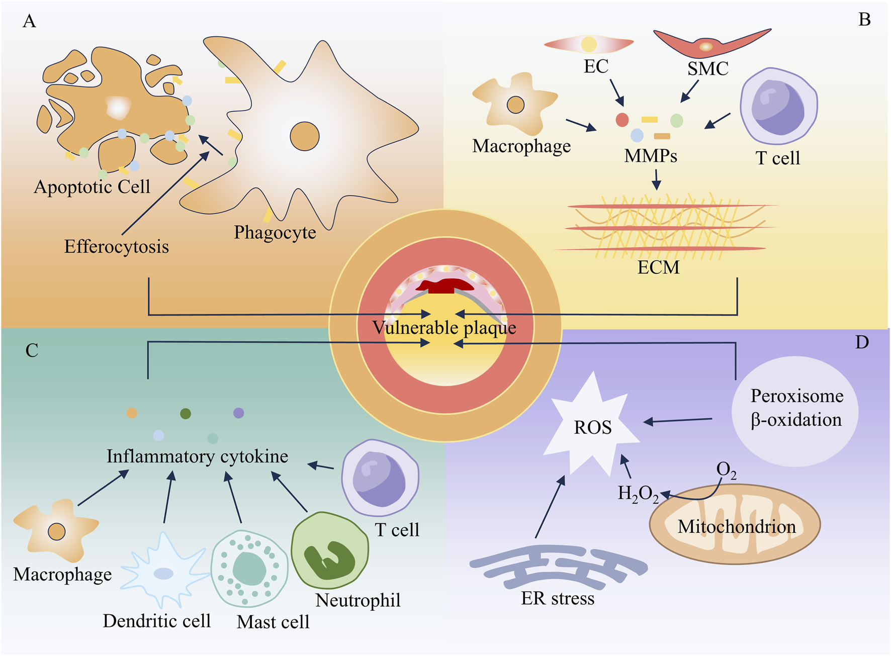
Mechanism of vulnerable plaque formation. (A) Apoptosis (B) Extracellular matrix remodeling (C) Inflammatory response (D) Oxidative stress.
AS can affect systemic arterial vessels such as the coronary arteries and peripheral arteries. Distinct disparities exist in the regulatory mechanisms governing plaque stability across different vascular territories, with the differentiation of angiogenesis patterns serving as the core demarcation. Coronary arteries are characterized by abundant bifurcations, low blood flow shear stress and prominent turbulent flow, which lead to a high degree of hypoxia within the plaques. This robustly activates the VEGF pathway, driving extensive proliferation of adventitial vasa vasorum and subintimal vascular plexuses. The resultant neovessels feature high density and diffuse distribution, markedly elevating the risk of plaque rupture. In contrast, peripheral arteries exhibit relatively stable blood flow shear stress and milder plaque hypoxia. Neovascularization in peripheral arterial plaques is mostly confined to the adventitia, with a density merely half that observed in coronary plaques. Additionally, peripheral arterial plaques display significant calcification and more uniform plaque stress distribution. Consequently, these plaques possess substantially greater overall stability. These differences provide a critical theoretical basis for the clinical implementation of targeted interventions against vulnerable plaques.
3.1 Cell apoptosis and efferocytosis impairment
One of the characteristics of vulnerable plaques is a large necrotic core covered by a thin fibrous cap, and the apoptosis of smooth muscle cells (SMCs) and macrophages is the leading cause (Silvestre-Roig et al., 2014; Yin et al., 2024). SMCs are the primary component of the fibrous cap, promoting its thickening by synthesizing matrix components, including collagen and elastin. In the fibrous cap of vulnerable plaques, the apoptosis of SMCs is exacerbated, which reduces the synthesis of matrix proteins—contributing to the formation of a thin fibrous cap and the expansion of the necrotic core (Rotllan et al., 2015). The apoptosis of SMCs is mainly induced by the continuous infiltration of inflammatory cells and protease-mediated changes in the extracellular environment. Additionally, the accumulation of degradation products from the fibrous cap matrix can also induce SMC apoptosis (von Wnuck et al., 2006). Macrophage apoptosis occurs in all stages of AS. In the early stage, it helps reduce plaque burden; however, in the late stage, it impairs plaque stability (Deng et al., 2025). As the disease progresses, a large number of apoptotic macrophages cannot be cleared promptly and accumulate in the plaque, leading to the expansion of the necrotic core and exacerbation of local inflammatory responses (Gautier et al., 2009). In the late stage of AS, the accumulation of free cholesterol (caused by impaired esterification function) and exposure to oxidized cholesterol trigger the unfolded protein response (UPR) (Moore and Tabas, 2011). When the UPR is chronically activated, it induces macrophage apoptosis (Thorp et al., 2009). Furthermore, impairment of macrophage autophagy can also induce excessive apoptosis (Liao et al., 2012).
When efferocytosis is impaired, these protective functions are weakened, thereby contributing to the development of AS (Schrijvers et al., 2005). Efferocytosis refers to the process by which phagocytes clear apoptotic cells, thereby preventing the accumulation of apoptotic cell debris and triggering the reprogramming of phagocytes toward an anti-inflammatory phenotype. In the early stages of AS, dying cells can be rapidly cleared; however, as the disease progresses, efferocytosis becomes defective (Yurdagul, 2021). Since chemotactic and phagocytic signals are usually abundant in advanced plaques, the defect in efferocytosis may result from impaired phagocytic function of macrophages within the lesion, rather than a lack of signals that attract phagocytes.
3.2 Remodeling of ECM
ECM remodeling constitutes an indispensable step in the formation of vulnerable atherosclerotic plaques. The ECM is a complex reticular scaffold structure composed of collagen fibers, elastin, fibronectin, laminin, and other components. These components are distributed in the basement membrane or interstitial matrix, providing structural support for tissues. In the early stage of AS, the ECM consists of laminin, polymerized type Ⅰ collagen, type Ⅲ collagen, type Ⅳ collagen, a small amount of fibronectin, and elastin fibers. In the late stage of AS, the ECM comprises monomeric type I collagen, fibronectin, fibrinogen, vitronectin, and osteopontin (Yurdagul et al., 2016; Ström et al., 2004). ECM remodeling affects plaque stability primarily by regulating the mechanical strength of the fibrous cap and vascular function. Collagen is the main component of the ECM and is particularly abundant in atherosclerotic plaques, among which type Ⅰ collagen accounts for the highest proportion (approximately 60%) (Holm et al., 2019). Studies on sirtuin 1 (SIRT1) have confirmed the importance of type Ⅰ collagen in plaque stability. Specifically, SIRT1 deacetylates regulatory factor X5 (RFX5), thereby relieving the inhibitory effect of RFX5 on the promoter activity of the collagen α2 chain (Xia et al., 2012). This process increases the expression of type Ⅰ collagen and prevents plaque rupture. Elastin enables blood vessels to adapt to changes in hemodynamics. In the intima of healthy arteries, the content of soluble elastin is high; however, its content decreases in ruptured plaques (Maedeker et al., 2016). A study has verified that inhibiting microRNA-181b can significantly increase elastin expression and stabilize plaques (Di Gregoli et al., 2017).
ECM remodeling is primarily driven by proteases, which represent a key process leading to the reduced mechanical strength of the fibrous cap (Newby, 2012). MMPs are a family of zinc-dependent endopeptidases that can degrade almost all other ECM components (Galis and Khatri, 2002; Li et al., 2014). In vulnerable plaques, in addition to endothelial cells, T cells, macrophages, and SMC can all increase the production of MMPs (Bäck et al., 2010). In AS, the expression of MMP-1 and MMP-8 is significantly upregulated, and their levels are even higher in vulnerable plaques (Molloy et al., 2004; Müller et al., 2014). As collagenases, MMP-1 and MMP-8 can degrade fibrillar collagen (Libby, 2013). In a model of AS with MMP-8 knockout, the plaque area was significantly reduced (Laxton et al., 2009). MMP-2 and MMP-9, as major gelatinases, can degrade a variety of ECM proteins, including collagen (Siefert and Sarkar, 2012). Compared with stable plaques, MMP-2 and MMP-9 in vulnerable plaques are 20 times higher (Guo ZY. et al., 2018). The aforementioned studies indicate that MMPs can regulate plaque stability by remodeling the ECM.
3.3 Inflammatory response
Inflammation plays a crucial role in plaque rupture, and vulnerable plaques exhibit obvious signs of inflammation (Aukrust et al., 2011). During the progression of AS, macrophages, dendritic cells (DCs), and mast cells can affect plaque stability through phenotypic changes and the secretion of cytokines. An extensive cohort study involving 200 human carotid plaques reported an association between CD163+ macrophages and the vulnerable plaque phenotype (Bengtsson et al., 2020). In vulnerable plaques, the number of mature dendritic cells is significantly increased. Due to the interaction between dendritic cells and regulatory T cells (Tregs), the migration of DCs and their adhesion to endothelial cells are directly inhibited, thereby impairing plaque stability (Dietel et al., 2013). The frequency of CD117+ mast cells is higher in unstable plaques than in stable ones (Joo et al., 2020). Mast cells are activated by binding to IgE attached to the Fc epsilon receptor (FcεR), which leads to the release of cytoplasmic granules containing proinflammatory factors, histamine, and proteases (Kritikou et al., 2019). As a crucial driver of AS, the understanding of inflammatory responses has been progressively deepened. Some researchers have proposed an inflammation-based 3-category classification of atherosclerotic plaques, namely active plaques, dormant plaques, and inactive plaques, analogous to the activity states of volcanoes (Miceli et al., 2025). Even clinically “stable” plaques may exhibit silent yet persistent immunometabolic and thromboinflammatory activities, thereby elevating residual cardiovascular risk. This evolving paradigm endorses immunomodulation as the cornerstone of precision cardiovascular medicine.
3.4 Oxidative stress
Oxidative stress serves as a vital initiating and promoting factor in the development of vulnerable plaques. Oxidative stress refers to a state where the body, upon exposure to various stimuli, generates excessive amounts of highly reactive molecules. When the degree of oxidation exceeds the scavenging capacity of antioxidants, an imbalance occurs between the oxidative and antioxidant systems, leading to tissue damage. Reactive oxygen species (ROS) are a group of oxygen-containing molecules or oxygen atoms with unpaired electrons, including superoxide anions, hydroxyl radicals, and hydrogen peroxide. Among these, superoxide anions are produced in the largest quantity. Studies have shown that the size of AS lesions is closely correlated with the rate of ROS production; inhibiting oxidative stress and ROS generation can suppress the formation of vulnerable plaques (Zhu et al., 2024). In vitro studies have shown that inhibiting ROS production can effectively prevent macrophage apoptosis, thereby reducing the risk of atherosclerotic plaque rupture (Fang et al., 2021). In vivo studies have demonstrated that targeted scavenging of ROS in AS model mice using nanoparticle therapy can significantly inhibit AS progression, providing a novel approach for the diagnosis and treatment of vulnerable plaques (Dai et al., 2022). Key catalytic enzymes involved in oxidative stress include nicotinamide adenine dinucleotide phosphate (NADPH) oxidase, xanthine oxidase, and endothelial nitric oxide synthase (NOS). In human atherosclerotic plaques, superoxide anions and their leading producers—NADPH oxidase and xanthine oxidase—are present in large quantities (Förstermann et al., 2017). Inhibiting nitric oxide (NO) derived from NOS is an effective strategy for reducing foam cell formation and limiting the progression of atherosclerotic plaques (Roy et al., 2021).
4 Angiogenesis and vulnerable plaques
Vasa vasorum originates from the branching points of arteries, extends longitudinally along the vascular wall, and subsequently forms circular arches surrounding the arterial lumen (Svetlikov et al., 2025). Since the diffusion of blood-borne nutrients from the central arterial lumen is limited to a distance of approximately 100 μm, the primary function of the vasa vasorum is to transport nutrients to the vascular wall (Cebral et al., 2025). Angiogenesis exerts distinct effects at different stages of atherosclerotic lesions: it primarily functions in tissue repair during the early phase but becomes a major pathogenic factor in the advanced stage. In the early stage of the AS, angiogenesis improves blood supply to ischemic regions, alleviates hypoxic injury to vascular endothelial cells, and delays the progression of endothelial dysfunction. As plaques advance, the newly formed blood vessels exhibit fragile structures that are prone to rupture and hemorrhage, triggering intraplaque hematoma and accelerating plaque instability or even rupture. In the advanced stage, the high permeability of neovessels increases the leakage of plasma proteins and lipids, further exacerbating the inflammatory response and lipid deposition within plaques, promoting plaque enlargement and lipid core expansion, and aggravating arterial stenosis. As AS progresses, intimal thickening causes the vascular wall thickness to exceed the critical threshold distance of 100 μm. This exceeds the limit of oxygen and nutrient supply to the media and hyperplastic intima, creating a hypoxic environment within the artery. Hypoxia further activates angiogenesis (Giannarelli et al., 2014). This activation of angiogenesis aims to improve the oxygen and nutrient supply to vascular tissues, serving as a permissive factor that allows plaque growth to continue after the critical intimal thickness is reached (Pan et al., 2024). While intraplaque microvessels contribute to compensatory blood supply, they are closely associated with plaque rupture. Compared with unruptured plaques, ruptured plaques exhibit increased microvessel density—this increase is particularly prominent in the shoulder regions of plaques. These findings suggest that angiogenesis is involved in the formation of vulnerable plaques and may serve as a key target for regulating plaque stability (Virmani et al., 2005). As presented in Figure 4, numerous studies have explored the role of angiogenesis in the development of vulnerable plaques.
FIGURE 4

Crosstalk between angiogenesis and vulnerable plaque phenotype.
4.1 Angiogenesis and plaque burden
Angiogenesis is an inevitable consequence of the progression of plaque burden. Meanwhile, the immature neovessels can induce intraplaque hemorrhage, expand the necrotic core, and exacerbate plaque burden. As mentioned earlier, the hypoxic microenvironment within the large necrotic core of vulnerable plaques can activate angiogenic pathways. While the neovessels formed via angiogenic pathways provide oxygen and nutrients to the plaque, they also facilitate the influx of lipids, leukocytes, and red blood cells. This influx leads to the expansion of the necrotic core and intraplaque hemorrhage, which increases the plaque burden and impairs plaque stability (Moreno et al., 2012; Kwon et al., 2015). Intraplaque hemorrhage is a critical determinant of plaque instability and is closely associated with the occurrence of future ischemic events (Faghani et al., 2025). It accelerates plaque progression by increasing the size of the necrotic core and the overall plaque volume, resulting from the accumulation of lipid-rich red blood cell membranes within the plaque (Zhao et al., 2022). Such hemorrhage originates from the rupture and leakage of structurally immature neovessels. Studies have found that 80% of intraplaque neovessels exhibit poor integrity, with loose endothelial junctions, insufficient pericyte coverage, and a lack of SMC support, making them prone to leakage (Sluimer et al., 2009). In human carotid plaques, macrophages are typically found surrounding hemorrhagic areas. These macrophages participate in the phagocytosis of red blood cells and iron, and can also secrete VEGF, further enhancing the permeability of neovessels (Guo L. et al., 2018). During the leakage process, blood cells infiltrate into the plaque core, and their cell membranes release free cholesterol. This free cholesterol promotes the formation of cholesterol crystals, which can erode the fibrous cap and protrude into the arterial lumen, potentially triggering embolism or thrombosis (Abela et al., 2009). Undoubtedly, the aforementioned blood cells, cholesterol, and other substances extravasated through immature neovessels further increase the burden on phagocytes within the plaque, exacerbate macrophage apoptosis, and augment plaque burden.
4.2 Angiogenesis and ECM remodeling
Both angiogenesis and plaque tissue matrix remodeling rely on the regulation of MMPs, making MMPs the mediator that bridges these processes. As previously mentioned, a variety of MMPs are involved in the formation of vulnerable plaques; they also act as driving factors for angiogenesis, while neovessels can, in turn, accelerate vascular remodeling by affecting MMPs. A range of angiogenic factors can induce the activation of MMPs. Stimulated by VEGF and bFGF, endothelial cells secrete vesicles containing MMP-2 and MMP-9, thereby regulating tip cell migration and lumen formation (Taraboletti et al., 2002). After inhibiting the expression of MMP-2, the proliferation and migration capabilities of endothelial cells decrease, and the growth of the new capillary network is suppressed (Cheng et al., 2007). MMP-1 can promote the expression of VEGFR2 and the proliferation of endothelial cells; it exerts this effect by stimulating serine/threonine protein kinases and activating the transcription factor nuclear factor-κB (Mazor et al., 2013). MMP-7 can regulate the VEGF pathway and indirectly promote angiogenesis by degrading soluble VEGFR1 (Libby et al., 2011; Ito et al., 2009). The aforementioned MMPs involved in angiogenesis can accelerate the degradation of the fibrous cap, thereby exacerbating plaque instability or rupture (Leclercq et al., 2007). At the same time, cells that extravasate through immature neovessels can release TNF-α, IL-8, and other cytokines, which further stimulate endothelial cells to produce MMP-2, MMP-8, and MMP-9, thereby regulating the processes of angiogenesis and vascular remodeling (Li et al., 2005; Akbari et al., 2023).
4.3 Angiogenesis and inflammatory response
Inflammation and angiogenesis in vulnerable plaques are interconnected. On the one hand, various inflammatory cells infiltrate the plaque and secrete inflammatory and growth factors, which promote the formation of new blood vessels. On the other hand, neovessels can facilitate the infiltration of blood components, such as leukocytes, into the plaque, thereby exacerbating the inflammatory response. This positive feedback loop impairs plaque stability, intensifies inflammation, and leads to the formation of more defective neovessels. Inflammation increases the local metabolic rate, characterized by elevated glucose uptake and oxygen consumption, which further aggravates local hypoxia. Hypoxia serves as a crucial stimulus for angiogenesis. A hallmark of plaques is the infiltration of inflammatory cells, such as macrophages. These cells can secrete VEGF and bFGF, actively participating in angiogenesis. Additionally, inflammatory cells can secrete classical inflammatory cytokines with pro-angiogenic functions, including IL-6, TNF-α, MCP-1, and IL-8. Beyond the aforementioned classical pro-angiogenic factors, inflammatory cells can secrete specific factors that promote angiogenesis. For instance, the macrophage-derived peptide PR39 can inhibit the ubiquitin-proteasome-dependent degradation of the HIF-1α, thereby accelerating the formation of vascular structures (Li et al., 2000). Numerous pro-angiogenic factors, in turn, can exacerbate the inflammatory response. VEGF can stimulate the migration of monocytes into the plaque (Chen et al., 2025). In hypercholesterolemic mice, infusion of low-dose VEGF increases the recruitment of monocytes from the bone marrow to the bloodstream, ultimately exacerbating the plaque burden (Celletti et al., 2001). bFGF can synergize with inflammatory factors, including TNF-α, to enhance the expression of adhesion molecules on activated endothelial cells, thereby promoting leukocyte recruitment (Zittermann and Issekutz, 2006). Intraplaque hemorrhage secondary to neovessel formation can activate platelets and promote the retention of leukocytes on the vascular endothelium, thereby further exacerbating inflammation (May et al., 2008).
Recent studies have highlighted the role of neutrophil extracellular traps (NETs) in neovascularization. In mouse models of choroidal neovascularization, the levels of NETs are elevated, and inhibition of NETs can significantly reduce the formation of new blood vessels. In vitro, NETs are capable of stimulating the proliferation, migration and tube formation of endothelial cells. The underlying mechanism involves the activation of endothelial cells via the TLR4/HIF-1α pathway, which in turn leads to the upregulation of MMP-9 and IL-1β—both of which are key mediators of angiogenesis (Zeng et al., 2023). The involvement of NETs in angiogenesis has been verified in various diseases such as tumors and pulmonary arterial hypertension (Yang et al., 2023; Aldabbous et al., 2016).
4.4 Angiogenesis and oxidative stress
Oxidative stress and angiogenesis together accelerate the formation of vulnerable plaques. The interaction between oxidative stress and angiogenesis centers on the VEGF signaling pathway. ROS generated during arterial endothelial injury upregulate VEGF expression; for example, hydrogen peroxide can induce VEGF expression in both SMCs and endothelial cells, thereby promoting angiogenesis (Bretón-Romero and Lamas, 2014). Additionally, after endothelial cells are stimulated by ox-LDL, they produce ROS that activate angiogenesis-related signaling pathways, including those involving p38 mitogen-activated protein kinase (p38MAPK) and extracellular regulated protein kinases 1/2 (ERK1/2). Antioxidants can block the activation of these aforementioned signaling pathways, thereby inhibiting angiogenesis (Dandapat et al., 2007; Camaré et al., 2015; Kim and Byzova, 2014). ROS also affect the activation, aggregation, and phosphorylation of VEGFR2 (Colavitti et al., 2002). Furthermore, oxidized products, such as oxidized phospholipids have been shown to interact with VEGFR2 and activate angiogenesis (Birukova et al., 2012). In summary, oxidative stress promotes angiogenesis by acting on upstream and downstream components of the VEGF/VEGFR2 signaling pathway. Conversely, intraplaque angiogenesis can further activate oxidative stress. Microhemorrhage caused by intraplaque neovessels is directly associated with oxidative stress (Yunoki et al., 2009; Nagy et al., 2010). Hemorrhage from immature neovessels releases hemoglobin and iron; these substances can promote the production of free radicals and ROS (e.g., via the Fenton reaction), which in turn inactivate nitric oxide and drive lipid peroxidation (Camaré et al., 2017). Additionally, VEGF can activate NADPH oxidase in endothelial cells, thereby promoting ROS generation (Huang and Nan, 2019).
5 Anti-angiogenic therapy in AS
Given the critical role of angiogenesis in the formation of vulnerable plaques, a large body of research has focused on this field, and preclinical studies have demonstrated promising application prospects. The number of compounds targeting VEGF and its upstream/downstream pathways to inhibit angiogenesis has increased significantly, opening up a new research direction for the treatment of AS. In the long term, using intraplaque angiogenesis inhibitors as an adjuvant therapy to statins for plaque stabilization has emerged as a potential research avenue.
Preclinical studies have demonstrated that antagonizing key pro-angiogenic factors and regulating endothelial cell metabolism are promising strategies for anti-angiogenesis. Among these, the antagonism of key pro-angiogenic factors has been the most extensively studied. In murine models, anti-VEGF compounds, such as endostatin and fumagillin, have been shown to alleviate AS and inhibit macrophage infiltration (Moulton et al., 1999; Griffith et al., 1998). Endostar, a recombinant human endostatin, is a broad-spectrum anti-angiogenic agent that can interfere with the pro-angiogenic effects of bFGF and VEGF (17). In recent years, researchers have observed that Endostar reduces vasa vasorum formation, vascular wall inflammatory responses, and AS progression in porcine AS models (Xu et al., 2015). In hypercholesterolemic rabbits, targeted delivery of fumagillin-loaded nanoparticles effectively blocks endothelial cell proliferation and reduces intraplaque neovascularization (Winter et al., 2006). Bevacizumab, an anti-VEGF monoclonal antibody, is a potent angiogenesis inhibitor that can suppress intraplaque neovascularization and reduce the volume of AS plaques (Stefanadis et al., 2007). In murine models, bevacizumab restores pericyte function by inducing the expression of platelet-derived growth factor-B, thereby reducing vascular leakage (Xiao et al., 2016). In New Zealand rabbit models, the implantation of bevacizumab-eluting stents in the iliac artery inhibits neovascularization without impairing endothelial repair (Wang et al., 2011). Axitinib is a potent and selective inhibitor of VEGFR tyrosine kinases. In AS mice, continuous intraperitoneal injection of axitinib for 6 weeks significantly reduces intraplaque neovascularization by 50% and decreases the incidence of intraplaque hemorrhage (Van der Veken et al., 2018). Additionally, animals in the axitinib-treated group exhibit improved cardiac function and a lower incidence of myocardial infarction. By targeting VEGFR, axitinib reduces intraplaque neovascularization and hemorrhage, and promotes plaque stabilization. Beyond antagonizing VEGF-related pathways, antagonists of other pro-angiogenic factors hold developmental potential. Statins, as foundational drugs for AS treatment, have been found to inhibit angiogenesis. In animal models of hypercholesterolemia, statins inhibit neovascularization (Wilson et al., 2002; Tian et al., 2013). In addition to antagonizing pro-angiogenic factors, regulating cellular metabolism has shown beneficial effects in cancer research, and this strategy holds value in AS. Endothelial cells under hypoxic conditions exhibit high glycolytic activity, with the ATP produced accounting for more than 85% of the total cellular ATP content (De Bock et al., 2013). In glycolysis, the conversion of fructose-6-phosphate to fructose-2,6-bisphosphate, which is regulated by 6-phosphofructo-2-kinase/fructose-2,6-bisphosphatase (PFKFBs), is one of the rate-limiting steps. Intraperitoneal injection of the small-molecule compound 3PO reduces capillary sprouting by inhibiting endothelial cell proliferation and migration (Schoors et al., 2014). Studies suggest that 3PO exerts its effects by blocking PFKFB3. The inhibition is partial (35%–40%) and transient, yet sufficient to reduce neovascularization. This characteristic confers potential safety, as complete and permanent inhibition of glycolysis could lead to ATP depletion and subsequent cytotoxicity (Granchi and Minutolo, 2012). Inhibiting carnitine palmitoyltransferase 1a (CPT1a), the rate-limiting enzyme in fatty acid oxidation (FAO), also reduces endothelial cell proliferation and inhibits vascular sprouting (Schoors et al., 2015). Studies have found that etomoxir, an irreversible inhibitor of CPT1a, inhibits vascular sprouting and exhibits favorable inhibitory effects on pathological angiogenesis in ocular disease models (Missiaen et al., 2017).
Although preclinical studies have brought new hope for anti-angiogenic therapy in the treatment of AS, there is still a lack of novel anti-angiogenic agents with no unacceptable adverse reactions for clinical application in AS treatment. This may be attributed to the dual role of VEGF in maintaining arterial function. Briefly, the action of VEGF exhibits distinct concentration dependence and spatiotemporal specificity. Under pathological conditions, as mentioned earlier, it contributes to the formation of vulnerable atherosclerotic plaques. In contrast, at physiological levels, VEGF can maintain plaque stability and delay lesion progression through multiple mechanisms: promoting the repair and regeneration of endothelial cells, improving local blood supply to plaques to reduce the formation of necrotic cores, and inhibiting the excessive proliferation of vascular smooth muscle cells (Dabravolski et al., 2022). Several clinical studies have investigated the effect of anti-angiogenic agents on AS. Studies on the use of anti-VEGF drugs in cancer treatment have reported hypertension as an adverse reaction (Versmissen et al., 2019). Another study indicated that anti-VEGF therapy can induce proteinuria, leading to renal injury in kidney transplant recipients (Cheungpasitporn et al., 2015). Beyond its role in angiogenesis, the VEGF signaling pathway is involved in multiple processes, including compensation and remodeling following myocardial injury. Anti-angiogenic therapy may cause endothelial dysfunction, reduce microvascular density, and trigger adverse reactions, including hypertension, myocardial ischemia, cardiomyopathy, thromboembolic diseases, and thrombotic microangiopathy (Xiao et al., 2016; Maurea et al., 2016). For instance, a meta-analysis focusing on the use of VEGFR inhibitors in cancer treatment demonstrated an increased risk of arterial thromboembolism (Choueiri et al., 2010). However, as mentioned earlier, statins can reduce the formation of neovascularization within atherosclerotic plaques (Koutouzis et al., 2007). Therefore, the potential harmful effects of long-term anti-angiogenic therapy may not constitute a major concern, and this issue remains to be further demonstrated.
6 Conclusion
Intraplaque angiogenesis is a consequence of the hypoxic environment in the hyperplastic intima induced by the progression of AS. It involves a series of complex processes, including endothelial cell sprouting, proliferation, migration, as well as lumen formation and maturation, among which the activation of VEGF-related pro-angiogenic signaling plays a dominant role. While intraplaque neovessels compensate for the loss of oxygen and nutrients to the intima, these neovessels exhibit loose endothelial junctions and insufficient pericyte coverage, making them highly prone to leakage. This leakage creates conditions for the entry of cholesterol, inflammatory cells, red blood cells, ECM, and other atherogenic molecules into the plaque. In turn, this further activates phenotypes associated with vulnerable plaque formation, such as cell apoptosis, ECM remodeling, inflammatory response, and oxidative stress.
Inhibiting intraplaque angiogenesis represents a highly promising novel pharmacological target, with the potential to stabilize vulnerable plaques. In the long term, this intervention strategy may lead to the development of new therapeutic approaches for patients who cannot derive sufficient benefits from existing lipid-lowering therapies. Although numerous studies have reported an association between angiogenesis and plaque stability, pharmacological interventions targeting this phenotype are still under exploration. In experimental animal models, angiogenesis inhibitors have demonstrated efficacy in delaying AS progression and stabilizing plaques. However, clinical trial results indicate that currently available anti-angiogenic drugs carry certain risks. Anti-angiogenic drugs applicable for AS treatment remain to be developed. Among these, inhibitors targeting pro-angiogenic factor pathways, as well as inhibitors of endothelial cell glycolysis and fatty acid oxidation pathways, show considerable promise.
Statements
Author contributions
X-ZH: Writing – original draft, Writing – review and editing. Y-TY: Writing – original draft, Writing – review and editing. J-MY: Writing – original draft, Writing – review and editing.
Funding
The author(s) declared that financial support was received for this work and/or its publication. This work was supported by grants from the Joint TCM Science and Technology Projects of National Demonstration Zones for Comprehensive TCM Reform (No. GZY-KJS-SD-2023-001).
Conflict of interest
The author(s) declared that this work was conducted in the absence of any commercial or financial relationships that could be construed as a potential conflict of interest.
Generative AI statement
The author(s) declared that generative AI was not used in the creation of this manuscript.
Any alternative text (alt text) provided alongside figures in this article has been generated by Frontiers with the support of artificial intelligence and reasonable efforts have been made to ensure accuracy, including review by the authors wherever possible. If you identify any issues, please contact us.
Publisher’s note
All claims expressed in this article are solely those of the authors and do not necessarily represent those of their affiliated organizations, or those of the publisher, the editors and the reviewers. Any product that may be evaluated in this article, or claim that may be made by its manufacturer, is not guaranteed or endorsed by the publisher.
References
1
Abela G. S. Aziz K. Vedre A. Pathak D. R. Talbott J. D. Dejong J. (2009). Effect of cholesterol crystals on plaques and intima in arteries of patients with acute coronary and cerebrovascular syndromes. Am. J. Cardiol.103, 959–968. 10.1016/j.amjcard.2008.12.019
2
Ackah E. Yu J. Zoellner S. Iwakiri Y. Skurk C. Shibata R. et al (2005). Akt1/protein kinase balpha is critical for ischemic and VEGF-Mediated angiogenesis. J. Clin. Invest115, 2119–2127. 10.1172/JCI24726
3
Acuña-Pilarte K. Koh M. Y. (2025). The HIF axes in cancer: angiogenesis, metabolism, and immune-modulation. Trends Biochem. Sci.10.1016/j.tibs.2025.06.005
4
Akbari T. Kazemi F. T. Fadaei R. Rostami R. Moradi N. Movahedi M. et al (2023). Evaluation of MMP-9, IL-6, TNF-α levels and peripheral blood mononuclear cells genes expression of MMP-9 and TIMP-1 in Iranian patients with coronary artery disease. J. Cardiovasc Thorac. Res.15, 223–230. 10.34172/jcvtr.2023.31844
5
Aldabbous L. Abdul-Salam V. Mckinnon T. Duluc L. Pepke-Zaba J. Southwood M. et al (2016). Neutrophil extracellular traps promote angiogenesis: evidence from vascular pathology in pulmonary hypertension. Arterioscler. Thromb. Vasc. Biol.36, 2078–2087. 10.1161/ATVBAHA.116.307634
6
Aukrust P. Sandberg W. J. Otterdal K. Vinge L. E. Gullestad L. Yndestad A. et al (2011). Tumor necrosis factor superfamily molecules in acute coronary syndromes. Ann. Med.43, 90–103. 10.3109/07853890.2010.523711
7
ávila-Gálvez M. Á. Vico-Padilla A. Schneider C. Espín J. C. González-Sarrías A. Giménez-Bastida J. A. (2025). Angiogenesis as a therapeutic target of (poly)phenols: tackling cancer and vascular-related complications. Mol. Nutr. Food Res.69, e70110. 10.1002/mnfr.70110
8
Bäck M. Ketelhuth D. F. Agewall S. (2010). Matrix metalloproteinases in atherothrombosis. Prog. Cardiovasc Dis.52, 410–428. 10.1016/j.pcad.2009.12.002
9
Basavaraju A. M. Shivanna N. Yadavalli C. Garlapati P. K. Raghavan A. K. (2021). Ameliorative effect of Ananas comosus on cobalt chloride-induced hypoxia in caco2 cells via HIF-1α, GLUT 1, VEGF, ANG and FGF. Biol. Trace Elem. Res.199, 1345–1355. 10.1007/s12011-020-02278-6
10
Bengtsson E. Hultman K. Edsfeldt A. Persson A. Nitulescu M. Nilsson J. et al (2020). CD163+ macrophages are associated with a vulnerable plaque phenotype in human carotid plaques. Sci. Rep.10, 14362. 10.1038/s41598-020-71110-x
11
Bergers G. Song S. (2005). The role of pericytes in blood-vessel formation and maintenance. Neuro Oncol.7, 452–464. 10.1215/S1152851705000232
12
Birukova A. A. Lee S. Starosta V. Wu T. Ho T. Kim J. et al (2012). A role for VEGFR2 activation in endothelial responses caused by barrier disruptive OxPAPC concentrations. PLoS One7, e30957. 10.1371/journal.pone.0030957
13
Björkegren J. Lusis A. J. (2022). Atherosclerosis: recent developments. Cell185, 1630–1645. 10.1016/j.cell.2022.04.004
14
Bretón-Romero R. Lamas S. (2014). Hydrogen peroxide signaling in vascular endothelial cells. Redox Biol.2, 529–534. 10.1016/j.redox.2014.02.005
15
Cai J. Pardali E. Sánchez-Duffhues G. Ten D. P. (2012). BMP signaling in vascular diseases. FEBS Lett.586, 1993–2002. 10.1016/j.febslet.2012.04.030
16
Cai Y. Pan J. Li Z. (2021). Mathematical modeling of intraplaque neovascularization and hemorrhage in a carotid atherosclerotic plaque. Biomed. Eng. Online20, 42. 10.1186/s12938-021-00878-4
17
Camaré C. Trayssac M. Garmy-Susini B. Mucher E. Sabbadini R. Salvayre R. et al (2015). Oxidized LDL-Induced angiogenesis involves sphingosine 1-phosphate: prevention by anti-s1p antibody. Br. J. Pharmacol.172, 106–118. 10.1111/bph.12897
18
Camaré C. Pucelle M. Nègre-Salvayre A. Salvayre R. (2017). Angiogenesis in the atherosclerotic plaque. Redox Biol.12, 18–34. 10.1016/j.redox.2017.01.007
19
Carmeliet P. Jain R. K. (2011). Molecular mechanisms and clinical applications of angiogenesis. Nature473, 298–307. 10.1038/nature10144
20
Cebral J. R. Mut F. Lohner R. Abdurakhmonov M. Ramezanpour M. Tobe Y. et al (2025). Analysis of the role of vasa vasorum in the oxygen transport to the aneurysm wall. Int. J. Numer. Method Biomed. Eng.41, e70051. 10.1002/cnm.70051
21
Celletti F. L. Waugh J. M. Amabile P. G. Brendolan A. Hilfiker P. R. Dake M. D. (2001). Vascular endothelial growth factor enhances atherosclerotic plaque progression. Nat. Med.7, 425–429. 10.1038/86490
22
Chen W. C. Chung C. H. Lu Y. C. Wu M. H. Chou P. H. Yen J. Y. et al (2018). BMP-2 induces angiogenesis by provoking integrin α6 expression in human endothelial progenitor cells. Biochem. Pharmacol.150, 256–266. 10.1016/j.bcp.2018.02.021
23
Chen M. Chen J. Liu Y. Wang X. Yao M. Chen J. et al (2025). Senescent macrophages promote age-related revascularization impairment by increasing antiangiogenic VEGF-a165b expression. Aging Cell24, e70059. 10.1111/acel.70059
24
Cheng X. W. Kuzuya M. Nakamura K. Maeda K. Tsuzuki M. Kim W. et al (2007). Mechanisms underlying the impairment of ischemia-induced neovascularization in matrix metalloproteinase 2-deficient mice. Circ. Res.100, 904–913. 10.1161/01.RES.0000260801.12916.b5
25
Cheng C. Chrifi I. Pasterkamp G. Duckers H. J. (2013). Biological mechanisms of microvessel formation in advanced atherosclerosis: the big five. Trends Cardiovasc Med.23, 153–164. 10.1016/j.tcm.2012.10.004
26
Cheungpasitporn W. Chebib F. T. Cornell L. D. Brodin M. L. Nasr S. H. Schinstock C. A. et al (2015). Intravitreal antivascular endothelial growth factor therapy may induce proteinuria and antibody mediated injury in renal allografts. Transplantation99 (99), 2382–2386. 10.1097/TP.0000000000000750
27
Choi B. K. Jo M. H. Shin H. J. Park S. J. (2025). Anti-angiogenic potential of marine streptomyces-derived lucknolide a on VEGF/VEGFR2 signaling in human endothelial cells. Molecules30, 30. 10.3390/molecules30050987
28
Choueiri T. K. Schutz F. A. Je Y. Rosenberg J. E. Bellmunt J. (2010). Risk of arterial thromboembolic events with sunitinib and sorafenib: a systematic review and meta-analysis of clinical trials. J. Clin. Oncol.28, 2280–2285. 10.1200/JCO.2009.27.2757
29
Colavitti R. Pani G. Bedogni B. Anzevino R. Borrello S. Waltenberger J. et al (2002). Reactive oxygen species as downstream mediators of angiogenic signaling by vascular endothelial growth factor receptor-2/KDR. J. Biol. Chem.277, 3101–3108. 10.1074/jbc.M107711200
30
Dabravolski S. A. Khotina V. A. Omelchenko A. V. Kalmykov V. A. Orekhov A. N. (2022). The role of the VEGF family in atherosclerosis development and its potential as treatment targets. Int. J. Mol. Sci.23, 23. 10.3390/ijms23020931
31
Dai Y. Sha X. Song X. Zhang X. Xing M. Liu S. et al (2022). Targeted therapy of atherosclerosis vulnerable plaque by ROS-scavenging nanoparticles and MR/fluorescence dual-modality imaging tracing. Int. J. Nanomedicine17, 5413–5429. 10.2147/IJN.S371873
32
Dandapat A. Hu C. Sun L. Mehta J. L. (2007). Small concentrations of oxLDL induce capillary tube formation from endothelial cells via LOX-1-dependent redox-sensitive pathway. Arterioscler. Thromb. Vasc. Biol.27, 2435–2442. 10.1161/ATVBAHA.107.152272
33
De Bock K. Georgiadou M. Carmeliet P. (2013). Role of endothelial cell metabolism in vessel sprouting. Cell Metab.18, 634–647. 10.1016/j.cmet.2013.08.001
34
Del V. D. Berta B. Roleder T. Malinowski K. Bastante T. Hermanides R. S. et al (2024). Increased incidence of adverse events in diabetes mellitus patients with combined multiple vulnerable plaque features: new insights from the COMBINE OCT-FFR trial. Eur. Heart J. Cardiovasc Imaging26, 38–48. 10.1093/ehjci/jeae210
35
Deng S. Liu Y. Wang Y. Zhang S. Chen X. Yu Z. et al (2025). Progress in macrophage immune regulation of atherosclerosis. Am. J. Transl. Res.17, 3261–3275. 10.62347/GMTC2479
36
Di Gregoli K. Mohamad A. N. Bianco R. White S. J. Newby A. C. George S. J. et al (2017). MicroRNA-181b controls atherosclerosis and aneurysms through regulation of TIMP-3 and elastin. Circ. Res.120, 49–65. 10.1161/CIRCRESAHA.116.309321
37
Dietel B. Cicha I. Voskens C. J. Verhoeven E. Achenbach S. Garlichs C. D. (2013). Decreased numbers of regulatory t cells are associated with human atherosclerotic lesion vulnerability and inversely correlate with infiltrated mature dendritic cells. Atherosclerosis230, 92–99. 10.1016/j.atherosclerosis.2013.06.014
38
Ding S. Lin N. Sheng X. Zhao Y. Su Y. Xu L. et al (2019). Melatonin stabilizes rupture-prone vulnerable plaques via regulating macrophage polarization in a nuclear circadian receptor RORα-dependent manner. J. Pineal Res.67, e12581. 10.1111/jpi.12581
39
Faghani S. Moassefi M. Albach E. Madhavan A. A. Mark I. T. Bathla G. et al (2025). Intraplaque hemorrhage volume and ischemic stroke risk. AJNR Am. J. Neuroradiol.46, 2215–2220. 10.3174/ajnr.A8889
40
Fang S. Sun S. Cai H. Zou X. Wang S. Hao X. et al (2021). IRGM/irgm1 facilitates macrophage apoptosis through ROS generation and MAPK signal transduction: irgm1(+/-) mice display increases atherosclerotic plaque stability. Theranostics11, 9358–9375. 10.7150/thno.62797
41
Ferrara N. (2002). Role of vascular endothelial growth factor in physiologic and pathologic angiogenesis: therapeutic implications. Semin. Oncol.29, 10–14. 10.1053/sonc.2002.37264
42
Fleg J. L. Stone G. W. Fayad Z. A. Granada J. F. Hatsukami T. S. Kolodgie F. D. et al (2012). Detection of high-risk atherosclerotic plaque: report of the NHLBI working group on current status and future directions. JACC Cardiovasc Imaging5, 941–955. 10.1016/j.jcmg.2012.07.007
43
Förstermann U. Xia N. Li H. (2017). Roles of vascular oxidative stress and nitric oxide in the pathogenesis of atherosclerosis. Circ. Res.120, 713–735. 10.1161/CIRCRESAHA.116.309326
44
Fukumura D. Gohongi T. Kadambi A. Izumi Y. Ang J. Yun C. O. et al (2001). Predominant role of endothelial nitric oxide synthase in vascular endothelial growth factor-induced angiogenesis and vascular permeability. Proc. Natl. Acad. Sci. U. S. A.98, 2604–2609. 10.1073/pnas.041359198
45
Galis Z. S. Khatri J. J. (2002). Matrix metalloproteinases in vascular remodeling and atherogenesis: the good, the bad, and the ugly. Circ. Res.90, 251–262. 10.1161/res.90.3.251
46
Gautier E. L. Huby T. Witztum J. L. Ouzilleau B. Miller E. R. Saint-Charles F. et al (2009). Macrophage apoptosis exerts divergent effects on atherogenesis as a function of lesion stage. Circulation119, 1795–1804. 10.1161/CIRCULATIONAHA.108.806158
47
Giannarelli C. Alique M. Rodriguez D. T. Yang D. K. Jeong D. Calcagno C. et al (2014). Alternatively spliced tissue factor promotes plaque angiogenesis through the activation of hypoxia-inducible factor-1α and vascular endothelial growth factor signaling. Circulation130, 1274–1286. 10.1161/CIRCULATIONAHA.114.006614
48
Gössl M. Versari D. Hildebrandt H. A. Bajanowski T. Sangiorgi G. Erbel R. et al (2010). Segmental heterogeneity of vasa vasorum neovascularization in human coronary atherosclerosis. JACC Cardiovasc Imaging3, 32–40. 10.1016/j.jcmg.2009.10.009
49
Goumans M. J. Liu Z. Ten D. P. (2009). TGF-beta signaling in vascular biology and dysfunction. Cell Res.19, 116–127. 10.1038/cr.2008.326
50
Granchi C. Minutolo F. (2012). Anticancer agents that counteract tumor glycolysis. ChemMedChem7, 1318–1350. 10.1002/cmdc.201200176
51
Griffith E. C. Su Z. Niwayama S. Ramsay C. A. Chang Y. H. Liu J. O. (1998). Molecular recognition of angiogenesis inhibitors fumagillin and ovalicin by methionine aminopeptidase 2. Proc. Natl. Acad. Sci. U. S. A.95, 15183–15188. 10.1073/pnas.95.26.15183
52
Gude N. A. Emmanuel G. Wu W. Cottage C. T. Fischer K. Quijada P. et al (2008). Activation of notch-mediated protective signaling in the myocardium. Circ. Res.102, 1025–1035. 10.1161/CIRCRESAHA.107.164749
53
Guo Z. Y. Zhang B. Yan Y. H. Gao S. S. Liu J. J. Xu L. et al (2018a). Specific matrix metalloproteinases and calcification factors are associated with the vulnerability of human carotid plaque. Exp. Ther. Med.16, 2071–2079. 10.3892/etm.2018.6424
54
Guo L. Akahori H. Harari E. Smith S. L. Polavarapu R. Karmali V. et al (2018b). CD163+ macrophages promote angiogenesis and vascular permeability accompanied by inflammation in atherosclerosis. J. Clin. Invest128, 1106–1124. 10.1172/JCI93025
55
Helisch A. Schaper W. (2003). Arteriogenesis: the development and growth of collateral arteries. Microcirculation10, 83–97. 10.1038/sj.mn.7800173
56
Herrington W. Lacey B. Sherliker P. Armitage J. Lewington S. (2016). Epidemiology of atherosclerosis and the potential to reduce the global burden of atherothrombotic disease. Circ. Res.118, 535–546. 10.1161/CIRCRESAHA.115.307611
57
Holm N. S. Tengryd C. Edsfeldt A. Brix S. Genovese F. Bengtsson E. et al (2019). A biomarker of collagen type i degradation is associated with cardiovascular events and mortality in patients with atherosclerosis. J. Intern Med.285, 118–123. 10.1111/joim.12819
58
Hou X. Z. Wu Q. Yang Y. T. Ye X. J. Lv Q. Y. Yang C. Y. et al (2025). The epidemiology and burden of atherosclerotic cardiovascular disease in China from 1990 to 2021: findings from the global burden of disease 2021. Front. Public Health13, 1529506. 10.3389/fpubh.2025.1529506
59
Huang Y. J. Nan G. X. (2019). Oxidative stress-induced angiogenesis. J. Clin. Neurosci.63, 13–16. 10.1016/j.jocn.2019.02.019
60
Huang K. Mi B. Xiong Y. Fu Z. Zhou W. Liu W. et al (2025). Angiogenesis during diabetic wound repair: from mechanism to therapy opportunity. Burns Trauma13, tkae052. 10.1093/burnst/tkae052
61
Hutchings G. Kruszyna A. Nawrocki M. J. Strauss E. Bryl R. Spaczyńska J. et al (2021). Molecular mechanisms associated with ROS-dependent angiogenesis in lower extremity artery disease. Antioxidants (Basel)10, 10. 10.3390/antiox10050735
62
Ito T. K. Ishii G. Saito S. Yano K. Hoshino A. Suzuki T. et al (2009). Degradation of soluble VEGF receptor-1 by MMP-7 allows VEGF access to endothelial cells. Blood113, 2363–2369. 10.1182/blood-2008-08-172742
63
Itoh N. Ornitz D. M. (2011). Fibroblast growth factors: from molecular evolution to roles in development, metabolism and disease. J. Biochem.149, 121–130. 10.1093/jb/mvq121
64
Joo S. P. Lee S. W. Cho Y. H. Kim Y. S. Seo B. R. Kim H. S. et al (2020). Vasa vasorum densities in human carotid atherosclerosis is associated with plaque development and vulnerability. J. Korean Neurosurg. Soc.63, 178–187. 10.3340/jkns.2019.0077
65
Kharitonenkov A. (2009). FGFs and metabolism. Curr. Opin. Pharmacol.9, 805–810. 10.1016/j.coph.2009.07.001
66
Kim Y. W. Byzova T. V. (2014). Oxidative stress in angiogenesis and vascular disease. Blood123, 625–631. 10.1182/blood-2013-09-512749
67
Koutouzis M. Nomikos A. Nikolidakis S. Tzavara V. Andrikopoulos V. Nikolaou N. et al (2007). Statin treated patients have reduced intraplaque angiogenesis in carotid endarterectomy specimens. Atherosclerosis192, 457–463. 10.1016/j.atherosclerosis.2007.01.035
68
Kritikou E. Depuydt M. de Vries M. R. Mulder K. E. Govaert A. M. Smit M. D. et al (2019). Flow cytometry-based characterization of mast cells in human atherosclerosis. Cells8, 8. 10.3390/cells8040334
69
Kurdi A. Roth L. Van der Veken B. Van Dam D. De Deyn P. P. De Doncker M. et al (2019). Everolimus depletes plaque macrophages, abolishes intraplaque neovascularization and improves survival in mice with advanced atherosclerosis. Vasc. Pharmacol.113, 70–76. 10.1016/j.vph.2018.12.004
70
Kwon T. G. Lerman L. O. Lerman A. (2015). The vasa vasorum in atherosclerosis: the vessel within the vascular wall. J. Am. Coll. Cardiol.65, 2478–2480. 10.1016/j.jacc.2015.04.032
71
Laxton R. C. Hu Y. Duchene J. Zhang F. Zhang Z. Leung K. Y. et al (2009). A role of matrix metalloproteinase-8 in atherosclerosis. Circ. Res.105, 921–929. 10.1161/CIRCRESAHA.109.200279
72
Leclercq A. Houard X. Loyau S. Philippe M. Sebbag U. Meilhac O. et al (2007). Topology of protease activities reflects atherothrombotic plaque complexity. Atherosclerosis191, 1–10. 10.1016/j.atherosclerosis.2006.04.011
73
Lee S. H. Jeong D. Han Y. S. Baek M. J. (2015). Pivotal role of vascular endothelial growth factor pathway in tumor angiogenesis. Ann. Surg. Treat. Res.89, 1–8. 10.4174/astr.2015.89.1.1
74
Lee C. Kim M. J. Kumar A. Lee H. W. Yang Y. Kim Y. (2025). Vascular endothelial growth factor signaling in health and disease: from molecular mechanisms to therapeutic perspectives. Signal Transduct. Target Ther.10, 170. 10.1038/s41392-025-02249-0
75
Li J. Post M. Volk R. Gao Y. Li M. Metais C. et al (2000). PR39, a peptide regulator of angiogenesis. Nat. Med.6, 49–55. 10.1038/71527
76
Li A. Varney M. L. Valasek J. Godfrey M. Dave B. J. Singh R. K. (2005). Autocrine role of interleukin-8 in induction of endothelial cell proliferation, survival, migration and MMP-2 production and angiogenesis. Angiogenesis8 (8), 63–71. 10.1007/s10456-005-5208-4
77
Lin J. Kakkar V. Lu X. (2014). Impact of matrix metalloproteinases on atherosclerosis. Curr. Drug Targets15, 442–453. 10.2174/1389450115666140211115805
78
Liao X. Sluimer J. C. Wang Y. Subramanian M. Brown K. Pattison J. S. et al (2012). Macrophage autophagy plays a protective role in advanced atherosclerosis. Cell Metab.15, 545–553. 10.1016/j.cmet.2012.01.022
79
Libby P. (2013). Collagenases and cracks in the plaque. J. Clin. Invest123, 3201–3203. 10.1172/JCI67526
80
Libby P. Folco E. (2011). Tension in the plaque: hypoxia modulates metabolism in atheroma. Circ. Res.109, 1100–1102. 10.1161/RES.0b013e31823bdb84
81
Liu H. Y. Zhou J. Tong H. Tang Y. Wang X. F. Zhou Q. C. (2017). Quantitative evaluation of atherosclerotic plaques and intraplaque neovascularization using contrast-enhanced ultrasound after treatment with atorvastatin in rabbits. Biomed. Pharmacother.92, 277–284. 10.1016/j.biopha.2017.04.068
82
Liu Z. L. Chen H. H. Zheng L. L. Sun L. P. Shi L. (2023). Angiogenic signaling pathways and anti-angiogenic therapy for cancer. Signal Transduct. Target Ther.8, 198. 10.1038/s41392-023-01460-1
83
Liu X. Zhang J. Yi T. Li H. Tang X. Liu D. et al (2025a). Decoding tumor angiogenesis: pathways, mechanisms, and future directions in anti-cancer strategies. Biomark. Res.13, 62. 10.1186/s40364-025-00779-x
84
Liu S. Zhang Y. Meng Y. Huang Q. Feng Z. Wen L. et al (2025b). Multi-omics analyses identify mannose phosphate isomerase-centered hypoxia-induced angiogenesis signature in colorectal cancer. J. Transl. Med.23, 1246. 10.1186/s12967-025-07291-8
85
Maedeker J. A. Stoka K. V. Bhayani S. A. Gardner W. S. Bennett L. Procknow J. D. et al (2016). Hypertension and decreased aortic compliance due to reduced elastin amounts do not increase atherosclerotic plaque accumulation in ldlr-/- mice. Atherosclerosis249, 22–29. 10.1016/j.atherosclerosis.2016.03.022
86
Maurea N. Coppola C. Piscopo G. Galletta F. Riccio G. Esposito E. et al (2016). Pathophysiology of cardiotoxicity from target therapy and angiogenesis inhibitors. J. Cardiovasc Med. Hagerst.17 (Suppl. 1), e19–e26. 10.2459/JCM.0000000000000377
87
May A. E. Seizer P. Gawaz M. (2008). Platelets: inflammatory firebugs of vascular walls. Arterioscler. Thromb. Vasc. Biol.28, s5–s10. 10.1161/ATVBAHA.107.158915
88
Mazor R. Alsaigh T. Shaked H. Altshuler A. E. Pocock E. S. Kistler E. B. et al (2013). Matrix metalloproteinase-1-mediated up-regulation of vascular endothelial growth factor-2 in endothelial cells. J. Biol. Chem.288, 598–607. 10.1074/jbc.M112.417451
89
Meurette O. Stylianou S. Rock R. Collu G. M. Gilmore A. P. Brennan K. (2009). Notch activation induces akt signaling via an autocrine loop to prevent apoptosis in breast epithelial cells. Cancer Res.69, 5015–5022. 10.1158/0008-5472.CAN-08-3478
90
Miceli G. Ciaccio A. M. Cuffaro M. Profita M. Tuttolomondo A. (2025). Immunology of atherosclerosis as an inflammatory disease: rethinking the dynamic immunoinflammatory activity beneath stability. Expert Rev. Clin. Immunol.21, 1517–1537. 10.1080/1744666X.2025.2575449
91
Missiaen R. Morales-Rodriguez F. Eelen G. Carmeliet P. (2017). Targeting endothelial metabolism for anti-angiogenesis therapy: a pharmacological perspective. Vasc. Pharmacol.90, 8–18. 10.1016/j.vph.2017.01.001
92
Mitsos S. Katsanos K. Koletsis E. Kagadis G. C. Anastasiou N. Diamantopoulos A. et al (2012). Therapeutic angiogenesis for myocardial ischemia revisited: basic biological concepts and focus on latest clinical trials. Angiogenesis15, 1–22. 10.1007/s10456-011-9240-2
93
Molloy K. J. Thompson M. M. Jones J. L. Schwalbe E. C. Bell P. R. Naylor A. R. et al (2004). Unstable carotid plaques exhibit raised matrix metalloproteinase-8 activity. Circulation110, 337–343. 10.1161/01.CIR.0000135588.65188.14
94
Monaco C. Mcnamara C. A. Slütter B. Foks A. C. Bekiranov S. Mulder W. et al (2025). Immunotherapy for atherosclerosis. Physiol. Rev.105, 2141–2230. 10.1152/physrev.00016.2024
95
Moore K. J. Tabas I. (2011). Macrophages in the pathogenesis of atherosclerosis. Cell145, 341–355. 10.1016/j.cell.2011.04.005
96
Moraes F. Paye J. Mac G. F. Zhuang Z. W. Zhang J. Lanahan A. A. et al (2013). Endothelial cell-dependent regulation of arteriogenesis. Circ. Res.113, 1076–1086. 10.1161/CIRCRESAHA.113.301340
97
Moreno P. R. Purushothaman M. Purushothaman K. R. (2012). Plaque neovascularization: defense mechanisms, betrayal, or a war in progress. Ann. N. Y. Acad. Sci.1254, 7–17. 10.1111/j.1749-6632.2012.06497.x
98
Moulton K. S. Heller E. Konerding M. A. Flynn E. Palinski W. Folkman J. (1999). Angiogenesis inhibitors endostatin or TNP-470 reduce intimal neovascularization and plaque growth in apolipoprotein e-deficient mice. Circulation99, 1726–1732. 10.1161/01.cir.99.13.1726
99
Müller A. Krämer S. D. Meletta R. Beck K. Selivanova S. V. Rancic Z. et al (2014). Gene expression levels of matrix metalloproteinases in human atherosclerotic plaques and evaluation of radiolabeled inhibitors as imaging agents for plaque vulnerability. Nucl. Med. Biol.41, 562–569. 10.1016/j.nucmedbio.2014.04.085
100
Nagasaki M. Kondo S. Mukudai Y. Kamatani T. Akizuki A. Yaso A. et al (2016). Clinicopathological implications of vascular endothelial growth factor 165b expression in oral squamous cell carcinoma stroma. Oncol. Rep.36, 573–581. 10.3892/or.2016.4826
101
Nagy E. Eaton J. W. Jeney V. Soares M. P. Varga Z. Galajda Z. et al (2010). Red cells, hemoglobin, heme, iron, and atherogenesis. Arterioscler. Thromb. Vasc. Biol.30, 1347–1353. 10.1161/ATVBAHA.110.206433
102
Newby A. C. (2012). Matrix metalloproteinase inhibition therapy for vascular diseases. Vasc. Pharmacol.56, 232–244. 10.1016/j.vph.2012.01.007
103
Pan N. Shi J. Du S. Qiu Z. Ran Q. Guo Y. et al (2024). Honokiol attenuates choroidal neovascularization by inhibiting the hypoxia-inducible factor-α/vascular endothelial growth factor axis via nuclear transcription factor-kappa b activation. Curr. Eye Res.49, 88–96. 10.1080/02713683.2023.2259634
104
Parma L. Peters H. Sluiter T. J. Simons K. H. Lazzari P. de Vries M. R. et al (2020). BFGF blockade reduces intraplaque angiogenesis and macrophage infiltration in atherosclerotic vein graft lesions in ApoE3*leiden mice. Sci. Rep.10, 15968. 10.1038/s41598-020-72992-7
105
Phan P. Saikia B. B. Sonnaila S. Agrawal S. Alraawi Z. Kumar T. et al (2021). The saga of endocrine FGFs. Cells10, 10. 10.3390/cells10092418
106
Plastino F. Pesce N. A. André H. (2021). MicroRNAs and the HIF/VEGF axis in ocular neovascular diseases. Acta Ophthalmol.99, e1255–e1262. 10.1111/aos.14845
107
Qi Z. Yan Z. Zhu K. Wang Y. Fan Y. Li T. et al (2023). Novel treatment from a botanical formulation si-miao-yong-an decoction inhibits vasa vasorum angiogenesis and stabilizes atherosclerosis plaques via the wnt1/β-catenin signalling pathway. Pharm. Biol.61, 1364–1373. 10.1080/13880209.2023.2249061
108
Räber L. Koskinas K. C. Yamaji K. Taniwaki M. Roffi M. Holmvang L. et al (2019). Changes in coronary plaque composition in patients with acute myocardial infarction treated with high-intensity statin therapy (IBIS-4): a serial optical coherence tomography study. JACC Cardiovasc Imaging12, 1518–1528. 10.1016/j.jcmg.2018.08.024
109
Roleder-Dylewska M. Gasior P. Hommels T. M. Roleder T. Berta B. Ang H. Y. et al (2023). Morphological characteristics of lesions with thin cap fibroatheroma-a substudy from the COMBINE (OCT-FFR) trial. Eur. Heart J. Cardiovasc Imaging24, 687–693. 10.1093/ehjci/jeac218
110
Rotllan N. Wanschel A. C. Fernández-Hernando A. Salerno A. G. Offermanns S. Sessa W. C. et al (2015). Genetic evidence supports a major role for akt1 in VSMCs during atherogenesis. Circ. Res.116, 1744–1752. 10.1161/CIRCRESAHA.116.305895
111
Roy A. Saqib U. Baig M. S. (2021). NOS1-mediated macrophage and endothelial cell interaction in the progression of atherosclerosis. Cell Biol. Int.45, 1191–1201. 10.1002/cbin.11558
112
Schoors S. De Bock K. Cantelmo A. R. Georgiadou M. Ghesquière B. Cauwenberghs S. et al (2014). Partial and transient reduction of glycolysis by PFKFB3 blockade reduces pathological angiogenesis. Cell Metab.19, 37–48. 10.1016/j.cmet.2013.11.008
113
Schoors S. Bruning U. Missiaen R. Queiroz K. C. Borgers G. Elia I. et al (2015). Fatty acid carbon is essential for dNTP synthesis in endothelial cells. Nature520, 192–197. 10.1038/nature14362
114
Schrijvers D. M. De Meyer G. R. Kockx M. M. Herman A. G. Martinet W. (2005). Phagocytosis of apoptotic cells by macrophages is impaired in atherosclerosis. Arterioscler. Thromb. Vasc. Biol.25, 1256–1261. 10.1161/01.ATV.0000166517.18801.a7
115
Semenza G. L. (2014). Oxygen sensing, hypoxia-inducible factors, and disease pathophysiology. Annu. Rev. Pathol.9, 47–71. 10.1146/annurev-pathol-012513-104720
116
Siefert S. A. Sarkar R. (2012). Matrix metalloproteinases in vascular physiology and disease. Vascular20, 210–216. 10.1258/vasc.2011.201202
117
Silvestre-Roig C. de Winther M. P. Weber C. Daemen M. J. Lutgens E. Soehnlein O. (2014). Atherosclerotic plaque destabilization: mechanisms, models, and therapeutic strategies. Circ. Res.114, 214–226. 10.1161/CIRCRESAHA.114.302355
118
Sluimer J. C. Kolodgie F. D. Bijnens A. P. Maxfield K. Pacheco E. Kutys B. et al (2009). Thin-walled microvessels in human coronary atherosclerotic plaques show incomplete endothelial junctions relevance of compromised structural integrity for intraplaque microvascular leakage. J. Am. Coll. Cardiol.53, 1517–1527. 10.1016/j.jacc.2008.12.056
119
Song P. Fang Z. Wang H. Cai Y. Rahimi K. Zhu Y. et al (2020). Global and regional prevalence, burden, and risk factors for carotid atherosclerosis: a systematic review, meta-analysis, and modelling study. Lancet Glob. Health8, e721–e729. 10.1016/S2214-109X(20)30117-0
120
Stefanadis C. Toutouzas K. Stefanadi E. Lazaris A. Patsouris E. Kipshidze N. (2007). Inhibition of plaque neovascularization and intimal hyperplasia by specific targeting vascular endothelial growth factor with bevacizumab-eluting stent: an experimental study. Atherosclerosis195, 269–276. 10.1016/j.atherosclerosis.2006.12.034
121
Ström A. Ahlqvist E. Franzén A. Heinegård D. Hultgårdh-Nilsson A. (2004). Extracellular matrix components in atherosclerotic arteries of Apo e/LDL receptor deficient mice: an immunohistochemical study. Histol. Histopathol.19, 337–347. 10.14670/HH-19.337
122
Su T. Wang Y. B. Han D. Wang J. Qi S. Gao L. et al (2017). Multimodality imaging of angiogenesis in a rabbit atherosclerotic model by GEBP11 peptide targeted nanoparticles. Theranostics7 (7), 4791–4804. 10.7150/thno.20767
123
Suzuki K. Kinoshita D. Niida T. Yuki H. Fujimoto D. Dey D. et al (2024). Computed tomography angiography characteristics of thin-cap fibroatheroma in patients with diabetes. J. Am. Heart Assoc.13, e033639. 10.1161/JAHA.123.033639
124
Svetlikov A. V. Shapovalov A. S. Pigarevsky P. V. Maltseva S. V. Lukin S. V. Gurevich V. S. (2025). The role of vasa vasorum in carotid stenosis. Int. Angiol.44, 203–211. 10.23736/S0392-9590.25.05445-8
125
Taraboletti G. D'Ascenzo S. Borsotti P. Giavazzi R. Pavan A. Dolo V. (2002). Shedding of the matrix metalloproteinases MMP-2, MMP-9, and MT1-MMP as membrane vesicle-associated components by endothelial cells. Am. J. Pathol.160, 673–680. 10.1016/S0002-9440(10)64887-0
126
Thorp E. Li G. Seimon T. A. Kuriakose G. Ron D. Tabas I. (2009). Reduced apoptosis and plaque necrosis in advanced atherosclerotic lesions of apoe-/- and ldlr-/- mice lacking CHOP. Cell Metab.9, 474–481. 10.1016/j.cmet.2009.03.003
127
Tian J. Hu S. Sun Y. Yu H. Han X. Cheng W. et al (2013). Vasa vasorum and plaque progression, and responses to atorvastatin in a rabbit model of atherosclerosis: contrast-enhanced ultrasound imaging and intravascular ultrasound study. Heart99, 48–54. 10.1136/heartjnl-2012-302775
128
Tziakas D. Chalikias G. Kapelouzou A. Tentes I. Schäfer K. Karayannakos P. et al (2013). Erythrocyte membrane cholesterol and lipid core growth in a rabbit model of atherosclerosis: modulatory effects of rosuvastatin. Int. J. Cardiol.170, 173–181. 10.1016/j.ijcard.2013.10.070
129
Ugusman A. Hisam N. Othman N. S. Anuar N. Hamid A. A. Kumar J. et al (2024). Pharmacological interventions for intraplaque neovascularization in atherosclerosis. Pharmacol. Ther.261, 108685. 10.1016/j.pharmthera.2024.108685
130
Van der Veken B. De Meyer G. Martinet W. (2018). Axitinib attenuates intraplaque angiogenesis, haemorrhages and plaque destabilization in mice. Vasc. Pharmacol.100, 34–40. 10.1016/j.vph.2017.10.004
131
Versmissen J. Mirabito C. K. Koolen S. Danser A. (2019). Vascular cardio-oncology: vascular endothelial growth factor inhibitors and hypertension. Cardiovasc Res.115, 904–914. 10.1093/cvr/cvz022
132
Vimalraj S. (2022). A concise review of VEGF, PDGF, FGF, notch, angiopoietin, and HGF signalling in tumor angiogenesis with a focus on alternative approaches and future directions. Int. J. Biol. Macromol.221, 1428–1438. 10.1016/j.ijbiomac.2022.09.129
133
Virmani R. Kolodgie F. D. Burke A. P. Finn A. V. Gold H. K. Tulenko T. N. et al (2005). Atherosclerotic plaque progression and vulnerability to rupture: angiogenesis as a source of intraplaque hemorrhage. Arterioscler. Thromb. Vasc. Biol.25, 2054–2061. 10.1161/01.ATV.0000178991.71605.18
134
von Wnuck L. K. Keul P. Ferri N. Lucke S. Heusch G. Fischer J. W. et al (2006). Integrin-mediated transcriptional activation of inhibitor of apoptosis proteins protects smooth muscle cells against apoptosis induced by degraded collagen. Circ. Res.98, 1490–1497. 10.1161/01.RES.0000229267.77982.0d
135
Wagenaar G. Moll G. N. (2025). Advances in the therapeutic potentials of ligands of the apelin receptor APJ. Eur. J. Pharmacol.991, 177302. 10.1016/j.ejphar.2025.177302
136
Wang Y. Zhou Y. He L. Hong K. Su H. Wu Y. et al (2011). Gene delivery of soluble vascular endothelial growth factor receptor-1 (sflt-1) inhibits intra-plaque angiogenesis and suppresses development of atherosclerotic plaque. Clin. Exp. Med.11, 113–121. 10.1007/s10238-010-0112-7
137
Wang P. Di X. Li F. Rong Z. Lian W. Sun G. et al (2025). Advancements in gene-based therapeutic angiogenesis for chronic limb-threatening ischemia. Hum. Gene Ther.36, 787–800. 10.1089/hum.2024.245
138
Wilson S. H. Herrmann J. Lerman L. O. Holmes D. J. Napoli C. Ritman E. L. et al (2002). Simvastatin preserves the structure of coronary adventitial vasa vasorum in experimental hypercholesterolemia independent of lipid lowering. Circulation105, 415–418. 10.1161/hc0402.104119
139
Winter P. M. Neubauer A. M. Caruthers S. D. Harris T. D. Robertson J. D. Williams T. A. et al (2006). Endothelial alpha(v)beta3 integrin-targeted fumagillin nanoparticles inhibit angiogenesis in atherosclerosis. Arterioscler. Thromb. Vasc. Biol.26, 2103–2109. 10.1161/01.ATV.0000235724.11299.76
140
Xia J. Wu X. Yang Y. Zhao Y. Fang M. Xie W. et al (2012). SIRT1 deacetylates RFX5 and antagonizes repression of collagen type i (COL1a2) transcription in smooth muscle cells. Biochem. Biophys. Res. Commun.428, 264–270. 10.1016/j.bbrc.2012.10.043
141
Xiang S. Gong X. Qiu T. Zhou J. Yang K. Lan Y. et al (2024). Insights into the mechanisms of angiogenesis in infantile hemangioma. Biomed. Pharmacother.178, 117181. 10.1016/j.biopha.2024.117181
142
Xiao L. Yan K. Yang Y. Chen N. Li Y. Deng X. et al (2016). Anti-vascular endothelial growth factor treatment induces blood flow recovery through vascular remodeling in high-fat diet induced diabetic mice. Microvasc. Res.105, 70–76. 10.1016/j.mvr.2016.01.005
143
Xu X. Mao W. Chai Y. Dai J. Chen Q. Wang L. et al (2015). Angiogenesis inhibitor, endostar, prevents vasa vasorum neovascularization in a swine atherosclerosis model. J. Atheroscler. Thromb.22, 1100–1112. 10.5551/jat.26906
144
Yahagi K. Kolodgie F. D. Otsuka F. Finn A. V. Davis H. R. Joner M. et al (2016). Pathophysiology of native coronary, vein graft, and in-stent atherosclerosis. Nat. Rev. Cardiol.13, 79–98. 10.1038/nrcardio.2015.164
145
Yang S. Sun B. Li J. Li N. Zhang A. Zhang X. et al (2023). Neutrophil extracellular traps promote angiogenesis in gastric cancer. Cell Commun. Signal21, 176. 10.1186/s12964-023-01196-z
146
Yin Z. Zhang J. Shen Z. Qin J. J. Wan J. Wang M. (2024). Regulated vascular smooth muscle cell death in vascular diseases. Cell Prolif.57, e13688. 10.1111/cpr.13688
147
Yunoki K. Naruko T. Komatsu R. Ehara S. Shirai N. Sugioka K. et al (2009). Enhanced expression of haemoglobin scavenger receptor in accumulated macrophages of culprit lesions in acute coronary syndromes. Eur. Heart J.30, 1844–1852. 10.1093/eurheartj/ehp257
148
Yurdagul A. J. (2021). Metabolic consequences of efferocytosis and its impact on atherosclerosis. Immunometabolism3, 3. 10.20900/immunometab20210017
149
Yurdagul A. J. Finney A. C. Woolard M. D. Orr A. W. (2016). The arterial microenvironment: the where and why of atherosclerosis. Biochem. J.473, 1281–1295. 10.1042/BJ20150844
150
Zeng J. Wang Y. Zhu M. Wu M. Zhou Y. Wang Q. et al (2023). Neutrophil extracellular traps boost laser-induced mouse choroidal neovascularization through the activation of the choroidal endothelial cell TLR4/HIF-1α pathway. FEBS J.290, 5395–5410. 10.1111/febs.16928
151
Zhao X. Q. Sun J. Hippe D. S. Isquith D. A. Canton G. Yamada K. et al (2022). Magnetic resonance imaging of intraplaque hemorrhage and plaque lipid content with continued lipid-lowering therapy: results of a magnetic resonance imaging substudy in AIM-HIGH. Circ. Cardiovasc Imaging15, e014229. 10.1161/CIRCIMAGING.122.014229
152
Zhao S. Yang C. Gao L. Fan X. Liu S. (2025). Unveiling the role of angiogenesis-related biomarkers in diabetic nephropathy via transcriptomics. Diabetol. Metab. Syndr.10.1186/s13098-025-02044-5
153
Zhu L. Liao Y. Jiang B. (2024). Role of ROS and autophagy in the pathological process of atherosclerosis. J. Physiol. Biochem.80, 743–756. 10.1007/s13105-024-01039-6
154
Zittermann S. I. Issekutz A. C. (2006). Basic fibroblast growth factor (bFGF, FGF-2) potentiates leukocyte recruitment to inflammation by enhancing endothelial adhesion molecule expression. Am. J. Pathol.168, 835–846. 10.2353/ajpath.2006.050479
155
Zorc-Pleskovič R. Pleskovič A. Vraspir-Porenta O. Zorc M. Milutinović A. (2018). Immune cells and vasa vasorum in the tunica media of atherosclerotic coronary arteries. Bosn. J. Basic Med. Sci.18, 240–245. 10.17305/bjbms.2018.2951
Summary
Keywords
angiogenesis, apoptosis, atherosclerosis, extracellular matrix, inflammation, oxidative stress, vulnerable plaque
Citation
Hou X-Z, Yang Y-T and Yao J-M (2026) Vulnerable plaques in atherosclerosis: focus on angiogenesis-associated phenotypic crosstalk. Front. Pharmacol. 16:1737140. doi: 10.3389/fphar.2025.1737140
Received
01 November 2025
Revised
20 December 2025
Accepted
23 December 2025
Published
13 January 2026
Volume
16 - 2025
Edited by
Xinjiang Cai, University of California, Los Angeles, United States
Reviewed by
Giuseppe Miceli, University of Palermo, Italy
Lei Qi, University of California, Los Angeles, United States
Updates
Copyright
© 2026 Hou, Yang and Yao.
This is an open-access article distributed under the terms of the Creative Commons Attribution License (CC BY). The use, distribution or reproduction in other forums is permitted, provided the original author(s) and the copyright owner(s) are credited and that the original publication in this journal is cited, in accordance with accepted academic practice. No use, distribution or reproduction is permitted which does not comply with these terms.
*Correspondence: Jian-Ming Yao, 250036581@qq.com
†These authors have contributed equally to this work
Disclaimer
All claims expressed in this article are solely those of the authors and do not necessarily represent those of their affiliated organizations, or those of the publisher, the editors and the reviewers. Any product that may be evaluated in this article or claim that may be made by its manufacturer is not guaranteed or endorsed by the publisher.