 Fang He1
 Yoshinori Matsumoto1*
 Yosuke Asano1Yuriko Yamamura1Takayuki Katsuyama1Jose La Rose2Nahoko Tomonobu3Ni Luh Gede Yoni Komalasari3Masakiyo Sakaguchi3Robert Rottapel2
 Jun Wada1
- 1Department of Nephrology, Rheumatology, Endocrinology and Metabolism, Okayama University Graduate School of Medicine, Dentistry and Pharmaceutical Sciences, Okayama, Japan
- 2Princess Margaret Cancer Center, University Health Network, University of Toronto, Toronto, ON, Canada
- 3Department of Cell Biology, Okayama University Graduate School of Medicine, Dentistry, and Pharmaceutical Sciences, Okayama, Japan
A Corrigendum on
RUNX2 Phosphorylation by Tyrosine Kinase ABL Promotes Breast Cancer Invasion
By He F, Matsumoto Y, Asano Y, Yamamura Y, Katsuyama T, Rose JL, Tomonobu N, Komalasari NLGY, Sakaguchi M, Rottapel R and Wada J (2021). Front. Oncol. 11:665273. doi: 10.3389/fonc.2021.665273
In the original article, there was a mistake in Figures 1A, B, D, F, 2D, 3B–D, F, 4D, S5A as published. We noticed that several qPCR data were not correctly normalized since the calculation program of the ΔCt method did not work. The corrected Figures 1A, B, D, F, 2D, 3B–D, F, 4D, S5A and the equivalent raw data in Figures S1–S4 and S5A appears below.
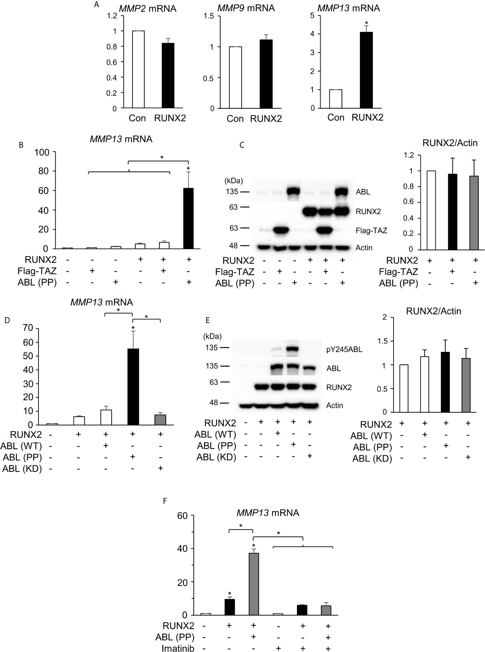
Figure 1 ABL kinase activity is required for RUNX2-mediated MMP13 expression. (A) Quantitative PCR analysis of MMP2, 9, and 13 mRNA expression in HEK293T cells transfected with RUNX2. n = 3. (B) Quantitative PCR analysis of MMP13 mRNA expression in HEK293T cells co-transfected with RUNX2 with or without TAZ or ABL (PP). n = 3. (C) HEK293T cells were co-transfected with RUNX2 with or without TAZ or ABL (PP). Whole cell lysates were probed with the indicated antibodies for Western blot analysis. (D) Quantitative PCR analysis of MMP13 mRNA expression in HEK293T cells co-transfected with RUNX2 with or without ABL (WT, PP or KD). n = 3. (E) HEK293T cells were co-transfected with RUNX2 with or without ABL (WT, PP or KD). Whole cell lysates were probed with the indicated antibodies for Western blot analysis. (F) Quantitative PCR analysis of MMP13 mRNA expression in HEK293T cells co-transfected with RUNX2 with or without ABL (PP) and cultured in the presence or absence of 10 μM imatinib for 24 hours. n = 3. P values were determined by the unpaired t-test (A) or ANOVA with Tukey–Kramer’s post hoc test (B–F). Data are presented as means ± SEM. *P < 0.05.
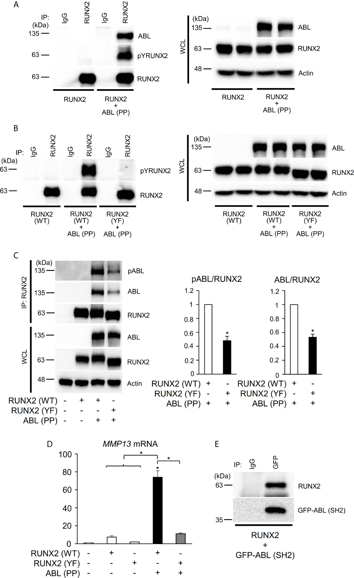
Figure 2 ABL binds to, phosphorylates, and activates RUNX2 through its SH2 domain. (A–C) HEK293T cells were co-transfected with wild-type (WT) or all tyrosine to phenylalanine mutant (YF) RUNX2 with or without ABL (PP). RUNX2 immune complexes were probed with an anti-phosphotyrosine (4G10), anti-pY245ABL, anti-ABL or anti-RUNX2 antibody. Whole cell lysates (WCL) were probed with the indicated antibodies for Western blot analysis. (D) Quantitative PCR analysis of MMP13 mRNA expression in HEK293T cells co-transfected with RUNX2 (WT or YF) with or without ABL (PP). n = 3. (E) HEK293T cells were co-transfected with RUNX2 with or without GFP-ABL (SH2). GFP-ABL (SH2) immune complexes were probed with an anti-RUNX2 or anti-GFP antibody. P values were determined by ANOVA with Tukey–Kramer’s post hoc test. Data are presented as means ± SEM. *P < 0.05.
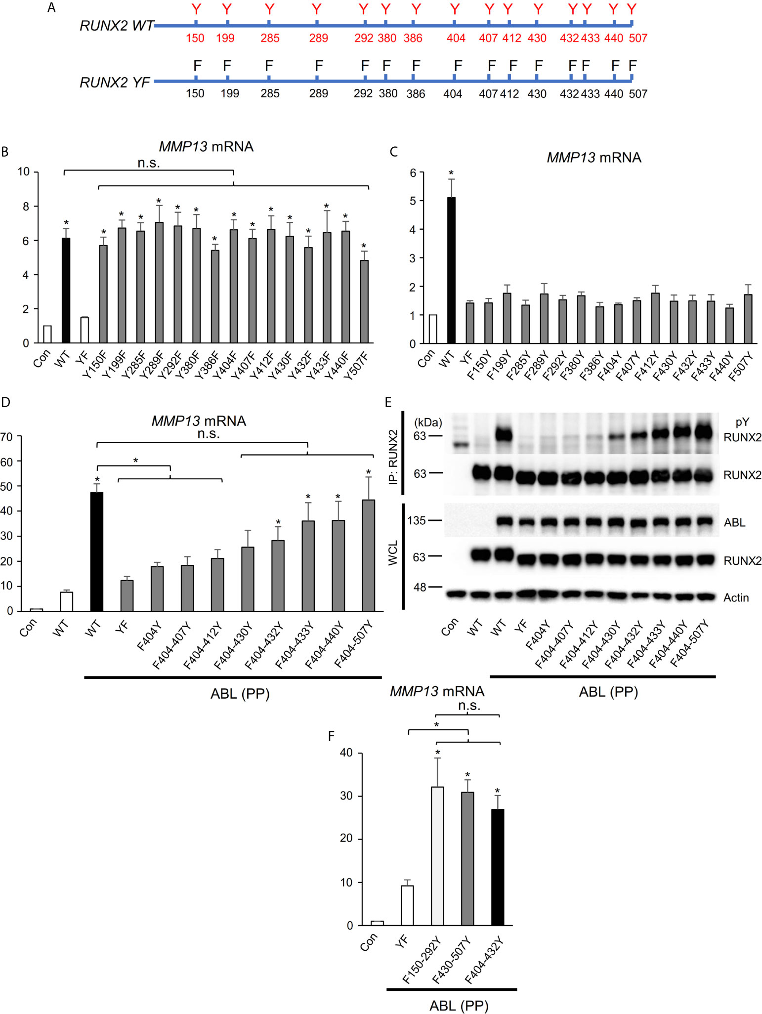
Figure 3 RUNX2 transcriptional activity is dependent on the number of its tyrosine residues phosphorylated by ABL. (A) Schematic models of RUNX2 (WT) and RUNX2 (YF). (B–D, F) Quantitative PCR analysis of MMP13 mRNA expression in HEK293T cells co-transfected with the indicated constructs. n = 3. (E) HEK293T cells were co-transfected with the indicated constructs and RUNX2 immune complexes were probed with an anti-4G10 or anti-RUNX2 antibody. P values were determined by ANOVA with Tukey–Kramer’s post hoc test. Data are presented as means ± SEM. *P < 0.05. ns, no significance.
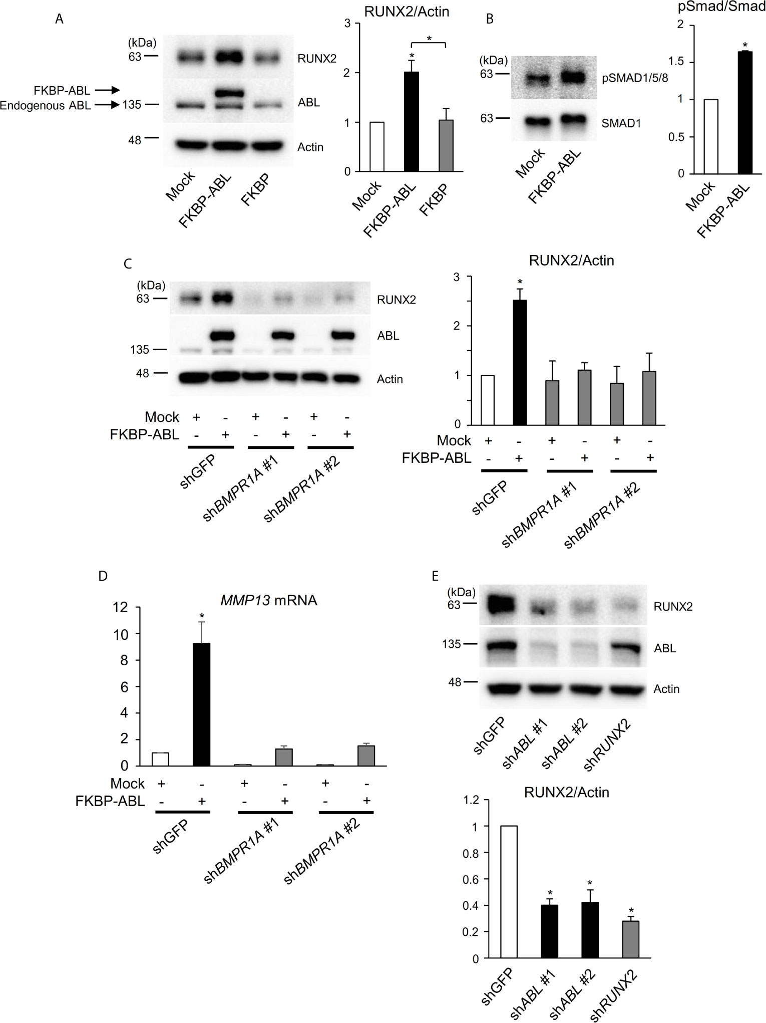
Figure 4 ABL regulates RUNX2 expression through control of the BMP-SMAD pathway. (A, B) Saos-2 cells were infected with an empty vector control or an FKBP-ABL- or FKBP-expressing retroviral vector. Whole cell lysates were probed with the indicated antibodies for Western blot analysis. (C) Saos-2 cells were infected with an empty vector control or an FKBP-ABL-expressing retroviral vector in the presence of shGFP or shBMPR1A. Whole cell lysates were probed with the indicated antibodies for Western blot analysis. (D) Quantitative PCR analysis of MMP13 mRNA expression in cells in (C). n = 3. (E) MDA-MB231 cells were infected with an shGFP-, shABL- or shRUNX2-expressing vector. Whole cell lysates were probed with the indicated antibodies for Western blot analysis. P values were determined by ANOVA with Tukey–Kramer’s post hoc test. Data are presented as means ± SEM. *P < 0.05.
The authors apologize for these errors and state that this does not change the scientific conclusions of the article in any way. The original article has been updated.
Supplementary Material
The Supplementary Material for this article can be found online at: https://www.frontiersin.org/articles/10.3389/fonc.2021.729192/full#supplementary-material
Supplementary Figure 1 | ABL kinase activity is required for RUNX2-mediated MMP13 expression. (A–D) Independent raw data of the panels shown in Figures 1A (A), 1B (B), 1D (C) and 1F (D).
Supplementary Figure 2 | ABL binds to, phosphorylates, and activates RUNX2 through its SH2 domain. (A) Independent raw data of the panel shown in Figure 2D.
Supplementary Figure 3 | RUNX2 transcriptional activity is dependent on the number of its tyrosine residues phosphorylated by ABL. (A–D) Independent raw data of the panels shown in Figures 3B (A), 3C (B), 3D (C) and 3F (D).
Supplementary Figure 4 | ABL regulates RUNX2 expression through control of the BMP-SMAD pathway. (A) Independent raw data of the panel shown in Figure 4D.
Supplementary Figure 5 | ABL-mediated RUNX2 expression and phosphorylation regulate breast cancer invasion. (A) Quantitative PCR analysis of MMP13 mRNA expression in MDA-MB231 cells infected with an shGFP-, shABL- or shRUNX2-expressing vector and the independent raw data. n = 3. (B) MDA-MB231 cells infected with an shGFP-, shABL- or shRUNX2-expressing vector were subjected to a Matrigel invasion assay, and invading cells in five independent regions were counted. Representative photographs were taken at 10 × magnification. (C) MDA-MB231 cells stably expressing luciferase were infected with an shGFP- or shABL-expressing vector and injected into the lateral tail veins of BALB/c-nu/nu female mice as described in the methods section. After 4 weeks, the presence of metastases was detected by IVIS, and regions of interest from displayed images were identified and quantified as total photon counts or photons/s. n = 6-7. (D) A representative image of H&E staining of the lungs from mice in (C). P values were determined by the unpaired t-test (C) or ANOVA with Tukey–Kramer’s post hoc test (A, B). Data are presented as means ± SEM. *P < 0.05.
Keywords: ABL—Abelson murine leukemia viral oncogene homolog, Runx2 (runt-related transcription factor 2), tyrosine, phosphorylation, invasion
Citation: He F, Matsumoto Y, Asano Y, Yamamura Y, Katsuyama T, Rose JL, Tomonobu N, Komalasari NLGY, Sakaguchi M, Rottapel R and Wada J (2021) Corrigendum: RUNX2 Phosphorylation by Tyrosine Kinase ABL Promotes Breast Cancer Invasion. Front. Oncol. 11:729192. doi: 10.3389/fonc.2021.729192
Received: 22 June 2021; Accepted: 28 June 2021;
Published: 20 July 2021.
Edited and reviewed by:
Xu Wang, Affiliated Hospital of Jiangsu University, ChinaCopyright © 2021 He, Matsumoto, Asano, Yamamura, Katsuyama, Rose, Tomonobu, Komalasari, Sakaguchi, Rottapel and Wada. This is an open-access article distributed under the terms of the Creative Commons Attribution License (CC BY). The use, distribution or reproduction in other forums is permitted, provided the original author(s) and the copyright owner(s) are credited and that the original publication in this journal is cited, in accordance with accepted academic practice. No use, distribution or reproduction is permitted which does not comply with these terms.
*Correspondence: Yoshinori Matsumoto, eW1hdHN1bW90b0Bva2F5YW1hLXUuYWMuanA=