- 1Radiation Oncology Center, Chongqing University Cancer Hospital, School of Medicine, Chongqing University, Chongqing, China
- 2Department of Oncology, The Fifth People 's Hospital of Chongqing, Chongqing, China
Background: Clinical target volume (CTV) delineation is a major focus in radiotherapy for nasopharyngeal carcinoma (NPC) and currently lacks a universally accepted standard across treatment centers. We proposed an individualized CTV delineation method for eccentric NPC and evaluated its feasibility based on the eccentric distance of the primary lesion.
Materials and methods: Ninety patients with eccentric NPC were included. Each treatment plan was replanned using the individualized CTV method for dosimetric comparison with the conventional CTV, to evaluate coverage, homogeneity, and conformity of CTV and PTV, sparing of organs at risk (OARs) and radiotherapy technique. Paired sample t-tests and nonparametric rank-sum tests were used to compare target coverage, homogeneity, conformity, and OAR dose parameters between the two approaches. Correlation analysis is used to evaluate the correlation between eccentric distance of primary lesion and OARs dose changes. Subgroup analysis is used to compare the PTV and OARs dose parameters of individualized CTV at different T stages or radiotherapy techniques.
Results: Our results showed that compared with conventional CTV, the volume of CTV decreased significantly (P< 0.05) through individualizing delineation for eccentric NPC, especially CTV1 volume (95.81 cm³ vs. 57.57 cm³, P < 0.001). Individualized CTV reduced the doses delivered to OARs, including the brainstem, spinal cord, optic chiasm, optic nerves, and contralateral temporal lobe, inner ear and so on (all P< 0.05). When the eccentric distance of the primary lesion was between 1.4 and 2.1 cm, the individualized CTV approach provided significant advantages in organ protection, such as contralateral optic nerve, temporal lobe and parotid gland. Additionally, Subgroup analysis showed that the dose-sparing benefit of individualized CTV was more pronounced in patients treated with VMAT (volumetric modulated arc therapy).
Conclusion: This study demonstrates the dosimetric advantages of individualized CTV delineation based on eccentric distance. Our prospective trial is currently ongoing for further research (NCT06167109).
Introduction
Nasopharyngeal Carcinoma (NPC) is a malignant tumor originating from the nasopharynx mucosa, which has a high incidence in the south of China, Southeast Asia, and North Africa. With the application of intensity-modulated radiotherapy (IMRT), the survival rate of NPC has significantly improved, with a 5-year local control rate of 85-90% (1, 2). However, acute complications such as hearing loss (47.4%) and xerostomia (83.0%) (2), and late complications such as temporal lobe injury (13.1%) and cranial nerve injury (14.0%) (3) still seriously affect the quality of life of patients. Complications following radiotherapy are mainly caused by excessive irradiation of the corresponding normal tissue range and dose. Therefore, reducing the toxic and side effects of patients has become a research hotspot recently, and the treatment mode has changed to individualization (4), such as asynchronous chemotherapy for early and low-risk patients, reducing the dose of radiotherapy and narrowing the range of irradiation.
There is still no recognized delineation standard for the delineation of clinical target volume (CTV). Restricted the anatomical structure around the nasopharynx to a certain distance based solely on experience would not meet the requirements of precise treatment (5). As a result, researchers have continued to optimize the CTV for these patients to minimize radiotherapy-related side effects without affecting survival rate. A retrospective study (6) found that simultaneously reducing the volume and dose of CTV did not affect local area control rates, but significantly reduced late-stage adverse reactions, particularly pulmonary infection, dysphagia, and xerostomia. Miao et al. (7) researched on 103 patients with stage I and II NPC. CTV2 was defined as the omission of the posterior third of the maxillary sinus and cavernous sinus, and patients with N0–1 were defined as the omission of the pterygopalatine fossa, lateral pterygoid muscle, post-styloid space, foramen ovale, and spinous process. The study achieved satisfactory 10-year LRFS (90.3%) and overall survival (91.2%). Only 1 patient had local recurrence 6.1 months after radiotherapy, 4 patients (3.9%) had acute grade 3 mucositis, and 1 patient (1.0%) had acute grade 3 radiation dermatitis. These results support the notion that appropriately reducing CTV volume does not impair the disease control rate. However, most studies have ignored the distribution characteristics of tumors when reducing the volume of CTV.
The application of individualized CTV delineation should also consider the location of the lesion. Most NPC patients present eccentricity, accounting for 72.4% of all NPC cases, and it was found that the spread of eccentricity lesions is concentrated in the ipsilateral pharyngeal recess. The ipsilateral invasion rate of most anatomical structures is significantly higher than that of the contralateral side, and the risk of bilateral invasion is low (10%) (8). The extreme typical case of eccentric NPC is unilateral NPC with a nasopharyngeal mass limited to one side of the nasopharynx and not exceeding the midline, which accounts for about 10% of all NPC cases (9). Li et al. (10) performed a pathological examination of contralateral pharyngeal recess (CPR) and contralateral posterior superior wall (CPSW) in 20 patients with unilateral NPC, and analyzed the factors related to contralateral tumor infiltration to explore the potential of reducing CTV. It was found that unilateral NPC rarely had CPR infiltration, so CTV did not include CPR in patients without lateral lymph node metastasis or EBV-DNA negative. According to the trend of tumor invasion, Sanford et al. (11) carried out individualized target delineation for NPC radiotherapy, excluding all traditional high-risk areas of NPC, and for unilateral NPC, excluding the contralateral parapharyngeal space and skull base channel. It was showed a 5-year local control rate of 94% and no recurrence of contralateral tissue structure. The optimization of the contralateral risk structure reduced the dose of the contralateral parotid gland, optic nerve, and cochlea in T1 patients by 54%, 50%, and 34%, respectively, and the dose of the optic chiasm and contralateral optic nerve in T4 patients decreased by 46% and 37%, respectively. Another study has summarized the local invasion characteristics of unilateral NPC through MRI, which is characterized by continuous invasion from the proximal to the distal end. The probability of contralateral parapharyngeal space and skull base channel invasion is very low (12). Therefore, conventional prophylactic irradiation should not be performed on the contralateral side with a low risk of invasion for patients with eccentric NPC, as this would reduce the volume of CTV, alleviate post-radiation complications, and improve the quality of life.
At present, there is little research on individualized treatment for eccentric NPC, as well as its protective effect on OARs. To determine whether our individualized CTV offers an advantage over conventional CTV, We are the first center to compare the dosimetry parameters of PTV and OARs in patients with eccentric NPC and evaluate radiotherapy techniques suitable for individualized CTV.
Materials and methods
Patients
From September 2021 to July 2023, 90 patients with newly treated eccentric NPC at Chongqing University Cancer Hospital were enrolled. Other inclusion criteria were: 1) meeting the definition of eccentric nasopharyngeal carcinoma (patients with a nasopharyngeal mass confIned to one side of the nasopharynx or did not exceed the midline.); 2) having no distant organ metastasis; 3) with baseline MRI data of the nasopharynx and neck.
Informed consent was obtained from all participants at baseline and all procedures were performed in accordance with our local guidelines and clinical regulations. The study was conducted in accordance with the Declaration of Helsinki, and the protocol was approved by the Ethics Committee of The Chongqing University Cancer Hospital (No. CZLLYJ0252). Our prospective trial is currently ongoing for further research (NCT06167109, 12/12/2023).
Clinical target volume delineation and prescription dose
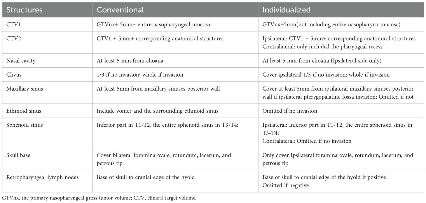
We redesigned a new plan based on the individualized CTV delineation, but it was not used for radiotherapy. Gross tumor volume(GTV) is the primary nasopharyngeal lesions, positive retropharyngeal lymph nodes (GTVnx), and cervical lymph nodes (GTVnd) by imaging examination. CTV1 and CTV2 represent high-risk and low-risk invasion areas, respectively. For individualized CTV, the contralateral CTV1 was defined as a subclinical disease consisting of a 5mm margin surrounding GTVnx(not including entire nasopharynx mucosa); the CTV2 was defined as potentially involved regions consisting of a 5mm margin surrounding the CTV1, and the contralateral CTV2 only included the pharyngeal recess. For conventional CTV, the CTV1 is defined as GTVnx+5mm+entire nasopharyngeal mucosa. The CTV2 is defined as the CTV1 + 5mm+corresponding anatomical structures. The delineation of the CTV is outlined in Table 1 and Figure 1.
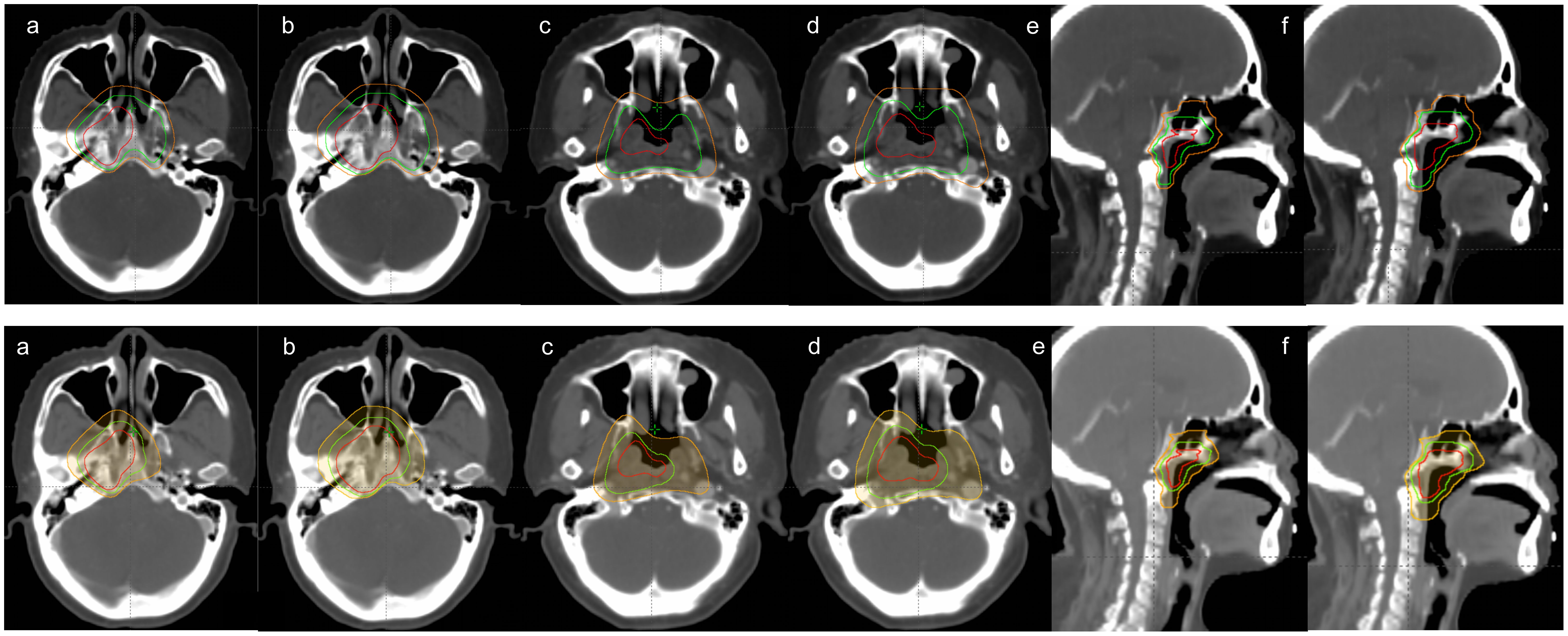
Figure 1. Comparison of conventional(up) and Individualized(down) CTV delineation. The images of (a), (c) and (e) represent the delineation of the GTV, CTV1 and CTV2, and the images of (b), (d) and(f) exhibit the delineation of PGTV, PTV1 and PTV2. The GTV and PGTV is in solid red, CTV1 and PTV1 is in solid green, and CTV2 and PTV2 is in solid orange.
The dose prescriptions of PGTVnx, PGTVnd, PTV1, and PTV2 were 69.96-74.2Gy, 69.96-74.2Gy,60.06-60.8Gy and 54.12-54.4Gy in 30-33fractions, respectively. All plans meet the following requirements: 100% of the prescribed dose should cover more than 95% of the target volume, the maximum dose point is located in the target area, and PTV should not receive more than 110% of the prescribed dose. In order to maintain consistency, the delineation of the target volume and OAR was performed by a doctor who specializes in radiotherapy for NPC. The dosimetric restriction of the OARs are shown in Supplementary Table 1. For example, the Dmax of the brainstem is 54Gy, the Dmax of the spinal cord is 45Gy, and the Dmean of the parotid gland is 26Gy.
Plan evaluation
90 patients with eccentric NPC underwent a revised radiotherapy plan, which was completed by senior medical physicists at our center. The Kappa analysis involved two radiation oncologists (with 8+, and 10+ years of experience, respectively), each independently reviewing the same 20 randomly selected cases. The evaluation process included standardized training on contouring criteria (Conventional and Individualized for CTV) prior to analysis. The prescribed doses were consistently maintained and dose and site validation was performed to meet clinical treatment criteria. The plan was evaluated according to the dose-volume histogram(DVH) generated after the optimization of the treatment planning system (TPS) plan.
The IMRT plans were generated in the Eclipse TPS (Version 15.6) based on multi-leaf collimators Millennium 120, and a 6MV X-ray in a Varian IX linear accelerator was used. The coplanar irradiation of 9 fields (200°, 240°, 280°, 320°, 0°, 40°, 80°, 120°, 160°) was used. The VMAT plans were generated in the Pinncle3 16.2 TPS (Version 16.2, Philips, Inc., USA). All plans used double-arcs, with a rotation angle of 181°to 179°and a bed angle of 0°. The TOMO plans were designed using the three-dimensional TPS of the Tomotherapy Planning Station with 6MV X-ray. The dose calculation was performed using the convolution superposition algorithm. The main planning parameters were: field width = 2.512cm, pitch = 0.287 and modulation factor = 2.2-3.5.
Dosimetric validation was performed using a square polymethyl methacrylate (PMMA) phantom with a PTW Freiburg ionization chamber (model TN30013). Measurements followed AAPM TG-51 protocols, with beam calibration at 6 MV (SSD = 100 cm, depth = 5 cm). Prior to clinical implementation, all treatment plans underwent comprehensive 3D dose verification utilizing the ArcCHECK dosimetry system. Clinical approval required fulfillment of pre-established quality assurance criteria, specifically a gamma index passing rate exceeding 95% under 3%/3 mm evaluation parameters.
PTV coverage
The dosimetric parameters were quantified to evaluate PTVs included V100%, Dmean, D2%, D95%, D98%, heterogeneity index (HI) and the conformity index (CI). V100% is the percentage of the volume of PTV1 and PTV2 covered by prescription isodose of PTV1 and PTV2; D2%, D95%, and D98% are doses that cover 2%, 95%, and 98% of the PTV volume, respectively, with D2% approximating the maximum dose and D98% approximating the minimum dose.
HI was calculated as HI= D5%/D95% to evaluate the homogeneity of the prescription dose in target volume (D5%: the dose received by 5% of the target volume; D95%: the dose received by 95% of the target volume). The smaller the HI, the better the homogeneity. CI was calculated as CI= Vt,ref/Vt×Vt,ref/Vref to evaluate the conformity of the prescription dose in target volume (Vt,ref: the target volume surrounded by the prescription dose; Vt: the target volume; Vref:the volume surrounded by the prescription dose). The ideal CI value is 1, and the closer it is to 1, the better the conformity.
Organs at risk
The maximum doses(Dmax) of the spinal cord, temporal lobe, optic chiasm, optic nerve, pituitary, lens, inner ear, and brainstem; the mean doses(Dmean) of the parotid gland, thyroid gland, larynx, pharyngeal constrictor, pituitary, oral cavity and inner ear; the D0.03ccPRV of the spinal cord, temporal lobe, optic chiasm, optic nerve and brainstem; and the V30 (the volume that received 30Gy) of parotid gland were analyzed and recorded.
Statistical analysis
Statistical analysis was performed using SPSS version 27. The descriptive analysis of the data is expressed in terms of means and standard deviations. The dosimetric parameters of the PTV and OAR were assessed and compared using the paired-sample t-test and nonparametric rank-sum test. Correlation analysis was used to evaluate the correlation between eccentric distance of primary lesion and OARs dose changes. Subgroup analysis was used to compare the PTV and OAR dosimetric parameters of individualized CTV at different T stages or radiotherapy techniques. The two-tailed P values < 0.05 are considered statistically significant.
Results
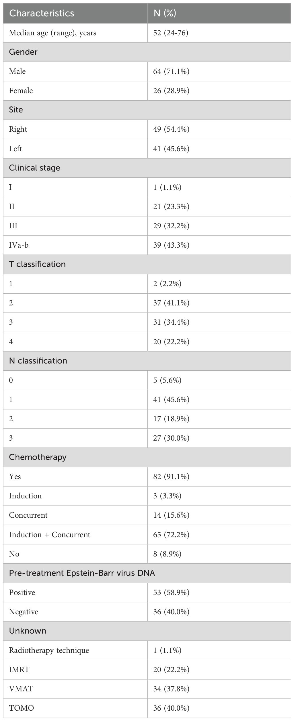
This study included 90 NPC patients with eccentric lesions. The median patient age was 52 years (24-76). There were 39, 31, and 20 patients in the T1-2, T3, and T4 stages, respectively. Histologically, 85(94%) patients had non-keratinized carcinoma, and 5(6%) had keratinized carcinoma. The number of patients treated with IMRT, VMAT, and TOMO were 20, 34, and 36 respectively. A total of 82 patients received chemotherapy, 65 of whom underwent concurrent chemoradiotherapy. All patients were staged according to the 8th AJCC staging system, and the detailed clinical data of patients are presented in Table 2. The Kappa analysis indicated good inter-physician consistency in target volume delineation (Supplementary Table 2).
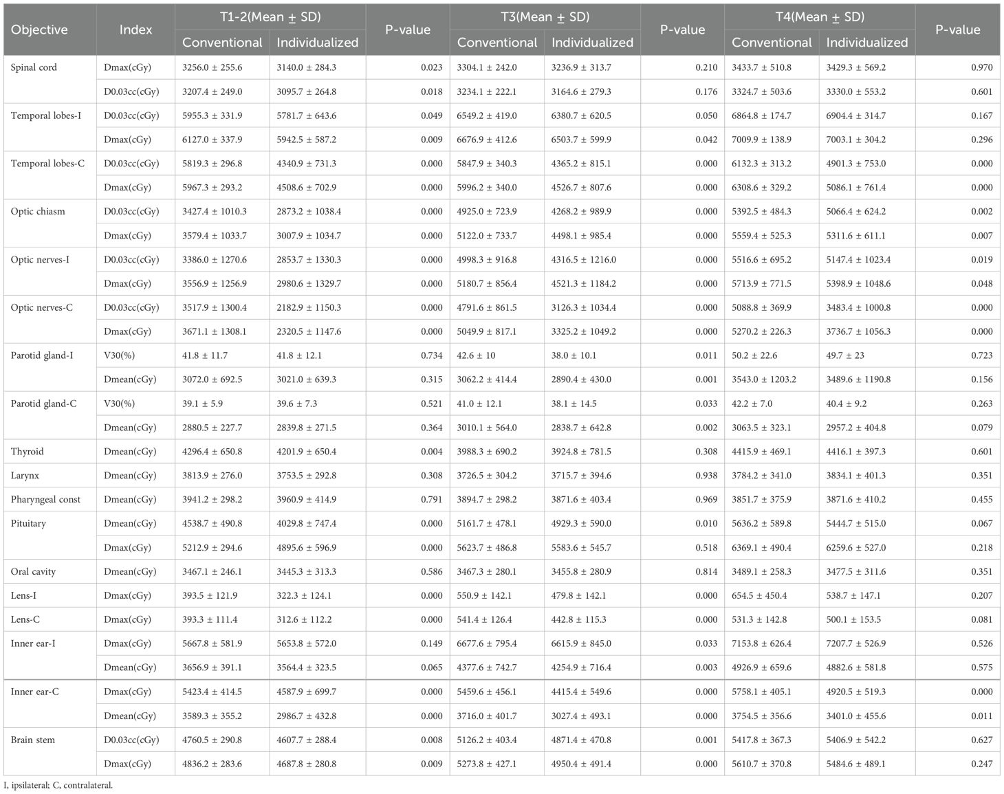
As shown in Figure 1 and Supplementary Table 3, the volume of individualized CTV significantly decreased compared to the conventional CTV (CTV1: 95.81 cm3 vs. 57.57 cm3, CTV2: 428.50 cm3 vs. 395.05 cm3; P< 0.001). Strikingly, we found that individualized CTV reduced the doses delivered to OARs generally, with a significant decrease in the D0.03 and Dmax of organs which belong to the priority level one, including the spinal cord, brainstem, optic chiasm, optic nerve, and contralateral temporal lobe (P< 0.05) (Supplementary Table 4, Figure 2). In addition, an obvious decrease (P< 0.05) was seen in the Dmean of the parotid gland, thyroid gland, pituitary and inner ear, the Dmax of the lens, pituitary, inner ear. However, there was no distinct difference among the doses of the larynx, pharyngeal const and oral cavity (Supplementary Table 4). Next, subgroup analysis was performed to compare the specific advantages of the two kinds of delineation schemes to different OARs in different T stages. In the group of stage T1-2, significant differences were observed in D0.03 and Dmax of the spinal cord, optic chiasm, optic nerve, brainstem, contralateral temporal lobe, Dmean of contralateral inner ear, and Dmax of the lens and pituitary (Table 3). The individualized CTV for patients in stage T3–4 also experienced a sharp decrease in D0.03 and Dmax of the optic chiasm, optic nerve and contralateral temporal lobe, Dmean of the contralateral inner ear, Dmax of the lens, but not differences of spinal cord, brainstem and pituitary (Table 3).
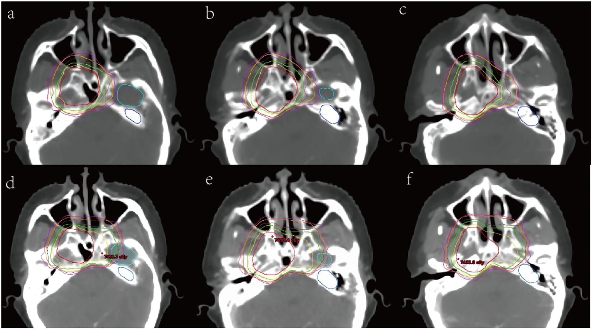
Figure 2. Dose distribution comparison images of conventional (down) and Individualized (up) CTV delineation. (a), (b) and (c) are individualized cross-sectional dose distribution maps, while (d), (e), and (f) are conventional cross-sectional dose distribution maps. The PGTV is in solid red, PTV1 is in solid green, and PTV2 is in solid orange. The 95% dose line of 69.96Gy is yellow, the 95% dose line of 60.06Gy is brown, and the 95% dose line of 54.12Gy is purple. The light blue structure is the left temporal lobe, and the dark blue structure is the left inner ear.
The distance from the tumor center to the midline of the body (L for short), which represented the eccentric extent of primary lesion, is positively correlated with the reduction of the CTV volume (Supplementary Table 5). Further, the dose reduction of the contralateral organs were positively correlated with L in different distance intervals (P<0.05). Especially, the dose the contralateral optic nerve, temporal lobe and parotid gland showed a dramatic reduction in the distance interval of 1.4-2.1cm (Figure 3).
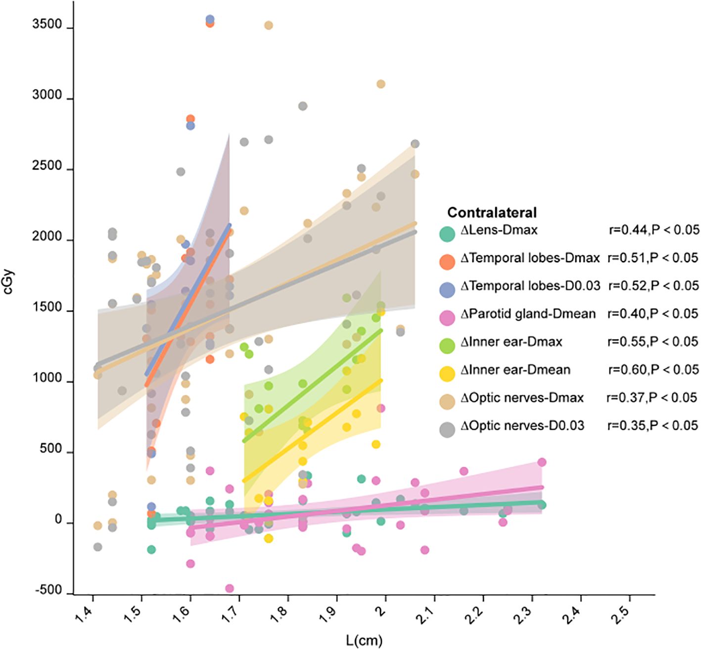
Figure 3. Correlation between L and the dose difference of the contralateral structure. This figure was drawn using ChiPlot (https://www.chiplot.online/) (accessed on May 2024).
The target coverage of the two groups of plans reached more than 95% of the prescription dose coverage, and the individualized PTV1 was better (Conventional vs. Individualized: 98.19% vs. 99.29%, P< 0.001, Supplementary Table 6). Although the target coverage of PTV2 was not statistically significant (P=0.125), it showed an increasing trend (Conventional vs. Individualized: 97.02% vs. 97.16%). The Dmean, D95, and D98 increased, and HI and CI decreased for individualized PTV1, while the Dmean, HI, and CI decreased for PTV2. There was no statistically significant difference in Mu and other indicators (P> 0.05, Supplementary Table 6).
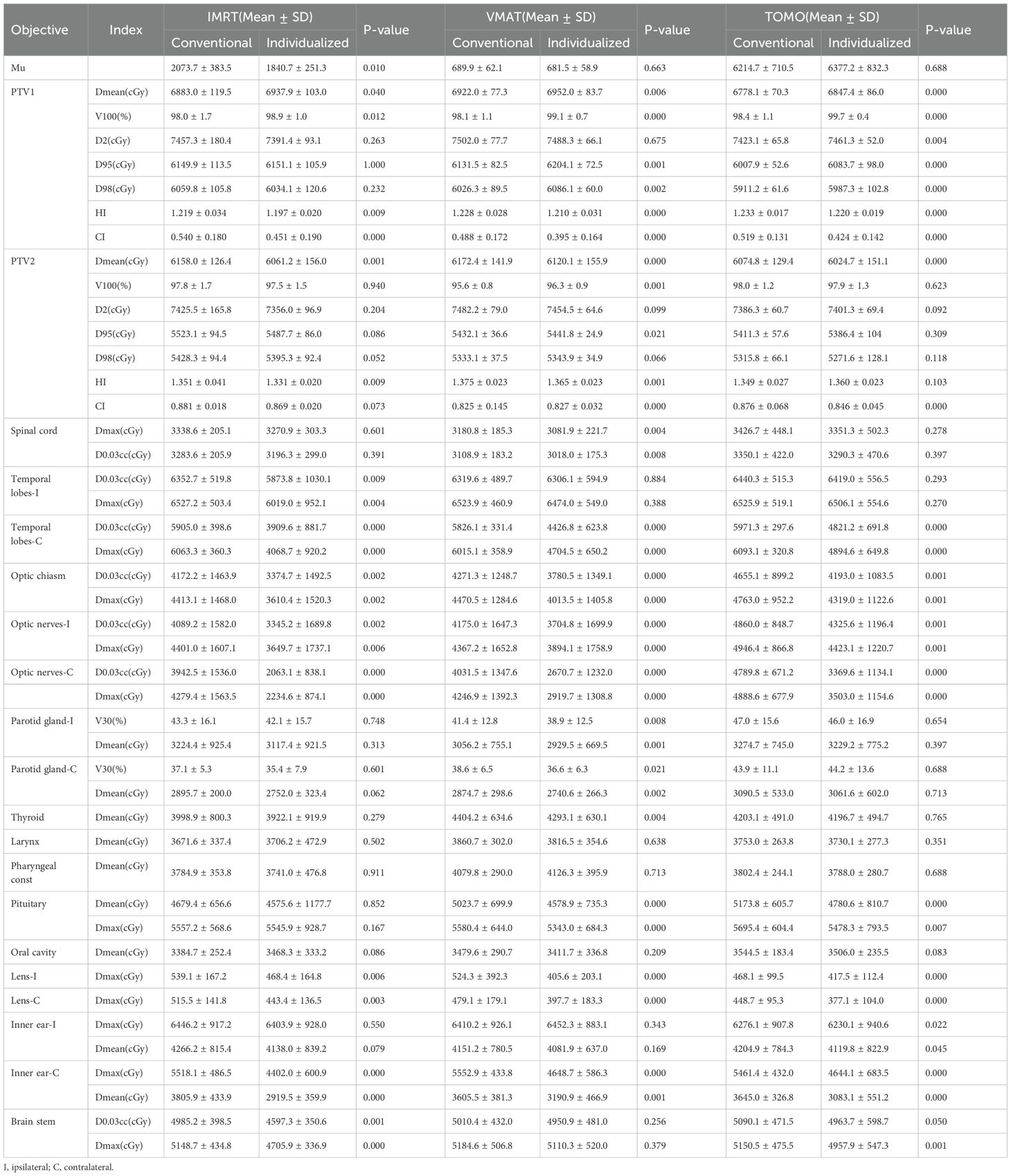
After comparison of different treatment planning system (TPS), the following conclusions can be drawn (Table 4): (1) under the IMRT treatment mode, the Mu of individualized CTV decreased, the Dmean and V100% of PTV1 increased, and HI and CI decreased; the HI and Dmean of PTV2 decreased; doses of OARs dropped in the temporal lobe, optic chiasm, optic nerve, lens, contralateral inner ear, and brainstem (P< 0.05); (2) under the VMAT treatment mode, the Dmean, V100%, D95, and D98 of PTV1 of individualized CTV increased, while HI and CI decreased; the Dmean and HI decreased for PTV2, while V100%, D95, and CI increased; doses of OARs dropped in spinal cord, contralateral temporal lobe, optic nerve, optic chiasm, parotid gland, pituitary, thyroid gland, lens, and contralateral inner ear (P< 0.05); (3) under the TOMO treatment mode, the Dmean, V100%, D2, D95, and D98 were increased and HI and CI decreased for PTV1 with individualized CTV, while Dmean and CI decreased for PTV2; doses of OARs dropped in contralateral temporal lobe, optic nerve, optic chiasm, pituitary, lens, inner ear, and brainstem (P< 0.05).
Discussion
Our individualized CTV delineation refers to previous experience in reducing the target volume, whether it is based on the invasion risk of NPC (11, 12) or confirmed through pathological examination (10). It has been found that for unilateral NPC, routine prophylactic radiation may not be required for the contralateral parapharyngeal space, cranial foramen, and contralateral pharyngeal recess. According to the study of the T stage and invasion pattern, local recurrence of NPC also follows the gradual infiltration pattern, similar to the primary invasion, usually in the ipsilateral cavernous sinus recurrence (13). Xie et al. (14) also delineated the individualized target area of unilateral NPC, excluding the contralateral parapharyngeal space and skull base channel. The 10-year local recurrence-free survival(LRFS), regional recurrence-free survival (RRFS), and overall survival (OS) reached 96.2%, 90.5%, and 84.7%, respectively, achieving good long-term local control and mild late toxicity. Late toxicity mainly occurred in the ipsilateral organs, and the incidence of hearing loss on the ipsilateral side of the tumor (10/95) was significantly higher than that on the contralateral side of the tumor (1/95).
Accordingly, we designed the individualized CTV delineation for eccentric NPC: the contralateral CTV1 is a 5mm geometrical extension that does not include the entire nasopharyngeal mucosa, and the contralateral CTV2 only includes the pharyngeal recess. Our results showed that compared with conventional CTV, the volume of individualized CTV was significantly decreased (P< 0.05). Individualized CTV reduced the doses delivered to OARs in general, including priority level one protective organs such as the spinal cord, brainstem, optic chiasm, optic nerve, contralateral temporal lobe, as well as the parotid gland, thyroid gland, pituitary, lens, and inner ear. As we know, radiation-induced tissue and nerve damage can cause adverse reactions such as xerostomia, vision loss, and hearing loss. Pituitary dysfunction is also a common complication, which increases in incidence from year to year and is lifelong. In addition, radiation-induced brain injury such as brainstem and temporal lobe can cause symptoms such as disturbance of consciousness, ataxia and epilepsy that can affect the quality of life for patients, and the injury is often irreversible. However, the larynx, Pharyngeal const, and oral cavity did not show significant dose downregulation due to being slightly further away from the scope of CTV reduction. In addition, the individualized CTV delineation also fully met the dose and range of target irradiation, ensuring the safety and efficacy of irradiation. All plans with individualized CTV achieved more than 95% of the target volume covered by the prescribed dose, with individualized PTV1 achieving better target volume coverage.
The prospective study published by Lin et al. (15) omits CTV1 and directly defines CTV2 as GTVnx+entire nasopharyngeal mucosa+8mm+corresponding anatomical structure. The 4-year LRFS and RRFS in this study reached 96.6% and 97.7%, respectively, and all local recurrences were intra-field recurrences, but their CTV delineation still uniformly covered adjacent structures regardless of tumor laterality and tumor stage. However, we discussed the exempted irradiation of the contralateral structure of the nasopharynx for eccentric lesions. Subgroup analysis of the T stage found that individualized CTV delineation of eccentric NPC has less benefit in the T4 stage, due to the limited contralateral structures that can be exempted in high stage patients. A study published this year (16) proposed a concept of CTV delineation including distance and substructure by combining the laterality of tumors and the propagation mode of NPC. The anatomical structures around the nasopharynx were defined as grade 1, grade 2, grade 3, and grade 4, respectively. And the CTV delineation was based on the combination of distance and substructure, When the nasopharyngeal tumor invades different substructures, the corresponding CTV contains different regions. The 5-year LRFS and OS rates for the overall population were 93.2% and 91.5%, respectively. Grade 3 late toxicities included xerostomia, subcutaneous fibrosis, and hearing impairment, with a total incidence of 1.5%. For the eccentric NPC, we also proposed a distance-based concept of delineation, where the contralateral structures is positively correlated at different distance intervals. When the distance between the tumor center and the midline of the body is in the range of 1.4-2.1cm, the individualized CTV delineation would be able to bring greater benefits to organ protection, such as contralateral optic nerve, temporal lobe and parotid gland. This phenomenon may be attributed to the anatomical characteristics of nasopharyngeal carcinoma (NPC), where eccentric tumors predominantly localize in the ipsilateral pharyngeal recess with low contralateral invasion risk (10%). When the tumor is located 1.4-2.1 cm from midline, its distance from the contralateral pharyngeal recess enables more effective sparing of contralateral organs at risk (Supplementary Figure 1).
The comparison between VMAT and IMRT in the treatment of NPC found that the target dose of VMAT was higher than that of IMRT, and the maximum dose to the brainstem, spinal cord, and optic nerve, as well as the incidence of late toxic side effects, were lower than those of IMRT (17). However, other studies have shown that IMRT has better conformity than VMAT, similar in the protection of OARs (18), and similar survival outcomes (19, 20). Refined the T stage showed that VMAT provides better OAR retention and HI in the early T stage (21, 22). Du et al. (23) summarized the results of 190 NPC patients treated by TOMO, showing that TOMO had low toxicity and good efficacy. Another study retrospectively compared the efficacy of TOMO and IMRT in the treatment of NPC and found no significant difference in 5-year OS between the two techniques (24). However, TOMO is superior to VMAT in HI and CI (25) and dosimetric parameters of target volume, brainstem, spinal cord, lens, and parotid gland for advanced NPC, and there is no significant difference in survival outcome (26). It can be seen that IMRT, VMAT, and TOMO have similar efficacy, but further studies are needed to determine whether they can effectively reduce the radiation dose to OARs. Our subgroup analysis results show that all three radiotherapy techniques can meet the PTV coverage of treatment needs, but compared with IMRT and VMAT, the prescription dose optimization of PTV1 in TOMO is better, which is consistent with the research conclusion of Chen et al (26). However, under the VMAT treatment mode, the dose of OARs was significantly reduced, including the spinal cord, parotid gland, and thyroid gland, which were not different from IMRT and TOMO. Additionally, VMAT is superior to IMRT in the protection of glands and also associated with lower in MUs and treatment times in this analysis, consistent with previous studies (18, 27). The advantage may stem from the staging of patients and different algorithms employed by the TPS. Variation in TPS results stems partly from different dose calculation algorithms; Eclipse typically uses AAA, while Pinnacle employs the Collapsed Cone Convolution (CCC) algorithm. Although CCC can overestimate dose near bone due to scaling water-derived kernel (28), its convolution/superposition approach generally models lateral electron scatter and energy transport more accurately in heterogeneous regions like the head and neck (with air cavities and bone interfaces) compared to pencil-beam-like algorithms such as AAA (29), which underestimates dose by missing lateral scatter. This superior handling of heterogeneity, combined with Pinnacle’s efficient convolution-based optimization for VMAT, underpins its enhanced performance in NPC planning: the algorithm enables precise dynamic MLC modulation for highly conformal doses around complex, concave targets, significantly improving OAR sparing (e.g., reducing mean parotid dose), while continuous arc optimization minimizes MUs and treatment time, reducing effects like tongue-and-groove. Thus, CCC’s accurate modeling in heterogeneous media and optimized VMAT delivery make it advantageous for NPC.
There are some limitations in our study. First, the radiotherapy plans we have redefined were completed by four senior physicists at our center, and it is still inevitable that there may be some differences in the completion of radiotherapy plans among different physicists. Second, the study of which radiotherapy technique is more appropriate for the individualized CTV of eccentric NPC was based on subgroup analysis, and the exploratory results cannot be directly used for clinical decision-making, and more studies are needed to confirm. While the subgroup analyses in Table 3 provide preliminary insights into potential effect modifiers, these findings should be interpreted with caution due to the inherent limitations of multiple comparisons. and we acknowledge that formal correction methods were not applied given the exploratory nature of these analyses. Finally, our comparison between conventional and individualized CTV delineation stays in dosimetric analysis without the support of survival data, which is not equivalent to clinical benefits, and the sample size was constrained by clinical availability. More prospective studies are needed to support the translation of the benefits of dose distribution into clinical advantages in reducing radiation toxicity and improving survival. Our center is also conducting a prospective study of the corresponding individualized CTV delineation.
Conclusion
Individualized CTV delineation for eccentric NPC can achieve significant reduction in the target volume and overall OAR dose, with a significant reduction in contralateral organs, and meet the prescription dose coverage required for treatment. We proposed a distance-based concept for the eccentric NPC, which is easy to be applied in the clinical practice. In addition, subgroup analysis showed that the OAR benefit of individualized CTV was more significant in the VMAT treatment mode. Our prospective trial is currently ongoing for further research on efficacy and feasibility of our individualized CTV method based eccentric distance (NCT06167109).
Data availability statement
The raw data supporting the conclusions of this article will be made available by the authors, without undue reservation.
Ethics statement
The studies involving humans were approved by Ethics Committee of The Chongqing University Cancer Hospital. The studies were conducted in accordance with the local legislation and institutional requirements. Written informed consent for participation was required from the participants or the participants’ legal guardians/next of kin in accordance with the national legislation and institutional requirements.
Author contributions
YS: Data curation, Formal Analysis, Writing – original draft. YuW: Methodology, Writing – original draft. MY: Software, Writing – original draft. XY: Data curation, Writing – review & editing. ML: Data curation, Writing – review & editing. BL: Supervision, Writing – review & editing. XS: Resources, Writing – review & editing. XZ: Methodology, Resources, Writing – review & editing. FW: Methodology, Writing – review & editing. CW: Resources, Writing – review & editing. MH: Resources, Writing – review & editing. JS: Funding acquisition, Methodology, Writing – review & editing. YiW: Funding acquisition, Methodology, Writing – review & editing.
Funding
The author(s) declare that financial support was received for the research and/or publication of this article. The current study was supported by grants from the National Natural Science Foundation of China (No. 81972857 to YiW; No.82373196 to J-DS), Chongqing Science and Health Joint Medical Research Project (No.2024ZDXM032 to YiW; No.2022ZDXM028 to J-DS), Natural Science Foundation of Chongqing City (No. cstb2023NSCQ-MSX0709 to J-DS), Chongqing Talent Program (No. cstc2021ycjh-bgzxm0023 to YiW), Key Laboratory Open Fund (No. cquchkfjj003 to XZ), Shapingba Technology Innovation and Application Development Project (No.2023128 to XS).
Acknowledgments
We thank all the members of the Radiation Oncology Translational Research Group (ROTRG) who participated in this study.
Conflict of interest
The authors declare that the research was conducted in the absence of any commercial or financial relationships that could be construed as a potential conflict of interest.
Generative AI statement
The author(s) declare that no Generative AI was used in the creation of this manuscript.
Publisher’s note
All claims expressed in this article are solely those of the authors and do not necessarily represent those of their affiliated organizations, or those of the publisher, the editors and the reviewers. Any product that may be evaluated in this article, or claim that may be made by its manufacturer, is not guaranteed or endorsed by the publisher.
Supplementary material
The Supplementary Material for this article can be found online at: https://www.frontiersin.org/articles/10.3389/fonc.2025.1587764/full#supplementary-material
References
1. Peng G, Wang T, Yang KY, Zhang S, Zhang T, Li Q, et al. A prospective, randomized study comparing outcomes and toxicities of intensity-modulated radiotherapy vs. conventional two-dimensional radiotherapy for the treatment of nasopharyngeal carcinoma. Radiother Oncol. (2012) 104:286–93. doi: 10.1016/j.radonc.2012.08.013
2. Chen L, Zhang Y, Lai SZ, Li WF, Hu WH, Sun R, et al. 10-year results of therapeutic ratio by intensity-modulated radiotherapy versus two-dimensional radiotherapy in patients with nasopharyngeal carcinoma. Oncologist. (2019) 24:e38–45. doi: 10.1634/theoncologist.2017-0577
3. McDowell L, Corry J, Ringash J, and Rischin D. Quality of life, toxicity and unmet needs in nasopharyngeal cancer survivors. Front Oncol. (2020) 10:930. doi: 10.3389/fonc.2020.00930
4. Yip PL, You R, Chen MY, and Chua MLK. Embracing personalized strategies in radiotherapy for nasopharyngeal carcinoma: beyond the conventional bounds of fields and borders. Cancers (Basel). (2024) 16:383. doi: 10.3390/cancers16020383
5. Hansen CR, Johansen J, Samsøe E, Andersen E, Petersen JBB, Jensen K, et al. Consequences of introducing geometric GTV to CTV margin expansion in DAHANCA contouring guidelines for head and neck radiotherapy. Radiother Oncol. (2018) 126:43–7. doi: 10.1016/j.radonc.2017.09.019
6. Liu WS, Tsai KW, Kang BH, Yang CC, Huang WL, Lee CC, et al. Simultaneous reduction of volume and dose in clinical target volume for nasopharyngeal cancer patients. Int J Radiat Oncol Biol Phys. (2021) 109:495–504. doi: 10.1016/j.ijrobp.2020.09.034
7. Miao J, Di M, Chen B, Wang L, Cao Y, Xiao W, et al. A prospective 10-year observational study of reduction of radiation therapy clinical target volume and dose in early-stage nasopharyngeal carcinoma. Int J Radiat Oncol Biol Phys. (2020) 107:672–82. doi: 10.1016/j.ijrobp.2020.03.029
8. Wu Z, Qi B, Lin FF, Zhang L, He Q, Li FP, et al. Characteristics of local extension based on tumor distribution in nasopharyngeal carcinoma and proposed clinical target volume delineation. Radiother Oncol. (2023) 183:109595. doi: 10.1016/j.radonc.2023.109595
9. Sun Y, Yu XL, Zhang GS, Liu YM, Tao CJ, Guo R, et al. Reduction of clinical target volume in patients with lateralized cancer of the nasopharynx and without contralateral lymph node metastasis receiving intensity-modulated radiotherapy. Head Neck. (2016) 38 Suppl 1:E468–72. doi: 10.1002/hed.24020
10. Li AC, Zhang YY, Zhang C, Wang DS, and Xu BH. Pathologic study of tumour extension for clinically localized unilateral nasopharyngeal carcinoma: Should the contralateral side be included in the clinical target volume? J Med Imaging Radiat Oncol. (2018) doi: 10.1111/1754-9485.12741
11. Sanford NN, Lau J, Lam MB, Juliano AF, Adams JA, Goldberg SI, et al. Individualization of clinical target volume delineation based on stepwise spread of nasopharyngeal carcinoma: outcome of more than a decade of clinical experience. Int J Radiat Oncol Biol Phys. (2019) 103:654–68. doi: 10.1016/j.ijrobp.2018.10.006
12. Wu Z, Zhang L, He Q, Li F, Ma H, Zhou Y, et al. Characteristics of locoregional extension of unilateral nasopharyngeal carcinoma and suggestions for clinical target volume delineation. Radiat Oncol. (2022) 17:52. doi: 10.1186/s13014-022-02017-2
13. Ou X, Yan W, Huang Y, He X, Ying H, Lu X, et al. Unraveling the patterns and pathways of local recurrence of nasopharyngeal carcinoma: evidence for individualized clinical target volume delineation. Radiat Oncol. (2023) 18:55. doi: 10.1186/s13014-023-02199-3
14. Xie DH, Wu Z, Li WZ, Cheng WQ, Tao YL, Wang L, et al. Individualized clinical target volume delineation and efficacy analysis in unilateral nasopharyngeal carcinoma treated with intensity-modulated radiotherapy (IMRT): 10-year summary. J Cancer Res Clin Oncol. (2022) 148:1931–42. doi: 10.1007/s00432-022-03974-7
15. Guo Q, Zheng Y, Lin J, Xu Y, Hu C, Zong J, et al. Modified reduced-volume intensity-modulated radiation therapy in non-metastatic nasopharyngeal carcinoma: A prospective observation series. Radiother Oncol. (2021) 156:251–7. doi: 10.1016/j.radonc.2020.12.035
16. Wang X, Huang N, Yip PL, Wang J, Huang R, Sun Z, et al. The individualized delineation of clinical target volume for primary nasopharyngeal carcinoma based on invasion risk of substructures: A prospective, real-world study with a large population. Radiother Oncol. (2024) 194:110154. doi: 10.1016/j.radonc.2024.110154
17. He L, Xiao J, Wei Z, He Y, Wang J, Guan H, et al. Toxicity and dosimetric analysis of nasopharyngeal carcinoma patients undergoing radiotherapy with IMRT or VMAT: A regional center’s experience. Oral Oncol. (2020) 109:104978. doi: 10.1016/j.oraloncology.2020.104978
18. Fung-Kee-Fung SD, Hackett R, Hales L, Warren G, and Singh AK. A prospective trial of volumetric intensity-modulated arc therapy vs conventional intensity modulated radiation therapy in advanced head and neck cancer. World J Clin Oncol. (2012) 3:57–62. doi: 10.5306/wjco.v3.i4.57
19. Chen BB, Huang SM, Xiao WW, Sun WZ, Liu MZ, Lu TX, et al. Prospective matched study on comparison of volumetric-modulated arc therapy and intensity-modulated radiotherapy for nasopharyngeal carcinoma: dosimetry, delivery efficiency and outcomes. J Cancer. (2018) 9:978–86. doi: 10.7150/jca.22843
20. Huang TL, Tsai MH, Chuang HC, Chien CY, Lin YT, Tsai WL, et al. Quality of life and survival outcome for patients with nasopharyngeal carcinoma treated by volumetric-modulated arc therapy versus intensity-modulated radiotherapy. Radiat Oncol. (2020) 15:84. doi: 10.1186/s13014-020-01532-4
21. Sun Y, Guo R, Yin WJ, Tang LL, Yu XL, Chen M, et al. Which T category of nasopharyngeal carcinoma may benefit most from volumetric modulated arc therapy compared with step and shoot intensity modulated radiation therapy. PLoS One. (2013) 8:e75304. doi: 10.1371/journal.pone.0075304
22. Wang Q, Qin J, Cao R, Xu T, Yan J, Zhu S, et al. Comparison of dosimetric benefits of three precise radiotherapy techniques in nasopharyngeal carcinoma patients using a priority-classified plan optimization model. Front Oncol. (2021) 11:646584. doi: 10.3389/fonc.2021.646584
23. Du L, Zhang XX, Feng LC, Chen J, Yang J, Liu HX, et al. Treatment of nasopharyngeal carcinoma using simultaneous modulated accelerated radiation therapy via helical tomotherapy: a phase II study. Radiol Oncol. (2016) 50:218–25. doi: 10.1515/raon-2016-0001
24. Wang YC, Li CC, and Chien CR. Effectiveness of tomotherapy vs linear accelerator image-guided intensity-modulated radiotherapy for localized pharyngeal cancer treated with definitive concurrent chemoradiotherapy: a Taiwanese population-based propensity score-matched analysis. Br J Radiol. (2018) 91:20170947. doi: 10.1259/bjr.20170947
25. Lu S, Fan H, Hu X, Li X, Kuang Y, Yu D, et al. Dosimetric comparison of helical tomotherapy, volume-modulated arc therapy, and fixed-field intensity-modulated radiation therapy in locally advanced nasopharyngeal carcinoma. Front Oncol. (2021) 11:764946. doi: 10.3389/fonc.2021.764946
26. Chen Q, Tang L, Zhu Z, Shen L, and Li S. Volumetric modulated arc therapy versus tomotherapy for late T-stage nasopharyngeal carcinoma. Front Oncol. (2022) 12:961781. doi: 10.3389/fonc.2022.961781
27. Hoyne C, Dreosti M, Shakeshaft J, and Baxi S. Comparison of treatment techniques for reduction in the submandibular gland dose: A retrospective study. J Med Radiat Sci. (2017) 64:125–30. doi: 10.1002/jmrs.203
28. Zhen H, Hrycushko B, Lee H, Timmerman R, Pompoš A, Stojadinovic S, et al. Dosimetric comparison of Acuros XB with collapsed cone convolution/superposition and anisotropic analytic algorithm for stereotactic ablative radiotherapy of thoracic spinal metastases. J Appl Clin Med Phys. (2015) 16:181–92. doi: 10.1120/jacmp.v16i4.5493
29. Chen D, Cai SB, Soon YY, Cheo T, Vellayappan B, Tan CW, et al. Dosimetric comparison between Intensity Modulated Radiation Therapy (IMRT) vs dual arc Volumetric Arc Therapy (VMAT) for nasopharyngeal cancer (NPC): Systematic review and meta-analysis. J Med Imaging Radiat Sci. (2023) 54:167–77. doi: 10.1016/j.jmir.2022.10.195
Keywords: eccentric NPC, clinical target volume, dosimetry, plan evaluation, radiotherapy techniques
Citation: Song Y, Wang Y, Yang M, Yu X, Li M, Long B, Shu X, Zhang X, Wang F, Wang C, Hu M, Sui J-D and Wang Y (2025) Individualization of clinical target volume delineation in eccentric nasopharyngeal carcinoma: a prospective comparative study. Front. Oncol. 15:1587764. doi: 10.3389/fonc.2025.1587764
Received: 04 March 2025; Accepted: 14 July 2025;
Published: 04 August 2025.
Edited by:
Sunil Dutt Sharma, Bhabha Atomic Research Centre (BARC), IndiaReviewed by:
Jia-Ming Wu, Wuwei Cancer Hospital of Gansu Province, ChinaChunyan Chen, Sun Yat-sen University Cancer Center (SYSUCC), China
Copyright © 2025 Song, Wang, Yang, Yu, Li, Long, Shu, Zhang, Wang, Wang, Hu, Sui and Wang. This is an open-access article distributed under the terms of the Creative Commons Attribution License (CC BY). The use, distribution or reproduction in other forums is permitted, provided the original author(s) and the copyright owner(s) are credited and that the original publication in this journal is cited, in accordance with accepted academic practice. No use, distribution or reproduction is permitted which does not comply with these terms.
*Correspondence: Jiang-Dong Sui, amlhbmdkb25nLnN1aUBjcXUuZWR1LmNu; Ying Wang, d2FuZ3kxMjNAY3F1LmVkdS5jbg==
†These authors have contributed equally to this work
 Yunrui Song
 Yuwei Wang1†
 Mengqi Yang
 Xiaolei Shu
 Xin Zhang
 Mengyu Hu
 Jiang-Dong Sui
 Ying Wang