Vacuum-assisted staged omphalocele reduction: A preliminary report
- 1Department of Pediatric Surgery, Marien Hospital Witten, Ruhr-University Bochum, Germany
- 2Department of Pediatric Surgery, Dr. von Hauner Children's Hospital, Ludwig-Maximilian-University, Munich, Germany
Introduction: Omphalocele represents a rare congenital abdominal wall defect. In giant omphalocele, due to the viscero-abdominal disproportion, gradual reintegration of eviscerated organs is often associated with medical challenges. We report our preliminary experience combining staged gravitational reduction with vacuum (VAC) therapy as a novel approach for treatment of giant omphalocele.
Patients and methods: Retrospective chart review of six patients (five females) born between September 2018 and May 2022 who underwent staged reduction of giant omphalocele in conjunction with VAC therapy was conducted. Treatment was performed at two German third-level Pediatric Surgery Departments. Biometric and periprocedural data were assessed. Main outcome measure was the feasibility of VAC therapy for giant omphalocele. Data are reported as median and interquartile range (Q1–Q3).
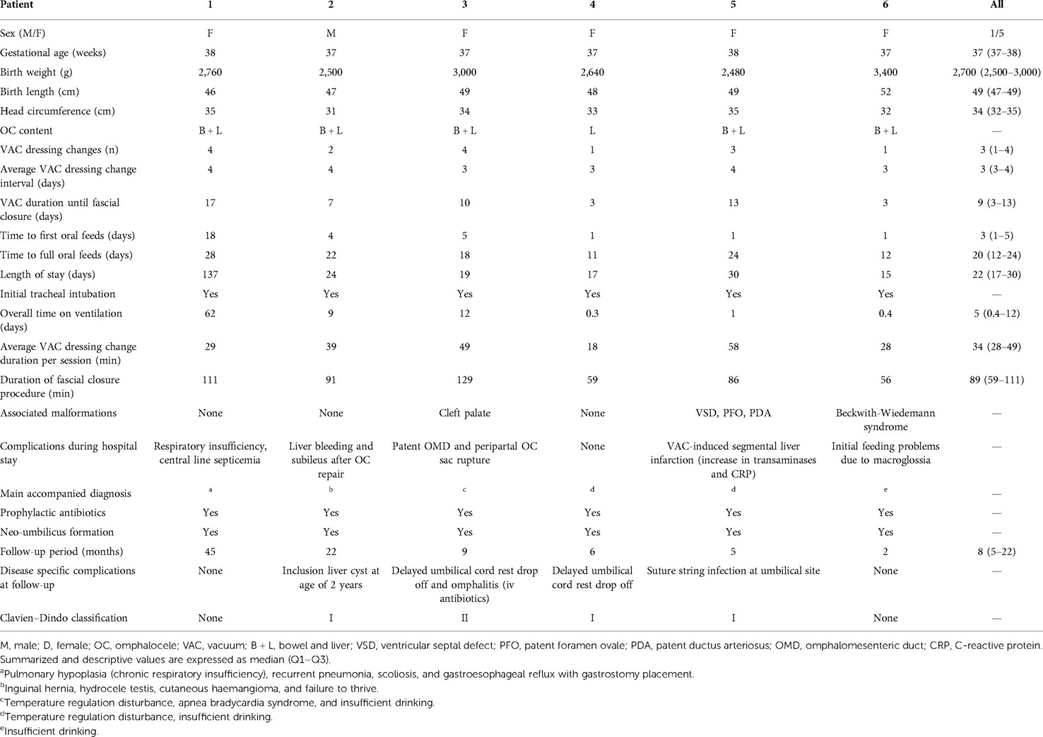
Results: Gestational age was 37 (37–38) weeks, and birth weight was 2700 (2500–3000) g. VAC dressing was changed every 3 (3–4) days until abdominal fascia closure at the age of 9 (3–13) days. Time to first/full oral feeds was 3 (1–5)/20 (12–24) days with a hospital stay of 22 (17–30) days. Follow-up was 8 (5–22) months and complications were of minor extent (none: n = 2; Clavien–Dindo I: n = 3; Clavien–Dindo II: n = 1), comprising a delayed neo-umbilical cord rest separation (n = 2) and/or concomitant neo-umbilical site infection (n = 2) with no repeat surgery.
Conclusion: In neonates with giant omphalocele, VAC constitutes a promising and technically feasible enhancement of the staged gravitational reduction method. This study shows evidence that VAC may accelerate restoration of the abdominal wall integrity in giant omphalocele, thus minimizing associated comorbidities inherent to a prolonged hospitalization.
Introduction
Omphalocele (OC) represents the most common entity of congenital abdominal wall defects. In OC, eviscerated abdominal organs herniate through the umbilical cord which is covered by a three-layered membrane consisting of peritoneum, Wharton's jelly, and amnion. The extent of evisceration often correlates with the abdominal wall defect size and may comprise intestine and solid organs as liver, spleen, bladder, or gonads. The current prevalence of OC is 1.24 per 10.000 live births (1). The primary intention in OC treatment is a timely return of abdominal organs back into the peritoneal cavity and closure of fascial and skin defect without compromising the visceral and systemic perfusion. In contrast to the treatment of giant OC, which are generally defined by a defect size larger than 5 cm and/or a herniation of more than 50% of liver, minor OC may often be managed by primary closure (2, 3). Giant OC often requires gradual reduction of the external peritoneal viscera in order to minimize the risk for cardiopulmonary complications prior to either surgical tension-free abdominal fascial closure or a nonoperative closure allowing for epithelization of the OC sac with delayed fascial closure. Recently, vacuum (VAC) therapy has been introduced for the treatment of complicated cases in OC (4–11). However, VAC therapy has mostly been implemented after failure of another primary OC repair method with only one case series by Aldridge et al. (12) in which VAC therapy was utilized by primary intention within first two days of life as an adjunct to the staged gravitational OC reduction until complete abdominal reintegration of giant OC with time to fascial closure lasting several months. In the present study, we aimed for describing our experiences with the staged OC reduction approach in adjunct with primary vacuum application started within first hours of life.
Patients and methods
Patients
This study comprises a retrospective two-institutional chart review of six neonates born between September 2018 and May 2022 with giant omphalocele [International Classification of Diseases, 10th Revision (ICD-10-GM); code Q79.2] who received a staged gravitational reduction of extracoelomic OC contents in combination with VAC therapy at the Departments of Pediatric Surgery in Munich (Bavaria, Germany; n = 2) and Witten (North Rhine-Westphalia, Germany; n = 4). Any minor OC amenable to primary reduction was considered a criterion for exclusion. Main outcome measure was the feasibility of VAC therapy in OC patients. Secondary outcome measures were demographic or procedural parameters as time to first or full (defined as the entire nutritional calories obtained enterally) enteral feeds. All VAC procedures were performed under either the first or last authors' supervision; both of which were familiar with the technique. Follow-up investigation was performed either by outpatient consultation or by personal communication. For quantification of complications, the Clavien–Dindo Classification was applied, as described elsewhere (13). This classification consists of seven grades (I, II, IIIa, IIIb, IVa, IVb, and V) and objectifies the therapy necessary to correct a specific complication. In the study, only grade I and II complications were observed with grade I representing any postoperative course deviation from normal without further interventions (i.e., surgery with the exception of wound infections opened at the bedside) and grade II complications representing any complications requiring pharmacological treatment with drugs different to that allowed for grade I complications, including the use of antibiotics. This study was approved by the Ethics Committee of Ruhr-University Bochum (registry no. 22-7547-MPG, date of approval: 06/21/2022) and parental consent was obtained in each case.
Methods
Technique
All patients included had prenatal diagnosis of OC and they were specially referred to the treating centers for delivery. Upon delivery, each patient was primarily taken care of by a consultant-level neonatologist and was immediately covered from neck downward with a sterile bowel bag (20″ × 20″; Steri-Drape™; 3M Deutschland GmbH Health Care Business, Neuss, Germany) followed by elective intubation with subsequent mechanical ventilation. After induction of anesthesia, integrity of OC was either confirmed or restored, as in the case of sac rupture. The VAC technique consisted of the following steps:
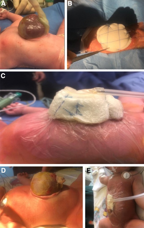
(1) White Foam (Vivano®Med; Mondomed NV, HAMONT-Achel, Belgium) was attached to the OC surface (Figure 1A) in a cylindric shape after trimming the umbilical cord.
(2) Cutaneous sagittal and horizontal tight four-point-fixation (traction sutures) of the foam cylinder (Figure 1B).
(3) Cavilon™ no sting barrier film (3M Deutschland GmbH Health Care Business, Neuss, Germany) application to degrease the skin.
(4) RENASYS Transparent Film adhesive drape (Smith & Nephew Orthopaedics GmbH, Tuttlingen, Germany) was applied covering the entire lower torso including the foam cylinder. By this, kinking of OC with consecutive vascular structure occlusion was prevented.
(5) RENASYS Soft Port connector (Smith & Nephew Orthopaedics GmbH, Tuttlingen, Germany) was attached to the adhesive foil.
(6) RENASYS TOUCH Device (Smith & Nephew Orthopaedics GmbH, Tuttlingen, Germany) was activated and a permanent negative pressure of −80 mmHg was exerted (Figure 1C).

Figure 1. (A) Giant omphalocele with eviscerated bowel and liver. (B) Cutaneous sagittal and horizontal four-point-fixation of foam cylinder. (C) Lateral view on attached VAC device. (D) Omphalocele after removal of VAC dressing. (E) Lateral view at VAC device after amnial plication maneuver and foam application just above the skin level.
There was no discontinuation of VAC therapy other than during VAC dressing changes (Figure 1D). VAC dressing change intervals were chosen at the surgeons' discretion. Procedure of VAC changing included a further reduction of extracoelomic contents with the amnial plication maneuver and a complete coverage of OC surface with foam. In all patients, amnial plication of OC contents was performed under continuous cardiorespiratory monitoring. Definite closure of fascia was carried out when all OC contents were reduced within the abdominal cavity without cardiopulmonary compromise or signs of abdominal compartment syndrome. Closure included removal of the OC membrane at the fascial level with consecutive vertical midline closure of fascia using VICRYL™ 2–0 (Johnson & Johnson Medical GmbH, Ethicon Deutschland, Norderstedt, Germany) sutures and skin closure with either continuous subcuticular running sutures MONOCRYL™ 5–0 (Johnson & Johnson Medical GmbH, Ethicon Deutschland, Norderstedt, Germany) or LEUKOSTRIP© S (4 × 38 mm; Smith & Nephew Orthopaedics GmbH, Tuttlingen, Germany). Neo-umbilicoplasty was performed at the lowest edge of the abdominal wall defect by preserving a 1–2 cm wide stripe of OC including the ligated umbilical arteries followed by a z-omphaloplasty, as adapted from Michel et al. (14). After abdominal wall closure, VAC therapy was continued at the skin level (Figure 1E) and then removed after 3–6 days.
Data analysis
Sampling and statistical analysis of data were performed using OriginPro 2021 (OriginLab, Northampton, MA, United States; RRID: SCR_014212). For descriptive statistics, the median and interquartile range was utilized. Categorical variables were presented as frequencies. The Kolmogorov–Smirnov test at a 0.05 significance level was used to confirm the normal distribution of numeric variables.
Results
Six patients underwent VAC-assisted reduction of OC with a female preponderance (n = 5; 83%). Individual basic demographic and procedural characteristics are enlisted in Table 1 and Supplementary Figure S1. Gestational age was 37 (37–38) weeks. Birth weight, length, and head circumference were 2700 (2500–3000) g, 49 (47–49) cm, and 34 (32–35) cm, respectively. The amount of VAC dressing procedures was 3 (1–4) per patient with a dressing change interval of 3 (3–4) days and a VAC therapy duration was 9 (3–13) days until tension-free closure of the abdominal fascia and skin. First oral feeding was initiated on day 3 (1–5) of life and full oral feeding was achieved at the age of 20 (12–24) days. Length of stay (LOS) was 22 (17–30) days. All patients were primarily intubated with 5 (0–12) days on ventilation. Prophylactic antibiotics were administered in each case, as was the creation of a neo-umbilicus. In patient 3, OC membrane rupture was repaired by continuous sutures and a patent omphalomesenteric duct (OMD) was ligated at its base. Postsurgical complications are enlisted in Table 1. The follow-up period was 8 (5–22) months and main complications were associated with neo-umbilical formation, namely, a delay in umbilical cord rest drop off (n =2) and/ or umbilical site infection (n = 2) with no case needing repeat surgery. Specifically, complications at follow-up were nonexistent (n = 2) or graded Clavien–Dindo I (n = 3) or Clavien–Dindo II (n = 1), respectively. All patients demonstrated an age-appropriate neurodevelopmental status and their weight, height, and head circumferences increased along normal centiles.

Table 1. Characteristics of patients undergoing vacuum-assisted staged reduction of giant omphalocele.
Discussion
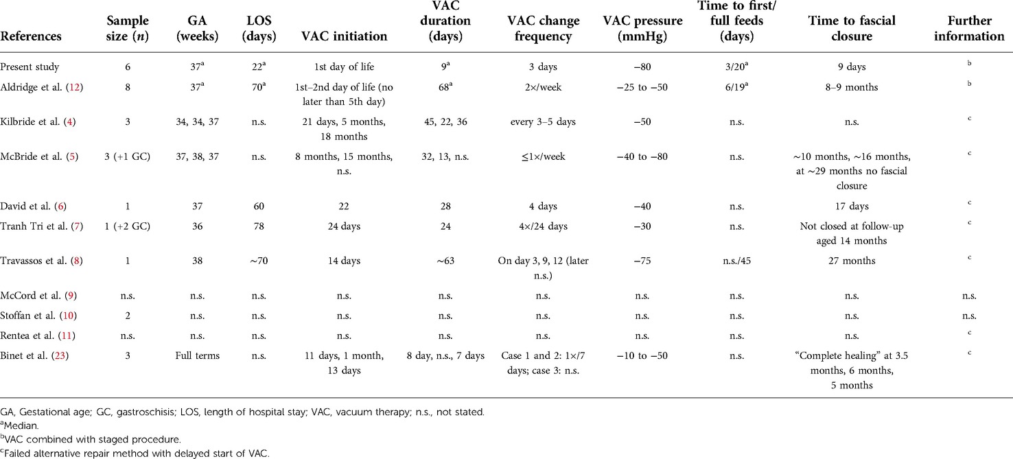
We present our preliminary experience with simultaneous VAC application as an improvement of the staged gravitational reduction technique in six patients with giant OC. No consensus as to the preferable surgical treatment of giant OC exists, which is mainly due to the heterogeneity of applied methods (15). Since its introduction in 1997 (16, 17), the vacuum technique has been subject to a large spectrum of applications in the adult population. However, it has not yet proven the same efficacy and safety in children (18). Nonetheless, the literature on the efficacy of vacuum therapy seems promising in terms of the treatment of complicated pediatric wounds as pressure ulcers, extremity wounds, surgical wound dehiscence, skin grafting, or complex abdominal defects (9–11, 19–21) and also congenital abdominal wall defects (5, 22). A review of the literature on VAC application associated with omphalocele revealed that VAC has mostly been implemented as secondary salvage therapy after failure of another primary OC repair method (4–11, 23) (Table 2). It is worth mentioning that reported cohort sizes were small ranging from one to three patients or were not reported at all (9, 11). Time to abdominal fascial closure ranged from 17 days to ∼29 months. Time to full enteral feeds was not documented in all cases, with only one exception (8). LOS ranged from 60 to 78 days (6–8, 12) or was also not reported (4, 5, 9–11, 23).
Presented procedure has only been reported once by another group (12) (Table 2) in an equivalent setting, characterized by eight patients of similar gestational age with start of VAC therapy within the first two days of life and comparable VAC change intervals, but with much longer duration until fascial closure (8–9 months vs. 9 days in our study) and LOS (70 days vs. 22 days in our study). Obtained durations until first and full oral feeding were comparable to the results obtained by Aldridge et al. (6 and 19 days vs. 3 and 20 days in our study). Noteworthy, in the present study, exerted vacuum levels were higher (−80 mmHg) than those used by Aldridge et al. (−25 to −50 mmHg). In this context, recommended negative pressure setting in congenital abdominal wall defects ranges from −50 to −75 mmHg (4, 24, 25). By vacuum levels exerted more positive than a certain threshold, an insufficient or decelerated reintegration of visceral contents could occur. This might be one explanation for the comparably fast reduction of eviscerated OC contents in our study compared to that by Aldridge et al. Of note, higher vacuum levels for OC reduction may also induce higher transient intra-abdominal pressure levels with increased risk for cardiorespiratory depression. However, we did not observe any signs of cardiorespiratory compromise within our continuously monitored cohort.
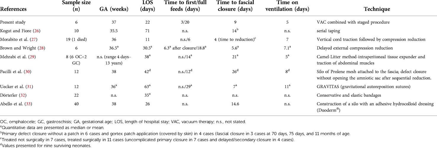
Comparing the different treatment strategies for giant omphaloceles (26–33) (Table 3) and the presented method, LOS was only shorter in one series by Morabito et al. (27), using the vertical cord traction followed by the compression reduction method. The time to full oral feeds of 20 days in our VAC method was slightly longer than in most of the comparative studies (27–30), or was not reported (26, 32, 33). With regard to time until fascial closure, only three groups (24) reported shorter periods than in the present study, ranging from 4 to 7 days utilizing vertical cord traction followed by compression reduction method (27), the delayed external compression reduction method (28), or the gravitational autoreposition suture method (31), respectively. In concordance with our data, Mehrabi et al. (29) also elicited a ventilation duration of 5 days by utilizing an intraperitoneal tissue expander and traction of abdominal muscles (camel-litter method) in six OC patients. In all other studies, durations of ventilation were longer, ranging from 7 to 11 days (27, 28, 30, 31), or were not even mentioned (26, 32, 33). Noteworthy, except one study by Abello et al. (33) utilizing a constructed silo with an adhesive hydrocolloid dressing in 40 neonates, neither of the reported studies had large sample sizes (ranging from 6 to 22 patients) and therefore associated malformations as pulmonary insufficiency may have large impact on average procedural parameters, as was the case in our first patient with pulmonary hypoplasia (PH) (Table 1). By excluding patient 1, median duration of ventilation decreased from 5 (0–12) days to 1 (0–9) day only. Over the course of the study, the individual ventilation duration decreased (supplementary Figure S1B), reflecting a possible learning curve. However, this has to be confirmed by larger studies.

Table 3. Literature data on biometric and procedural parameters for different treatment strategies regarding giant omphaloceles.
The presented VAC method combines some advantages, as being noninvasive in terms of OC sac preservation until fascial closure. Thus, the achieved intestinal nontouch procedure also diminishes risk for development of intra-abdominal adhesions. In addition, VAC application promotes a gentle pressure elevation by intra-abdominal dead space obliteration with a decreased risk for cardiorespiratory compromise. It is important to mention that the VAC method may reach its limits in those 36%–57% (34, 35) of cases with giant OC that present with associated pulmonary hypertension or PH. Since this condition may worsen with increasing abdominal pressure, a careful cardiorespiratory monitoring under reduction of OC contents is mandatory. Given that our method was well tolerated in patient 1 with PH, we do not consider this comorbidity an exclusion criterion at this early stage of experience.
In our series, the VAC method permitted an early enteral feeding. In general, VAC is supposed to minimize bacterial biioburden (24). Consecutive VAC changes might be performed under less invasive (awake caudal) modes of anesthesia without intubation at bedside, thus lowering the risk for associated side effects in this vulnerable cohort. Furthermore, the presented method can easily be learned and may be advantageous in case of complications, such as a patent OMD or an OC sac rupture as seen in patient 3. By reducing time until fascial closure, risks for associated morbidities inherent to a prolonged hospitalization in this delicate age group may be mitigated by presented VAC method. Finally, repair of OC with simultaneous neo-umbilicoplasty is of high relevance regarding the patient's satisfaction and self-identification (36–38). In general, an umbilicus located in the midline at two-thirds of the distance from the symphysis to xiphoid is considered cosmetically acceptable (39). In our series, the position of the neo-umbilicus was dictated by the umbilical arteries and thus at the anatomically correct position.
Limitations
The main limitations of this preliminary report were the small cohort size and the limited follow-up period. As a consequence, randomized controlled trials comparing the presented VAC technique to other OC reduction techniques are needed.
Conclusions
In neonates with giant OC, VAC constitutes a promising and technically feasible enhancement of the staged gravitational reduction method when primary closure seems questionable. Even in complicated cases with OC membrane defects or associated intestinal malformations, the presented technique seems to be safe and effective. Only in one previous series, VAC has been used in a similar manner for primary OC treatment, but with longer time until fascial closure. In summary, this study underlines the importance of VAC as an enhancement enabling an accelerated restoration of abdominal wall integrity in neonates with giant OC within their first 2 weeks of life, thus minimizing associated comorbidities inherent to a prolonged hospitalization. Therefore, we provide evidence that VAC is a promising technique to improve the treatment of OC and should be investigated further in future studies.
Data availability statement
The raw data supporting the conclusions of this article will be made available by the authors, without undue reservation.
Ethics statement
The studies involving human participants were reviewed and approved by Ethics Committee of Ruhr-University Bochum (registry no. 22-7547-MPG, date of approval: 06/21/2022). Written informed consent to participate in this study was provided by the participants' legal guardian/next of kin. Written informed consent was obtained from the individual(s), and minor(s)' legal guardian/next of kin, for the publication of any potentially identifiable images or data included in this article.
Author contributions
MN designed study, collected and analyzed data, treated patients, and wrote the manuscript. AR collected data, treated patients, and wrote parts of the manuscript. EW collected data, treated patients, and conceptualized and critically revised the manuscript. LP and MA treated patients and critically revised the manuscript. JH evolved method, conceptualized study, treated patients, and critically revised the manuscript. All authors contributed to the article and approved the submitted version.
Funding
We acknowledge support by the Open Access Publication Funds of the Ruhr-Universität Bochum.
Conflict of interest
The authors declare that the research was conducted in the absence of any commercial or financial relationships that could be construed as a potential conflict of interest.
Publisher's note
All claims expressed in this article are solely those of the authors and do not necessarily represent those of their affiliated organizations, or those of the publisher, the editors and the reviewers. Any product that may be evaluated in this article, or claim that may be made by its manufacturer, is not guaranteed or endorsed by the publisher.
Supplementary material
The Supplementary Material for this article can be found online at: https://www.frontiersin.org/articles/10.3389/fped.2022.1053568/full#supplementary-material.
References
1. EUROCAT: Prevalence Charts and Tables—Prevalence per 10,000 Births. Omphalocele—2013 to 2020. Available online: https: //eu-rd-platform.jrc.ec.europa.eu/eurocat/eurocat-data/prevalence_en (Accessed: July 18, 2022).
2. van Eijck FC, Hoogeveen YL, van Weel C, Rieu PNMA, Wijnen RMH. Minor and giant omphalocele: long-term outcomes and quality of life. J Pediatr Surg. (2009) 44:1355–9. doi: 10.1016/j.jpedsurg.2008.11.034
3. Campos BA, Tatsuo ES, Miranda ME. Omphalocele: how big does it have to be a giant one? J Pediatr Surg. (2009) 44:1474–5. doi: 10.1016/j.jpedsurg.2009.02.060
4. Kilbride KE, Cooney DR, Custer MD. Vacuum-assisted closure: a new method for treating patients with giant omphalocele. J Pediatr Surg. (2006) 41:212–5. doi: 10.1016/j.jpedsurg.2005.10.003
5. McBride CA, Stockton K, Storey K, Kimble RM. Negative pressure wound therapy facilitates closure of large congenital abdominal wall defects. Pediatr Surg Int. (2014) 30:1163–8. doi: 10.1007/s00383-014-3545-3
6. David VL, Neagu MC, Sosoi A, Stanciulescu MC, Horhat FG, Stroescu RF, et al. Combined staged surgery and negative-pressure wound therapy for closure of a giant omphalocele. Case Rep Pediatr. (2021) 2021:5234862. doi: 10.1155/2021/5234862
7. Thanh Tri T, Minh Duc N, Phi Duy H, Thanh Thien N, Nguyen An Thuan L, et al. A case series describing vacuum-assisted closure for complex congenital abdominal wall defects. Clin Ter. (2021) 172:273–7. doi: 10.7417/CT.2021.2331
8. Travassos D, van Eerde A, Kramer W. Management of a giant omphalocele with non–cross-linked intact porcine-derived acellular dermal matrix (strattice) combined with vacuum therapy. European J Pediatr Surg Rep. (2015) 03:061–3. doi: 10.1055/s-0035-1549364
9. McCord SS, Naik-Mathuria BJ, Murphy KM, McLane KM, Gay AN, Bob Basu C, et al. Negative pressure therapy is effective to manage a variety of wounds in infants and children. Wound Repair Regen. (2007) 15:296–301. doi: 10.1111/j.1524-475X.2007.00229.x
10. Stoffan AP, Ricca R, Lien C, Quigley S, Linden BC Use of negative pressure wound therapy for abdominal wounds in neonates and infants. J Pediatr Surg. (2012) 47:1555–9. doi: 10.1016/j.jpedsurg.2012.01.014
11. Rentea RM, Somers KK, Cassidy L, Enters J, Arca MJ Negative pressure wound therapy in infants and children: a single-institution experience. J Surg Res. (2013) 184:658–64. doi: 10.1016/j.jss.2013.05.056
12. Aldridge B, Ladd AP, Kepple J, Wingle T, Ring C, Kokoska ER. Negative pressure wound therapy for initial management of giant omphalocele. Am J Surg. (2016) 211:605–9. doi: 10.1016/j.amjsurg.2015.11.009
13. Dindo D, Demartines N, Clavien P-A. Classification of surgical complications. Ann Surg. (2004) 240:205–13. doi: 10.1097/01.sla.0000133083.54934.ae
14. Michel JL, Kassir R, Harper L, Gavage L, Frade F, Clermidi P, et al. ZORRO: z omphaloplasty repair for omphalocele. J Pediatr Surg. (2018) 53:1424–7. doi: 10.1016/j.jpedsurg.2018.04.003
15. van Eijck FC, Aronson DA, Hoogeveen YL, Wijnen RMH. Past and current surgical treatment of giant omphalocele: outcome of a questionnaire sent to authors. J Pediatr Surg. (2011) 46:482–8. doi: 10.1016/j.jpedsurg.2010.08.050
16. Argenta LC, Morykwas MJ. Vacuum-assisted closure: a new method for wound control and treatment: clinical experience. Ann Plast Surg. (1997) 38:563–76. doi: 10.1097/00000637-199706000-00002
17. Morykwas MJ, Argenta LC, Shelton-Brown EI, McGuirt W. Vacuum-assisted closure: a new method for wound control and treatment. Ann Plast Surg. (1997) 38:553–62. doi: 10.1097/00000637-199706000-00001
18. Santosa KB, Keller M, Olsen MA, Keane AM, Sears ED, Snyder-Warwick AK. Negative-pressure wound therapy in infants and children: a population-based study. J Surg Res. (2019) 235:560–8. doi: 10.1016/j.jss.2018.10.043
19. McGarrah B. Using negative pressure therapy for wound healing in the extremely low-birth-weight infant (micropreemie). J Wound Ostomy Continence Nurs. (2015) 42:409–12. doi: 10.1097/WON.0000000000000139
20. Lopez G, Clifton-Koeppel R, Emil S. Vacuum-assisted closure for complicated neonatal abdominal wounds. J Pediatr Surg. (2008) 43:2202–7. doi: 10.1016/j.jpedsurg.2008.08.067
21. Gutierrez IM, Gollin G. Negative pressure wound therapy for children with an open abdomen. Langenbecks Arch Surg. (2012) 397:1353–7. doi: 10.1007/s00423-012-0923-y
22. Choi WW, McBride CA, Kimble RM. Negative pressure wound therapy in the management of neonates with complex gastroschisis. Pediatr Surg Int. (2011) 27:907–11. doi: 10.1007/s00383-011-2868-6
23. Binet A, Gelas T, Jochault-Ritz S, Noizet O, Bory JP, Lefebvre F, et al. VAC® therapy a therapeutic alternative in giant omphalocele treatment: a multicenter study. J Plast Reconstr Aesthet Surg. (2013) 66:e373–5. doi: 10.1016/j.bjps.2013.05.010
24. Baharestani M, Amjad I, Bookout K, Fleck T, Gabriel A, Kaufman D, et al. V.A.C.® therapy in the management of paediatric wounds: clinical review and experience. Int Wound J. (2009) 6:1–26. doi: 10.1111/j.1742-481X.2009.00607.x
25. Gabriel A, Gollin G. Management of complicated gastroschisis with porcine small intestinal submucosa and negative pressure wound therapy. J Pediatr Surg. (2006) 41:1836–40. doi: 10.1016/j.jpedsurg.2006.06.050
26. Kogut KA, Fiore NF. Nonoperative management of giant omphalocele leading to early fascial closure. J Pediatr Surg. (2018) 53:2404–8. doi: 10.1016/j.jpedsurg.2018.08.018
27. Morabito A, Owen A, Bianchi A. Traction-compression-closure for exomphalos major. J Pediatr Surg. (2006) 41:1850–3. doi: 10.1016/j.jpedsurg.2006.06.044
28. Brown MF, Wright L. Delayed external compression reduction of an omphalocele (DECRO): an alternative method of treatment for moderate and large omphaloceles. J Pediatr Surg. (1998) 33:1113–6. doi: 10.1016/S0022-3468(98)90542-5
29. Mehrabi V, Mehrabi A, Kadivar M, Soleimani M, Fallahi A, Khalilzadeh N. Staged repair of giant recurrent omphalocele and gastroschesis “camel-litter method”—a new technique. Acta Med Iran. (2012) 50:388–94. PMID: 2283711722837117
30. Pacilli M, Spitz L, Kiely EM, Curry J, Pierro A. Staged repair of giant omphalocele in the neonatal period. J Pediatr Surg. (2005) 40:785–8. doi: 10.1016/j.jpedsurg.2005.01.042
31. Uecker M, Petersen C, Dingemann C, Fortmann C, Ure BM, Dingemann J. Gravitational autoreposition for staged closure of omphaloceles. Eur J Pediatr Surg. (2020) 30:045–50. doi: 10.1055/s-0039-1693727
32. Dörterler ME. Management of giant omphalocele leading to early fascial closure. Cureus. (2019) 11(10):e5932. doi: 10.7759/cureus.5932
33. Abello C, Harding CA, Rios AP, Guelfand M. Management of giant omphalocele with a simple and efficient nonsurgical silo. J Pediatr Surg. (2021) 56:1068–75. doi: 10.1016/j.jpedsurg.2020.12.003
34. Liu TX, Du LZ, Ma XL, Chen Z, Shi LP. Giant omphalocele associated pulmonary hypertension: a retrospective study. Front Pediatr. (2022) 10:940289. doi: 10.3389/fped.2022.940289
35. Hutson S, Baerg J, Deming D st, Peter SD, Hopper A, Goff DA. High prevalence of pulmonary hypertension complicates the care of infants with omphalocele. Neonatology. (2017) 112:281–6. doi: 10.1159/000477535
36. Koivusalo A, Lindahl H, Rintala RJ. Morbidity and quality of life in adult patients with a congenital abdominal wall defect: a questionnaire survey. J Pediatr Surg. (2002) 37:1594–601. doi: 10.1053/jpsu.2002.36191
37. Davies BW, Stringer MD. The survivors of gastroschisis. Arch Dis Child. (1997) 77:158–60. doi: 10.1136/adc.77.2.158
38. Lindham S. Long-Term results in children with omphalocele and gastroschisis—a follow-up study. Eur J Pediatr Surg. (1984) 39:164–7. doi: 10.1055/s-2008-1044202
Keywords: giant omphalocele, congenital abdominal wall defect, staged omphalocele reduction, vacuum therapy, VAC
Citation: Nissen M, Romanova A, Weigl E, Petrikowski L, Alrefai M and Hubertus J (2022) Vacuum-assisted staged omphalocele reduction: A preliminary report. Front. Pediatr. 10:1053568. doi: 10.3389/fped.2022.1053568
Received: 25 September 2022; Accepted: 28 October 2022;
Published: 24 November 2022.
Edited by:
Pablo Andrés Lobos, Italian Hospital of Buenos Aires, ArgentinaReviewed by:
Luzia Toselli, Fundacion Hospitalaria, ArgentinaEmmanuelle Seguier, Meir Medical Center, Israel
© 2022 Nissen, Romanova, Weigl, Petrikowski, Alrefai and Hubertus. This is an open-access article distributed under the terms of the Creative Commons Attribution License (CC BY). The use, distribution or reproduction in other forums is permitted, provided the original author(s) and the copyright owner(s) are credited and that the original publication in this journal is cited, in accordance with accepted academic practice. No use, distribution or reproduction is permitted which does not comply with these terms.
*Correspondence: Matthias Nissen matthias.nissen@rub.de
Specialty Section: This article was submitted to Pediatric Surgery, a section of the journal Frontiers in Pediatrics
 Matthias Nissen
 Anna Romanova1