- 1Department of Traditional Chinese Medicine, Shenzhen Children's Hospital, Shenzhen, China
- 2Department of Pediatrics, Beijing Anzhen Nanchong Hospital of Capital Medical University & Nanchong Central Hospital, Nanchong, Sichuan, China
- 3School of Basic Medicine and Clinical Pharmacy, China Pharmaceutical University, Nanjing, China
Background: Childhood asthma has a significant effect on growth and development. Traditional Chinese Medicine (TCM) has notable advantages in asthma treatment; however, a modern scientific basis for the differentiation of cold and heat syndromes in asthma remains lacking.
Methods: This study employed non-targeted metabolomics to analyze the plasma metabolic profiles in children aged 5–14 years with cold or heat syndrome asthma. Plasma metabolites were examined to identify and compare metabolic differences among children with asthma and healthy controls, as well as between cold and heat asthma syndromes, with the aim of uncovering potential biomarkers and providing a foundation for differential diagnosis.
Results: Of the 92 participants, 48 had cold syndrome asthma, 14 had heat syndrome asthma, and 30 were healthy controls. A total of 50 differential plasma metabolites were identified between the TCM asthma syndrome groups and healthy controls in both positive and negative ion modes. These metabolites were primarily phospholipids and amino acids enriched in the lipid metabolism, amino acid metabolism, and glucose metabolism pathways. Furthermore, 18 differential metabolites were identified between the cold and heat asthma groups, with significant enrichment in the amino acid metabolic pathways. Notably, 36 common differential metabolites that mainly were lipids, amino acids and its related metabolites between cold asthma and heat asthma, cold asthma and the healthy group, and heat asthma and the healthy group were identified of which can be considered as biomarkers.
Conclusions: Lipids, amino acids, and their associated metabolic pathways have been identified as potential biomarkers for distinguishing cold and heat asthma syndromes in children. These findings contribute to the modern interpretation of TCM syndrome differentiation and may support the evaluation of the therapeutic effects of TCM-based asthma treatment.
1 Introduction
Bronchial asthma, commonly referred to as asthma, is the most prevalent chronic respiratory disease in children. Data show that its incidence is increasing annually worldwide (1, 2). However, owing to its recurrent nature, it seriously affects the physical and mental health of children and imposes a heavy economic burden on both families and society (3). Moreover, the pathogenesis of asthma is diverse and complex, influenced by climate change, environmental pollution, and genetic heterogeneity (4, 5), and involves chronic airway inflammation (6), immune responses, airway hyperresponsiveness, and genetic factors (7). Currently, the management and control of asthma remain inadequate, particularly in low-resource settings (2). Therefore, this issue warrants extensive research.
In Traditional Chinese Medicine (TCM) theory, asthma is classified under the category of “Xiao disease.” According to TCM guidelines and expert consensus, pediatric asthma is a recurrent lung disease characterized by wheezing, coughing, and rales in the throat. Clinical episodes present symptoms, such as throat rales, shortness of breath, coughing, chest tightness, and prolonged expiration. In severe cases, patients may be unable to lie down and may experience dyspnea, open-mouth breathing, shoulder lifting, body shaking, lip cyanosis, and irritability. Symptoms often occur or worsen in the early morning or at night (8–10). This disease corresponds to bronchial asthma in children in Western medicine (10). Asthma can be divided into three distinct phases: exacerbation, relocation, and remission. The exacerbation phase is characterized by a sudden onset of symptoms, such as wheezing, coughing, shortness of breath, chest tightness, or the rapid worsening of existing symptoms. This phase corresponds to the acute attack stage in Western medicine, and includes cold asthma syndrome, heat asthma syndrome, and external cold with internal heat syndrome (8). Cold and heat asthma are the most common clinical phenotypes.
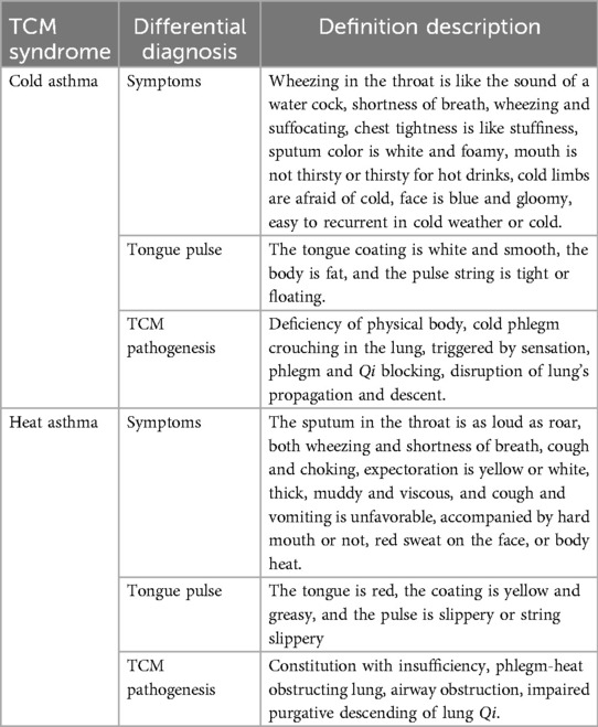
The TCM differentially diagnostic criteria for cold asthma and heat asthma can be seen in Table 1 (8, 11). Compared to adults, TCM holds that children have immature organs and insufficient Qi, making them more vulnerable to external pathogens. The Plain Questions—Theory of Wind states, “Wind is the root of all diseases.” In TCM theory, “wind evil” is a type of pathogenic xie Qi, characterized by sudden onset, rapid changes, and migration of symptoms—resembling natural wind. When wind evil invades the human body, it can affect organs such as the lungs, spleen, and kidneys, leading to various diseases. Wind-related syndromes can be classified as external (originating from nature) or internal (resulting from visceral dysfunctions). In children, the lungs regulate fluid distribution, the spleen governs transportation and transformation, and the kidneys lack the ability to efficiently vaporize fluids. These deficiencies contribute to phlegm accumulation and the formation of asthma roots. Clinically, TCM categorizes asthma into cold and heat syndromes based on these manifestations (12). TCM has been widely used to treat asthma (13). Despite its notable efficacy and safety, a universally accepted TCM diagnostic standard is currently lacking. Therefore, a comprehensive and scientific explanation of the nature and connotations of TCM syndromes is essential.
Metabolomics is the study of the types, quantities, and variations in endogenous metabolites in organisms following stimulation or disturbance (14). Metabolites are the end products of gene and protein activities and can promptly, sensitively, and accurately reflect the functional state of an organism in response to external stimuli. As such, they are considered “biological phenotypes” of physiological functions (15). In recent years, increasing evidences have shown that metabolomics can be applied to study the pathogenesis, diagnosis, and treatment of asthma and other diseases (16–20). Some studies have applied metabolomics to explore the TCM syndromes of asthma (12), indicating significant metabolic disturbances in patients with asthma. However, few studies have specifically focused on TCM syndromes in pediatric asthma. Therefore, investigating potential biomarkers associated with TCM asthma syndromes is of great significance for clinical diagnosis.
In this study, we aimed to identify plasma biomarkers in children with asthma during acute exacerbation and screen for differential metabolites that may help elucidate the pathogenesis of asthma. We recruited children diagnosed with cold and heat asthma syndromes based on the TCM criteria from Shenzhen Children's Hospital, along with healthy children as a control group. Plasma samples were collected and analyzed using liquid chromatography–mass spectrometry (LC-MS)-based metabolomics.
2 Materials and methods
2.1 Study population and study design
A total of 62 patients aged 5–14 years with asthma were recruited from Shenzhen Children's Hospital between January 2016 and September 2017. Asthma subtypes were classified based on the diagnostic criteria for pediatric bronchial asthma and TCM syndrome classification for pediatric asthma. The final diagnosis was made through unanimous agreement between the two chief physicians.
The exclusion criteria were as follows: (1) patients whose diagnosis did not meet the inclusion criteria; (2) those with confirmed congenital or acquired metabolic disorders; (3) patients with comorbid conditions, such as toxic encephalopathy, respiratory failure, or heart failure; (4) individuals with diagnosed mental illness; (5) patients with severe primary diseases affecting the heart, liver, kidneys, or hematopoietic system; and (6) patients undergoing treatment with glucocorticoids, immunosuppressants, or participating in clinical drug trials.
Thirty age- and sex-matched children without any evidence of asthma were recruited as healthy controls. This study was approved by the Ethics Committee of Shenzhen Children's Hospital. Written informed consent was obtained from all participants and their legal guardians.
2.2 Sample collection and processes
Two milliliters of morning fasting venous blood were collected from each participant using a disposable heparin anticoagulant tube. The samples were centrifuged at 3,000 rpm for 10 min, and the resulting plasma was transferred to cryopreservation tubes and stored at −80 °C to avoid repeated freeze–thaw cycles.
Prior to the analysis, the plasma samples were thawed at room temperature. Then, 40 μl of plasma was transferred to a new tube and 120 μl of cold precipitant (dichloromethane/methanol, 3:1, v/v) was added. The mixture was vortexed for 1 min, left to stand at room temperature for 10 min, and stored at −20 °C overnight. The next day, the samples were centrifuged at 4,000 rpm for 30 min at 4 °C. From each sample, 25 μl of the supernatant was transferred to a new centrifuge tube and 225 μl of a lipid complex diluent (isopropanol:acetonitrile:water, 2:1:1, v/v/v) was added.
For quality control (QC), 20 μl of each sample was pooled to obtain a QC sample. Before initiating the analysis, ten QC samples were run to equilibrate the instrument. Subsequently, one QC sample was injected for every 10 test samples, and three QC samples were run at the end of the sequence to monitor the system stability.
2.3 Untargeted metabolomics analysis
Untargeted metabolomic analysis was performed using liquid chromatography coupled with mass spectrometry. Chromatographic separation was carried out on an ACQUITY Ultra-Performance Liquid Chromatography (UPLC) system (2777C, Waters, UK). The samples were analyzed in the order determined by the instrument sequence. A reversed-phase ACQUITY UPLC HSS T3 column (100 mm × 2.1 mm, 1.8 μm, Waters, UK) was used. The column oven temperature was maintained at 40 °C. The mobile phases consisted of solvent A (water with 0.1% formic acid) and solvent B (acetonitrile with 0.1% formic acid) at a flow rate of 0.4 ml/min. Gradient elution was programmed as follows: 0–1 min, 99% A; 1–3 min, 1%–15% B; 3–6 min, 15%–50% B; 6–9 min, 50%–95% B; 9–10 min, 95% B; and 10.1–12 min, 99% A. The injection volume for each sample was 10 μl.
Mass spectrometry analysis was conducted using a Xevo G2-XS QTOF system (Waters, UK) operated in both the positive and negative ion modes. In positive ion mode, the capillary voltage and sample cone voltage were set to 0.25 kV and 40 V, respectively. In the negative-ion mode, the voltage was set to 2 kV and 40 V. Data were acquired in the centroid MSE mode. The TOF mass range was set to 50–1,200 Da with a scan time of 0.2 s. For MS/MS detection, all precursor ions were fragmented at 20–40 eV, with a scan time of 0.2 s. During the acquisition, a lock mass (LE) signal was collected every 3 s to ensure mass accuracy. In addition, one QC sample was analyzed every 10 experimental samples to monitor the stability and reproducibility of the LC-MS system.
2.4 Data collection and metabolite identification
Untargeted raw data were processed and identified using the commercial software, Progenesis QI (version 2.2). Local polynomial regression fitting for signal correction based on quality control (QC) sample data, referred to as QC-based robust LOESS signal correction (QC-RSC), was used to adjust the real sample signals. This method is recognized as an effective approach for signal correction in metabolomic data analyses (21). An internal standard, in combination with the QC-RSC approach, was used to calibrate the peak areas of the plasma samples. After normalization, the data acquired from both the positive and negative ion modes were merged into a single dataset for downstream metabolite analysis.
Multivariate statistical analyses, including principal component analysis (PCA) and partial least-squares discriminant analysis (PLS-DA), were performed using MetaboAnalyst or metaX software (22). The key performance metrics for evaluating the PLS-DA model included R2Y and Q2 values. R2Y indicates the proportion of variation in the dependent variable Y explained by the model, whereas Q2 reflects the predictive ability of the model. In general, R2Y and Q2 values closer to 1 suggest a more robust model, with Q2 > 0.5 indicating good predictive performance.
Variable Importance in Projection (VIP) scores were extracted from the PLS-DA model to evaluate the contribution of individual metabolites to the group discrimination. These VIP values were used to identify differentially expressed metabolites.
Univariate statistical analyses, including t-tests and fold-change analysis, were conducted to further evaluate metabolite differences. The resulting p-values were adjusted using the false discovery rate (FDR) method to obtain q-values. Differential ions were identified based on the combined criteria of VIP ≥1, fold-change ≥1.2 or ≤0.83, and q-value <0.05.
Differential metabolites identification were firstly identified via Progenesis QI (version2.2) software, and further validated using the Human Metabolome Database (HMDB, https://hmdb.ca/) (23). Metabolite candidates were refined based on MSE fragmentation data, and non-endogenous metabolites were excluded. Finally, a t-test was performed on the relative abundance of endogenous metabolites, and those with P < 0.05 were considered significantly different.
2.5 Pathway analysis
Pathway enrichment analysis was performed using the MetaboAnalyst database (https://www.metaboanalyst.ca/, version 4.0) (24). Metabolic pathway analysis was conducted to identify specific metabolites and explore the potential pathogenesis of bronchial asthma as well as the metabolic differences between the cold asthma and heat asthma groups.
3 Results
3.1 Participant characteristics
A total of 92 participants were included, comprising 62 children with asthma and 30 healthy controls. Among children with asthma, 48 were diagnosed with cold syndrome asthma (cold asthma group) and 14 with heat syndrome asthma (heat asthma group). Healthy children served as the controls.
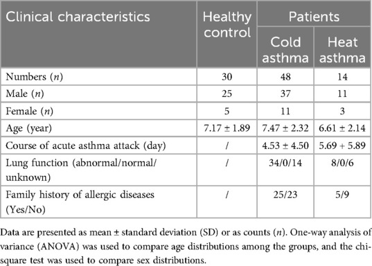
There were no significant differences between the groups in terms of age, sex, or clinical examination results. Additionally, no significant differences were observed in the duration of acute asthma exacerbation, lung function, or family history of allergic diseases (Table 2).
3.2 Plasma profile of non-targeted metabolomics analysis
Plasma metabolites were analyzed in the healthy control, cold asthma, and heat asthma groups. Principal component analysis in both positive and negative ion modes revealed a discernible separation trend between the healthy control group and the cold asthma group, as well as between the healthy control group and the heat asthma group. However, complete separation was not observed (Figures 1a,b,e,f). In contrast, PLS-DA demonstrated clear separation between the groups, indicating significant differences in plasma metabolite profiles between the healthy control group and the cold asthma group, as well as between the healthy control group and the heat asthma group during asthma exacerbation (Figures 1c,d,g,h).
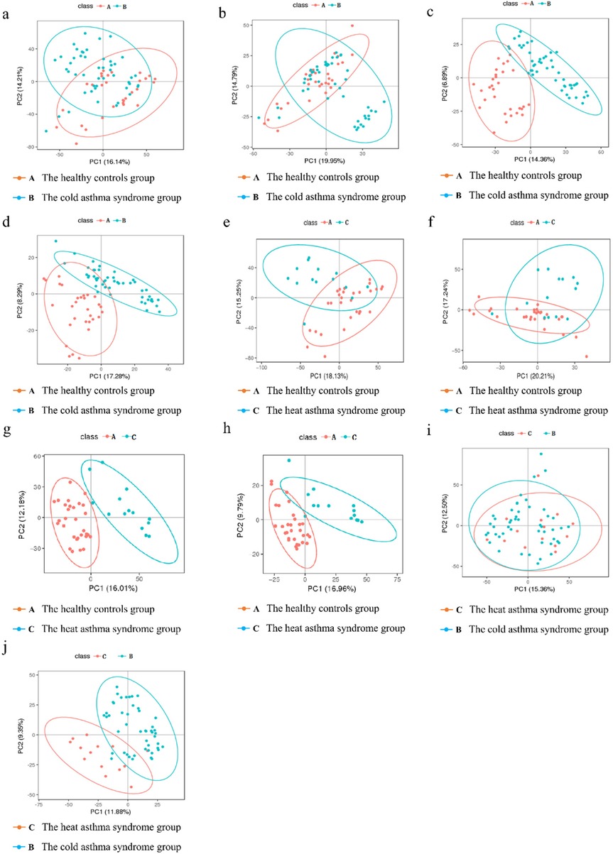
Figure 1. Multivariate analysis of plasma metabolites in different groups. (a,b) PCA plots in the positive ion mode (a) and negative ion mode (b) between the healthy controls group and the cold asthma group. (c,d) PLS-DA plots in positive ion mode [(c) R2Y = 0.819, Q2 = 0.683] and negative ion mode [(d) R2Y = 0.741, Q2 = 0.481] between the healthy controls and cold asthma groups. (e,f) PCA plots in positive ion mode (e) and negative ion mode (f) between the healthy controls group and the heat-asthma group. (g,h) PLS-DA plots in positive ion mode [(g) R2Y = 0.842, Q2 = 0.707] and negative ion mode [(h) R2Y = 0.812, Q2 = 0.575] between the healthy controls and heat asthma groups. (i,j) PCA (i) and PLS-DA [(j) R2Y = 0.616, Q2 = 0.627] plots between the cold and heat asthma groups in the positive ion mode. (In the figures, class A represents the healthy controls group; class B represents the cold asthma syndrome group; class C represents the heat asthma syndrome group). In the figure, PC1 and PC2 represent the first and second principal components, respectively. The number in parentheses represents the proportion of the principal component that can integrate the original information. The score graph reflects the distribution of each sample in the coordinate system composed of PC1 and PC2, mainly observing the trend of inter group separation.
Additionally, we compared the metabolite profiles of the cold and heat asthma groups. Few differential ions were identified in the negative ion mode; therefore, the subsequent analysis focused on the positive ion mode. PCA revealed no obvious separation trend between the cold and heat asthma groups (Figure 1i), whereas PLS-DA showed a distinct separation (Figure 1j), suggesting substantial metabolic differences between the two asthma syndromes. These findings indicate that plasma metabolites are differentially expressed not only between asthmatic and healthy children, but also between children with cold and heat asthma syndromes.
3.3 Identification of the differential metabolites
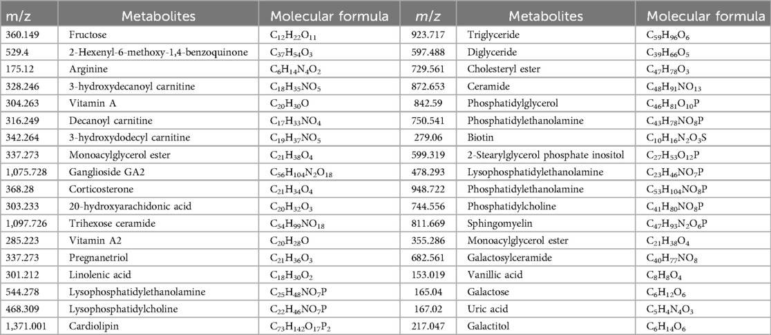
Total ion chromatogram (TIC) results under positive and negative ions model of differential metabolites identification and the quality control samples were presented in the Supplementary Figure S1). Differential metabolites between the healthy control group and the cold asthma group were identified using the criteria of fold change ≥1.2 or ≤0.83 and q-value <0.05, respectively (Figures 2a,b). A total of 50 differential metabolites were identified: 33 in positive ion mode and 17 in negative ion mode. As shown in Table 3, compared to the healthy control group, plasma levels of trihexosylceramide, diglyceride, triglyceride, and prostaglandin A1 were significantly elevated in the cold asthma group. In contrast, the levels of metabolites such as fructose, vanillic acid, uric acid, fumarate acetoacetate, galactose alcohol, and inositol phosphate were significantly decreased.
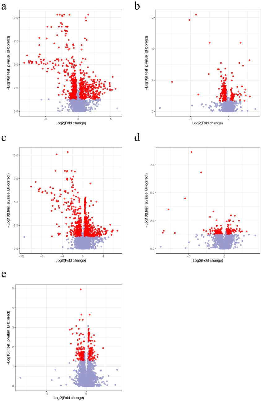
Figure 2. Volcano plots of the identification of differential ions in the plasma samples between the healthy control, cold, and heat asthma groups in the positive and negative ion modes. Volcano plot of the differential ions in the positive ion mode (a) and negative ion mode (b) between the healthy control and cold asthma groups. Volcano plot of the differential ions in positive mode (c) and negative ion mode (d) between the healthy control and heat asthma groups. (e) Volcano plot of the differential ions in positive ion mode between the cold and heat asthma groups. The red points in the heat plots represent the difference multiples of the differential metabolites that were both ≥1.2 or ≤0.83, and q-value <0.05. The blue points represent other metabolites.
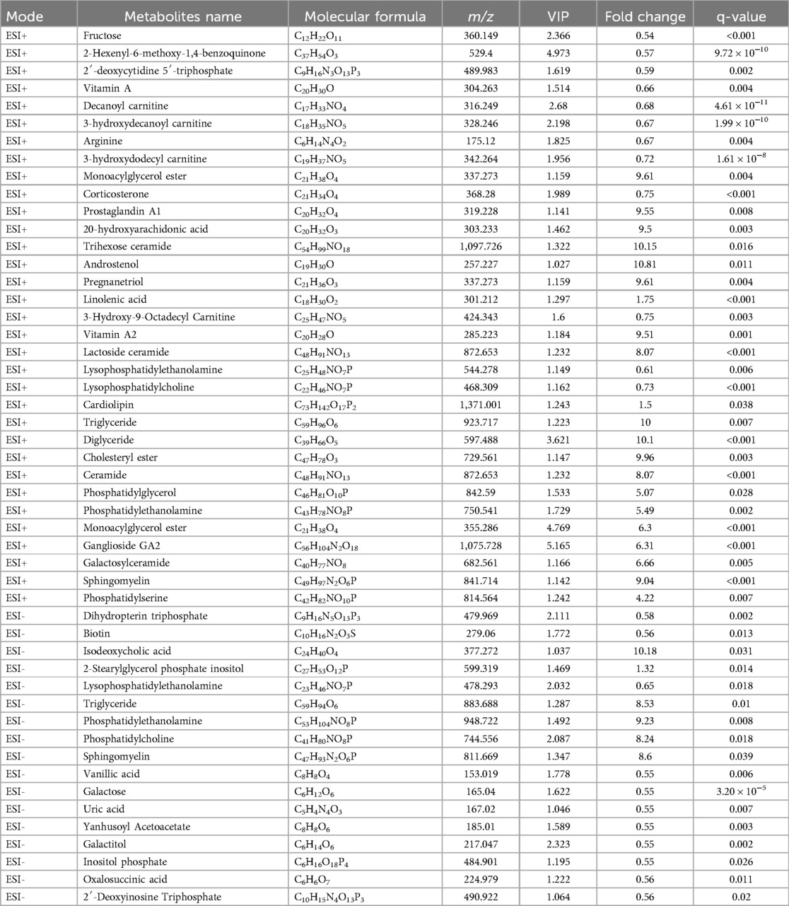
Table 3. List of differentially expressed metabolites in children with cold asthma and healthy controls.
In the comparison between the healthy control group and the heat asthma group, 50 differential metabolites were detected: 34 in positive ion mode and 16 in negative ion mode (Figures 2c,d). Compared with the healthy control group, metabolites, including androstenediol, triglyceride, diacylglycerol, and cholesteryl ester, were significantly upregulated in the heat asthma group. The levels of fructose, galactose, uric acid, tetrahydropyridine dicarboxylate, and galactose alcohol were significantly reduced (Table 4).
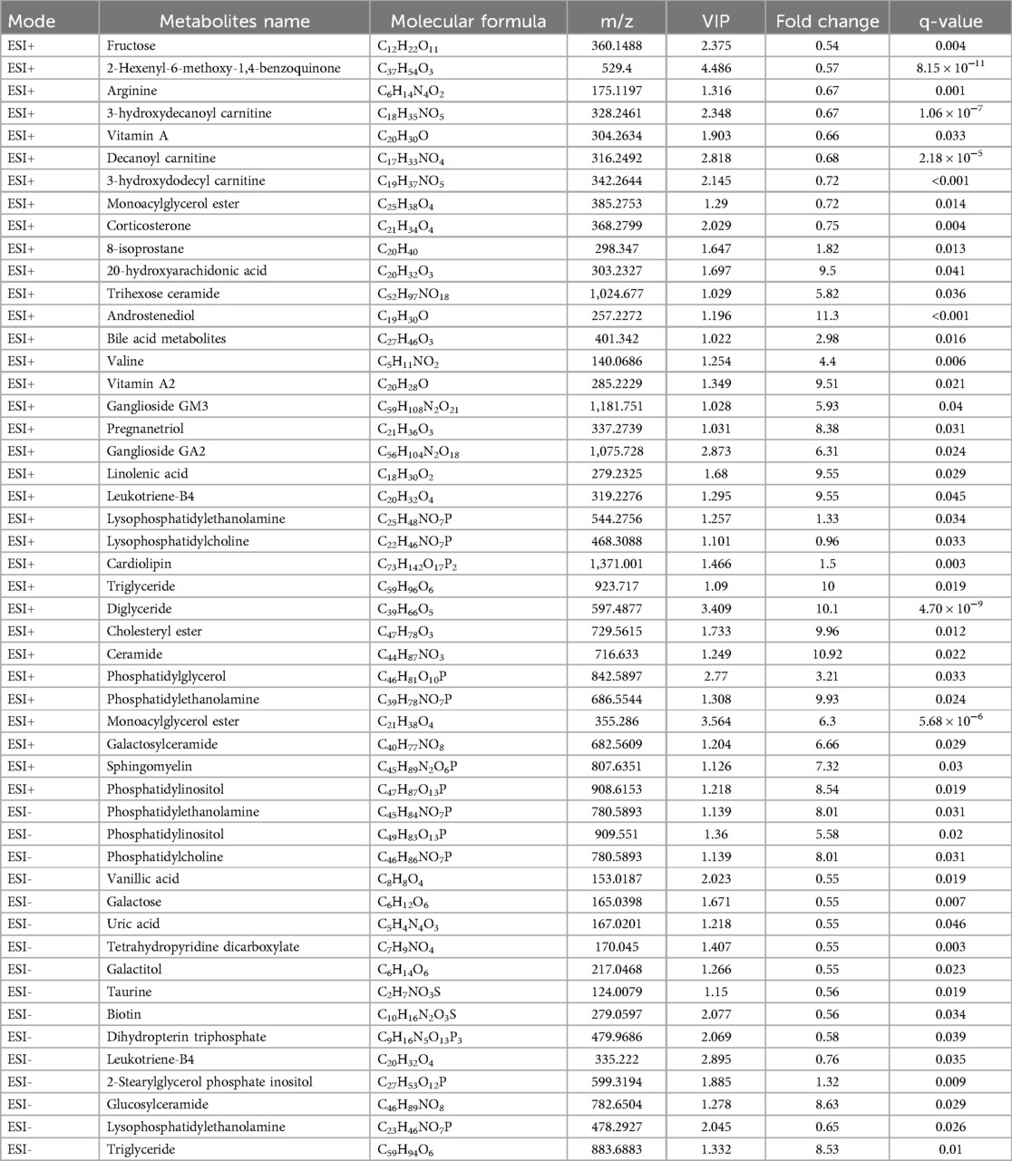
Table 4. List of differentially expressed metabolites in children with heat asthma and healthy controls.
Due to the limited number of differential metabolites identified between the cold and heat asthma groups in the negative ion mode (Supplementary Figures S1D,F), further analysis was conducted exclusively in the positive ion mode (Figure 2e). A total of 18 differential metabolites were identified. Among them, vitamin A, androstenol, and triglycerides were significantly upregulated in the heat asthma group compared with in those the cold asthma group (Table 5).
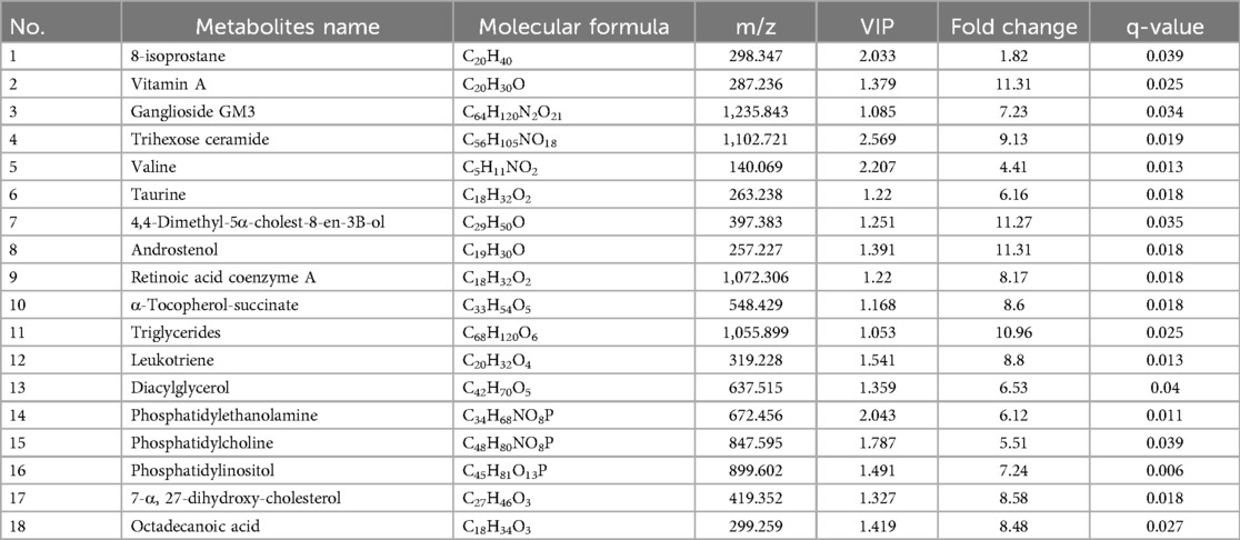
Table 5. List of differentially expressed metabolites in children with cold asthma and heat asthma syndrome.
3.4 Identification of differentially enriched metabolic pathways
To identify differentially enriched metabolic pathways, MetaboAnalyst 4.0 platform (https://www.metaboanalyst.ca/) was used. In the positive ion mode, six metabolic pathways were found to be significantly altered in the healthy control and cold asthma groups. These included the sphingolipid, retinol, glycerophospholipid, arachidonic acid, linolenic acid, arginine, and proline metabolism pathways (Figure 3a, Table 6). Six additional differential pathways were identified in negative ion mode: glycerophospholipid metabolism, biotin metabolism, phosphoinositide metabolism, tyrosine metabolism, folate biosynthesis, and galactose metabolism (Figure 3b, Table 6).
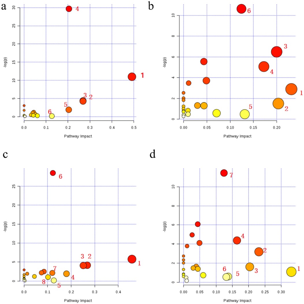
Figure 3. Bubble charts of differential metabolic pathways between asthma subtypes and healthy controls in both ionization modes. (a,b) Differential metabolic pathways between the healthy control and the cold asthma groups in positive ion mode (a) and negative ion mode (b). (c,d) Differential metabolic pathways between the healthy control group and the heat asthma group in the positive ion mode (c) and negative ion mode (d).

Table 6. Differential metabolic pathways between children with cold asthma and the healthy controls in both the positive and negative ion modes.
Eight differential metabolic pathways were identified in the positive ion mode between the healthy control and heat asthmatic groups. These included sphingolipids, retinol, glycerophospholipid, pantothenate and CoA biosynthesis, arginine and proline, arachidonic acid, glycine/serine/threonine, and starch and sucrose metabolism (Figure 3c, Table 6). Seven altered pathways were observed in negative ion mode: taurine metabolism, glycerophospholipid metabolism, biotin metabolism, sphingolipid metabolism, phosphoinositide metabolism, folate biosynthesis, and galactose metabolism (Figure 3d, Table 7).
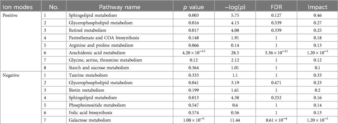
Table 7. Differential metabolic pathways between children with heat asthma and healthy controls in both positive and negative ion modes.
Further analysis of the differential pathways between the cold asthma and heat asthma groups revealed seven significantly enriched metabolic pathways: glycerophospholipid metabolism, arachidonic acid metabolism, retinol metabolism, glycosylation biosynthesis, glycine/serine/threonine metabolism, valine/leucine/isoleucine metabolism, and the protobiliary acid biosynthesis pathway (Figure 4, Table 8).
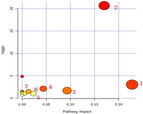
Figure 4. Bubble chart of differential metabolic pathways between cold asthma and heat asthma in the positive ion mode.

Table 8. Differential metabolic pathways between children with cold asthma and heat asthma syndrome in the positive ion mode.
In summary, sphingolipid metabolism, retinol metabolism, glycerophospholipid metabolism, and arachidonic acid metabolism pathways distinguished both cold and heat asthma groups from healthy controls. Meanwhile, metabolites in glycerophospholipid metabolism and glycosylation biosynthesis pathways may serve as distinguishing biomarkers between cold and heat asthma syndromes.
4 Discussion
This study presents a comprehensive untargeted metabolomic analysis of plasma samples from children with cold asthma syndrome, heat asthma syndrome, and healthy controls. Several metabolites were differentially expressed between the groups. These findings may help to elucidate the underlying mechanisms of cold and heat asthma syndromes and support the identification of potential biomarkers for TCM syndrome differentiation.
Fifty metabolites were identified as significantly altered in patients with cold asthma syndrome compared with healthy controls. These changes included changes in fructose, lipids and their derivatives, amino acids, phospholipids, and related metabolites. Notably, the levels of trihexosylceramide, androstenediol, triglyceride, diglyceride, and isodeoxycholic acid were elevated by more than 10-fold in the cold asthma group relative to controls. Additionally, levels of monoacylglycerides, prostaglandin A1, 20-hydroxyarachidonic acid, pregnanetriol, vitamin A2, cholesteryl ester, phosphatidylethanolamine (PE), and sphingomyelin (SM) were increased by more than 9-fold in children with cold asthma syndrome compared to healthy children.
Kyoto Encyclopedia of Genes and Genomes (KEGG) pathway analysis revealed that these differential metabolites were primarily enriched in sphingolipid, glycerophospholipid, arachidonic acid (AA), arginine and proline, and tyrosine metabolism. Previous studies have shown that lipid metabolism is closely associated with airway hyper-responsiveness and an increased risk of asthma and wheezing attacks (25). Free fatty acid receptor 1 (GPR40), which is expressed on airway smooth muscle cells, functions as a sensor for medium- and long-chain free fatty acids. Its activation can promote airway smooth muscle cell proliferation and p70S6 K phosphorylation via the MEK/ERK and PI3 K/Akt signaling pathways, thereby aggravating asthma severity (26).
In the present study, we also observed significant changes in plasma lipid metabolites in children with both cold and heat asthma syndromes. Consistent with our results, Jiang et al. (16) reported disturbed lipid metabolism in asthmatic patients, characterized by significantly higher levels of triglycerides, ceramides (Cer), PE, SM, phosphatidylinositol (PI), phosphatidylglycerol (PG), and lysophosphatidylcholine (LPC), than in healthy controls. Similarly, Wang et al. (17, 18) found significant alterations in plasma levels of phosphatidic acid (PA), PG, PE, phosphatidylcholine (PC), PI, and phosphatidylserine in patients with asthma. Furthermore, several metabolomic studies have demonstrated that the incidence and severity of asthma in children are closely associated with abnormalities in fatty acid, glycerophospholipid, and sphingolipid metabolism (27). Our findings are consistent with those of previous research and further support the presence of metabolic dysregulation in pediatric asthma.
However, to date, no study has directly linked lipid metabolism with TCM syndromes to provide mechanistic insights supporting the TCM theory. According to TCM, typical clinical manifestations of cold asthma syndrome include wheezing in the throat, shortness of breath, chest tightness, white frothy sputum, absence of thirst or a preference for warm drinks, cold limbs, bluish facial complexion, and a tendency for symptom exacerbation upon exposure to cold environments. These manifestations may reflect the underlying metabolic disturbances in the affected children. As asthma is primarily a respiratory disease, most symptoms are concentrated in the airway. However, children with cold asthma syndrome often present with cold intolerance, peripheral coldness, and a dull or cyanotic complex, suggesting reduced metabolic activity and impaired systemic homeostasis. Exposure to cold or other environmental triggers can cause acute exacerbations. Our current study found that children with cold asthma syndrome exhibited significant metabolic alterations, with markedly increased plasma levels of metabolites, such as androstenediol, triglycerides, diglycerides, isodeoxycholic acid, monoacylglycerides, and prostaglandin A1.
Similarly, in the heat asthma syndrome group, plasma levels of androstenediol, triglycerides, diglycerides, and ceramide were elevated more than 10-fold compared to controls. Moreover, 20-hydroxyarachidonic acid, vitamin A2, linolenic acid, leukotriene B4, cholesteryl esters, and PE increased by more than 9-fold. KEGG pathway enrichment analysis indicated that these differential metabolites were associated with sphingolipid, glycerophospholipid, taurine, arginine and proline, glycine, serine, and threonine metabolism.
Adipose tissue functions as a metabolically active endocrine organ and plays a central role in the regulation of energy balance and metabolic homeostasis. The hydrolysis of triglycerides and the release of free fatty acids (FFAs) from adipocytes, especially when insulin-mediated, can activate the MAPK and PI3 K signaling pathways. This promotes smooth muscle proliferation in the airway, exacerbating asthma symptoms (26).
In TCM, heat asthma syndrome is characterized by symptoms such as loud phlegm sounds in the throat, shortness of breath, persistent coughing with thick yellow or white sputum, a dry or bitter taste in the mouth, facial flushing, sweating, and body heat. Our findings suggest that children with heat asthma also exhibit marked metabolic imbalance. The differentially expressed metabolites in this group included androstenediol, triglycerides, diglycerides, ceramide, 20-hydroxyarachidonic acid, vitamin A2, linolenic acid, and leukotriene B4, all of which may contribute to the pathophysiological basis of the heat syndrome as interpreted through TCM principles.
A comparative analysis between children with cold asthma and those with heat asthma syndromes revealed that several metabolites were significantly elevated in the cold asthma group. Specifically, the levels of vitamin A, 4,4-dimethyl-5α-cholyl-8-ene-3β-alcohol, androstenediol, and triglycerides were more than 10-fold higher in patients with cold asthma syndrome than in those with heat asthma syndrome. Additionally, trihexosylceramide, retinoic acid coenzyme A, α-tocopherol succinate, leukotriene, 7-α,27-dihydroxycholesterol, and octadecanoic acid were elevated by more than 8-fold in the cold asthma group.
Pathway enrichment analysis indicated that these differential metabolites are primarily involved in glycerophospholipid metabolism, arachidonic acid, retinol, glycosylation, glycine/serine/threonine, valine/leucine/isoleucine, and primary bile acid biosynthesis. Wang et al. (28) reported that cysteine and glycine-rich protein 2 (Csrp2) plays a key role in the phenotypic switching of airway smooth muscle cells (ASMCs). Upregulation of Csrp2 inhibits the PDGF-BB-induced transition of ASMCs to a synthetic/proliferative phenotype via modulation of the Yes-associated protein (YAP)/transcriptional coactivator with PDZ-binding motif (TAZ) signaling axis, thereby contributing to airway remodeling in asthma. Kelly et al. (29) conducted a systematic review of 21 studies utilizing metabolomics techniques and concluded that asthma pathogenesis is closely associated with various metabolic pathways, including amino acid metabolism, glutamate-glutamine cycle, hypoxia response, immune and inflammatory signaling, lipid metabolism, oxidative stress, and tricarboxylic acid (TCA) cycle. These findings support the notion that correcting metabolic imbalance may be a promising therapeutic approach for asthma. Furthermore, Gao et al. (30) used ultra-high-performance liquid chromatography–mass spectrometry to analyze serum samples from children with asthma and bronchiolitis. Their results showed significant alterations in the serum levels of phosphatidylethanolamine, saturated and monounsaturated fatty acids, and bile acids in asthmatic children.
In our comparative analysis of cold and heat asthma syndromes, we identified several common differentially expressed serum metabolites, including triglycerides, phosphatidylethanolamine, and ceramides. These shared biomarkers suggest that despite differences in syndrome classification, both cold and heat asthma share core respiratory pathophysiological features. However, distinct metabolic profiles between the two syndromes likely reflect underlying differences in etiology and clinical presentation, as described in TCM theory.
Compared with Western medicine, Traditional Chinese Medicine (TCM) has been widely applied in the treatment of asthma, chronic obstructive pulmonary disease (COPD), and other diseases owing to its holistic and systemic approach. Clinical practice has demonstrated that TCM offers notable advantages, including significant efficacy and favorable safety profile. TCM provides unique therapeutic benefits in asthma. According to TCM theory, asthma falls under the category of “Xiao” (喘证). However, the classification of bronchial asthma into different syndrome types in TCM is often subjective and heavily reliant on individual physician experience and lacks an objective and standardized basis for syndrome differentiation. This subjectivity has hindered the establishment of standardized diagnostic and treatment protocols. Moreover, relatively few studies have systematically explored the theoretical underpinnings and biological validation of TCM syndromes, particularly those related to asthma.
In our study, we observed significant differences in sphingolipid metabolites, including ganglioside GA2, ganglioside GM3, galactosylceramide, and sphingomyelin, between patients with asthma and healthy controls. Sphingolipids are complex lipids comprising a sphingosine backbone, long-chain fatty acids, and polar head groups. They are classified as sphingomyelins, gangliosides, and glycosphingolipids. Alterations in sphingolipids, such as ceramides, have been shown to influence the biosynthesis of long-chain fatty acids, including palmitic acid, which is associated with asthma severity (31). Notably, sphingosine-1-phosphate and ceramide, key metabolites derived from sphingomyelin, have been shown to interact directly with bronchial smooth muscle cells, promoting contraction and contributing to airway remodeling (32, 33).
In addition, our findings demonstrated that the levels of several glycerophospholipids, including lysophosphatidylethanolamine, lysophosphatidylcholine, cardiolipin, PE, PI, and PC, were significantly altered in the plasma of asthma syndrome groups compared with controls. Glycerophospholipids are essential components of cellular membranes and are involved in the synthesis of pulmonary surfactants. Approximately 85% of phospholipids in alveolar surfactants consist of phosphatidylcholine, which plays a critical role in pulmonary defense and is implicated in chronic airway inflammation and increased airway responsiveness (34).
Asai et al. (35) used LC-MS-based metabolomics to analyze bronchoalveolar lavage fluid from asthma patients and found elevated levels of lysophosphatidylcholine and phospholipase A2 activity compared to healthy controls. Lysophosphatidylcholine exerts strong pro-inflammatory effects and contributes to alveolar epithelial cell injury. The latest research indicated that lysophosphatidylcholine may serve as novel lipid mediators of severe asthma involving to corticosteroid resistance and correlating with oxidative stress (36), of which once again proves the importance of the current research findings.
Interestingly, in our comparative analysis of the cold and heat asthma syndrome groups, we also found differential expressions of PE, PC, and PI, suggesting that alterations in the glycerophospholipid metabolism pathway may serve as potential biomarkers for distinguishing between cold and heat asthma syndromes.
Significant alterations in amino acid metabolic pathways were observed between the cold and heat asthma groups. Arginine, a semi-essential amino acid, plays a key role in asthma pathophysiology. Studies have shown that excessive arginine methylation can trigger asthma attacks (37), and the levels of asymmetric dimethylarginine and methylarginine are significantly elevated in patients with severe asthma (38). Arginine is metabolized by arginase to produce endogenous nitric oxide (NO), which modulates airway responsiveness (39). Several studies have demonstrated a correlation between endogenous NO levels and airway hyper-reactivity (40, 41). Conversely, arginase activity has been shown to mitigate airway hyper-responsiveness by reducing systemic arginine levels (42).
Proline, a non-essential cyclic amino acid, also contributes to asthma pathogenesis. The collagen-derived peptide proline-glycine-proline has been shown to induce neutrophil chemotaxis, a key feature of airway inflammation (43). Glycine reduces lung injury by attenuating the recruitment of inflammatory cells and suppressing the release of inflammatory mediators (44). Serine, which participates in the biosynthesis of purines, pyrimidines, glycerophospholipids, and sphingolipids, is involved in the oxidative stress pathways linked to asthma (45). Furthermore, plasma levels of branched-chain amino acids (BCAAs), such as valine, leucine, and isoleucine, were found to be significantly elevated in patients with asthma compared to healthy controls (46). Taken together, the differences in plasma metabolites between children with cold and heat asthma syndromes are not limited to lipids, such as triglycerides, diglycerides, and ceramides, but also extend to amino acids, including arginine, proline, and valine, and their related metabolic pathways.
Overall, our study suggests that serum metabolite profiling in children with cold- and heat-asthma syndromes has the potential to identify biomarkers that support the diagnosis and differentiation of TCM syndromes. These findings could assist TCM practitioners in achieving more accurate syndrome classification for childhood asthma. Our results demonstrate that systemic metabolic disturbances, particularly in lipid and amino acid pathways, are evident in pediatric asthma and that these disturbances differ between cold and heat syndromes. Table 9 summarized the common differential metabolites between cold asthma and heat asthma, cold asthma and the healthy group, and heat asthma and the healthy group. These common differential metabolites were mainly lipids, amino acids and its related metabolite such as triglyceride, diglyceride and arginine, ceramide that can be considered as biomarkers. In addition, through comparative analysis, we also concluded in Table 10 that the other potential plasma differential metabolites biomarkers and the metabolic pathways can be used in differential diagnosis of cold and heat asthma syndromes.
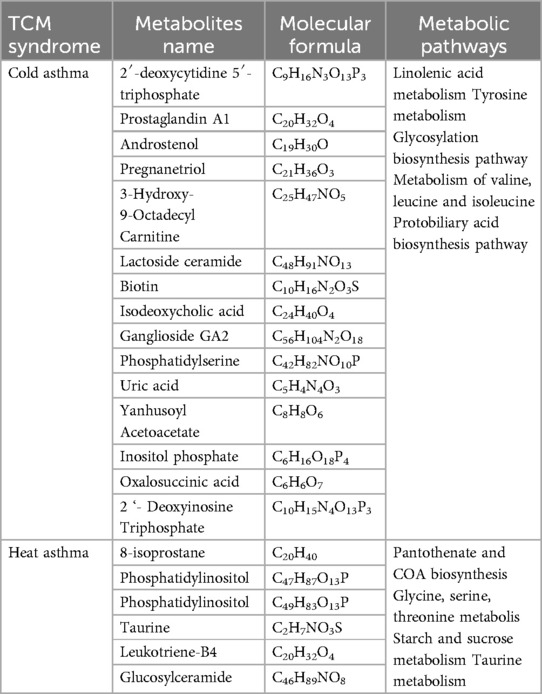
Table 10. The other potential plasma differential metabolites biomarkers and related metabolic pathways between cold and heart asthma.
In addition, attentions should also be paid to the dramatically change of arachidonic acid (AA) metabolic pathway and retinol metabolism. Asthma, as a chronic disease, is characteristic with airway inflammation, airway hyperresponsiveness, mucus overproduction, remodeling, and narrowing of the airway. Studies have demonstrated that inflammatory characteristics of asthma may be related to the release of Th2 cytokines (IL-4, IL-5, IL-13 and IL-33) mediated by the regulation of arachidonic acid metabolic pathway (17, 18, 47). Meanwhile, the cyclooxygenase (COX) and lipoxygenase (LOX) pathways involved in AA metabolism are also involved in lipid synthesis and metabolism (17, 18, 48). These evidences indicated that AA metabolism may link to the lipid synthesis and metabolism during the asthma onset process, but there still lack of evidence used to explain its relationship with the classification of TCM asthma syndromes. Another important issue is retinol metabolism. Retinol, also named vitamin A, was change significantly in both syndromes despite distinct clinical presentations in the current study. As is well known, vitamins including fat-soluble and water-soluble kinds, a type of trace organic substances, are indispensable to human healthy and primarily obtained from food, mainly participate in biochemical reactions and regulate metabolic functions (49, 50). In our study, vitamin A and vitamin A2 were identified as differential metabolites, and retinol metabolism is one of the differential metabolic pathways. Dominika et al. systematically summarized that the role of vitamins in the pathogenesis of asthma including the influence of vitamins on asthma and its typical symptoms (51). They also summarized the correlation between vitamin intake and levels and the risk of asthma in both pre- and postnatal life. Hu and Sang reported that no significant correlation between vitamin A intake and asthma risk in children, nor between serum vitamin A levels and asthma risk (52,53). Taken together, the evidence regarding vitamins and asthma is still controversial due to the complex pathogenesis of asthma. In other words, the intake of vitamin and its balances can be considered as an intervention to prevent asthma indicated by our study and others.
However, this study has several limitations. First, the sample size was relatively small, which may have limited the generalizability of the findings and may bring bias to the research results. Second, the absence of a drug intervention group prevented us from evaluating the therapeutic effect of TCM treatment and its influence on the identified metabolic markers. In the future studies, we not only should expand the sample size, but also increase the study before and after the intervention of TCM formulas (such as classic formulas). At the same time, we should use high-throughput technologies, and combine leverage big data with artificial intelligence technology to construct a plasma biomarker model for asthma patients with cold or heat asthma syndrome based on the key findings, and conduct large-scale and multi- center clinical data validation studies. Moreover, mature in vivo animal models and in vitro cell experiments are also necessary performed to elucidate in-depth on its mechanisms, providing comprehensive theoretical basis for the clinical treatment of asthma patients with different TCM syndromes. Finally, the comparison between cold and heat asthma syndromes was primarily based on positive-ion mode data. Limited information is available from the negative ion mode, which may have omitted additional relevant metabolic alterations.
5 Conclusion
Our study provides important insights into the plasma differential metabolites associated with TCM-defined asthma syndromes in children, particularly distinguishing between cold and heat syndromes. These findings contribute to the modern scientific understanding of TCM syndrome classification in asthma. The common lipids, amino acids, and their associated metabolic pathways have emerged as potential biomarkers for the differential diagnosis of cold and heat syndromes in TCM. Key lipid-related metabolites and pathways include triglycerides, diglycerides, sphingomyelin, phosphatidylcholine, sphingolipid metabolism, glycerophospholipid metabolism, and phosphoinositide metabolism. Significant contributors of amino acids include taurine, valine, and arginine, along with the glycine/serine/threonine metabolism, taurine metabolism, and arginine/proline metabolism pathways. These results enhance our understanding of metabolic dysregulation in children with cold or heat asthma syndromes and offer a scientific basis for exploring its pathogenesis, improving clinical syndrome differentiation, and evaluating the therapeutic efficacy of TCM interventions. Future studies should further explore amino acid metabolism in children with different TCM asthma syndromes and identify characteristic amino acid markers that reflect TCM syndrome differentiation with greater objectivity.
Data availability statement
The raw data supporting the conclusions of this article will be made available by the authors, without undue reservation.
Ethics statement
The studies involving humans were approved by The ethics committee of Shenzhen Children's Ethics Committee. The studies were conducted in accordance with the local legislation and institutional requirements. Written informed consent for participation in this study was provided by the participants' legal guardians/next of kin.
Author contributions
YS: Conceptualization, Data curation, Investigation, Writing – review & editing. LH: Visualization, Writing – original draft, Writing – review & editing. WY: Writing – review & editing. ZC: Investigation, Writing – review & editing. DC: Investigation, Writing – review & editing. XD: Methodology, Writing – review & editing. LW: Funding acquisition, Project administration, Resources, Supervision, Writing – review & editing.
Funding
The author(s) declare that financial support was received for the research and/or publication of this article. This study was funded by the National Natural Science Foundation of China (No. 81574019).
Acknowledgments
We are very grateful for the research platform provided by Shenzhen Children's Hospital and the sample omics testing and analysis services provided by the Shenzhen Huada Gene Company.
Conflict of interest
The authors declare that the research was conducted in the absence of any commercial or financial relationships that could be construed as a potential conflict of interest.
Generative AI statement
The author(s) declare that no Generative AI was used in the creation of this manuscript.
Publisher's note
All claims expressed in this article are solely those of the authors and do not necessarily represent those of their affiliated organizations, or those of the publisher, the editors and the reviewers. Any product that may be evaluated in this article, or claim that may be made by its manufacturer, is not guaranteed or endorsed by the publisher.
Supplementary material
The Supplementary Material for this article can be found online at: https://www.frontiersin.org/articles/10.3389/fped.2025.1549431/full#supplementary-material
References
1. Asher MI, Rutter CE, Bissell K, Chiang CY, El Sony A, Ellwood E, et al. Worldwide trends in the burden of asthma symptoms in school-aged children: global asthma network phase I cross-sectional study. Lancet. (2021) 398(10311):1569–80. doi: 10.1016/s0140-6736(21)01450-1
2. García-Marcos L, Chiang C-Y, Asher MI, Marks GB, El Sony A, Masekela R, et al. Asthma management and control in children, adolescents, and adults in 25 countries: a global asthma network phase I cross-sectional study. Lancet Glob Health. (2023) 11(2):e218–28. doi: 10.1016/s2214-109x(22)00506-x
3. Shang B, Li X, Xu Y, Ren W, Wang J, Xing C, et al. Clinical characteristics and economic burden of asthma in China: a multicenter retrospective study. Iran J Allergy Asthma Immunol. (2023) 22(3):290–8. doi: 10.18502/ijaai.v22i3.13057
4. von Mutius E, Smits HH. Primary prevention of asthma: from risk and protective factors to targeted strategies for prevention. Lancet. (2020) 396(10254):854–66. doi: 10.1016/s0140-6736(20)31861-4
5. Deng L, Chen X, Ma P, Wu Y, Okoye CO, Du D, et al. The combined effect of oxidative stress and TRPV1 on temperature-induced asthma: evidence in a mouse model. Environ Pollut. (2024) 344:123313. doi: 10.1016/j.envpol.2024.123313
6. Britt RD Jr, Ruwanpathirana A, Ford ML, Lewis BW. Macrophages orchestrate airway inflammation, remodeling, and resolution in asthma. Int J Mol Sci. (2023) 24(13):10451. doi: 10.3390/ijms241310451
7. Raby KL, Michaeloudes C, Tonkin J, Chung KF, Bhavsar PK. Mechanisms of airway epithelial injury and abnormal repair in asthma and COPD. Front Immunol. (2023) 14:1201658. doi: 10.3389/fimmu.2023.1201658
8. Zhao X, Qin Y-H, Wang Y-P, Wang M-Q, Xie Z, Chen J, et al. The guideline for the diagnosis and treatment of pediatric asthma in Chinese medicine (revised). J. Nanjing Univ Tradit Chin Med. (2022) 38(6):476–82. doi: 10.14148/j.issn.1672-0482.2022.0476
9. Professional Committee of Respiratory Diseases of the Chinese Association of Integrative Medicine. Experts consensus on diagnosis and treatment of bronchial asthma by integrative medicine. Chin J Int Tradit Western Med. (2023) 43(1):12–20. doi: 10.7661/j.cjim.20221019.212
10. The Subspecialty Group of Respiratory, the Society of Pediatrics, Chinese Medical Association; the Editorial Board, Chinese Journal of Pediatrics; China Medicine Education Association Committee on Pediatrics. Guidelines for the diagnosis and optimal management of asthma in children (2025). Chin J Pediatr. (2025) 63(04):324–37. doi: 10.3760/cma.j.cn112140-20250124-00074
11. Li J-S, Wang Z-W. Diagnostic criteria for traditional Chinese medicine syndromes in bronchial asthma (2016 edition). J Tradit Chin Med. (2016) 57(22):1978–80. doi: 10.13288/j.11-2166/r.2016.22.022
12. Sun W, Liu K, Yang R, Guo L, Gao P. A traditional Chinese medicine characteristic therapy for bronchial asthma: moxibustion. J Vis Exp. (2023) 195:e65119. doi: 10.3791/65119
13. Zhou B-W, Liu H-M, Jia X-H. The role and mechanisms of traditional Chinese medicine for airway inflammation and remodeling in asthma: overview and progress. Front Pharmacol. (2022) 13:917256. doi: 10.3389/fphar.2022.917256
14. Mildau K, Ehlers H, Meisenburg M, Del Pup E, Koetsier RA, Torres Ortega LR, et al. Effective data visualization strategies in untargeted metabolomics. Nat Prod Rep. (2024) 42(6):982–1019. doi: 10.1039/d4np00039k
15. Chen C, Wang J, Pan D, Wang X, Xu Y, Yan J, et al. Applications of multi-omics analysis in human diseases. MedComm (2020). (2023) 4(4):e315. doi: 10.1002/mco2.315
16. Jiang T, Dai L, Li P, Zhao J, Wang X, An L, et al. Lipid metabolism and identification of biomarkers in asthma by lipidomic analysis. Biochim Biophys Acta Mol Cell Biol Lipids. (2021) 1866(2):158853. doi: 10.1016/j.bbalip.2020.158853
17. Wang S, Tang K, Lu Y, Tian Z, Huang Z, Wang M, et al. Revealing the role of glycerophospholipid metabolism in asthma through plasma lipidomics. Clin Chim Acta. (2021) 513:34–42. doi: 10.1016/j.cca.2020.11.026
18. Wang B, Wu L, Chen J, Dong L, Chen C, Wen Z, et al. Metabolism pathways of arachidonic acids: mechanisms and potential therapeutic targets. Signal Transduct Target Ther. (2021) 6(1):94. doi: 10.1038/s41392-020-00443-w
19. Roshan Lal T, Cechinel LR, Freishtat R, Rastogi D. Metabolic contributions to pathobiology of asthma. Metabolites. (2023) 13(2):212. doi: 10.3390/metabo13020212
20. Kim J, Shin H-W. Exploring biomarkers in asthma: insights from serum metabolomics. Allergy Asthma Immunol Res. (2024) 16(3):211. doi: 10.4168/aair.2024.16.3.211
21. Dunn WB, Broadhurst D, Begley P, Zelena E, Francis-McIntyre S, Anderson N, et al. Procedures for large-scale metabolic profiling of serum and plasma using gas chromatography and liquid chromatography coupled to mass spectrometry. Nat Protoc. (2011) 6(7):1060–83. doi: 10.1038/nprot.2011.335
22. Wen B, Mei Z, Zeng C, Liu S. Metax: a flexible and comprehensive software for processing metabolomics data. BMC Bioinformatics. (2017) 18(1):183. doi: 10.1186/s12859-017-1579-y
23. Wishart DS, Guo A, Oler E, Wang F, Anjum A, Peters H, et al. HMDB 5.0: the human metabolome database for 2022. Nucleic Acids Res. (2022) 50(D1):D622–31. doi: 10.1093/nar/gkab1062
24. Chong J, Soufan O, Li C, Caraus I, Li S, Bourque G, et al. Metaboanalyst 4.0: towards more transparent and integrative metabolomics analysis. Nucleic Acids Res. (2018) 46(W1):W486–94. doi: 10.1093/nar/gky310
25. Henderson GC. Plasma free fatty acid concentration as a modifiable risk factor for metabolic disease. Nutrients. (2021) 13(8):2590. doi: 10.3390/nu13082590
26. Matoba A, Matsuyama N, Shibata S, Masaki E, Emala CW Sr, Mizuta K. The free fatty acid receptor 1 promotes airway smooth muscle cell proliferation through MEK/ERK and PI3K/akt signaling pathways. Am J Physiol Lung Cell Mol Physiol. (2018) 314(3):L333–48. doi: 10.1152/ajplung.00129.2017
27. Rago D, Pedersen CT, Huang M, Kelly RS, Gürdeniz G, Brustad N, et al. Characteristics and mechanisms of a sphingolipid-associated childhood asthma endotype. Am J Respir Crit Care Med. (2021) 203(7):853–63. doi: 10.1164/rccm.202008-3206OC
28. Wang H, Zhang Y, Zhong B, Geng Y, Hao J, Jin Q, et al. Cysteine and glycine-rich protein 2 retards platelet-derived growth factor-BB-evoked phenotypic transition of airway smooth muscle cells by decreasing YAP/TAZ activity. Cell Biochem Funct. (2023) 42(1):e3896. doi: 10.1002/cbf.3896
29. Kelly RS, Dahlin A, McGeachie MJ, Qiu W, Sordillo J, Wan ES, et al. Asthma metabolomics and the potential for integrative omics in research and the clinic. Chest. (2017) 151(2):262–77. doi: 10.1016/j.chest.2016.10.008
30. Gao XH, Yin TL, Li Y, Shi SF, Liu FL, Li MJ, et al. Assessment of serum metabolism and eicosanoid profiling in pediatric asthma and bronchiolitis via liquid chromatography-mass spectrometry. Rapid Commun Mass Spectrom. (2025) 39(4):e9955. doi: 10.1002/rcm.9955
31. Yoshida K, Morishima Y, Ishii Y, Mastuzaka T, Shimano H, Hizawa N. Abnormal saturated fatty acids and sphingolipids metabolism in asthma. Respir Investig. (2024) 62(4):526–30. doi: 10.1016/j.resinv.2024.04.006
32. Lai WQ, Wong WS, Leung BP. Sphingosine kinase and sphingosine 1-phosphate in asthma. Biosci Rep. (2011) 31(2):145–50. doi: 10.1042/bsr20100087
33. Terashita T, Kobayashi K, Nagano T, Kawa Y, Tamura D, Nakata K, et al. Administration of JTE013 abrogates experimental asthma by regulating proinflammatory cytokine production from bronchial epithelial cells. Respir Res. (2016) 17(1):146. doi: 10.1186/s12931-016-0465-x
34. Haczku A, Atochina EN, Tomer Y, Cao Y, Campbell C, Scanlon ST, et al. The late asthmatic response is linked with increased surface tension and reduced surfactant protein B in mice. Am J Physiol Lung Cell Mol Physiol. (2002) 283(4):L755–765. doi: 10.1152/ajplung.00062.2002
35. Asai A, Okajima F, Nakajima Y, Nagao M, Nakagawa K, Miyazawa T, et al. Involvement of rac GTPase activation in phosphatidylcholine hydroperoxide-induced THP-1 cell adhesion to ICAM-1. Biochem Biophys Res Commun. (2011) 406(2):273–7. doi: 10.1016/j.bbrc.2011.02.032
36. Kume H, Kazama K, Sato R, Sato Y. Possible involvement of lysophospholipids in severe asthma as novel lipid mediators. Biomolecules. (2025) 15(2):182. doi: 10.3390/biom15020182
37. Lara A, Khatri SB, Wang Z, Comhair SA, Xu W, Dweik RA, et al. Alterations of the arginine metabolome in asthma. Am J Respir Crit Care Med. (2008) 178(7):673–81. doi: 10.1164/rccm.200710-1542OC
38. Jung J, Kim SH, Lee HS, Choi GS, Jung YS, Ryu DH, et al. Serum metabolomics reveals pathways and biomarkers associated with asthma pathogenesis. Clin Exp Allergy. (2013) 43(4):425–33. doi: 10.1111/cea.12089
39. Morris CR. Arginine and asthma. Nestle Nutr Inst Workshop Ser. (2013) 77:1–15. doi: 10.1159/000351365
40. Aytekin M, Dweik RA. Nitric oxide and asthma severity: towards a better understanding of asthma phenotypes. Clin Exp Allergy. (2012) 42(5):614–6. doi: 10.1111/j.1365-2222.2012.03976.x
41. Scott JA, Grasemann H. Arginine metabolism in asthma. Immunol Allergy Clin North Am. (2014) 34(4):767–75. doi: 10.1016/j.iac.2014.07.007
42. Xu W, Comhair SAA, Janocha AJ, Lara A, Mavrakis LA, Bennett CD, et al. Arginine metabolic endotypes related to asthma severity. PLoS One. (2017) 12(8):e0183066. doi: 10.1371/journal.pone.0183066
43. Snelgrove RJ, Jackson PL, Hardison MT, Noerager BD, Kinloch A, Gaggar A, et al. A critical role for LTA4H in limiting chronic pulmonary neutrophilic inflammation. Science. (2010) 330(6000):90–4. doi: 10.1126/science.1190594
44. Loureiro CC, Duarte IF, Gomes J, Carrola J, Barros AS, Gil AM, et al. Urinary metabolomic changes as a predictive biomarker of asthma exacerbation. J Allergy Clin Immunol. (2014) 133(1):261–3.e5. doi: 10.1016/j.jaci.2013.11.004
45. Fitzpatrick AM, Park Y, Brown LA, Jones DP. Children with severe asthma have unique oxidative stress-associated metabolomic profiles. J Allergy Clin Immunol. (2014) 133(1):258–61.e8. doi: 10.1016/j.jaci.2013.10.012
46. Zhang X, Fan H-R, Li Y-Z, Xiao X-F, Liu R, Qi J-W, et al. Development and application of network toxicology in safety research of Chinese materia medica. Chin Herb Med. (2015) 7(1):27–38. doi: 10.1016/s1674-6384(15)60016-8
47. Ito E, Hayashizaki R, Hosaka T, Yamane T, Miyata J, Isobe Y, et al. Eosinophils and pleural macrophages counter regulate IL-33-elicited airway inflammation via the 12/15-lipoxygenase pathway. Front Immunol. (2025) 16:1565670. doi: 10.3389/fimmu.2025.1565670
48. Luo Y, Jin M, Lou L, Yang S, Li C, Li X, et al. Role of arachidonic acid lipoxygenase pathway in asthma. Prostaglandins Other Lipid Mediat. (2022) 158:106609. doi: 10.1016/j.prostaglandins.2021.106609
49. Fitzpatrick TB, Basset GJ, Borel P, Carrari F, DellaPenna D, Fraser PD, et al. Vitamin deficiencies in humans: can plant science help? Plant Cell. (2012) 24(2):395–414. doi: 10.1105/tpc.111.093120
50. Yuan Y, Chen L. Transporters in vitamin uptake and cellular metabolism: impacts on health and disease. Life Metab. (2025) 4(3):loaf008. doi: 10.1093/lifemeta/loaf008
51. Zajac D, Wojciechowski P. The role of vitamins in the pathogenesis of asthma. Int J Mol Sci. (2023) 24(10):8574. doi: 10.3390/ijms24108574
52. Hu J, Sang J, Hao F, Liu L. Association between vitamin A and asthma: a meta-analysis with trial sequential analysis. Front Pharmacol. (2023) 14:1100002. doi: 10.3389/fphar.2023.1100002
Keywords: children asthma, cold asthma, heat asthma, TCM syndromes, metabolomics, plasma biomarkers
Citation: Sun Y, Huang L, Yao W, Chen Z, Cui D, Ding X and Wan L (2025) Plasma lipids, amino acids, and their metabolic pathways as potential biomarkers for differential diagnosis of cold and heat syndrome asthma in children: a preliminary study. Front. Pediatr. 13:1549431. doi: 10.3389/fped.2025.1549431
Received: 21 December 2024; Accepted: 9 July 2025;
Published: 23 July 2025.
Edited by:
Izolde Bouloukaki, University of Crete, GreeceReviewed by:
Amit Krishna De, Sister Nivedita University, IndiaLinjing Deng, Jiangsu University, China
Zhe Chen, Affiliated Kunshan Hospital of Jiangsu University, China
Chaofang Lei, Beijing University of Chinese Medicine, China
Copyright: © 2025 Sun, Huang, Yao, Chen, Cui, Ding and Wan. This is an open-access article distributed under the terms of the Creative Commons Attribution License (CC BY). The use, distribution or reproduction in other forums is permitted, provided the original author(s) and the copyright owner(s) are credited and that the original publication in this journal is cited, in accordance with accepted academic practice. No use, distribution or reproduction is permitted which does not comply with these terms.
*Correspondence: Xuansheng Ding, MTAyMDAzMDc0OUBjcHUuZWR1LmNu; Lisheng Wan, d2xzaDE5NjkxMDE3QDE2My5jb20=
†Yi Sun and Lianzhan Huang contributed equally and shared the first authorship.
 Yi Sun1,2,†
 Lianzhan Huang
 Xuansheng Ding
 Lisheng Wan