- 1School of Medicine and Surgery, University of Milano-Bicocca, Monza, Italy
- 2Biostatistics and Clinical Epidemiology, Fondazione IRCCS San Gerardo dei Tintori, Monza, Italy
- 3Pediatrics, Fondazione IRCCS San Gerardo dei Tintori, Monza, Italy
Introduction: Coeliac disease (CD) manifests more frequently in individuals with Down syndrome (DS) and its prevalence varies across different studies. This study aims to assess the prevalence of CD in children with DS and to describe their clinical, serological, and histological features. A secondary aim was to analyze the time needed for the normalization of anti-transglutaminase IgA (TGA-IgA) and anti-endomysium IgA (EMA-IgA) levels in DS compared to non-syndromic (NS) children.
Materials and methods: This retrospective monocentric cohort study included patients with DS under 18 years of age, diagnosed with CD between 2005 and 2022. Each DS patient was matched for year of birth and sex with two NS celiac children. Follow-up was 6-, 12- and 24-months post-diagnosis.
Results: The prevalence of CD in 770 children with DS was 7.5% (95% CI: 5.8%–9.6%). 57 children with CD and DS were compared with 114 CD NS matched controls (total sample size = 171). DS demonstrated less symptoms than 114 NS CD children (26% vs. 79%, P < 0.001). In the CD DS group 81% had anti-TGA levels 10 times higher the upper limit of normal, compared to 72% in the control group. Among patients with CD and DS, 93% had histological damage equal to 3rd grade of Marsh-Oberhuber classification at diagnosis. The velocity of normalization of anti-TGA was higher in patients without DS (P = 0.005).
Discussion: This study reinforces the higher prevalence of CD in DS, emphasizing the necessity for routine screening, even in asymptomatic individuals. Despite less symptomatic presentation, patients with DS exhibited elevated antibody levels and severe histological damage. Clinicians should expect a prolonged time for antibody normalization following gluten-free diet in DS, mirroring potential challenges in diet adherence and altered immune responses.
Introduction
Coeliac Disease (CD) is an autoimmune enteropathy triggered by gluten ingestion in genetically predisposed individuals (1). The association between CD and Down Syndrome (DS) has been already extensively assessed; however, prevalence of CD in patients with DS varies considerably across the studies (range 0%–19%) (2–5). In a recent meta-analysis (6) including 4,000 patients with DS the prevalence of biopsy-confirmed diagnosis of CD was assessed at 5.8%, a considerably higher percentage compared to the general population (1% in Western countries) (7, 8). The prevalence of CD achieved 4.6% in an Italian cohort of patients with DS assessed over 20 years ago by Bonamico (9). To the best of our knowledge, no additional data focused on national cohorts have been published subsequently. Moreover, only few studies have assessed the specificities of CD in children with DS compared to non-syndromic (NS) otherwise healthy coeliac children. Furthermore, the time of normalization of IgA antibodies to transglutaminase (TGA) following gluten-free diet (GFD) has been studied in children with CD (10, 11), but no published data are available for patients with DS.
The primary aim of this study was to assess the prevalence of CD in a pediatric cohort of patients with DS and to describe its clinical, serological, and histological features. In addition, we aimed at reporting the DS-specific trendlines of TGA- IgA and anti-endomysium IgA (EMA-IgA) decrease over time, compared to NS children following GFD.
Materials and methods
Design of the study
We conducted a retrospective, monocentric cohort study. Clinical records of children with DS under the age of 18 followed by the Pediatric Genetics Outpatient Clinic of Fondazione IRCCS San Gerardo dei Tintori Hospital (Monza, Italy) were retrieved and reviewed by medical staff. All children with DS diagnosed with CD between January 1st, 2005, and December 31st, 2022, were eligible. Coeliac NS age- and gender-matched controls were identified from the Pediatric Gastroenterology Outpatient Clinic of the same Institution. In detail, we selected two NS coeliac patients with superimposable age and gender for each children with DS enrolled. Scheduled follow-up evaluations were performed 6- and 12-months following CD diagnosis and annually thereafter, until the patients turned 18 years. Detailed medical history collection, complete physical examination and CD serology were assessed upon every follow-up visit. The 6–12- and 24-month timepoints were considered for the present study. Informed consent was obtained by parents or legal tutor for each patient. All clinical and laboratorial data were collected and stored in a single excel worksheet anonymously. The study was approved by the Ethic Committee (n° 3896). Results are reported according to the STROBE checklist (12).
Patients with Down syndrome
In our Institution, from 2005 onwards, all patients with DS underwent an annual work up comprehensive of a follow-up clinical evaluation and laboratory assessment of CD-specific serology. The following data were collected for all patients with DS diagnosed with CD: age, sex, family history consistent with CD, associated symptoms (e.g., variation in appetite, faltering growth, abdominal pain, anemia, constipation, diarrhea, fatigue, vomiting, recurrent infections), anthropometric data (weight, height and body mass index - BMI), concomitant autoimmune disease (hyper/hypothyroidism, diabetes mellitus, psoriasis, vitiligo) and specific serology for CD, ie total IgA, TGA IgA and EMA IgA. When complete IgA deficiency was detected (defined as a total serum IgA level < 5 mg/dl), anti-TG IgG antibodies were considered.
Patients without Down syndrome
The same clinical and serological data were collected for NScontrols. Specific growth charts for DS (13) and NS patients (14) were used from the CDC (Centers for Disease Control and Prevention).
Celiac Disease: diagnostic criteria
The diagnosis of CD was made according to the European Society for Paediatric Gastroenterology Hepatology and Nutrition (ESPGHAN) guidelines (15–17) (see Supplementary Table S1). Over the observation period, our laboratory employed different assays to detect TGA-IgA. In detail, the historical Enzyme-linked Immuno Assay (ELISA) method was more recently replaced by chemiluminescence immunoassay (CLIA). On the other hand, EMA IgA have been always detected via immunofluorescent test. Biopsy samples were taken during upper endoscopy from the bulb (at least 1 biopsy) and from either the second or third portion of duodenum (at least 4 biopsies). The procedure was performed under deep sedation. Histopathological findings were classified according to Marsh-Oberhuber classification (18). Endoscopy was performed for all patients with IgA deficiency.
Statistical analysis
Qualitative variables were represented as absolute number and percentage. Quantitative variables were reported as median and first-third quartiles (Q1, Q3). Mann–Whitney U nonparametric test was conducted to compare the quantitative variables among patients with and without DS and Fisher test for categorical variables. Follow-up time was computed as time between the CD diagnosis to time of last available visit. The time between the CD diagnosis and the time to the first normalization of anti-TGA and EMA was computed to estimate the percentage of normalization by the Kaplan–Meier estimator. Patients lost to follow-up before normalization were censored at their last visit. A multivariable Cox model stratified for period of diagnosis (before/after 2012 to account for the change in the method of detection of TGA-IgA) was also used to evaluate the variables associated with the velocity of normalization of anti-TGA in patients with DS. The velocity of normalization in patients with and without DS was compared by the Wald test with robust Huber sandwich estimator from the Cox model accounting for matched set, with and without prespecified (by a clinical point of view) confounders. Proportionality of hazards was checked by the Schoenfeld residuals. Type I error was fixed at 0.05 and R-cran software (version 4.3.1) was used for the analyses.
Results
Study population and prevalence of Celiac Disease in Down syndrome
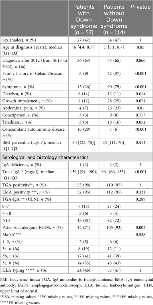
From January 1st, 2005, to December 31st, 2022, 770 patients with DS were admitted to the Pediatric Genetics' Clinic, among which 58 received a diagnosis of CD, with an overall prevalence of 7.5% (95% confidence interval, CI: 5.8%–9.6%). In the same period, 848 children were diagnosed with CD by the Pediatric Gastroenterology Clinic. Only one patient in the DS group could not be matched by date of birth and sex with any NS, leaving a final sample of 57 CD DS and 114 CD NS controls. Main clinical features of DS compared with NS children were reported in Table 1.

Table 1. Main clinical, serological and histology characteristics of celiac patients with and without Down syndrome at diagnosis.
Comparison of clinical, histological, and serological features of Celiac Disease in children with Down syndrome and non-syndromic children
In the DS group the frequency of CD in a first degree relative was 9% compared to 37% of NS. Symptoms at time of diagnosis were lower in the DS groups as compared with NS (26% vs. 79%, P < 0.001), whereas the incidence of autoimmune disease was higher (DS 28% vs. NS 6% P < 0.001). Thyroid disease was the most common autoimmune comorbidity among children with DS (Supplementary Table S2). In DS group, 81% of the anti-TGA levels were 10 times higher than the upper limit of normal (ULN), compared to 72% in NS. An intestinal biopsy sample was taken in 74% of all DS vs. 92% in the control group. Among patients with DS, 93% had histological damage equal to 3rd grade of Marsh-Oberhuber classification while 2 (5%) presented a 1st–2nd grade of Marsh-Oberhuber with anti-TGA levels below 10 times the ULN. In NS patients 6 (6%) presented a 1st–2nd grade of Marsh-Oberhuber (4 with anti-TGA levels below 10 times the ULN and 2 with anti-TGA levels over 10 times the ULN).
Considering the whole sample, the median follow-up was 747 days (first-third quartile 685–802), with a total of 32 patients lacking the 2 years follow-up visit. Supplementary Figure S1 reports the flow of patients among different BMI percentile classes over the 2 years follow-up, showing a reduction of patients in the class of BMI below the 5th percentile in the first year, with a subsequent small increase, without relevant differences among the two groups.
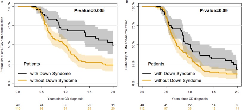
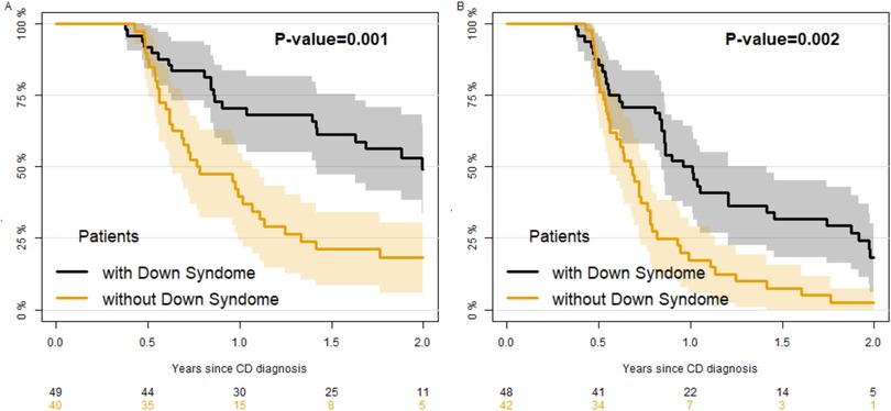
Figure 1 reports the percentage of children with positive (unnormal) anti-TGA (panel A) and EMA (panel B) by time since diagnosis. The median time to normalization of anti-TGA was significantly higher in DS (727 days, 95% CI 516–805) compared to NS patients (356 days, 95% CI: 263–403) (Figure 1, panel A, robust Wald test P = 0.005). The normalization of EMA was quicker, requiring a median time of 370 days (95% CI 308–516) for DS and 263 (95% CI: 239,311) for NS patients (Figure 1, panel B), without a significant difference between the two groups (robust Wald test P = 0.09). As children with DS perform yearly screening on CD, while others do not, we performed a secondary analysis selecting a more homogeneous group of controls including only children with a familial CD (and thus who probably had undergone antibody testing for screening). In those controls (Figure 2) the velocity of normalization of both anti-TGA and of EMA was higher, resulting in a significant difference between patients with and without DS (P = 0.001 and 0.002 respectively for anti-TGA and EMA). When we evaluated the factors associated with the velocity of anti-TGA normalization (Table 2, model A) in patients with DS, we found that the level of anti-TGA at diagnosis was associated with the velocity of normalization, in particular patients with anti-TGA values higher than 10 times the ULN showed a slower rate of normalization as compared with patients with values lower than 7 times the ULN (HR = 0.05, 95% CI 0.01–0.24). Over the total sample, (including patients with and without DS, Table 2 model B) level of anti-TGA at diagnosis and sex were both associated with velocity, with males being quicker in normalization of anti-TGA. As far as the comparison between patients with and without DS, the normalization of anti-TGA was slower in patients with DS as compared to patients without it (HR = 0.63, 95% CI: 0.40–1.001, P = 0.051).

Figure 1. Kaplan–Meier estimate of the percentage of patients positive to anti-TGA (panel A) and anti-EMA (panel B) since Celiac Disease diagnosis in children with and without Down syndrome.

Figure 2. Sensitivity analysis: Kaplan–Meier estimate of the percentage of patients positive to anti-TGA (panel A) and anti-EMA (panel B) since Celiac Disease diagnosis in children with Down syndrome as compared with children without Down syndrome but with a familiarity for Celiac Disease.

Table 2. Results on the Cox regression model on TGA normalization since Celiac Disease diagnosis among children with Celiac Disease with positive TGA at diagnosis.
Discussion
In the present analysis, the prevalence of CD in children and adolescents with DS was as high as 7.5%, remarkably greater than the occurrence reported in the historical Italian cohort assessed by Bonamico and colleagues (9). Several reasons may be hypothesized to support this apparent discrepancy, such as a deeper awareness of the clinical spectrum CD, a different serologic approach employed to diagnose CD [antigliadin antibodies (AGA) and EMA by Bonamico vs. TGA and EMA in the present analysis] and the systematic screening strategy introduced in our Centre from 2005 onwards. In addition, despite a patchy geographical distribution worldwide, a growing body of epidemiological studies held in our Country have shed light on the progressive increase of the prevalence of CD in school age children over the last decades (from 0.88% in 1999–2000 to 1.65% in 2017–2020) (19). The data recorded in patients with DS may simply mirror the trendlines of the general pediatric population, though the underlying causes of this phenomenon still need to be clarified.
The question of whether screening for CD is beneficial in the light of a higher incidence among individuals with DS remains a subject of controversy. The ESPGHAN 2020 (17) and the National Institute for Health and Clinical Excellence (NICE) guidelines (20) promote lab screening in populations exposed to a higher risk of developing CD, including DS, but the best timing for scheduled screening remains unclear. Conversely, the American Academy of Pediatrics (AAP) guidelines about health monitoring strategies for children with DS do not recommend systematic screening for CD due to the lack of indisputable evidence about its potential benefits (21).
The prevalence of diarrhea (14%), weight loss (12%), and abdominal pain (7%) observed in this study is superimposable to the findings reported by published literature about patients with DS (9, 22, 23). In some analyses (9, 24), the occurrence of symptoms may be overestimated, as testing for TGA and EMA antibodies were performed exclusively on symptomatic patients. The lower occurrence of clinical findings among patients with DS may be regarded as an expected outcome in a population screened yearly for CD, with the serological diagnosis anticipating the onset of reported symptoms. In addition, gastrointestinal disorders are reported in 50% of children with DS (23), therefore gastrointestinal symptoms may not be reliable indicators for identifying potential cases of CD in this population. In our study, a clinically driven prescription of serological work-up only among symptomatic patients would have led to remarkable underestimation of the prevalence of CD among patients with DS, as only 26% of affected children would have been diagnosed. Accordingly, we find reasonable to suggest a yearly lab screening for CD in DS and to test individuals with new onset of suggestive signs and symptoms.
Even if the BMI percentile median values were unchanged among patients with DS, we witnessed a progressive decrease in the number of patients with low BMI following the prescription of GFD. This data agrees with those published by Nisihara and colleagues (25) on children with DS.
As already extensively reported (9, 22, 26), our study highlights a greater occurrence of autoimmune disorders among celiac patients with DS, compared to NS controls (DS 28% vs. NS 6%). In our study, the most frequently observed autoimmune pathologies were thyroid disorders with a reported prevalence of 19%. The immune dysregulation in DS seems to be correlated with the combination of an increase in the expression of proinflammatory cytokines, an aberrant expression of B lymphocytes and an increase in the production autoantibodies directed against the central nervous system, gastrointestinal tract, pancreas, and thyroid (27).
Regarding family history of CD, our study showed a significantly lower prevalence in the CD diagnosed DS population compared to the control group (9% vs. 37%). To the best of our knowledge, no published studies report the frequency of family history of CD in Down children.
From a diagnostic perspective, 81% of patients with DS showed an anti-TGA titer 10-fold or greater the ULN. Most patients with DS were asymptomatic upon diagnosis and 93% of those undergoing intestinal biopsy showed a severe histological damage. These data are in consistent with the published literature, that reports a poor association between clinically relevant complaints and antibody titers or the degree of histologic damage (28), but a reliable agreement between serological data and histologic damage (28, 29). To the best of our knowledge, our study is the first specifically focused on CD in children with DS.
An additional element of novelty is the longer-lasting time needed to achieve antibodies normalization in DS compared to otherwise healthy controls. Our explanation for this phenomenon is the increased production of autoantibodies targeting the gastrointestinal tract, observed in individuals with DS (27), and the potential difficult adherence to GFD in this population. In a recent work published by Sbravati et al. (11), conducted on the general population, they found a time needed before the normalization of antibodies titer around 9 months from the beginning of the diet, irrespectively of the presence symptoms at diagnosis, with a longer time in those individuals with co-occurrent autoimmune diseases (e.g., diabetes or thyroiditis).
Given its retrospective nature, we are aware of some flaws affecting our analysis. Firstly, we did not have the chance to systematically collect data about the improvement gastrointestinal symptoms following GFD. Moreover, patients with DS were selected through the annual screening, so the probability to detect the antibody positivity before the surge of symptoms was higher than in NS. A multicentric prospective assessment is warranted in order to corroborate our outcomes achieved.
This study confirms a higher prevalence of CD in children and adolescents with DS and highlight the key diagnostic role of CD screening also among asymptomatic patients. Interestingly, even though the majority of DS are asymptomatic and detected through screening, they show higher serologic titers compared to NS controls. Moreover, the average degree of histopathological involvement was superimposable between Down patients and NS controls. This is one of a few works comparing clinical and serological characteristics between Down and non-Down patients. Moreover, it's the first analysis on the antibodies trend since diagnosis in patients with DS. Clinicians should expect a longer time of TGA IgA normalization after the start of a GFD. This should be anticipated to the families to avoid unuseful worries and blood samples to children with DS.
Data availability statement
The raw data supporting the conclusions of this article will be made available by the authors, without undue reservation.
Ethics statement
The studies involving humans were approved by Ethic Local Committee Board n° 3896. The studies were conducted in accordance with the local legislation and institutional requirements. Written informed consent for participation in this study was provided by the participants' legal guardians/next of kin.
Author contributions
ML: Data curation, Writing – review & editing, Conceptualization, Supervision, Writing – original draft. PR: Writing – original draft, Methodology, Formal analysis, Writing – review & editing, Data curation. CF: Methodology, Writing – review & editing, Conceptualization. AL: Data curation, Conceptualization, Writing – review & editing. LP: Data curation, Writing – original draft, Formal analysis. AC: Writing – review & editing, Supervision, Conceptualization. RP: Supervision, Writing – review & editing, Conceptualization. MV: Data curation, Formal analysis, Methodology, Conceptualization, Writing – review & editing. AB: Writing – review & editing, Supervision. GZ: Supervision, Data curation, Methodology, Writing – review & editing, Writing – original draft, Conceptualization.
Funding
The author(s) declare that financial support was received for the research and/or publication of this article. Open access funding was provided by the Italian Ministry of Health, ‘Ricerca Corrente’.
Conflict of interest
The authors declare that the research was conducted in the absence of any commercial or financial relationships that could be construed as a potential conflict of interest.
The author(s) declared that they were an editorial board member of Frontiers, at the time of submission. This had no impact on the peer review process and the final decision.
Generative AI statement
The author(s) declare that no Generative AI was used in the creation of this manuscript.
Publisher's note
All claims expressed in this article are solely those of the authors and do not necessarily represent those of their affiliated organizations, or those of the publisher, the editors and the reviewers. Any product that may be evaluated in this article, or claim that may be made by its manufacturer, is not guaranteed or endorsed by the publisher.
Supplementary material
The Supplementary Material for this article can be found online at: https://www.frontiersin.org/articles/10.3389/fped.2025.1595256/full#supplementary-material
References
1. Catassi C, Verdu EF, Bai JC, Lionetti E. Coeliac disease. Lancet. (2022) 399(10344):2413–26. doi: 10.1016/S0140-6736(22)00794-2
2. Bentley D. A case of down’s syndrome complicated by retinoblastoma and celiac disease. Pediatrics. (1975) 56(1):131–3. doi: 10.1542/peds.56.1.131
3. Liu E, Wolter-Warmerdam K, Marmolejo J, Daniels D, Prince G, Hickey F. Routine screening for celiac disease in children with down syndrome improves case finding. J Pediatr Gastroenterol Nutr. (2020) 71(2):252–6. doi: 10.1097/MPG.0000000000002742
4. Velasco-Benítez CA, Moreno-Giraldo LJ. Celiac disease in children with down syndrome. Rev Chil Pediatr. (2019) 90(6):589–97. doi: 10.32641/rchped.v90i6.925
5. Ostermaier KK, Weaver AL, Myers SM, Stoeckel RE, Katusic SK, Voigt RG. Incidence of celiac disease in down syndrome: a longitudinal, population-based birth cohort study. Clin Pediatr (Phila). (2020) 59(12):1086–91. doi: 10.1177/0009922820941247
6. Du Y, Shan LF, Cao ZZ, Feng JC, Cheng Y. Prevalence of celiac disease in patients with down syndrome: a meta-analysis. Oncotarget. (2017) 9(4):5387–96. doi: 10.18632/oncotarget.23624
7. Al-Toma A, Volta U, Auricchio R, Castillejo G, Sanders DS, Cellier C, et al. European Society for the study of coeliac disease (ESsCD) guideline for coeliac disease and other gluten-related disorders. United Eur Gastroenterol J. (2019) 7(5):583–613. doi: 10.1177/2050640619844125
8. Singh P, Arora A, Strand TA, Leffler DA, Catassi C, Green PH, et al. Global prevalence of celiac disease: systematic review and meta-analysis. Clin Gastroenterol Hepatol. (2018) 16(6):823–836.e2. doi: 10.1016/j.cgh.2017.06.037
9. Bonamico M, Mariani P, Danesi HM, Crisogianni M, Failla P, Gemme G, et al. Prevalence and clinical picture of celiac disease in Italian down syndrome patients: a multicenter study. J Pediatr Gastroenterol Nutr. (2001) 33(2):139–43. doi: 10.1097/00005176-200108000-00008
10. Sansotta N, Alessio MG, Norsa L, Previtali G, Ferrari A, Guerra G, et al. Trend of antitissue transglutaminase antibody normalization in children with celiac disease started on gluten-free diet: a comparative study between chemiluminescence and ELISA serum assays. J Pediatr Gastroenterol Nutr. (2020) 70(1):37–41. doi: 10.1097/MPG.0000000000002519
11. Sbravati F, Cosentino A, Lenzi J, Fiorentino M, Ambrosi F, Salerno A, et al. Antitissue transglutaminase antibodies’ normalization after starting a gluten-free diet in a large population of celiac children—a real-life experience. Dig Liver Dis. (2022) 54(3):336–42. doi: 10.1016/j.dld.2021.06.026
12. von Elm E, Altman DG, Egger M, Pocock SJ, Gøtzsche PC, Vandenbroucke JP. The strengthening the reporting of observational studies in epidemiology (STROBE) statement: guidelines for reporting observational studies. J Clin Epidemiol. (2008) 61(4):344–9. doi: 10.1016/j.jclinepi.2007.11.008
13. Zemel BS, Pipan M, Stallings VA, Hall W, Schadt K, Freedman DS, et al. Growth charts for children with down syndrome in the United States. Pediatrics. (2015) 136(5):e1204–11. doi: 10.1542/peds.2015-1652
14. Kuczmarski RJ, Ogden CL, Guo SS, Grummer-Strawn LM, Flegal KM, Mei Z, et al. 2000 CDC growth charts for the United States: methods and development. Vital Health Stat. (2002) 11(246):1–190.
15. Working Group of European Society of Paediatric Gastroenterology and Nutrition. Revised criteria for diagnosis of coeliac disease. Arch Dis Child. (1990) 65(8):909–11. doi: 10.1136/adc.65.8.909
16. Husby S, Koletzko S, Korponay-Szabó IR, Mearin ML, Phillips A, Shamir R, et al. European Society for pediatric gastroenterology, hepatology, and nutrition guidelines for the diagnosis of coeliac disease. J Pediatr Gastroenterol Nutr. (2012) 54(1):136–60. doi: 10.1097/MPG.0b013e31821a23d0
17. Husby S, Koletzko S, Korponay-Szabó I, Kurppa K, Mearin ML, Ribes-Koninckx C, et al. European Society paediatric gastroenterology, hepatology and nutrition guidelines for diagnosing coeliac disease 2020. J Pediatr Gastroenterol Nutr. (2020) 70(1):141–56. doi: 10.1097/MPG.0000000000002497
18. Oberhuber G, Granditsch G, Vogelsang H. The histopathology of coeliac disease: time for a standardized report scheme for pathologists. Eur J Gastroenterol Hepatol. (1999) 11(10):1185–94. doi: 10.1097/00042737-199910000-00019
19. Lionetti E, Pjetraj D, Gatti S, Catassi G, Bellantoni A, Boffardi M, et al. Prevalence and detection rate of celiac disease in Italy: results of a SIGENP multicenter screening in school-age children. Dig Liver Dis. (2023) 55(5):608–13. doi: 10.1016/j.dld.2022.12.023
20. National Institute for Health and Care Excellence (NICE). Coeliac Disease: Recognition, Assessment and Management. London: NICE (2015). Available online at: https://www.nice.org.uk/guidance/ng20
21. Bull MJ, Trotter T, Santoro SL, Christensen C, Grout RW, Burke LW, et al. Health supervision for children and adolescents with down syndrome. Pediatrics. (2022) 149(5):e2022057010. doi: 10.1542/peds.2022-057010
22. Chung H, Green PHR, Wang TC, Kong XF. Interferon-driven immune dysregulation in down syndrome: a review of the evidence. J Inflamm Res. (2021) 14:5187–200. doi: 10.2147/JIR.S280953
23. Bermudez BEBV, de Oliveira CM, de Lima Cat MN, Magdalena NIR, Celli A. Gastrointestinal disorders in down syndrome. Am J Med Genet A. (2019) 179(8):1426–31. doi: 10.1002/ajmg.a.61258
24. Costa Gomes R, Cerqueira Maia J, Fernando Arrais R, André Nunes Jatobá C, Auxiliadora Carvalho Rocha M, Edinilma Felinto Brito M, et al. The celiac iceberg: from the clinical spectrum to serology and histopathology in children and adolescents with type 1 diabetes mellitus and down syndrome. Scand J Gastroenterol. (2016) 51(2):178–85. doi: 10.3109/00365521.2015.1079645
25. Nisihara RM, Bonacin M, da Silva Kotze LM, de Oliveira NP, Utiyama S. Monitoring gluten-free diet in coeliac patients with down’s syndrome. J Hum Nutr Diet. (2014) 27(Suppl 2):1–3. doi: 10.1111/jhn.12137
26. Ram G, Chinen J. Infections and immunodeficiency in down syndrome. Clin Exp Immunol. (2011) 164(1):9–16. doi: 10.1111/j.1365-2249.2011.04335.x
27. Malle L, Patel RS, Martin-Fernandez M, Stewart OJ, Philippot Q, Buta S, et al. Autoimmunity in down’s syndrome via cytokines, CD4T cells and CD11c+ B cells. Nature. (2023) 615(7951):305–14. doi: 10.1038/s41586-023-05736-y
28. Pais WP, Duerksen DR, Pettigrew NM, Bernstein CN. How many duodenal biopsy specimens are required to make a diagnosis of celiac disease? Gastrointest Endosc. (2008) 67(7):1082–7. doi: 10.1016/j.gie.2007.10.015
Keywords: Down syndrome, Celiac Disease, children, screening, anti-transglutaminase antibodies, anti-endomysium antibodies
Citation: Lattuada M, Rebora P, Fossati C, Lazzerotti A, Paolini L, Cattoni A, Panceri R, Valsecchi MG, Biondi A and Zuin G (2025) Features of Celiac Disease in children and adolescents with Down syndrome: a single-center experience of annual screening. Front. Pediatr. 13:1595256. doi: 10.3389/fped.2025.1595256
Received: 17 March 2025; Accepted: 2 July 2025;
Published: 17 July 2025.
Edited by:
Nafiye Urganci, Şişli Hamidiye Etfal Education and Research Hospital, TürkiyeReviewed by:
Nilton Carlos Machado, Sao Paulo State University, BrazilShailendra Katwal, National Trauma Center, Nepal
Eyal Zifman, Meir Medical Center, Israel
Copyright: © 2025 Lattuada, Rebora, Fossati, Lazzerotti, Paolini, Cattoni, Panceri, Valsecchi, Biondi and Zuin. This is an open-access article distributed under the terms of the Creative Commons Attribution License (CC BY). The use, distribution or reproduction in other forums is permitted, provided the original author(s) and the copyright owner(s) are credited and that the original publication in this journal is cited, in accordance with accepted academic practice. No use, distribution or reproduction is permitted which does not comply with these terms.
*Correspondence: Giovanna Zuin, Z2lvdmFubmEuenVpbkBpcmNjcy1zYW5nZXJhcmRvLml0
 Martina Lattuada
 Paola Rebora
 Chiara Fossati3
 Lucia Paolini
 Alessandro Cattoni
 Andrea Biondi