 Ambily Francis1,2
 Immanuel Alex Pandian
 J. Anitha
- 1Department of Electronics and Communication Engineering, Karunya Institute of Technology and Sciences, Coimbatore, India
- 2Department of Electronics and Communication Engineering, Sahrdaya College of Engineering and Technology, Kodakara, India
- 3Department of Computer Science and Engineering, Karunya Institute of Technology and Sciences, Coimbatore, India
Introduction
Alzheimer's disease is a highly terrible condition for both the victims and their loved ones to endure. It is a degenerative neurological condition that steadily worsens until the patient is no longer able to do daily tasks. Even though there is no complete cure for the illness, several medications can slow its progression. After the age of 60, every person's brain, notably the hippocampus, begins to shrink (1, 2). But, for some unknown reasons, the shrinking rate will be higher for some people. In this context, the four cognitive stages of the human brain are classified as cognitively normal (CN), mild cognitive impairment convertible (MCIc), mild cognitive impairment non-convertible (MCInc), and Alzheimer's disease (AD) (3, 4). The stages are differentiated in terms of the shrinkage rate of the hippocampus. The hippocampus frequently shrinks as people age. Alzheimer's disease is clearly shown by the hippocampus's unusually rapid rate of atrophy. Cerebrospinal fluid (CSF) will occupy the gray matter area as the hippocampus shrinks. As a result, gray matter falls short of the level needed to maintain proper function. Therefore, the increasing CSF volume is yet another sign of Alzheimer's disease. The healthy brain and Alzheimer's disease affected brain are given in Figure 1 (*Credit: Adapted from illustration by Stacy Jannis/Alzheimer's Association, Link: https://www.alz.org/alzheimers-dementia/what-is-alzheimers/brain_tour/credits). The main determinants for classifying various stages of cognition are shrinkage of the hippocampus, growth of the brain's ventricles, and the change from gray matter to CSF (5, 6). The normal aging stage that will not lead to AD is known as mild cognitive impairment non-convertible stage. In this stage memory problems due to normal aging can be identified. However, Alzheimer's disease will never develop from this stage. The early stage of Alzheimer's disease with a significant rate of brain shrinking is known as the mild cognitive impairment convertible stage. The patients with mild cognitive impairment convertible stage will become Alzheimer's patients within years. In the field of early detection of AD, many algorithms address the classification of AD and CN. However, if the system accurately distinguishes between MCIc and MCInc, early detection of AD can be asserted. So the classification between MCIc and MCInc deserves more attention in this field.
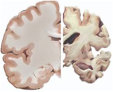
Figure 1. Healthy brain (Left) and Alzheimer's affected brain (Right)*.
Clinical practices of early diagnosis of Alzheimer's disease
To better comprehend the aberrant state of the human brain, doctors are running clinical trials. Clinical diagnoses are made using anecdotal evidence. The most often used technique for making a clinical diagnosis is the mini-mental state examination. Patients who are undergoing a mini-mental state assessment fill out a questionnaire designed to reflect any brain disorders, such as Alzheimer's disease. For the accurate diagnosis of disease, many conversations with clinicians are necessary.
For the screening, diagnosis, and subsequent management of Alzheimer's disease patients, doctors must be able to quickly and accurately identify the symptoms and pathology of the disease that are associated with Alzheimer's disease. Additionally, it enables patients and others who are caring for them to make necessary lifestyle modifications that may prolong the maintenance of their quality of life. Unfortunately, identifying Alzheimer's disease in its early stages in clinical practice can be difficult and is hampered by several obstacles, including time restrictions on clinicians, difficulty accurately diagnosing Alzheimer's pathology, and the fact that patients and healthcare professionals frequently write off symptoms as being a normal part of aging (7).
Computer based early diagnosis of Alzheimer's disease
Clinical exams and MRI scan reports may not be able to detect the early stage of AD in patients. If the early illness indications are discovered before the victim notices any symptoms, early detection of Alzheimer's disease gives a great possibility of prompt treatment. The researchers should integrate and evaluate the necessary data for the crucial improvement of medical diagnostic systems. Numerous contributions by researchers addressed the challenges related to the early detection of AD. In recent studies, MRI images are evaluated and processed to find signs of Alzheimer's disease in its early stages (8, 9). Doctors can use the successfully developed system as a clear channel of communication to identify AD in its early stages.
Machine learning techniques, both supervised and unsupervised, are applied to the analysis of medical images. Using supervised learning algorithms, labeled data is categorized using domain knowledge from an expert and pertinent feature data. The unsupervised learning algorithms work on unlabeled data. The supervised and unsupervised deep learning models are capable of automatically creating feature extractors and extracting discriminative features from train data. The rest of this section is structured as follows. Early diagnosis of AD using conventional feature extraction methods are deliberated in section Early diagnosis based on conventional feature extraction methods. Section Early diagnosis of Alzheimer's disease based on deep learning techniques describes related works on early diagnosis of AD using deep learning techniques. In section Early diagnosis of Alzheimer's disease based on pre-trained models, related works for early diagnosis of AD based on pre-trained models are discussed. The basic working flow of three computer-based methods for the early diagnosis of Alzheimer's disease are portrayed in Figure 2.
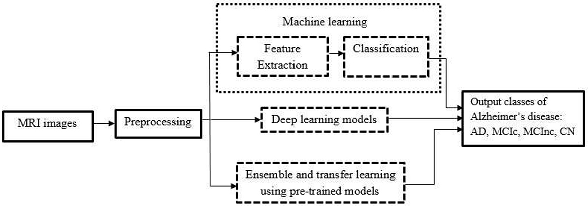
Figure 2. Basic working flow of computer-based methods.
Early diagnosis based on conventional feature extraction methods
Feature descriptors play an important role in medical image analysis. Medical image processing applications that explicitly use feature descriptors include disease diagnosis (10, 11), medical joint photographic experts group image steganography (12), object recognition, and segmentation. The two types of feature descriptors are global feature descriptors and local feature descriptors. Global features are those that apply to the entire image. Local features are provided by patches of an image. For any application involving the analysis of medical images using conventional feature extraction methods, global features are insufficient as they cannot provide the spatial information of images. For early Alzheimer's disease identification, many works suggested novel local feature descriptors.
Texture features are highly important low-level features that give significant details about specific regions of medical images. The pixel intensities of image patches are used to obtain the texture features. Numerous texture descriptors, including the local binary pattern, the scale-invariant feature transform, and the speed- up robust features, are used to interpret MRI images. The well-known descriptor, local binary pattern (LBP) is proposed for describing texture qualities (13). A high classification accuracy is achieved at the expense of a significant calculation time by using texture features taken from an elliptical neighborhood (14). Two-dimensional and three-dimensional advanced local binary patterns are combined to obtain accurate multi-class Alzheimer's disease prediction (15). The computational time taken for the processing of high-dimension features is the major limitation of this method. The textural features are identified by considering the voxel neighbors of MRI image (16–19).
Scale-invariant feature transform (SIFT) is another interesting local feature descriptor. The scale invariance and rotation invariance are the important characteristics of SIFT. Both the frequency domain and the spatial domain are utilized for extracting SIFT features. SIFT is effectively used for the early detection of AD (20–23). According to the discussions in the aforementioned articles, the benefit is that SIFT did not alter with variations in illumination. However, the gradients of each voxel along the path must be calculated. Hence the computation must be regarded as intensive.
The Hessian matrix serves as the foundation for the Speeded Up Robust Features (SURF) which shares many characteristics with SIFT (24). SURF performs admirably in computer vision applications and has been researched for use in medical image applications (5, 25, 26). The primary feature of SURF is the enhanced speed that the integral filter produces. As previously stated, one key sign of AD is the increasing CSF volume in the hippocampus. CSF volume computation is used to carry out early identification of AD (5). The CSF volume computation is done based on the number of CSF voxels in the hippocampal area. Circular harmonic functions (CHF) offers great image description independent of scale, position, and illumination for the early diagnosis of AD (27, 28).
After preprocessing, MRI images are sampled with nine volumes of interest in brain regions related to AD. Intensity and texture features are extracted from the interest of volume. The support vector machine classifier trains on the features. Non-linear registration systems can be made better even when the method offers tolerable accuracy (29). Clinical and texture characteristics are integrated to identify the mild cognitive impairment convertible stage (MCIc). The key benefit is that MCI (Mild cognitive impairment) and AD have been classified using the entire brain's MRI texture with binary logistic regression (30). A feature selection technique that makes use of a multivariate general linear model is proposed for the MCIc vs. CN classification. The modest intensity fluctuations from CN to MCIc are produced with the use of a general linear model. Additionally, multivariate adaptive regression splines are utilized as a classifier (31).
The orientation surrounding each voxel of eight local regions of MRI image including white matter and gray matter is calculated using a 3D local directional pattern. The algorithm is less susceptible to illumination and noise (11). The features obtained from many modalities are combined using multi- kernel SVM. In this method hyper graph-based regularization is used for AD vs. MCI classification. The method provides superior classification accuracy than existing multi-modality strategies, according to the results. The algorithm's primary flaw is that all hyperedge weights are set to 1 without considering various hyperedges (32). It is possible to identify Alzheimer's disease more quickly by analyzing data sets of medical records using the machine learning algorithms such as Logistic Regression, Decision Tree, Random Forest, Naive Bayes and variants of support vector machine to identify Alzheimer's infection (33).
Early diagnosis of Alzheimer's disease based on deep learning techniques
In recent years, deep learning methods have been prominent in many medical sectors. For efficient and precise categorization, deep learning techniques will abstract important features from the images. The methods aid medical professionals in early disease diagnosis. These methods enhance researchers' abilities to engage in studies that examine medical image approaches for disease diagnosis. Shortly, deep learning may replace traditional medical diagnostic techniques because of its learning capabilities and cost-effectiveness. A sizable number of convolutional neural network (CNN) models are combined to successfully anticipate various stages of AD. The classification performance is good as a result of the integration of Bagging, Boosting, and Random Forest algorithms (34). The review (35) listed and examined the most recent studies in the area of early Alzheimer's disease diagnosis using deep learning algorithms. Using 2D MRI slices, the work (36) proposes a CNN-based method for extracting discriminatory characteristics from structural MRI, intending to categorize Alzheimer's disease and healthy people.
The algorithm (37) uses functional MRI images in addition to medical data like age, gender, and genetic information. Using stacked autoencoders and functional MRI time-series data or correlation coefficient data, the deep neural network is trained. The research (38) reviews a number of deep learning based algorithms used in the early identification of Alzheimer's disease. The approach also offers a flexible foundation for repeatable testing. The strengths of hessian matrix and local binary pattern are utilized with the use of convolutional neural network (4). The algorithm provides best classification accuracy for AD vs. CN. But the Classification accuracy of MCIc vs. MCInc can be improved.
Early diagnosis of Alzheimer's disease based on pre-trained models
Transfer learning and ensemble learning are typically expressed in computer vision by employing pre-trained models. The innovative approaches for dealing with the issue of training data and test data having different distributions include transfer learning and ensemble learning. Pre-trained models can be used as the foundation for new models for the early diagnosis of AD. Using a neural network model, transfer learning is utilized to improve the precision and computing efficiency of image categorization. In transfer learning models, pre-trained neural networks are used as feature extractors, and the output of the pre-trained network is given to a new classification layer that is trained on data particular to the task. By integrating many previously trained models with new classification layers, ensemble transfer learning models (39, 40) are utilized to increase performance at the cost of increased model complexity. Without much computational complexity, the squeeze and excitation network affects the channel attention process by strengthening and weakening each feature channel. The algorithm (41) illustrates that non-biomedical pre-trained models, such as ResNet, learn cross-domain characteristics that enable the model to extract critical low-level properties from MRI scans to improve classification accuracy. The suggested method guarantees effective data augmentation before learning. The augmentation helps to regularize the model.
A 3D multi-channel feature maps based on Voxception-Resnet is created for the classification of AD and CN (42). Data augmentation is done before feature map creation. The dataset used in this algorithm is diffusion MRI images. The work (43) employs the VGG-16 pre-trained model, a non-biomedical model, to learn cross-domain characteristics and boost accuracy. The method achieves exceptional three-class classification accuracy and offers a mathematical model based on VGG-16 transfer learning. The method (44) uses 3D DenseNet to learn both hippocampus segmentation and classification features that based on deep CNN. Alzheimer's checking web application is proposed based on transfer learning of pre-trained models such as VGG19 (45).
Discussions
As discussed in the previous sections, AD develops gradually that takes years for symptoms to physically manifest in a patient, thus clinical approaches for an early diagnosis of the disease are insufficient. Physical symptoms of AD in its early stages will resemble those of a typical aged person in many ways. Clinical approaches are not very reliable for separating mild cognitive impairment into convertible and non-convertible stages in the context of early diagnosis.Early diagnosis is made using MRI scans using image processing techniques. The early diagnosis of AD utilizing MRI and Positron Emission Tomography (PET) image modalities is made possible by numerous algorithms (46, 47). Algorithms that need manual feature extraction to take a lot of time and have high computational complexity. Additionally, hardware implementation of such a system necessitates an extremely complicated system. Also, these kinds of systems only provide very poor MCIc vs. MCInc classification accuracy. The distinction between phases MCIc and MCInc is very difficult because of very slight voxel changes. Thus, the difference between MCIc and MCInc images may be described by a high-end feature description.
For the early diagnosis of AD, deep learning algorithms play a significant role. Numerous algorithms are developed for deep learning-based early diagnosis. Reduced computational complexity, less calculation time, low dimension features and good differentiation capability between MCIc vs. MCInc should be the goal of the computer-based diagnostic system. The pre-trained models successfully adapt the MRI images to predict the early phase of AD (48). The high-dimension features of pre-trained models cause complexity in the physical implementations later. But with pre-trained models, MCIc vs. MCInc classification accuracy is fairly good at the cost of computational complexity, processing time, and high dimension features.
Many investigations tried to classify AD vs. CN, and AD vs. CN vs. MCI (not specifically MCIc or MCInc). But only less focus is given to the classification of MCI vs. MCInc. Even though early Alzheimer's disease detection has been the subject of countless research, it is still challenging to identify the specific traits that can detect the disease in its earliest stages (49). The most salient classification for the early identification of AD is MCIc vs. MCInc. The objective of the upcoming efforts is to increase the classification precision of MCIc vs. MCInc. Due to extremely slight voxel changes, the differentiation between phases of MCIc and MCInc is exceedingly laborious. According to the published researches, MCIc vs. MCInc categorization accuracy ranges from 55 to 75% (38, 50, 51). Further study is necessary due to the low categorization accuracy for MCIc vs. MCInc. Unique learning strategies that discriminate between mild cognitive impairment convertible and nonconvertible stages will accelerate the classification accuracy of MCIc vs. MCInc. The reliable, fully automated system for the early diagnosis of AD can be a boon to the aged society in the near future.
Author contributions
AF, IP, and JA contributed to the study conception and design, literature review, interpretation, and manuscript preparation. All authors contributed to the article and approved the submitted version.
Acknowledgments
The authors express their sincere gratitude to the Karunya Institute of Technology and Sciences for the research facilities that have been extended.
Conflict of interest
The authors declare that the research was conducted in the absence of any commercial or financial relationships that could be construed as a potential conflict of interest.
Publisher's note
All claims expressed in this article are solely those of the authors and do not necessarily represent those of their affiliated organizations, or those of the publisher, the editors and the reviewers. Any product that may be evaluated in this article, or claim that may be made by its manufacturer, is not guaranteed or endorsed by the publisher.
References
1. Bethlehem RA, Seidlitz J, White SR, Vogel JW, Anderson KM, Adamson C, et al. Brain charts for the human lifespan. Nature. (2022) 604:525–33. doi: 10.1038/s41586-022-04554-y
2. Harris TC, de Rooij R, Kuhl E. The shrinking brain: cerebral atrophy following traumatic brain injury. Ann Biomed Eng. (2019) 47:1941–59. doi: 10.1007/s10439-018-02148-2
3. Salvatore C, Cerasa A, Battista P, Gilardi MC, Quattrone A, Castiglioni I, et al. Magnetic resonance imaging biomarkers for the early diagnosis of alzheimer's disease: a machine learning approach. Front Neurosci. (2015) 9:307. doi: 10.3389/fnins.2015.00307
4. Francis A, Pandian IA. Early detection of alzheimer's disease using local binary pattern and convolutional neural network. Multimed Tools Appl. (2021) 80:29585–600. doi: 10.1007/s11042-021-11161-y
5. Ben Ahmed O, Benois-Pineau J, Allard M, Ben Amar C, Catheline G. Classification of alzheimer's disease subjects from mri using hippocampal visual features. Multimed Tools Appl. (2015) 74:1249–66. doi: 10.1007/s11042-014-2123-y
6. Diogo VS, Ferreira HA, Prata D. Early diagnosis of alzheimer's disease using machine learning: a multi-diagnostic, generalizable approach. Alzheimer's Res. (2022) 14:1–21. doi: 10.1186/s13195-022-01047-y
7. Porsteinsson A, Isaacson R, Knox S, Sabbagh M, Rubino I. Diagnosis of early alzheimer's disease: Clinical practice in 2021. J Prevent Alzheimer's Dis. (2021) 8:371–86. doi: 10.14283/jpad.2021.23
8. Singh N, Soni N, Kapoor A, et al. Automated detection of alzheimer disease using mri images and deep neural networks-a review. arXiv. arXiv:2209.11282. (2022). doi: 10.48550/arXiv.2209.11282
9. Arafa DA, Moustafa HED, Ali-Eldin AM, Ali HA. Early detection of alzheimer's disease based on the state-of-the-art deep learning approach: a comprehensive survey. Multimed Tools Appl. (2022) 1–42. doi: 10.1007/s11042-022-11925-0
10. Hannun AY, Rajpurkar P, Haghpanahi M, Tison GH, Bourn C, Turakhia MP, et al. Cardiologist-level arrhythmia detection and classification in ambulatory electrocardiograms using a deep neural network. Nat Med. (2019) 25:65–9. doi: 10.1038/s41591-018-0268-3
11. Yan S, Song C, Zheng B. 3d local directional patterns for early diagnosis of Alzheimer's disease. J Eng. (2019) 2019:530–5. doi: 10.1049/joe.2018.9412
12. Liao X, Yin J, Guo S, Li X, Sangaiah AK. Medical jpeg image steganography based on preserving inter-block dependencies. Comp Electrical Eng. (2018) 67:320–9. doi: 10.1016/j.compeleceng.2017.08.020
13. Ojala T, Pietikainen M, Maenpaa T. Multiresolution gray-scale and rotation invariant texture classification with local binary patterns. IEEE Trans Pattern Anal Mach Intell. (2002) 24:971–87. doi: 10.1109/TPAMI.2002.1017623
14. Nanni L, Lumini A, Brahnam S. Local binary patterns variants as texture descriptors for medical image analysis. Artif Intell Med. (2010) 49:117–25. doi: 10.1016/j.artmed.2010.02.006
15. Sarwinda D, Bustamam A. Detection of alzheimer's disease using advanced local binary pattern from hippocampus and whole brain of mr images. In: 2016 International Joint Conference on Neural Networks (IJCNN) (IEEE) (2016) p. 5051–6. doi: 10.1109/IJCNN.2016.7727865
16. Oliver A, Llado' X, Freixenet J, Mart'i J. False positive reduction in mammographic mass detection using local binary patterns. In: International Conference on Medical Image Computing and Computer-Assisted Intervention (Springer) (2007) p. 286–293. doi: 10.1007/978-3-540-75757-3_35
17. Unay D, Ekin A, Jasinschi R. Medical image search and retrieval using local binary patterns and klt feature points. In: 2008 15th IEEE International Conference on Image Processing (IEEE) (2008) p. 997–1000. doi: 10.1109/ICIP.2008.4711925
18. Chang CW, Ho CC, Chen JH. Adhd classification by a texture analysis of anatomical brain mri data. Front Syst Neurosci. (2012) 6:66. doi: 10.3389/fnsys.2012.00066
19. Oppedal K, Engan K, Aarsland D, Beyer M, Tysnes OB, Eftestøl T. Using local binary pattern to classify dementia in MRI. In: 2012 9th IEEE International Symposium on Biomedical Imaging (ISBI) 235 (IEEE) (2012) p. 594–7. doi: 10.1109/ISBI.2012.6235618
20. Yang M, Yuan Y, Li X, Yan P. Medical image segmentation using descriptive image features. BMVC (Citeseer). (2011) 1–11. doi: 10.5244/C.25.94
21. Castellani U, Rossato E, Murino V, Bellani M, Rambaldelli G, Perlini C, et al. Classification of schizophrenia using feature-based morphometry. J Neural Transm. (2012) 119:395–404. doi: 10.1007/s00702-011-0693-7
22. Mizotin M, Benois-Pineau J, Allard M, Catheline G. Feature-based brain mri retrieval for alzheimer disease diagnosis. In: 2012 19th IEEE International Conference on Image Processing (IEEE) (2012) p. 1241–4. doi: 10.1109/ICIP.2012.6467091
23. Mondal P, Mukhopadhyay J, Sural S, Bhattacharyya PP. 3d-sift feature based brain atlas generation: An application to early diagnosis of alzheimer's disease. In: 2014 International Conference on Medical Imaging, m-Health and Emerging Communication Systems (MedCom) (IEEE) (2014) p. 342–347. doi: 10.1109/MedCom.2014.7006030
24. Bay H, Tuytelaars T, Gool LV. Surf: Speeded up robust features. In: European conference on computer vision (Springer) (2006) p. 404–417. doi: 10.1007/11744023_32
25. Sargent D, Chen CI, Tsai CM, Wang YF, Koppel D. Feature detector and descriptor for medical images. Med Imag 2009: Image Proc (SPIE). (2009) 7259:991–8. doi: 10.1117/12.811210
26. Lecron F, Benjelloun M, Mahmoudi S. Descriptive image feature for object detection in medical images. In: International Conference Image Analysis and Recognition (Springer) (2012) p. 331–338. doi: 10.1007/978-3-642-31298-4_39
27. Sorgi L, Cimminiello N, Neri A. Keypoints selection in the gauss laguerre transformed domain. BMVC. (2006) 539–547. doi: 10.5244/C.20.56
28. Sorokin DV, Mizotin MM, Krylov AS. Gauss-laguerre keypoints extraction using fast hermite projection method. In: International Conference Image Analysis and Recognition (Springer) (2011) p. 284–293. doi: 10.1007/978-3-642-21593-3_29
29. Chincarini A, Bosco P, Gemme G, Esposito M, Rei L, Squarcia S, et al. Automatic temporal lobe atrophy assessment in prodromal ad: Data from the descripa study. Alzheimer's & Dementia. (2014) 10:456–67. doi: 10.1016/j.jalz.2013.05.1774
30. Luk CC, Ishaque A, Khan M, Ta D, Chenji S, Yang YH, et al. Alzheimer's disease: 3-dimensional mri texture for prediction of conversion from mild cognitive impairment. Alzheimer's & Dementia. (2018) 10:755–63. doi: 10.1016/j.dadm.2018.09.002
31. Çevik A, Weber GW, Eyüboğlu BM, Oguz KK. Voxel-mars: a method for early detection of alzheimer's disease by classification of structural brain mri. Ann Operations Res. (2017) 258:31–57. doi: 10.1007/s10479-017-2405-7
32. Shao W, Peng Y, Zu C, Wang M, Zhang D, Initiative ADN, et al. Hypergraph based multi-task feature selection for multimodal classification of alzheimer's disease. Computerized Medical Imag Graphics. (2020) 80:101663. doi: 10.1016/j.compmedimag.2019.101663
33. Kishore P, Kumari CU, Kumar M, Pavani T. Detection and analysis of alzheimer's disease using various machine learning algorithms. Materials Today: proceedings. (2021) 45:1502–8. doi: 10.1016/j.matpr.2020.07.645
34. Pan D, Zeng A, Jia L, Huang Y, Frizzell T, Song X. Early detection of alzheimer's disease using magnetic resonance imaging: a novel approach combining convolutional neural networks and ensemble learning. Front Neurosci. (2020) 14:259. doi: 10.3389/fnins.2020.00259
35. Al-Shoukry S, Rassem TH, Makbol NM. Alzheimer's diseases detection by using deep learning algorithms: a mini-review. IEEE Access. (2020) 8:77131–41. doi: 10.1109/ACCESS.2020.2989396
36. Al-Khuzaie FE, Bayat O, Duru AD. Diagnosis of alzheimer disease using 2d mri slices by convolutional neural network. Appl Bionics Biomech. (2021) 2021. doi: 10.1155/2021/6690539
37. Ju R, Hu C, Li Q, et al. Early diagnosis of alzheimer's disease based on resting-state brain networks and deep learning. IEEE/ACM Trans. Comput. Biol. Bioinform. (2017) 16:244–57. doi: 10.1109/TCBB.2017.2776910
38. Wen J, Thibeau-Sutre E, Diaz-Melo M, Samper-Gonza'lez J, Routier A, Bottani S, et al. Convolutional neural networks for classification of alzheimer's disease: overview and reproducible evaluation. Med Image Analy. (2020) 63:101694. doi: 10.1016/j.media.2020.101694
39. Fathi S, Ahmadi M, Dehnad A. Early diagnosis of alzheimer's disease based on deep learning: a systematic review. Comput Biol Med. (2022) 105634. doi: 10.1016/j.compbiomed.2022.105634
40. Francis A, Pandian IA. Early detection of alzheimer's disease using ensemble of pre-trained models. In: 2021 International Conference on Artificial Intelligence and Smart Systems (ICAIS) (IEEE). (2021) p. 692–696. doi: 10.1109/ICAIS50930.2021.9395988
41. Valliani A, Soni A. Deep residual nets for improved alzheimer's diagnosis. In: Proceedings of the 8th ACM International Conference on Bioinformatics, Computational Biology, and Health Informatics. (2017) p. 615–615. doi: 10.1145/3107411.3108224
42. McCrackin L. Early detection of alzheimer's disease using deep learning. In: Canadian Conference on Artificial Intelligence (Springer) (2018) p. 355–359. doi: 10.1007/978-3-319-89656-4_40
43. Jain R, Jain N, Aggarwal A, Hemanth DJ. Convolutional neural network based alzheimer's disease classification from magnetic resonance brain images. Cogn Syst Res. (2019) 57:147–59. doi: 10.1016/j.cogsys.2018.12.015
44. Liu M, Li F, Yan H, Wang K, Ma Y, Shen L, et al. A multi-model deep convolutional neural network for automatic hippocampus segmentation and classification in alzheimer's disease. Neuroimage. (2020) 208:116459. doi: 10.1016/j.neuroimage.2019.116459
45. Helaly HA, Badawy M, Haikal AY. Deep learning approach for early detection of alzheimer's disease. Cognitive Computat. (2021) 1–17.
46. Lu D, Popuri K, Ding GW, Balachandar R, Beg MF. Multimodal and multiscale deep neural networks for the early diagnosis of alzheimer's disease using structural mr and fdg-pet images. Sci Rep. (2018) 8:1–13. doi: 10.1038/s41598-018-22871-z
47. Sherin A, Rajeswari R. Computer-aided diagnosis system for alzheimer's disease using positron emission tomography images. Interdisciplinary Sci. (2021) 13:433–42. doi: 10.1007/s12539-020-00409-0
48. Shanmugam JV, Duraisamy B, Simon BC, Bhaskaran P. Alzheimer's disease classification using pre-trained deep networks. Biomed Signal Process Control. (2022) 71:103217. doi: 10.1016/j.bspc.2021.103217
49. Kavitha C, Mani V, Srividhya S, Khalaf OI, Romero CAT. Early-stage alzheimer's disease prediction using machine learning models. Front Public Health. (2022) 10. doi: 10.3389/fpubh.2022.853294
50. Oh K, Chung YC, Kim KW, Kim WS, Oh IS. Classification and visualization of alzheimer's disease using volumetric convolutional neural network and transfer learning. Sci Rep. (2019) 9:1–16. doi: 10.1038/s41598-019-54548-6
Keywords: deep learning, pre-trained network, mild cognitive impairment (MCI), features, cognitively normal (CN), Alzheimer's disease
Citation: Francis A, Pandian IA and Anitha J (2022) A boon to aged society: Early diagnosis of Alzheimer's disease–An opinion. Front. Public Health 10:1076472. doi: 10.3389/fpubh.2022.1076472
Received: 21 October 2022; Accepted: 14 November 2022;
Published: 01 December 2022.
Edited by:
Steven Fernandes, Creighton University, United StatesReviewed by:
Belfin Robinson, University of North Carolina at Chapel Hill, United StatesCopyright © 2022 Francis, Pandian and Anitha. This is an open-access article distributed under the terms of the Creative Commons Attribution License (CC BY). The use, distribution or reproduction in other forums is permitted, provided the original author(s) and the copyright owner(s) are credited and that the original publication in this journal is cited, in accordance with accepted academic practice. No use, distribution or reproduction is permitted which does not comply with these terms.
*Correspondence: J. Anitha, YW5pdGhhX2pAa2FydW55YS5lZHU=