 Aboudou Habirou Kifouly
 Ngemani Obase Bekindaka
 Kaltun Said Ali1
 Juliana Rume
- 1Pan African University Life and Earth Sciences Institutes (Including Health and Agriculture), Ibadan, Oyo, Nigeria
- 2Department of Obstetrics and Gynecology, University College Hospital, University of Ibadan, Ibadan, Oyo, Nigeria
- 3Department of Animal Health and Production, University of Abomey-Calavi, Abomey-Calavi, Atlantique, Benin
Introduction: This study aims to access for the first time in the Benin Republic, the characteristics of the health status and the serological prevalence of Chlamydia abortus in pregnant women. Enzootic Abortion of Ewes (EAE) is a bacterial illness that can harm sheep by producing abortions and miscarriages in pregnant women.
Methods: About six municipalities under two governorates were concerned and around 420 pregnant women were enrolled for the survey (210 participants in each governorate). Among of this enrollment, 385 participants were concerned for serological test.
Results: Our result showed that this survey among pregnant women at the Sakété-Ifangni health zone hospital revealed that 125 participants (59.52%) had been exposed to potentially infected animals or products, with 40% having touched items from sick animals. Overall, 65.24% of animal owners were unsure whether they had been exposed. As much as 28 to 38% of the women farmed alongside their husbands, which frequently led to direct contact with aborted products. The consumption of milk from small ruminants was 26.67%. This consumption was associated with the risk of Chlamydia abortus. Half of them had experienced pregnancy complications. Knowledge of Chlamydia abortus varied from 16 to 68.5%. Proximity to small ruminant farms increases the risk of infection. Awareness among healthcare professionals needs to be improved. Although, the serological prevalence observed was relatively low (1.30%), it reveals a significant past exposure to the pathogen, especially in rural or cross-border areas such as the majority of the municipalities involved in this study.
Conclusion: This data constitutes an epidemiology alert, justifying the introduction of additional methods such as PCR to access the active circulation and refine prevention strategies.
Highlights
• The survey of pregnant women in our study revealed that tests for Chlamydia abortus infection are never administered, and worse still, other midwives and care assistants, as well as pregnant women, have never been aware of this infection.
• This observation was confirmed not only by the presence of numerous risk factors contributing to the suspicion of this disease in the environment of these pregnant women, but also by the serological analysis which revealed positive cases of Chlamydia abortus in the present study.
• Through this study, we suggested to the hospital administrative authorities that they require the Chlamydia abortus test during antenatal examinations.
1 Introduction
Chlamydia abortus (C. abortus) is a non-motile obligate intracellular Gram-negative pathogenic bacterium, belonging to the Chlamydiales family. C. abortus infects mainly ruminants, especially sheep and goats, and less frequently cattle, pigs and horses; however, it can also affect humans, being of particular concern in pregnant women (1, 2). C. abortus is known as the causative agent of enzootic abortion of ewes (EAE) or ovine enzootic abortion (OEA) which represents one of the most common causes of ovine and caprine infectious abortion worldwide, along with other infectious agents such as Campylobacter sp., Toxoplasma sp., Listeria sp., Salmonella sp., Border disease virus and Cache Valley virus (3, 4). Abortion occurs in the later stages of pregnancy, as C. abortus is able to progressively colonize the placenta, causing damage and affecting the fetus(es) to varying degrees (5). The infection can result in fetal loss (abortion), the birth of stillborn or weak lambs or, in some cases of unaffected animals; presence of a weak lamb with a healthy twin is also not uncommon (6). Breeders can incur great economic losses if numerous cases occur in a farm (abortion storm), usually when the infection first affects a naïve flock (7). Another important aspect is the spread of this enzootic infection to humans, which can develop as severe disease, especially in pregnant women (1), generally affecting female farmers, abattoir workers and veterinarians. However, environmental contamination with the bacteria released by abortion products or infected animals may also play a crucial role in disease spread, interspecies cross-over and adaptation (8). Indeed, abortion products, in particular vaginal fluids, placentas, dead/aborted lambs, fleeces and still born/infected lambs are all characterized by a high bacterial load and represent a significant risk, both for naïve animals and for humans (9). Infected and aborted animals shed the organism in placenta, uterine discharge, and feces, contaminating soils and generating infective aerosols that can be inhaled (9). Different types of flock management also influence the extent of environmental contamination and the spread of the pathogen. In intensively managed flocks, where animals are kept in smaller enclosures, there is a higher incidence of C. abortus, as the contamination is concentrated in confined spaces. Conversely, in extensively managed flocks, where animals are kept over larger areas, a lower incidence of the pathogen is observed, likely because animals are less exposed to contaminated environments (3). In addition, C. abortus can survive in the environment even in unfavorable conditions from a few days to a few months, thanks to the presence of a spore-like cell wall, which gives it considerable resilience (7). This resistance seems to be directly connected to the greater possibility for the bacterium to come into contact and infect many animal species, farmed or wild ones, and consequently to spread more easily to humans (1). Specific aspects of this will be discussed later.
The objective of this study was to assess for the first time, the characteristics of the health status and the serological prevalence of C. abortus in pregnant women in Benin, primarily in the governorate of Ouémé and Plateau.
2 Materials and methods
2.1 Period and type of study
This is a cross-sectional survey with a descriptive aim, conducted during the period from January to April 2023. This study followed the EQUATOR guidelines especially the extends STARD checklist.
2.2 Study area
The Ouémé department, with Porto-Novo as its capital, is characterized by its economic dynamism and fertile agricultural areas. The Plateau department, with its capital in Pobè, is distinguished by its hilly terrain, cultural richness, preservation of traditions, and traditional craftsmanship. Both regions encompass several municipalities, three (Ifangni, Sakété, and Adja-Ouèrè in Plateau department (Category 1), and Avrankou, Akpro-Missérété, and Dangbo in department of Ouémé; Category 2) of which have been included in this study. The Sakété-Ifangni health zone hospital, situated in Sakété within the Plateau region of Benin, serves as the focal point for this study.
2.3 Recruitment of participants
Pregnant women from prenatal clinics, health facilities, or maternity wards were recruited. This survey on enzootic sheep sickness was carried out from pregnant women attending prenatal clinics at the Sakété-Ifangni health zone hospital. These ladies were from diverse towns, the vast majority of which are within our research region. Avrankou, Akpro-Missérété, Dangbo, Adja-Ouèrè, Sakété, and Ifangni were among them.
2.4 Questionnaire
The questionnaire was created to gather information about the pregnant woman’s history of C. abortus infection, symptoms, risk factors, and sexual risk behaviors. The questionnaire also asked pregnant women about their knowledge and attitudes of C. abortus infection. The data were registered using KoboCollect. With that, more than 70 pregnant women were questioned by localities (06 localities in total) and around a total 420 were interrogated about their health status and their knowledge regarding C. abortus infections. The information was subdivided into two classifications for relevant comparisons. The category 1 is related to 3 municipalities of Plateau governorate which was: Ifangni, Sakété and Adja-Ouèrè and the second governorate were composed also 3 municipalities such as Avrankou, Akpro-Missérété and Dangbo.
2.5 Eligibility criteria
Inclusion criteria for pregnant women in a Chlamydia abortus study:
• Be pregnant, confirmed by ultrasound or a positive pregnancy test.
• Have a gestational age between 12 and 28 weeks.
• Be 18 years of age or older.
• Have given informed consent to participate in the study.
• Have regular access to health care during pregnancy.
• Pregnant women in the first trimester of pregnancy.
Exclusion criteria for pregnant women in a Chlamydia abortus study:
• Pregnant women in the second or third trimester of pregnancy
• Have been treated for chlamydia within the last 6 months.
• Have known HIV, hepatitis B, or hepatitis C infection.
• Have other sexually transmitted infections.
• Be on treatment for other chronic conditions that could affect pregnancy, such as diabetes or high blood pressure.
• History of pregnancy complications, such as pre-eclampsia or gestational diabetes
• Use of antibiotics in the 4 weeks before study inclusion
• Antibiotic use in the 30 days before study entry
• Diagnosis of C. trachomatis at the time of the study
• Alcohol or illicit drug use during pregnancy.
2.6 Sampling
The number of participants that were tested for C. abortus (via these clinical manifestations) were 385 pregnant women. Blood was drawn from the veins of the arm or wrist in dry tubes labeled with the patient’s medical record number. These tubes were then brought to the hospital laboratory and centrifuged at 3000 rpm for 5 min. This enabled us to separate the serum from the remainder of the blood and collect it in Eppendorf tubes labeled with the dry tube number.
2.7 Laboratory analysis
Diagnostic work has been carried out within the serology unit of the Bohicon Veterinary Laboratory. To test the sera, indirect ELISA kits provided by ID, VET companies (Montpellier, France) for C. abortus (chlamydiosis) were used. The tests were carried out following the manufacturer’s recommendations. Plate reading was performed using the ELISA reader (Chromate Inc. Germany) at 450 nm. For this zoonotic disease, the test was validated when the mean value of the optical density of the positive samples was greater than 0.350 and the ratio between the mean of the optical densities of the positive samples (ODpc) and those of the negative controls (ODnc) was greater than 3.
2.8 Statistical analysis
The data collected were analyzed to determine the prevalence of C. abortus infection, associated risk factors were calculated using the chi-square test, and knowledge and perceptions of infection by pregnant women were calculated using the student t-test.
3 Results
3.1 Distribution of pregnant women across different age
Table 1 shows the distribution of pregnant women across the different age groups, for both categories 1 and 2. In category 1, the largest number of pregnant women were in the 25–40 age group, accounting for 42.86% of the total. In category 2, the largest group of pregnant women were also in the 25–40 age group, representing 32.38% of the total. Notably, there is a significant difference in the percentages of pregnant women between the two categories in these age groups, with a p-value of 0.004, indicating a statistically significant difference between the two categories.

Table 1. Distribution of pregnant women across different age.
3.2 Factors influencing pregnancy risks among pregnant women
Table 2 presents on Factors Influencing Pregnancy Risks among pregnant women. Significant correlations were observed between the factors (such as indirect exposure to sick animals, housing type, farmer classification, previous illnesses, and the presence of sick animal products) and pregnancy risks among pregnant women. Indirect contact with sick animals was significantly more prevalent in category 2 (73.3%) compared to category 1 (59.5%; p = 0.000). Similarly, there was a higher incidence of indirect exposure to infected animal products in category 2 (47.1%) than in category 1 (40.5%), though not statistically significant (p = 0.053). Additionally, hygiene practices, such as disinfection after contact, were significantly less common in category 2 (18.1%) than in category 1 (24.3%; p = 0.005). Living near a livestock farm was also significantly less frequent in category 2 (31.9%) than in category 1 (39.1%; p = 0.026). Furthermore, mixed breeding’s were significantly more prevalent in category 2 (37.6%) than in category 1 (27.6%; p = 0.006). Underlying diseases were significantly less common in category 2 (33.3%) compared to category 1 (40.0%; p = 0.040). In summary, these findings suggested riskier exposures but better hygiene practices in category 1, while category 2 appears to have overall better health outcomes, indicating the necessity for targeted preventive measures to combat a likely incidence of C. abortus infection.
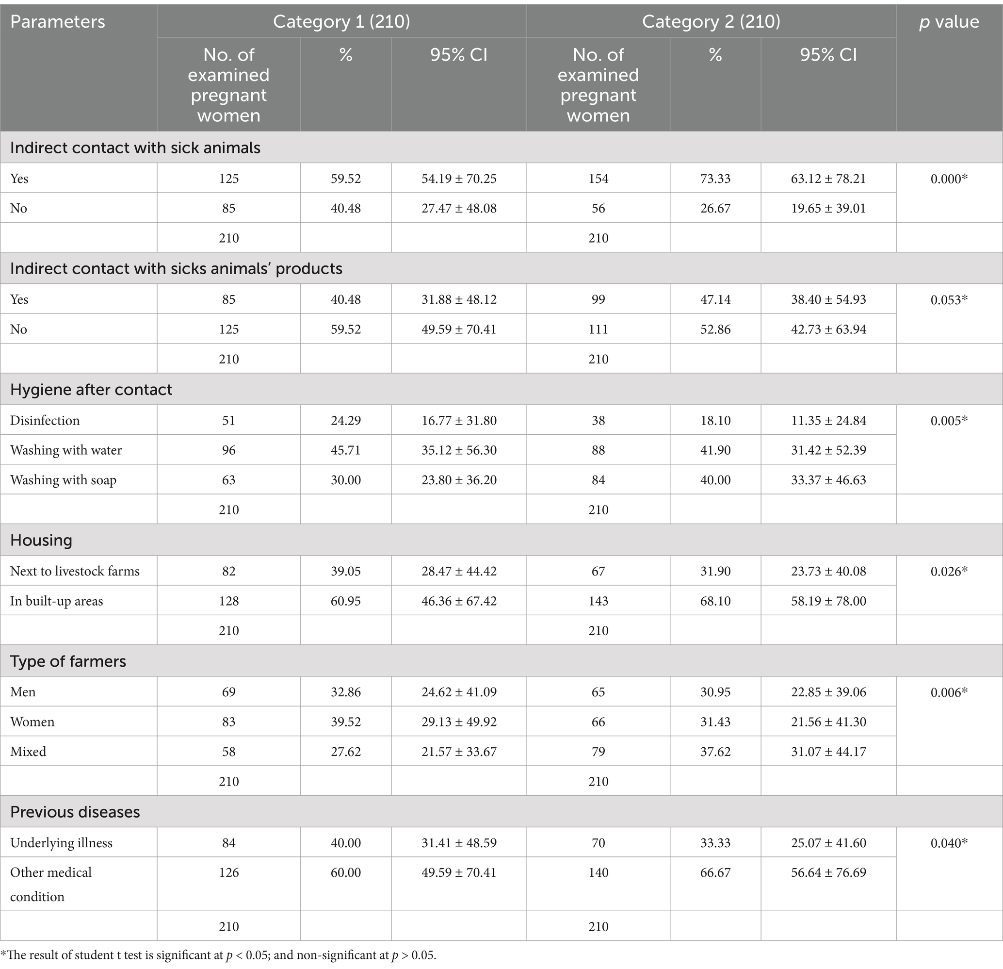
Table 2. Factors influencing pregnancy risks among pregnant women.
3.3 Factors influencing awareness of Chlamydia abortus on pregnancy
Table 3 compares categories on zoonotic disease prevention (C. abortus) in pregnant women. Preventive measures against zoonoses, included C. abortus, were significantly more prevalent in category 2 (62.9%) than in category 1 (54.8%; p = 0.015). Similarly, knowledge of available treatments for C. abortus was significantly higher in category 2 (64.3%) than in category 1 (56.2%; p = 0.014). History of genitourinary infections was also significantly more common in category 2 (58.1%) than in category 1 (50.5%; p = 0.025). Obstetric complications were more frequent in category 2 (59.1%) than in category 1 (51.9%; p = 0.035). However, knowledge of C. abortus and its associated risks was lower in category 2 (39.1%) compared to category 1 (46.2%), significantly (p = 0.034). In summary, despite improvements in prevention and clinical management, C. abortus infection possibly remained a concern in category 2, emphasized the need for enhanced education and screening.
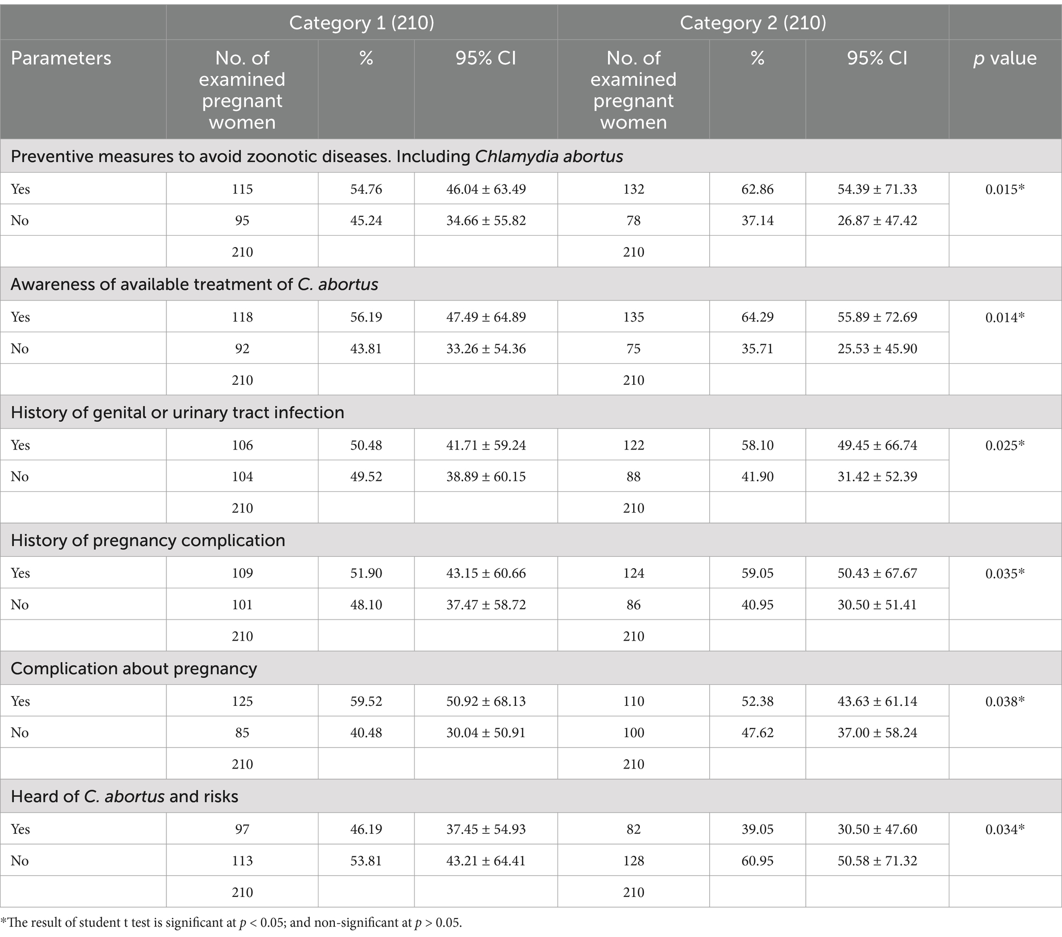
Table 3. Factors influencing awareness of C. abortus on pregnancy complications among the pregnant women examined.
3.4 Associated risks factors of Chlamydia abortus among pregnant women
Table 4 shows pregnant women differences in interactions, farm proximity, milk consumption, risk awareness, and symptoms. Significant p-values indicate impact on outcomes of pregnancy. Women who frequently visited small ruminant farms were significantly more prevalent in category 1 (35.7%) than in category 2 (33.3%; p = 0.013). Similarly, women living within 5 km of these farms were significantly more prevalent in category 2 (39.5%) than in category 1 (30.0%; p = 0.019). Regular consumption of small ruminant milk was also significantly higher in category 2 (23.8%) than in category 1 (14.8%; p = 0.009). Potential exposure to infected animals was reported by 34.8% of women in category 1 compared to 28.1% in category 2, significantly (p = 0.032). However, awareness of the risk of C. abortus infection was significantly higher in category 2 (41.9%) than in category 1 (34.8%; p = 0.04). Additionally, a history of abortions was significantly more common in category 2 (57.6%) than in category 1 (51.4%; p = 0.036). In summary, certain interactions with small ruminants seemed to elevate the risk of C. abortus infection in pregnant women, underscoring the need for improved education and preventive healthcare measures.
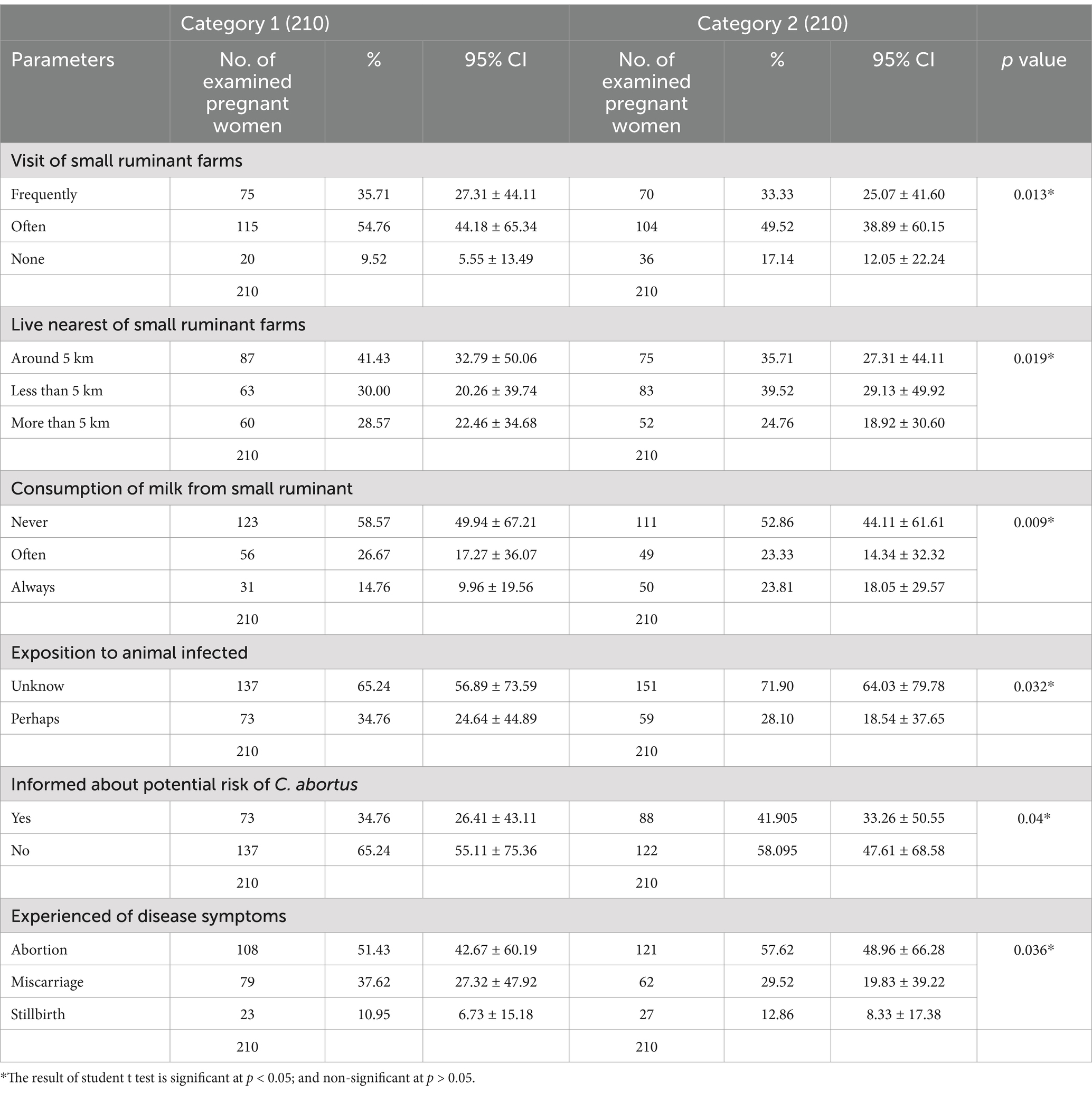
Table 4. Associated risks factors of C. abortus among pregnant women.
3.5 Prevalence of Chlamydia abortus
Table 5 shows the prevalence of C. abortus in pregnant women in different regions. The overall incidence was relatively low, with only 5 positive cases out of a total of 385 women tested (1.3%). Ifangni and Sakété municipalities had the highest incidences, at 1.49 and 2.67%, respectively. Conversely, Adja-Ouèrè and Dangbo municipalities have reported no positive cases. Despite the low seroprevalence, it underscored the presence of C. abortus among the demographic of pregnant women in these regions.
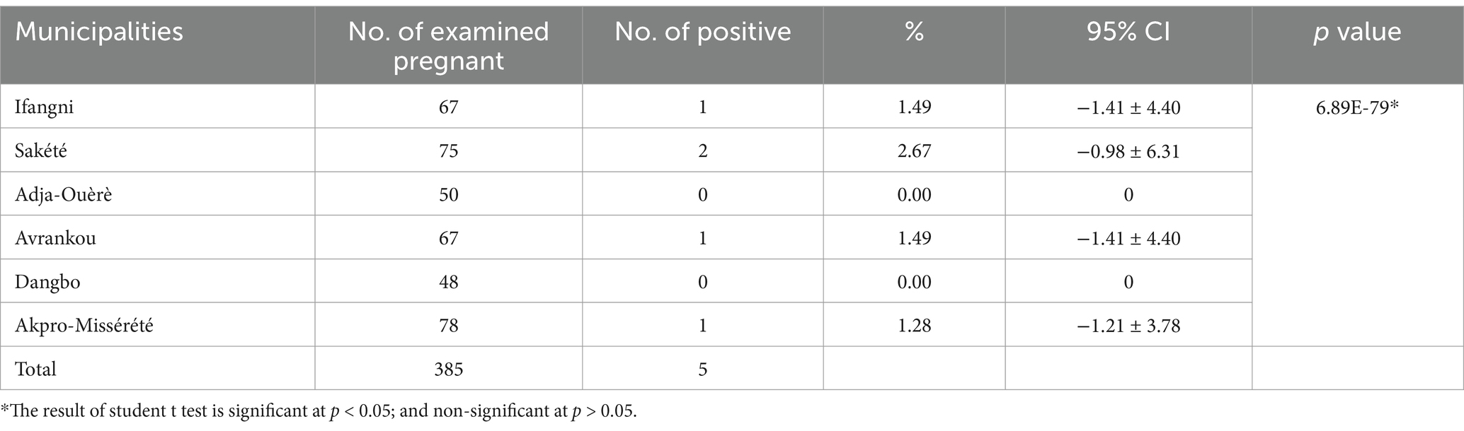
Table 5. Prevalence of C. abortus cases among expectant women.
4 Discussion
The survey of pregnant women attending an antenatal clinic for the first time highlights the proportion of pregnant women surveyed who had been exposed to animals suspected of being infected or to potentially contaminated animal products such as milk or blood in the course of their work or daily activities. The results show that more than half of the pregnant women surveyed (59.52%) had been exposed to sick animals indirectly, and almost half of these women (40%) said that they had at least touched sick animal products (blood, milk, saliva, snot, etc.) indirectly as part of their work or daily activities. This observation can be justified by the fact that nearly 40% of the pregnant women surveyed were livestock owners and that the majority of them (65.24% and 72% in both categories) did not know whether or not they were exposed to infected animals; while nearly 28% and 38% of these women also practiced animal husbandry jointly with their husbands, and because of the proximity, nearly 52% and 58% in both categories stated that they often touched aborted products. The prevalence of women reporting contact with diseased or dead animal products is relatively higher than that reported by (10), who found that 26% of women interviewed in their study had been in contact with dead animal products. However, regarding the consumption of small ruminant milk, the results presented indicate that only 26.67% of the women surveyed responded that they often consumed it. However, this proportion may be underestimated as a large number of women did not answer this question. Nevertheless, this prevalence is lower than that reported by (11), who found that 29.8% of the women surveyed in their study had consumed goat’s or sheep’s milk. In addition, a study by (12) in Germany showed that consumption of raw goat’s or sheep’s milk was a significant risk factor for C. abortus infection in pregnant women. This study suggests that the consumption of milk from small ruminants may play an important role in the transmission of C. abortus to pregnant women.
Interesting results on the medical history of the pregnant women interviewed with regard to C. abortus were collected during this survey. It is of concern that some of the women surveyed had a history of genital or urinary tract infections, as this may increase the risk of infection during pregnancy. In terms of symptoms, it was found that almost half of the women had experienced abortions, miscarriages and stillbirths in both departments during their maternity career. Our results are similar to those obtained by (13) who showed that 25% of pregnant women had a C. abortus infection, but lower than those found by (14) who showed that 43.3% of pregnant women infected with C. abortus presented symptoms such as vaginal discharge, abdominal pain or vaginal bleeding. These symptoms may be associated with C. abortus infection, but it is important to note that they may also be caused by other infections or pathologies. Finally, a study by (5) showed that a history of C. abortus infection was associated with an increased risk of complications during pregnancy, such as spontaneous abortion or preterm labor. The findings were concerning, revealing that half of the women had experienced pregnancy complications potentially linked to infection, while most lacked adequate awareness of Chlamydia abortus during pregnancy.
These results are in agreement with several other studies conducted on this subject. A study conducted by (15) found that only 16% of pregnant women surveyed were aware of the possibility of transmission of C. abortus from mother to child during pregnancy. Similarly, a study by (16) found that only 28% of pregnant women surveyed were aware of C. abortus and its consequences for the health of mother and child. However, other studies have reported higher rates of knowledge than the 20.52% found. For example, a study by (17) found that 67.8% of pregnant women surveyed were aware of C. abortus. Similarly, a study by (18) showed that 68.5% of pregnant women surveyed were aware of Chlamydia abortus and its impact on the health of mother and child.
Interesting information on the proximity and frequency of visits by pregnant women to small ruminant farms has been collected. However, a study by (19) revealed that small ruminants are important reservoirs of C. abortus, which can be transmitted to pregnant women through direct or indirect contact. The results of the present study are consistent with this observation, as more than half of the women surveyed responded that they lived near small ruminant farms. In addition, another study by (20) also examined the prevalence of C. abortus in small ruminants in Hungary. The authors found that the prevalence of infection was higher in small ruminant farms located in rural areas, which is consistent with the results presented in our study. In addition, a study by (1) evaluated the efficacy of different vaccines against C. abortus in small ruminants. The authors emphasized the need to control infections in small ruminants to prevent zoonotic transmission. Accordingly, raising awareness among pregnant women about the risks linked to close contact and regular visits to small ruminant farms is crucial. The results mentioned also that the majority of pregnant women are not well informed about the risks and means of preventing C. abortus during pregnancy, or about the advice given by healthcare professionals. These results are in line with other studies conducted on the subject. A study conducted by (21) among pregnant women in an antenatal clinic in Italy showed that only 33.5% of women knew about C. abortus and its risks during pregnancy. Similarly, a study conducted by (22) in the UK showed that only 25.6% of pregnant women were aware of C. abortus. In addition, these findings also highlight the need to improve knowledge of C. abortus among healthcare professionals, so that they can provide adequate advice and information to pregnant women. Previous studies have also highlighted the need for adequate training of healthcare professionals on awareness, screening and treatment of C. abortus during pregnancy (21), (22).
In addition, the study found a serological prevalence of 1.30%, i.e. five pregnant women out of the 385 examined, with positive results. These women were isolated for observation, revealing the presence of symptoms previously listed as well as a clinical history related to sexually transmitted infections and other genital infections. These observations corroborate previous findings by (23) and (24), both of whom identified this pathogen. Ortega identified it in a woman working in a laboratory, while Pichon linked it to a woman presenting with pelvic pain during her ante-natal consultation. This study highlights the urgent need to incorporate medical examinations targeting C. abortus into antenatal consultations for pregnant women in hospitals in the health zones of the Ouémé and Plateau governorates.
5 Conclusion
This study provides the first serological assessment of Chlamydia abortus among pregnant women in Benin. Despite a low prevalence (1.30%), the high exposure rates and associated risk factors highlight significant public health concerns. These findings call for improved awareness, targeted surveillance using molecular tools, and enhanced preventive strategies, especially in rural and cross-border areas.
Data availability statement
The raw data supporting the conclusions of this article will be made available by the authors, without undue reservation.
Ethics statement
The studies involving humans were approved by Institute for Advanced Medical Research and Training under assigned number: UI/EC/22/0023, College of Medicine, University of Ibadan, Nigeria. The studies were conducted in accordance with the local legislation and institutional requirements. The participants provided their written informed consent to participate in this study.
Author contributions
AK: Conceptualization, Data curation, Formal analysis, Investigation, Methodology, Resources, Software, Visualization, Writing – original draft, Writing – review & editing. NB: Conceptualization, Formal analysis, Methodology, Software, Writing – review & editing. KA: Data curation, Investigation, Validation, Writing – review & editing. JR: Conceptualization, Formal analysis, Methodology, Software, Validation, Visualization, Writing – review & editing. MO: Writing – review & editing.
Funding
The author(s) declare that no financial support was received for the research and/or publication of this article.
Acknowledgments
We would like to thank the staff and pregnant women of the Sakété-Ifangni health zone hospital for their willingness to collaborate with us in collecting data and providing advice. We would also like to thank the Bohicon veterinary laboratory for its support in analyzing samples. We would also like to thank the Bohicon veterinary laboratory for its support and collaboration in the laboratory analyses.
Conflict of interest
The authors declare that the research was conducted in the absence of any commercial or financial relationships that could be construed as a potential conflict of interest.
Generative AI statement
The author(s) declare that no Gen AI was used in the creation of this manuscript.
Publisher’s note
All claims expressed in this article are solely those of the authors and do not necessarily represent those of their affiliated organizations, or those of the publisher, the editors and the reviewers. Any product that may be evaluated in this article, or claim that may be made by its manufacturer, is not guaranteed or endorsed by the publisher.
References
1. Longbottom, D, and Coulter, LJ. Animal chlamydioses and zoonotic implications. J Comp Pathol. (2003) 128:217–44. doi: 10.1053/jcpa.2002.0629
2. OIE. Enzootic abortion of ewes (ovine chlamydiosis) In: Manual of diagnostic tests and vaccines for terrestrial animals. OIE Reference Laboratories. (2018). 1456–65.
3. Aitken, ID, and Longbottom, D. Chlamydial abortion In: ID Aitken, editor. Disease of Sheep, vol. 74, 4th Edn. Blackwell Publishing. (2007)
4. Elhaig, MM, Selim, A, Mandour, AS, Schulz, C, and Hoffmann, B. Prevalence and molecular characterization of peste des petits ruminants virus from Ismailia and Suez, northeastern Egypt, 2014–2016. Small Rumin Res. (2018) 169:94–8. doi: 10.1016/j.smallrumres.2018.07.001
5. Buxton, D, Anderson, IE, Longbottom, D, Livingstone, M, Wattegedera, S, and Entrican, G. Ovine chlamydial abortion: characterization of the inflammatory immune response in placental tissues. J Comp Pathol. (2002) 127:133–41. doi: 10.1053/jcpa.2002.0573
6. Arif, ED, Saeed, NM, and Rachid, SK. Isolation and identification of chlamydia abortus from aborted ewes in sulaimani province, northern Iraq. Pol J Microbiol. (2020) 69:1–7. doi: 10.33073/pjm-2020-009
7. Kerr, K, Entrican, G, McKeever, D, and Longbottom, D. Immunopathology of Chlamydophila abortus infection in sheep and mice. Res Vet Sci. (2005) 78:1–7. doi: 10.1016/j.rvsc.2004.08.004
8. Burnard, D, and Polkinghorne, A. Chlamydial infections in wildlife–conservation threats and/or reservoirs of ‘spill-over’ infections? Vet Microbiol. (2016) 196:78–84. doi: 10.1016/j.vetmic.2016.10.018
9. Essig, A, and Longbottom, D. Chlamydia abortus: new aspects of infectious abortion in sheep and potential risk for pregnant women. Curr Clin Microbiol Rep. (2015) 2:22–34. doi: 10.1007/s40588-015-0014-2
10. Bowen, RA, Spears, P, Storz, J, and Deidel, GE. Mechanisms of infertility in genital tract infections due to Chlamydia psittaci transmitted through contaminated semen. J Infect Dis. (1978) 138:95–8.
11. Ebani, VV, Bertelloni, F, and Mani, P. Molecular survey on zoonotic tick-borne bacteria and chlamydiae in feral pigeons (Columba livia domestica). Asian Pac J Trop Med. (2016) 9:324–7. doi: 10.1016/j.apjtm.2016.03.005
12. Kauffold, J, Henning, K, Bachmann, R, Hotzel, H, and Melzer, F. The prevalence of chlamydiae of bulls from six bull studs in Germany. Anim Reprod Sci. (2007) 102:111–21. doi: 10.1016/j.anireprosci.2006.10.013
13. Rousset, G, Meusburger, H, Hotzel, H, Neunteufel, W, Dierich, MP, and Würzner, R. Chlamydophila abortus pelvic inflammatory disease among pregnant women. Emerg Infect Dis. (2017) 9:1642–4. doi: 10.3201/eid0912.020566
14. Di Francesco, A, Baldelli, R, Donati, M, Cotti, C, Bassi, P, and Delogu, M. Evidence for Chlamydiaceae and Parachlamydiaceae in a wild boar (Sus scrofa) population in Italy. Vet Ital. (2013) 49:119–22.
15. Waddell, N, and Karatzias, T. The perception of Chlamydia abortus transmisssion between mother and baby in pregnancy. Br J Midwifery. (2016) 27:578–88. doi: 10.12968/bjom.2019.27.9.578
16. Fakari, H, Murie, L, and Jones, J. Existence of Chlamydia abortus from mother to baby in pregnancy and its consequences: case reports. Scott Med J. (2015) 60:21–4. doi: 10.1177/0036933015619284
17. Bakshi, H, Oakeshott, P, and Kerry, SR. Chlamydia related bacteria (Chlamydiales) in early pregnancy: community-based cohort study. Clin Microbiol Infect. (2017) 23:119.e9–119.e14. doi: 10.1016/j.cmi.2016.10.011
18. Adhikari, M, Leardon, A, and Jonas, D. The abortion and health controversy: a comprehensive study in Chlamydia abortus and pregnant women. Public Heal Res. (2016) 6:26–9. doi: 10.1177/2050312118807624
19. Rodolakis, A, and Laroucau, K. Chlamydiaceae and chlamydial infections in sheep or goats. Vet Microbiol. (2015) 181:107–18. doi: 10.1016/j.vetmic.2015.07.010
20. Papp, D, Cecilia, JM, Galarza, VB, and Machado, A. Prevalence of chlamydia abortus among small ruminants in Hungary. Front Cell Infect Microbiol. (2018) 10:1–13. doi: 10.3389/fcimb.2020.00303
21. Rank, RG, and Yeruva, L. Hidden in plain sight: chlamydial gastrointestinal infection and its relevance to persistence in human genital infection. Infect Immun. (2014) 82:1362–71. doi: 10.1128/IAI.01244-13
22. Paudel, S, Parajuli, N, and Shrestha, P. Awareness regarding reproductive health among young adults at a higher secondary sch ool in Lalitpur. J Kathmandu Med Coll. (2017) 6:6–11. doi: 10.3126/jkmc.v6i2.19804
23. Ortega, N, Caro, MR, Gallego, MC, Murcia-Belmonte, A, Álvarez, D, del Río, L, et al. Isolation of Chlamydia abortus from a laboratory worker diagnosed with atypical pneumonia. Ir Vet J. (2016) 69:8–70. doi: 10.1186/s13620-016-0067-4
Keywords: pregnant women, Chlamydia abortus, serology, prevalence, Benin
Citation: Kifouly AH, Bekindaka NO, Ali KS, Rume J and Okunlola M (2025) First assessment of the health status of pregnant women, detection of prevalence and risk factors associated with enzootic ovine abortion disease among pregnant women in southern Benin. Front. Public Health. 13:1532390. doi: 10.3389/fpubh.2025.1532390
Edited by:
Hrishikesh Pandit, National Heart, Lung, and Blood Institute (NIH), United StatesReviewed by:
Aman Ullah Khan, University of Veterinary and Animal Sciences, PakistanAmar Nasir, University of Veterinary and Animal Sciences, Pakistan
Copyright © 2025 Kifouly, Bekindaka, Ali, Rume and Okunlola. This is an open-access article distributed under the terms of the Creative Commons Attribution License (CC BY). The use, distribution or reproduction in other forums is permitted, provided the original author(s) and the copyright owner(s) are credited and that the original publication in this journal is cited, in accordance with accepted academic practice. No use, distribution or reproduction is permitted which does not comply with these terms.
*Correspondence: Aboudou Habirou Kifouly, a2lmb3VseWFkaXNzYUBnbWFpbC5jb20=