- O.N. Allen Laboratory for Soil Microbiology, Department of Soil Science, University of Wisconsin–Madison, Madison, WI, USA
The bacterium Delftia sp. Cs1-4 produces novel extracellular structures (nanopods) in conjunction with its growth on phenanthrene. While a full genome sequence is available for strain Cs1-4, genetic tools that could be applied to study phenanthrene degradation/nanopod production have not been reported. Thus, the objectives of this study were to establish such tools, and apply them for molecular analysis of nanopod formation or phenanthrene degradation. Three types of tools were developed or validated. First, we developed a new expression system based on a strong promoter controlling expression of a surface layer protein (NpdA) from Delftia sp. Cs1-4, which was ca. 2,500-fold stronger than the widely used lactose promoter. Second, the Cre-loxP system was validated for generation of markerless, in-frame, gene deletions, and for in-frame gene insertions. The gene deletion function was applied to examine potential roles in nanopod formation of three genes (omp32, lasI, and hcp), while the gene insertion function was used for reporter gene tagging of npdA. Lastly, pMiniHimar was modified to enhance gene recovery and mutant analysis in genome-wide transposon mutagenesis. Application of the latter to strain Cs1-4, revealed several new genes with potential roles in phenanthrene degradation or npdA expression. Collectively, the availability of these tools has opened new avenues of investigation in Delftia sp. Cs1-4 and other related genera/species with importance in environmental toxicology.
Introduction
Bacteria of the genus Delftia mediate a diversity of processes important in environmental toxicology, including xenobiotic biodegradation and biotransformation of heavy metals (Vacca et al., 2005; De Gusseme et al., 2010; Juarez-Jimenez et al., 2010; Leibeling et al., 2010; Paulin et al., 2010; Zhang et al., 2010; Morel et al., 2011; Yang et al., 2011). Additionally, Delftia spp. have been identified as endobionts in a variety of organisms including humans and, in the latter case, some are emerging as opportunistic pathogens (Hail et al., 2011; Preiswerk et al., 2011). Genome sequence data will be an essential resource for identification of functions in Delftia spp. that are key to these activities, and one recently completed genome is that of the phenanthrene degrader Delftia sp. Cs1-4.
In addition to its abilities as a phenanthrene degrader, strain Cs1-4 is noteworthy as the organism in which new extracellular structures, termed nanopods, were discovered (Shetty et al., 2011). Nanopods are tubular elements that contain outer membrane vesicles (OMV) within a sheath composed of a surface layer protein (SLP). The latter was termed Nanopod protein A (NpdA), and mutants lacking this protein were unable to form nanopods. Proteomic analyses of nanopods revealed a variety of proteins that were associated with these structures, two being outer membrane protein 32 (Omp32) and hemolysin co-regulated protein (Hcp). These proteins were of interest as we hypothesized that they, along with NpdA, could have key roles in nanopod structure. For Omp32, this hypothesis was based on its occurrence of OMV in nanopods, and Omp32 being the major protein in the outer membrane of strain Cs1-4 (Shetty et al., 2011). The protein Hcp, which is part of the recently discovered type 6 secretion system (T6SS), can self-assemble into ca. 10 nm diameter rings, which subsequently stack into ca. 100 nm tubes (Mougous et al., 2006; Ballister et al., 2008). The functions of such tubes are unknown, but in the case of nanopods, we hypothesized that they could have a structural role in nanopod formation, perhaps forming an inner core. One other gene/protein of interest in nanopod formation was lasI, which is involved in quorum sensing via the acyl homoserine lactone (AHL) synthase it encodes. Its potential connection to nanopod formation was based on two observations: (1) the increased abundance of nanopods in late-growth phase of phenanthrene-grown cultures (Shetty et al., 2011), and (2) the close association of the lone genomic copy of lasI with the phenanthrene degradation gene cluster. Thus, we hypothesized that nanopod production may be regulated by quorum sensing.
Testing of the above-described hypotheses has been hindered by a lack of genetic tools that have been developed for use in Delftia spp. The objectives of this study were thus to develop such tools, and apply them for molecular analysis of nanopod formation or phenanthrene degradation. Three types of tools were developed and/or validated. First, a new expression system was developed based on a strong promoter (controlling npdA expression) from Delftia sp. Cs1-4. Second, the Cre-loxP gene deletion system was validated for generation of markerless, in-frame, gene deletions. Third, pMiniHimar was modified to enhance gene recovery and mutant analysis in genome-wide transposon mutagenesis.
Materials and Methods
Bacterial Strains, Plasmids, and Growth Conditions
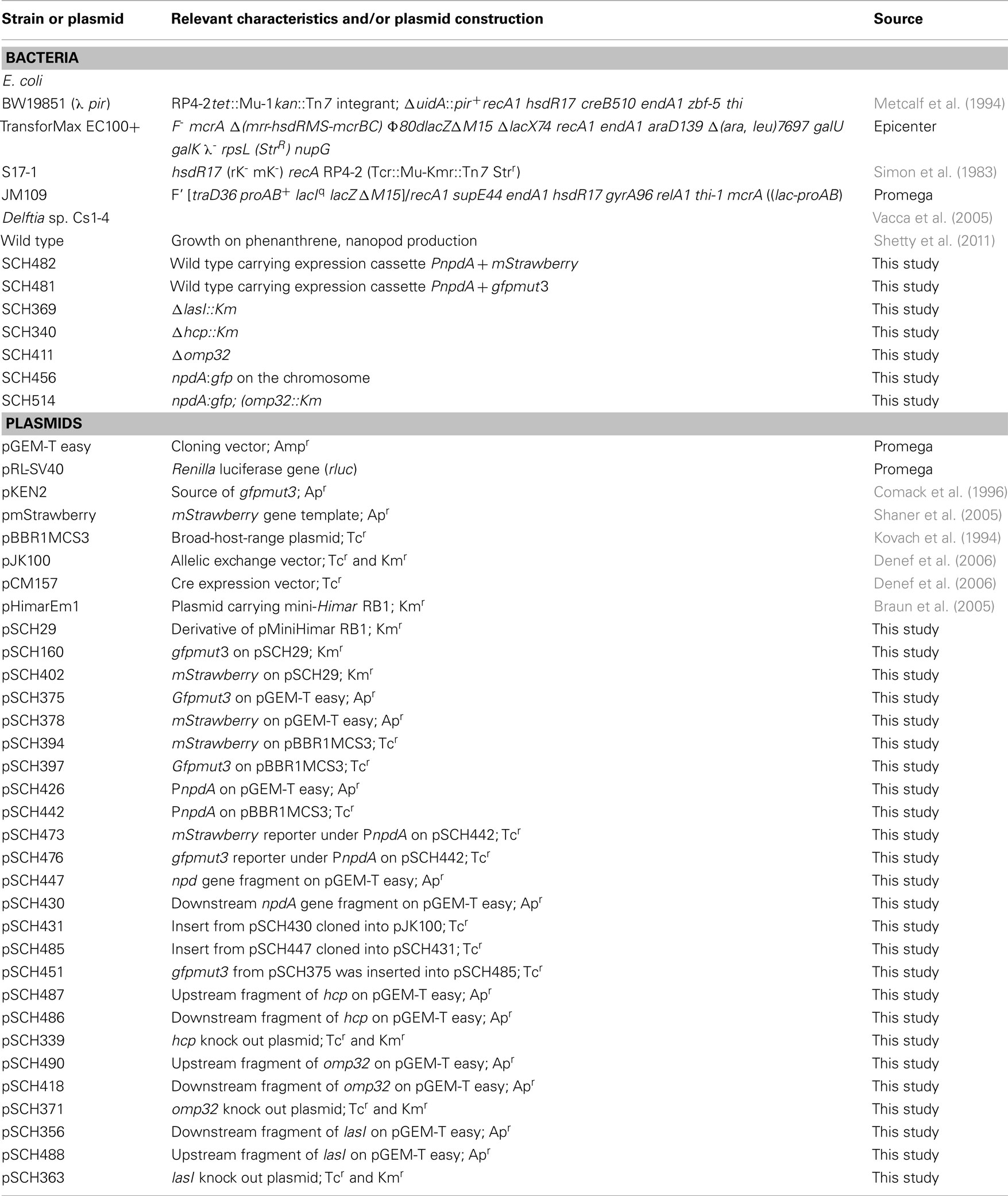
Bacterial strains and plasmids used in this work are listed in Table 1. E. coli JM109 was used for cloning. For conjugation, donor strains were either E. coli BW19851 (λ pir) or E. coli S17 (λ pir) and recipient strains were either E. coli TransforMax EC100+ (for propagation of constructs) or Delftia sp. Cs1-4. E. coli strains were routinely grown in Luria-Bertani (LB) broth at 37°C. Mineral salt medium (MSM; Hickey and Focht, 1990) containing phenanthrene as the sole carbon source (1 mg/mL) was routinely used for Delftia sp. Cs1-4 culture. Liquid cultures were grown with shaking (ca. 200 rpm) at either 25°C (strain Cs1-4) or 37°C (E. coli). For solid LB media, Bacto-Agar (Difco, Detroit, MI, USA) was added to a final concentration of 15 g/L. For E. coli, antibiotics were added when required at 100 μg/mL (ampicillin, Ap), 50 μg/mL (kanamycin, Km), or 10 μg/mL (tetracycline, Tc). Kanamycin and tetracycline were used in some Delftia sp. Cs1-4 cultures, and in these cases were added at 300 and 40 μg/mL, respectively.
DNA Manipulations
Genomic DNA was prepared using a genomic DNA extraction kit (Promega, Madison, WI, USA), and plasmid DNA was purified with the QIAprep spin miniprep kit (QIAGEN, Germantown, MD, USA). Restriction and modification enzymes were purchased from Promega (Madison, WI, USA) or New England Biolabs (Beverly, MA, USA). Klenow fragment or T4 DNA polymerase (Promega) was used to fill in recessed 3′ ends and to trim protruding 3′ ends of incompatible restriction sites. All PCR amplifications were done with the Failsafe PCR system (Epicenter Technology, Madison, WI, USA). Amplicons were separated in 0.7–1.0% (w/v) agarose gels, and DNA fragments were purified with the QIAquick gel extraction system (QIAGEN). Ligation mixtures were transformed into E. coli JM109 (Promega), and transformants were plated onto LB plates with appropriate antibiotic selection. Resistant colonies were isolated, and then screened for the acquisition of plasmids. All constructs were sequenced to verify structure. For conjugal transfer of plasmids from E. coli to Delftia sp. Cs1-4, LB-grown cultures of both cells were harvested (mid-log phase) by centrifugation, washed with LB and then equal amounts (ca. 1012 cells of each strain) were mixed, and spotted onto LB plates containing 5 mM CaCl2. Following overnight incubation at 22°C, cells were then scraped off of the plates, diluted, and plated on LB plates containing the appropriate antibiotics.
Transcription Start Site Determination
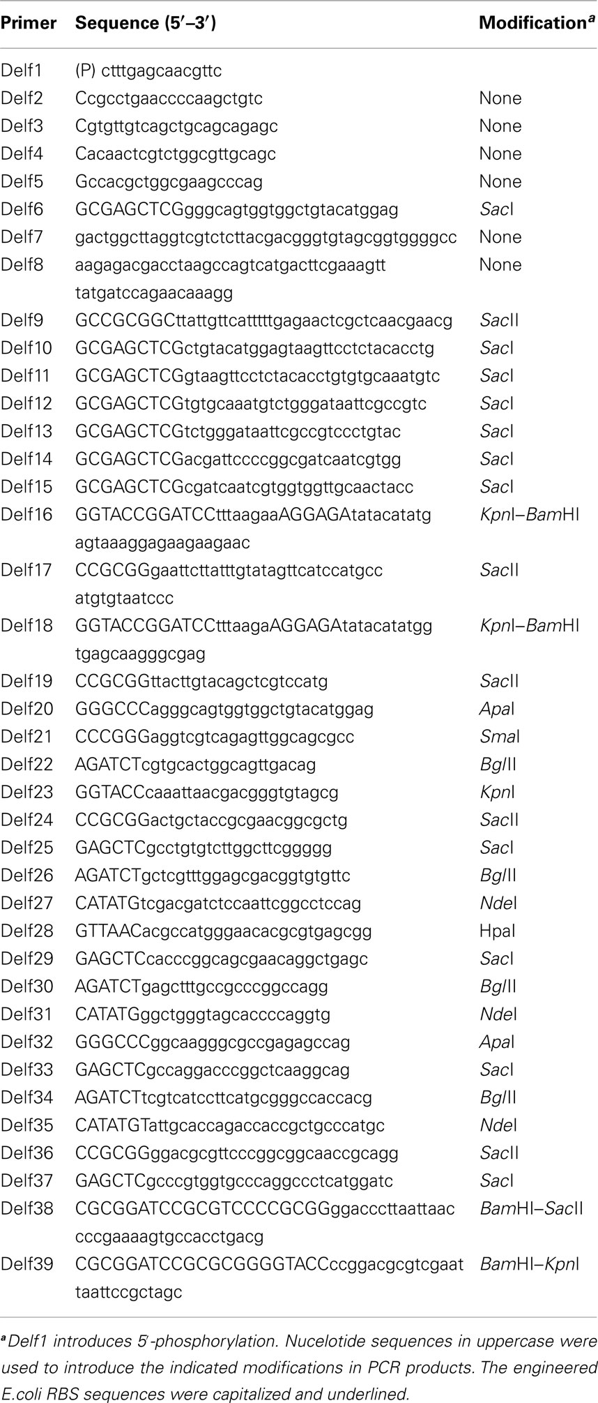
Total RNA was isolated from phenanthrene-grown strain Cs1-4 cells, and purified of genomic DNA by DNase I digestion. Analysis by 5′-RACE was done using TaKaRa 5′-full RACE Core set under conditions recommended by the supplier (TaKaRa). Reverse transcription (RT) was done with a 5′-phosphorylated RT primer (Delf1; Table 2). After RT, mRNA was digested with RNaseH, and then cDNA was concatenated using T4 RNA ligase. The region of interest was then amplified via nested PCR using two sets of primers to regions of npdA. In the first PCR, RT products were used as template, and amplified with primers Delf2 and Delf3 (Table 2). In the second PCR, template was a 10-fold dilution of the round one PCR product, and amplification was done using primers Delf4 and Delf5 (Table 2). The 5′-RACE products were isolated, purified, ligated into pGEM-T easy and then sequenced.
The npdA fragment including the non-coding and partial structural gene regions was amplified with primers Delf6 and Delf7 (Table 2) using strain Cs1-4 genomic DNA as template. The Renilla luciferase (rluc) gene was amplified from pRL-SV40 using primers Delf8 and Delf9 (Table 2). These fragments were fused via overlap PCR. To analyze the structure of the putative npdA promoter, deletion derivatives of non-coding fragments upstream of npdA were amplified by employing the same PCR strategy as described above, except using different N-terminal primers, namely Delf10, Delf11, Delf12, Delf13, Delf14, and Delf15 (Table 2). The above amplicons were inserted in pGEM-T easy, released from this vector by SacI and SacII digestion, and inserted into the same sites of pBBR1MCS-3 to create the deletion series. The reporter vector was then conjugated into strain Cs1-4.
Construction of Strong Expression System and Fluorescent Protein Reporter Vectors
Genes encoding green fluorescent protein and red fluorescent protein were amplified from pKEN2 and pmStrawberry using the primers Delf16/Delf17 and Delf18/Delf19, respectively, and engineered via PCR to contain an E. coli ribosome binding site on the 5′-end (Table 2). The amplicons were cloned into pGEM-T easy (pSCH374 and pSCH378, respectively), gfpmut3 was then released by ApaI and SacII digestion, and inserted into the same sites on pBBR1MCS3 (pSCH397). The mStrawberry gene was cut from pSC378 by digestion with KpnI and SacII, and inserted into KpnI/SacII sites on pBBR1MCS3 (pSCH395).
A strong expression system controlled by PnpdA was constructed as follows. The PnpdA region (genome position 5862152–5862685) was amplified from strain Cs1-4 genomic DNA using primers Delf20 and Delf21 (Table 2). The amplicon was then cloned into pGEM-T easy (pSCH426), released by digestion with ApaI and SmaI, and inserted into the same sites on pBBR1MCS3 (pSCH442). Green fluorescent protein (GFP, gfpmut3) and red fluorescent protein (RFP, mStrawberry) marker genes were released from pSCH374 and pSCH378 by digestion with SacII, cloned into pSCH442 and transformed into E. coli JM109. Colonies with strong green (pSCH476) and red (pSCH473) fluorescence were recovered, and orientation of reporter genes was confirmed by sequencing. These plasmids were next conjugated into Delftia sp. Cs1-4, leading to strains SCH481 (pSCH476) and SCH482 (pSCH473).
Construction of gfp Reporter Vector for Chromosaomal Tagging of npdA
To transcriptionally tag npdA, gfp was inserted immediately downstream of npdA using the Cre-loxP recombination method of Denef et al. (2005). An npdA fragment with the stop codon (genome position 5860670–5861289) was amplified using primers Delf22 and Delf23 (Table 2). The downstream fragment of npdA (genome positions 5860066–5860809) was amplified using primers Delf24 and Delf25 (Table 2). These fragments were then cloned into pGEM-T easy (pSCH447 and pSCH430). The downstream fragment from pSCH430 was released by digestion with SacII and SacI and inserted into the same sites on pJK100 (pSCH431). The npdA fragment from pSCH447 was released by NdeI and KpnI digestion, and then inserted into the same sites on pSCH431 (pSCH485). The gfpmut3 gene was released from pSCH375 by KpnI and NotI digestion, and assembled into the same sites on pSCH485 (pSCH451). Conjugation of pSCH451 into strain Cs1-4 gave Kmr/Tcs colonies, which were recovered for further analysis. The Cre-expressing vector, pCM157, was next introduced into a selected colony (SCH483) in order to remove Km resistance, leading to strain SCH484 (Kms/Tcr). Curing of pCM157 from SCH484 was done by serial transfers in LB medium. A selected colony (Kms/Tcs) with green fluorescence was then confirmed for the correct construct by PCR and sequencing (SCH456).
Mutant Construction
To knock out lasI, its upstream (strain Cs1-4 genome positions 1950815–1951882) and downstream (strain Cs1-4 genome positions 1952504–1953573) fragments were amplified with primers Delf26/Delf27 and Delf28/Delf29, respectively (Table 2). The amplicons were gel purified and cloned into pGEM-T easy (pSCH488 and pSCH356). The upstream fragments were released by BglII/NdeI digestion, and downstream fragments were released by ApaI/SacI from pGEM-T easy and then sequentially assembled on the same sites on pJK100 (pSCH363). To knock out hcp, upstream (strain Cs1-4 genome position 3366999–3367911) and downstream fragments (strain Cs1-4 genome position 3368229–3369041) were amplified using PCR primers Delf30/Delf31 and Delf32/Delf33, respectively (Table 2). The amplicons were gel purified and cloned into pGEM-T easy (pSCH487 and pSCH486). These fragments were sequentially assembled on the same sites on pJK100 (pSCH339) using the same strategy as described above. To knock out omp32, upstream (Cs1-4 genome positions 1041477–1042202) and downstream (Cs1-4 genome position 1044310–1045032) fragments were amplified with primers Delf34/Delf35 and Delf36/Delf37 (Table 2). The amplicons were gel purified and cloned into pGEM-T easy vector (pSCH490 and pSCH418). These fragments were sequentially assembled on the same sites on pJK100 (pSCH371). Each of the three constructs (pSCH363, pSCH339, pSCH371) was introduced into strain Cs1-4 by conjugation, and Tcs/Kmr transconjugants were selected, leading to strains SCH369, SCH340, and SCH389, respectively.
Genome-Wide Transposon Mutagenesis
Modification of pHimarEm1 was done to introduce additional unique KpnI–BamHI–SacII restriction sites, to remove the erythromycin resistance gene and to insert genes encoding GFP and RFP. To do so, PCR was done with pHimarEm1 DNA as template, and using forward primer Delf38 and reverse primer Delf39 (Table 2). The amplicon was digested with BamHI, self-ligated and transformed into E. coli S17 λpir. The gfpmut3 fragment was digested with KpnI and SacII from pSCH375 and inserted into pSC29 at the same restriction sites (pSCH160). The promoterless mStrawberry fragment was then released from pSCH378 by KpnI and SacII digestion, inserted into pSCH29 at the same restriction sites (pSCH402), and then introduced into strain SCH456 by conjugation. The Km-resistant colonies were randomly picked and replicated in 96-well plates containing MSM with either pyruvate and or phenanthrene as the carbon source. After incubation with shaking (24 h), the OD600 and GFP fluorescence were determined (see below).
Reporter Assays
Renilla luciferase assays were done as described in our prior work (Chen et al., 2009) using a commercially available kit (Promega) according to the manufacturer’s protocol. Quantitative analysis of fluorescent protein production was done using a Synergy 2 plate reader with the following conditions (all 0.2-s interval, 22°C): GFP, excitation at 485 nm, emission 510 nm; RFP, excitation at 574 nm, emission at 596 nm. All measurements were corrected for background with wild type (WT) Delftia sp. Cs1-4 cells.
DNA Sequence and Sequence Analysis
The complete genome sequence of Delftia sp. Cs1-4 was deposited in Genbank as accession NC(015563.1. All constructs were sequenced by the dideoxy termination method using an Applied Biosystems (Foster City, CA, USA) 3730 × l DNA Analyzer available at the University of Wisconsin-Madison, Biotechnology Center. GenBank database searches were carried out using the National Center for Biotechnology Information BLAST-N web server.
Results
Analysis of npdA Promoters in Delftia sp. Cs1-4 and Development of Strong Expression System
Three TSS were identified for npdA, and were located at (nucleotide) −34-bp (A), −56-bp(G), and −172-bp (A), respectively upstream of the npdA start codon (Figure 1A). Three putative promoter motifs, PnpdA1 (TCCTCT-N15-TGTCTG), PnpdA2 (TAGGGG-N15-TACGAT), and PnpdA3 (TACGAT-N17-TGGTGG) situated at −38, −61, and −180-bp, respectively were identified (Figure 1A). Serial deletion of non-coding regions upstream of npdA was done to establish involvement in npdA regulation of one or more of the three putative promoters. There was no significant difference in levels of gene expression between the WT and D1 (npdA −220 bp; Figure 1B). However, further deletion of an 11-bp fragment from D1 (D2, npdA −209 bp) yielded a ca. 20% decrease in Rluc activity relative to the WT (Figure 1B). Since the D2 construct carried the putative −35 motif in PnpdA1, we inferred the fragment (−220 to −209 bp) was also important for npdA expression. Deletion of the −35 region of PnpdA1 (D3, npdA −190 bp) decreased Rluc activity by >40% compared to the WT. Construct D4 (npdA −180 bp) had only ca. 20% Rluc activity. The latter contained a deletion that originated at −180 bp, and thus had the entire PnpdA1 region disrupted, indicating that PnpdA1 was the most important promoter for driving npdA expression. A further deletion (D5, npdA −67 bp) that removed the –35 bp motif in PnpdA2 retained ca. 5% of WT level. Removing the PnpdA2 region (D6, npdA −54) reduced Rluc activity to background levels.
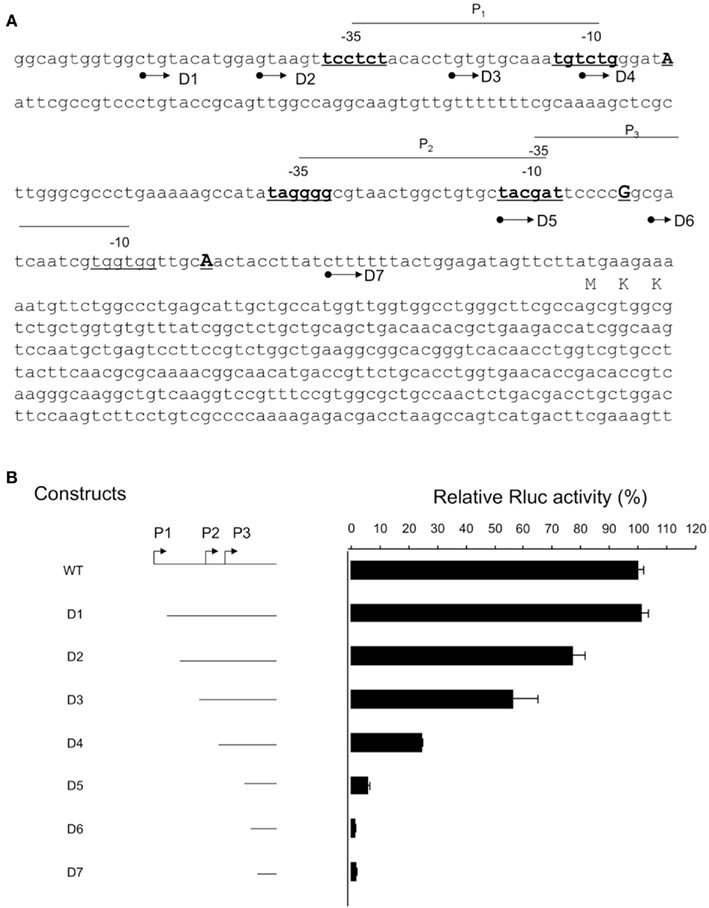
Figure 1. Analysis of npdA promoter regions. (A) Putative −10- and −35-bp motifs are indicated with P1, P2 and P3. Transcription start points are capitalized and underlined. Arrows indicate positions of deletions (D1-7). (B) Effect of serial deletion on rluc expression. Results were normalized to Rluc activity of the wild-type. Reactions were done in triplicate, and standard deviations are indicated by error bars.
To test the utility of the PnpdA expression system, the genes encoding a GFP and RFP were inserted downstream of the PnpdA cassette, which contained the 220-bp fragment described above. Transformants appeared green or red under ambient light, indicating strong expression of gfp and mstrawberry, respectively. The apparent high-level expression of these proteins was non-toxic to Delftia sp. Cs1-4, as growth of cultures expressing GFP or RFP was not distinguishable from that of the WT (Figure 2A). Production of GFP and RFP followed similar patterns, with levels increasing with culture growth, achieving stable accumulations upon reaching stationary phase (Figure 2B). In the absence of antibiotic selection, the expression vector was stable in Delftia sp. Cs1-4 for at least 56 generations (Figure 2C).

Figure 2. Growth and fluorescence characteristics of the GFP and RFP reporter strains. (A) SCH481 (PnpdA:gfp, circle), SCH482 (PnpdA:mStrawberry, diamond), and WT (square) were adjusted to the same cell density and cultured for 30 h. (B) Fluorescence determination for GFP (circle) and RFP (diamond) reporter strains. Values in (A) and (B) are means of measurements made from triplicate cultures, and error bars indicate standard deviation) (C) Stability test of the expression vector (pSCH477, PnpdA:gfp) in Delftia sp. Cs1-4. The strain SCH481 (PnpdA:gfp) was serially transferred in LB medium and the CFU were determined at generations of 16, 36 and 56. The blank bar is without tetracycline addition and the black one is supplemented with tetracycline.
Gene Deletion and Genome-Wide Mutagenesis
For generation of gene knockouts, the vector was used to target omp32, hcp, and lasI. Deletion of all three genes was successful, and confirmed by PCR and/or Southern hybridization. However, none of the gene deletions resulted in a loss of nanopod production, and only the Δomp32 mutant exhibited phenotypes different from that of the WT. In whole cell protein profiles, the latter mutant showed a loss of the predominant band corresponding to Omp32 (Shetty et al., 2011) and appearance of two other proteins, also identified as porins (Figure 3A). The Δomp32 mutant had an irregular cell shape (Figure 3B), and its growth was impaired on both pyruvate and phenanthrene, but the impact of Omp32 loss appeared to be greater with the latter substrate (Figures 3C,D).
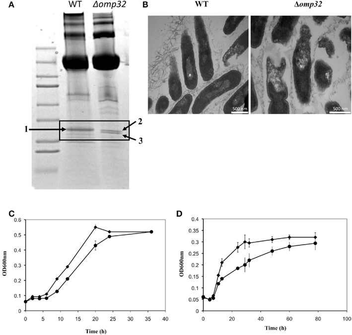
Figure 3. Characterization of the Delftia sp. Cs1-4 Δomp32 mutant. (A) Protein profiles of the wild type (WT) and mutant. Boxed area indicates the region of the Omp32 band in the WT. In the mutant, arrows indicate two bands identified as different porins, which were not detected in the WT. (B) Transmission electron micrographs of strain Cs1-4 biofilm cells grown on phenanthrene illustrating the mutant’s cellular deformities. (C) and (D) Growth of the WT (diamonds) and mutant (circles) on the indicated substrate.
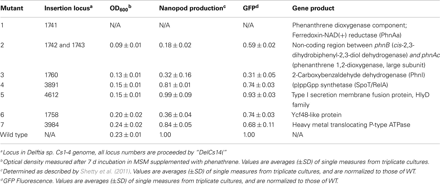
Following conjugal delivery to Delftia sp. Cs1-4, the transposition frequency of pMiniHimar was ca. 2 × 10⁻⁵ to 5 × 10⁻⁶ per recipient, a frequency comparable to those reported for Shewanella oneidensis, Geobacter sulfurreducens, and B. pseudomallei (Choi et al., 2008; Rollefson et al., 2009). From the 13,000 colonies screened, seven mutants were recovered that were impaired in either growth on phenanthrene (Mutants 1–6; Table 3) or in npdA expression (Mutant 7; Table 3). For the former, three mutants had insertions in the gene cluster encoding the phenanthrene catabolic pathway. Of these, Mutant 3 was intriguing as the gene bearing the insertion was predicted to encode an Ycf48 homolog. For Mutants 5 and 7, insertions were in genes outside of the phenanthene degradation cluster, and were predicted to encode a SpoT/RelA-type (p)ppGpp synthetase, and a HylD Family, type I secretion membrane fusion protein, respectively.
Discussion
Promoters proceeding SLP genes are among the most potent in many bacteria. For example, in Lactobacillus acidophilus, the strength of the SLP gene promoter is roughly twice that controlling the lactate dehydrogenase gene (Boot et al., 1996). Strong promoters may be needed for genes encoding SLP, as SLP are typically among the most abundant cellular proteins, as is the case with NpdA in strain Cs1-4 (Shetty et al., 2011). Thus, to develop a strong expression system, we focused on identification of the npdA promoter.
Collectively, the serial deletion analyses indicated that at least 220 bp upstream of npdA were required for maximal, log phase expression of npdA in strain Cs1-4 growing on phenanthrene. The presence within this region of multiple putative promoters is a feature that appears to be common for genes encoding SLP. For example, the SLP-encoding genes of Lactobacillus brevis ATCC 8287 (Hynönen et al., 2010), Aeromonas salmonicida (Chu et al., 1993), and Bacillus stearothermophilus ATCC 12980 (Jarosch et al., 2000) had at least two promoters, while in Bacillus brevis three promoters were arranged tandemly upstream of the cwp operon (Adachi et al., 1989). The reason(s) why SLP genes have multiple promoters are unknown. Possibly, these could be needed to respond to a variety of stimuli that could affect the expression of SLP genes (Sleytr and Messner, 1983; Adachi et al., 1989; Soual-Hoebeke et al., 1999). As yet, specific functions for the S-layer in Delftia sp. strain Cs1-4 are unknown, however, some involvement in phenanthrene degradation is a possibility as mutants lacking NpdA (and consequently the S-layer) are impaired in their ability to grow on this compound (unpublished data).
Expression systems based on well-characterized promoters such as Plac or Ptac are widely used (Dykxhoorn et al., 1996), but have had limited success in the Burkholderiales (Lefebre and Valvano, 2002). Likewise, for strain Cs1-4, Rluc was weakly expressed under control of Plac, as Rluc activity was ca. 2,500-fold lower than that from PnpdA:rluc. An alternative approach is to use promoters that originate from the Burkholderiales, and one example is the promoter regulating expression of small ribosomal protein S12 (Prsp). The latter promoter has been successfully utilized in Burkholderia xenovorans LB400 (Yu and Tsang, 2006) and in B. cepacia (Lefebre and Valvano, 2002). However, in strain Cs1-4, gene expression under Prsp was poor, and not significantly different from that of Plac (data not shown). Thus, demonstration of PnpdA as a strong promoter functional in Delftia sp. Cs1-4 has provided a much-needed tool for genetic analyses of this organism, and potentially other related bacteria.
The Δomp32 mutant had an irregular cell shape (Figure 3B), suggesting that Omp32 may have a key role in establishment of cell envelope structure, as shown for other outer membrane proteins (Lazar and Kolter, 1996; Watts and Hunstad, 2008). Analysis of the Δhcp mutant demonstrated that, as opposed to our hypothesis, Hcp did not have a structural function essential for nanopod formation. However, Western blot data indicated that Hcp was associated in some manner with nanopods as the majority of this protein accumulated in the >50-nm diameter fraction along with nanopods (data not shown). It is possible that Hcp was secreted separately from nanopods, and formed extracellular structures that were co-purified with nanopods. If so, such structures were not discernable in samples imaged by transmission electron microscope. Alternatively, Hcp may be associated with nanopods as cargo carried by OMV. In this case, Hcp may function as a virulence factor that may be employed by strain Cs1-4 in interactions with competing bacteria, as has been shown for T6SS in other bacteria (Schwarz et al., 2010; Leung et al., 2011; Records, 2011). Lastly, for the ΔlasI mutant, the absence of any detectable change in the formation of nanopods suggested that the process was not affected by quorum sensing, at least in the sense that it was regulated by AHL produced by a canonical AHL synthetase. This finding is noteworthy as it helps to narrow the spectrum of possible mechanisms that may control nanopod production.
Efficient targeting for gene inactivation is critical for functional genomic studies and, in bacteria, two widely used systems for generating in-frame, unmarked deletions are those based on sacB counter selection (Jäger et al., 1995; Chen et al., 2010), and Cre-loxP system (Denef et al., 2006; Choi et al., 2008). For strain Cs1-4, the sacB system proved unsuccessful; merodiploids (first recombination) were recovered at a high frequency, but these were not effectively resolved as Delftia sp. Cs1-4 grew in YT agar medium containing 5–15% (wt/vol) sucrose (data not shown). Similar observations have been reported for Streptomyces lividans and some Burkholderia strains, which carry an intrinsic sacBC operon. Alternatively, Cre-loxP system was successfully adapted for gene deletion or insertion, and was an efficient way for recycling antibiotic markers in Delftia sp. Cs1-4. To our knowledge, this is the first report of the Cre-loxP system being used for gene deletion analysis in Delftia spp.
Of the mutants recovered from genome-wide mutagenesis, three were of particular interest as they may encode new functions associated with nanopod production and/or phenanthrene degradation. One of these putatively encoded an Ycf48-like protein. In phototrophs, Ycf48 functions in the assembly and repair of Photosystem II (Komenda et al., 2008; Rengstl et al., 2011). Activities of an Ycf48-like protein that may be related to phenanthrene degradation are unknown, but, given the significant reduction (ca. 64%) in nanopod produced by this mutant, it’s interesting to speculate that it may have a role in the assembly of these structures. The putative spoT/relA mutant, had an insertion in a (p)ppGpp synthetase. The alarmone (p)ppGpp primarily governs the stringent response to amino acid starvation (Martinez-Costa et al., 1998; Åberg et al., 2006; Gomez-Escribano et al., 2008; Abranches et al., 2009) and, since growth of the spoT/relA mutant on pyruvate was not impaired, the effect of the mutation appeared related to use of phenanthrene as a carbon source. The third gene of interest, encoding an HlyD-like protein, was clustered with other genes predicted to encode pili formation. But, it remains to be determined how amino acid starvation and pili formation may be connected to phenanthrene degradation. Mutant 7 was not impaired in growth on phenanthrene, but did show decreased expression of npdA, and a depressed level of nanopod production. The protein predicted for the locus bearing the insertion contained a heavy-metal-associated domain that is also found in a number of proteins that transport or detoxify heavy metals; the relation of such a protein to npdA expression and nanopod formation remains to be determined.
Minitransposons are widely used for genome-wide mutagenesis in Gram-negative and Gram-positive bacteria (Lampe et al., 1999; Youderian et al., 2003; Maier et al., 2006; Choi et al., 2008) and, compared to other minitransposons, pMiniHimar is advantageous as it does not require host-specific factors for transposition, it lacks site specificity and the transposase is not introduced into the chromosome, thus enhancing insertion stability. The transposition frequency of pMiniHimar was sufficient (>5 × 10⁻⁶ per recipient) for saturation mutagenesis of the strain Cs1-4 genome. In the present study, pMiniHimar RB1 was modified by adding unique restriction sites for insertion of additional genetic elements. In our tests, these elements were promoterless gfpmut3 and mStrawberry, and the resultant vectors can be utilized for random generation of genomic transcriptional fusions. Such vectors can provide a convenient way to conduct genome-wide investigations of gene expression levels under selected conditions (de Lorenzo et al., 1990; Hahn et al., 1991; Boyle-Vavra and Seifert, 1995; Velayudhan et al., 2007).
Conclusion
The present report outlined the development of tools needed for genetic manipulation of Delftia sp. Cs1-4. These tools included a new expression cassette (PnpdA-based) that can be used for tagging of chromosomal genes as well as for complementation of knockout mutants, and a pMiniHimar transposon modified to enhance gene recovery and mutant analysis. The effectiveness in Delftia sp. of the Cre-loxP for gene deletion was also demonstrated. These tools were developed and validated for manipulation of Delftia sp. Cs1-4, but could also be applied to other related genera and species with importance in environmental toxicology.
Conflict of Interest Statement
The authors declare that the research was conducted in the absence of any commercial or financial relationships that could be construed as a potential conflict of interest.
Acknowledgments
These studies were funded by a National Science Foundation grant to William J. Hickey (MCB0920664). Sequencing and annotation of the Delftia sp. Cs1-4 genome was done by the U.S. Department of Energy Joint Genome Institute, through the Community Sequencing Project (CSP795673 to William J. Hickey). The work conducted by the U.S. Department of Energy Joint Genome Institute is supported by the Office of Science of the U.S. Department of Energy under contract No. DE-AC02-05CH11231.
References
Åberg, A., Shingler, V., and Balsalobre, C. (2006). (p)ppGpp regulates type 1 fimbriation of Escherichia coli by modulating the expression of the site-specific recombinase FimB. Mol. Microbiol. 60, 1520–1533.
Abranches, J., Martinez, A. R., Kajfasz, J. K., Chavez, V., Garsin, D. A., and Lemos, J. A. (2009). The molecular alarmone (p)ppGpp mediates stress responses, vancomycin tolerance, and virulence in Enterococcus faecalis. J. Bacteriol. 191, 2248–2256.
Adachi, T., Yamagata, H., Tsukagoshi, N., and Udaka, S. (1989). Multiple and tandemly arranged promoters of the cell wall protein gene operon in Bacillus brevis 47. J. Bacteriol. 171, 1010–1016.
Ballister, E. R., Lai, A. H., Zuckermann, R. N., Cheng, Y., and Mougous, J. D. (2008). In vitro self-assembly from a simple protein of tailorable nanotubes building block. Proc. Natl. Acad. Sci. U.S.A. 105, 3733–3738.
Boot, H., Kolen, C., Andreadaki, F., Leer, R., and Pouwels, P. (1996). The Lactobacillus acidophilus S-layer protein gene expression site comprises two consensus promoter sequences, one of which directs transcription of stable mRNA. J. Bacteriol. 178, 5388–5394.
Boyle-Vavra, S., and Seifert, H. S. (1995). Shuttle mutagenesis: a mini-transposon for producing PhoA fusions with exported proteins in Neisseria gonorrhoeae. Gene 155, 101–106.
Braun, T. F., Khubbar, M. K., Saffarini, D. A., and Mcbride, M. J. (2005). Flavobacterium johnsoniae gliding motility genes identified by mariner mutagenesis. J. Bacteriol. 187, 6943–6952.
Chen, S., Bleam, W. F., and Hickey, W. J. (2009). Simultaneous analysis of bacterioferritin gene expression and intracellular iron status in Pseudomonas putida KT2440 by using a rapid dual luciferase reporter assay. Appl. Environ. Microbiol. 75, 866–868.
Chen, S., Bleam, W. F., and Hickey, W. J. (2010). Molecular analysis of two bacterioferritin genes, bfra and bfrß, in the model rhizobacterium Pseudomonas putida KT2440. Appl. Environ. Microbiol. 76, 5335–5343.
Choi, K.-H., Mima, T., Casart, Y., Rholl, D., Kumar, A., Beacham, I. R., and Schweizer, H. P. (2008). Genetic tools for select-agent-compliant manipulation of Burkholderia pseudomallei. Appl. Environ. Microbiol. 74, 1064–1075.
Chu, S., Gustafson, C. E., Feutrier, J., Cavaignac, S., and Trust, T. J. (1993). Transcriptional analysis of the Aeromonas salmonicida S-layer protein gene vapA. J. Bacteriol. 175, 7968–7975.
Cormack, B. P., Valdivia, R. H., and Falkow, S. (1996). FACS-Optimized mutants of the green fluorescent protein (GFP). Gene 173, 33–38.
De Gusseme, B., Vanhaecke, L., Verstraete, W., and Boon, N. (2010). Degradation of acetaminophen by Delftia tsuruhatensis and Pseudomonas aeruginosa in a membrane bioreactor. Water Res. 45, 1829–1837.
de Lorenzo, V., Herrero, M., Jakubzik, U., and Timmis, K. N. (1990). Mini-Tn5 transposon derivatives for insertion mutagenesis, promoter probing, and chromosomal insertion of cloned DNA in Gram-negative eubacteria. J. Bacteriol. 172, 6568–6572.
Denef, V. J., Klappenbach, J. A., Patrauchan, M. A., Florizone, C., Rodrigues, J. L. M., Tsoi, T. V., Verstraete, W., Eltis, L. D., and Tiedje, J. M. (2006). Genetic and genomic insights into the role of benzoate-catabolic pathway redundancy in Burkholderia xenovorans LB400. Appl. Environ. Microbiol. 72, 585–595.
Denef, V. J., Patrauchan, M. A., Florizone, C., Park, J., Tsoi, T. V., Verstraete, W., Tiedje, J. M., and Eltis, L. D. (2005). Growth substrate- and phase-specific expression of biphenyl, benzoate, and C1 metabolic pathways in Burkholderia xenovorans LB400. J. Bacteriol. 187, 7996–8005.
Dykxhoorn, D. M., St. Pierre, R., and Linn, T. (1996). A set of compatible tac promoter expression vectors. Gene 177, 133–136.
Gomez-Escribano, J. P., Martín, J. F., Hesketh, A., Bibb, M. J., and Liras, P. (2008). Streptomyces clavuligerus relA-null mutants overproduce clavulanic acid and cephamycin C: Negative regulation of secondary metabolism by (p)ppGpp. Microbiology 154, 744–755.
Hahn, D. R., Solenberg, P. J., and Baltz, R. H. (1991). Tn5099, a xylE promoter probe transposon for Streptomyces spp. J. Bacteriol. 173, 5573–5577.
Hail, D., Lauziere, I., Dowd, S. E., and Bextine, B. (2011). Culture independent survey of the microbiota of the glassy-winged sharpshooter (Homalodisca vitripennis) using 454 pyrosequencing. Environ. Entomol. 40, 23–29.
Hickey, W. J., and Focht, D. D. (1990). Degradation of mono-, di-, and trihalogenated benzoic acids by Pseudomonas aeruginosa JB2. Appl. Environ. Microbiol. 56, 3842–3850.
Hynönen, U., Åvall-Jääskeläinen, S., and Palva, A. (2010). Characterization and separate activities of the two promoters of the Lactobacillus brevis S-layer protein gene. Appl. Microbiol. Biotechnol. 87, 657–668.
Jäger, W., Schäfer, A., Kalinowski, J., and Fühler, A. (1995). Isolation of insertion elements from Gram-positive Brevibacterium, Corynebacterium and Rhodococcus strains using the Bacillus subtilis sacB gene as a positive selection marker. FEMS Microbiol. Lett. 126, 1–6.
Jarosch, M., Egelseer, E. M., Mattanovich, D., Sleytr, U. B., and Sára, M. (2000). S-layer gene sbsC of Bacillus stearothermophilus ATCC 12980: molecular characterization and heterologous expression in Escherichia coli. Microbiology 146, 273–281.
Juarez-Jimenez, B., Manzanera, M., Rodelas, B., Martinez-Toledo, M. V., Gonzalez-Lopez, J., Crognale, S., Pesciaroli, C., and Fenice, M. (2010). Metabolic characterization of a strain (BM90) of Delftia tsuruhatensis showing highly diversified capacity to degrade low molecular weight phenols. Biodegradation 21, 475–489.
Komenda, J., Nickelsen, J., Tichy, M., Prasil, O., Eichacker, L. A., and Nixon, P. J. (2008). The cyanobacterial homologue of HCF136/YCF48 is a component of an early photosystem II assembly complex and is important for both the efficient assembly and repair of photosystem II in Synechocystis sp. PCC 6803. J. Biol. Chem. 283, 22390–22399.
Kovach, M., Phillips, R., Elzer, P., Roop, R., and Peterson, K. (1994). pBBR1MCS: a broadhost-range cloning vector. Biotechniques 16, 800–802.
Lampe, D. J., Akerley, B. J., Rubin, E. J., Mekalanos, J. J., and Robertson, H. M. (1999). Hyperactive transposase mutants of the Himar1 mariner transposon. Proc. Natl. Acad. Sci. U.S.A. 96, 11428–11433.
Lazar, S. W., and Kolter, R. (1996). SurA assists the folding of Escherichia coli outer membrane proteins. J. Bacteriol. 178, 1770–1773.
Lefebre, M. D., and Valvano, M. A. (2002). Construction and evaluation of plasmid vectors pptimized for constitutive and regulated gene expression in Burkholderia cepacia complex isolates. Appl. Environ. Microbiol. 68, 5956–5964.
Leibeling, S., Taubert, M., Seifert, J., Von Bergen, M., Harms, H., and Muller, R. H. (2010). Adaptation of the herbicide-degrading strain Delftia acidovorans MC1 through carbonylation of RdpA as key enzyme. J. Biotechnol. 150, S262–S263.
Leung, K. Y., Siame, B. A., Snowball, H., and Mok, Y. K. (2011). Type VI secretion regulation: crosstalk and intracellular communication. Curr. Opin. Microbiol. 14, 9–15.
Maier, T. M., Pechous, R., Casey, M., Zahrt, T. C., and Frank, D. W. (2006). In vivo himar1-based transposon mutagenesis of Francisella tularensis. Appl. Environ. Microbiol. 72, 1878–1885.
Martinez-Costa, O. H., Fernandez-Moreno, M. A., and Malpartida, F. (1998). The relA/spoT-homologous gene in Streptomyces coelicolor encodes both ribosome-dependent (p)ppGpp synthesizing and -degrading activities. J. Bacteriol. 180, 4123–4132.
Metcalf, W. W., Jiang, W., and Wanner, B. L. (1994). Use of the reprep technique for allele replacement to construct new Escherichia coli hosts for maintenance of R6Kγ origin plasmids at different copy numbers. Gene 138, 1–7.
Morel, M. A., Ubalde, M. C., Brana, V., and Castro-Sowinski, S. (2011). Delftia sp. JD2: a potential Cr(VI)-reducing agent with plant growth-promoting activity. Arch. Microbiol. 193, 63–68.
Mougous, J. D., Cuff, M. E., Raunser, S., Shen, A., Zhou, M., Gifford, C. A., Goodman, A. L., Joachimiak, G., Ordonez, C. L., Lory, S., Walz, T., Joachimiak, A., and Mekalanos, J. J. (2006). A virulence locus of Pseudomonas aeruginosa encodes a protein secretion apparatus. Science 312, 1526–1530.
Paulin, M. M., Nicolaisen, M. H., and Sorensen, J. (2010). Abundance and expression of enantioselective rdpA and sdpA dioxygenase genes during degradation of the racemic herbicide (R,S)-2-(2,4-dichlorophenoxy)propionate in soil. Appl. Environ. Microbiol. 76, 2873–2883.
Preiswerk, B., Ullrich, S., Speich, R., Bloemberg, G. V., and Hombach, M. (2011). Human infection with Delftia tsuruhatensis isolated from a central venous catheter. J. Med. Microbiol. 60, 246–248.
Records, A. R. (2011). The type VI secretion system: a multipurpose delivery system with a phagel-ike machinery. Mol. Plant Microbe Interact. 24, 751–757.
Rengstl, B., Oster, U., Stengel, A., and Nickelsen, J. (2011). An intermediate membrane subfraction in cyanobacteria is involved in an assembly network for photosystem II biogenesis. J. Biol. Chem. 286, 21944–21951.
Rollefson, J. B., Levar, C. E., and Bond, D. R. (2009). Identification of genes involved in biofilm formation and respiration via mini-Himar transposon mutagenesis of Geobacter sulfurreducens. J. Bacteriol. 191, 4207–4217.
Schwarz, S., Hood, R. D., and Mougous, J. D. (2010). What is type VI secretion doing in all those bugs? Trends Microbiol. 18, 531–537.
Shaner, N. C., Campbell, R. E., Steinbach, P. A., Giepmans, B. N. G., Palmer, A. E., and Tsien, R. Y. (2004). Improved monomeric red, orange and yellow fluorescent proteins derived from Discosoma sp. red fluorescent protein. Nat. Biotech. 22, 1567–1572.
Shetty, A., Chen, S., Tocheva, E. I., Jensen, G. J., and Hickey, W. J. (2011). Nanopods: a new bacterial structure and mechanism for deployment of outer membrane vesicles. PLoS One 6, e20725. doi: 10.1371/journal.pone.0020725
Simon, R., Priefer, U., and Puhler, A. (1983). A broad host range mobilization system for in vivo genetic engineering: transposon mutagenesis in Gram-negative bacteria. Nat. Biotechnol. 1, 784–791.
Sleytr, U. B., and Messner, P. (1983). Crystalline surface layers on bacteria. Annu. Rev. Microbiol. 37, 311–339.
Soual-Hoebeke, E., Sousa-D’Auria, C. D., Chami, M., Baucher, M.-F., Guyonvarch, A., Bayan, N., Salim, K., and Leblon, G. (1999). S-layer protein production by Corynebacterium strains is dependent on the carbon source. Microbiology 145, 3399–3408.
Vacca, D. J., Bleam, W. F., and Hickey, W. J. (2005). Isolation of soil bacteria adapted to degrade humic acid sorbed phenanthrene. Appl. Environ. Microbiol. 71, 3797–3805.
Velayudhan, J., Castor, M., Richardson, A., Main-Hester, K. L., and Fang, F. C. (2007). The role of ferritins in the physiology of Salmonella enterica sv. Typhimurium: a unique role for ferritin B in iron-sulphur cluster repair and virulence. Mol. Microbiol. 63, 1495–1507.
Watts, K. M., and Hunstad, D. A. (2008). Components of SurA required for outer membrane biogenesis in uropathogenic Escherichia coli. PLoS ONE 3, e3359. doi: 10.1371/journal.pone.0003359
Yang, Z. Y., Ni, Y., Lu, Z. Y., Liao, X. R., Zheng, Y. G., and Sun, Z. H. (2011). Industrial production of S-2,2-dimethylcyclopropanecarboxamide with a novel recombinant R-amidase from Delftia tsuruhatensis. Process Biochem. 46, 182–187.
Youderian, P., Burke, N., White, D. J., and Hartzell, P. L. (2003). Identification of genes required for adventurous gliding motility in Myxococcus xanthus with the transposable element mariner. Mol. Microbiol. 49, 555–570.
Yu, M., and Tsang, J. S. H. (2006). Use of ribosomal promoters from Burkholderia cenocepacia and Burkholderia cepacia for improved expression of transporter protein in Escherichia coli. Protein Expr. Purif. 49, 219–227.
Keywords: genetic manipulation, Delftia sp. Cs1-4, nanopods, phenanthrene, polynuclear aromatic hydrocarbons, biodegradation, surface layer protein
Citation: Chen S and Hickey WJ (2011) Development of tools for genetic analysis of phenanthrene degradation and nanopod production by Delftia sp. Cs1-4. Front. Microbio. 2:187. doi: 10.3389/fmicb.2011.00187
Received: 19 July 2011;
Accepted: 22 August 2011;
Published online: 12 October 2011.
Edited by:
Jeremy Semrau, The University of Michigan, USAReviewed by:
Steven Ripp, University of Tennessee, USAJong-In Han, Korean Advanced Institute of Science and Technology, South Korea
Copyright: © 2011 Chen and Hickey. This is an open-access article subject to a non-exclusive license between the authors and Frontiers Media SA, which permits use, distribution and reproduction in other forums, provided the original authors and source are credited and other Frontiers conditions are complied with.
*Correspondence: William J. Hickey, O.N. Allen Laboratory for Soil Microbiology, Department of Soil Science, University of Wisconsin–Madison, 1525 Observatory Drive, Madison, WI 53706, USA. e-mail:d2poaWNrZXlAd2lzYy5lZHU=
 Shicheng Chen
