- 1College of Animal Science and Technology, Henan Agricultural University, Zhengzhou, China
- 2Henan Delin Biological Product Co. Ltd., Xinxiang, China
- 3Henan Puai Feed Co. Ltd., Zhoukou, China
Introduction: Mycotoxins in animal diets cause a lot of economic loss in animal husbandry annually. The objective of this experiment was to evaluate the effect of combination of Aspergillus niger culture and glycyrrhizic acid (CANCGA) on alleviating multi-mycotoxin toxicity for broiler production performance and nutrient metabolism.
Methods: A total of 500 one-day-old male broilers were randomly divided into 10 groups, 5 replications in each group and 10 broilers in each replication. The feeding period was 21 d. The dietary treatment included group A (the basal diet as the control group); group B (0.03 mg/kg aflatoxin B1 (AFB1) + 0.15 mg/kg zearalenone (ZEN) + 1.5 mg/kg deoxynivalenol (DON), low-dose mycotoxin diet); group C (0.07 mg/kg AFB1 + 0.5 mg/kg ZEN + 3.0 mg/kg DON, high-dose mycotoxin diet); groups D, E and F (basal diet supplemented with 0.2, 0.4 and 0.6 g/kg CANCGA, respectively); groups G, H and I (low-dose mycotoxin diet supplemented with 0.2, 0.4 and 0.6 g/kg CANCGA, respectively); group J (high-dose mycotoxin diet supplemented with 0.4 g/kg CANCGA).
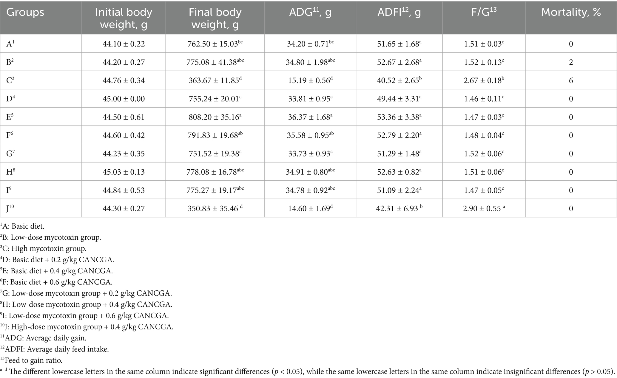
Results: The results demonstrated that broiler mortality in groups B and C was 2 and 6%, which in other groups was zero, indicating that CANCGA addition in diets could decrease broiler mortality caused by multi-mycotoxins. Average daily weight (ADG), metabolic rates of protein and phosphorus were significantly declined, while the ratio of daily feed intake and daily gain were significantly increased when dietary mycotoxin concentration was increased (p < 0.05). Compared with the control group, low-dose mycotoxin in diet could increase serum alanine aminotransferase (ALT) and aspartate aminotransferase (AST) activity (p < 0.05), while decrease serum total protein (TP), albumin (ALB) and total cholesterol (TC) levels (p < 0.05). However, CANCGA addition could effectively reverse the above results. Compared with the low-dose mycotoxin group, the addition of 0.4 g/kg CANCGA could decrease serum ALT, AST, alkaline phosphatase (ALP), glucose (GLU), triglyceride (TG) and high-density lipoprotein (HDL) levels (p < 0.05), while increase ALB, TC levels and ALB/Globulin (GLB) (p < 0.05), indicating that CANCGA addition was able to reduce oxidative stress of broilers induced by multi-mycotoxins. The contents of residual AFB1, ZEN and DON in broiler excreta were significantly increased in the low-dose mycotoxin group (p < 0.05), compared to the control group; however, CANCGA addition could decrease AFB1, ZEA and DON contents in broiler excreta. Serum metabolomics showed that metabolites such as creatine, N-acetyl-L-phenylalanine and linoleic acid as well as metabolic pathways related to glycine, serine, threonine, cysteine, methionine, selenium compounds and linoleic acid metabolisms were regulated by CANCGA addition to alleviate nutrient metabolic disorders caused by multi-mycotoxins.
Discussion: In conclusion, CANCGA was found to be effective in alleviating multi-mycotoxin toxicity for broilers’ growth performance through reducing oxidative stress and positively regulating nutrient metabolisms.
1 Introduction
Mycotoxins were toxic metabolites produced by Aspergillus spp., Penicillium spp., Fusarium spp. and other fungi during their growth process (1). Over 300 kinds of mycotoxins were identified that pose harm to both humans and animals (2). Among them, the most potent and wide-distribution mycotoxins included aflatoxin B1 (AFB1), zearalenone (ZEN) and deoxynivalenol (DON) (3). These mycotoxins could be generated at many stages including crop cultivation, processing, transportation and storage of grains and their by-products due to variations in environmental ask temperature and humidity (4, 5), posing significant health risks to both humans and animals. In addition, the main component of an animals’ diet consists mainly of grains, which are a favorable substrate for mycotoxin-producing fungal species. These ingredients have high inclusion rates in animal compound feed, and if contaminated, could be a source of contamination of the final products (6, 7). Up to 88% of 74,821 samples of diets and feed ingredients (e.g., corn, wheat and soybeans) collected from 100 countries were polluted with multi-mycotoxins, in which AFB1, ZEN and DON were detected in 23, 64 and 45% of the samples, respectively (8). It was reported that 17,316 samples of feed and feed raw materials from all over the world were analyzed for contamination with aflatoxins, ochratoxin A, zearalenone, deoxynivalenol and fumonisins, in which 72% of the samples were tested to be positive for at least one mycotoxin and 38% were found to be co-contaminated (9).
The presences of mycotoxins are associated with carcinogenicity, teratogenicity, hepatotoxicity, nephrotoxicity, embryotoxicity and immunosuppression in animals (10–13). Moreover, their occurrence could reduce body weight and feed conversion rates, increase incidences of diarrhea and mortality in poultry (14, 15). AFB1 was demonstrated to significantly disrupt hepatic lipid and protein metabolism in animals, which can lead to liver function damage and affect production performance (16–18). ZEN, an estrogen analog, competed with endogenous estrogen for receptor binding sites upon entering the organism, results in reproductive toxicity (19–21). DON contamination in animal feeds causes impairment of intestinal barrier function (22, 23), leading to intestinal flora disorders in animals (24). Once DON is absorbed into the body, it inhibits protein synthesis (1, 25, 26). However, mycotoxin contamination in feedstuffs or diets typically involves multiple toxin types rather than single toxin. It was observed that low concentration of multiple mycotoxins has a greater detrimental impact on livestock than high concentration of single mycotoxin (27–29). Coexistence of AFB1, ZEN and DON in poultry diets could lead to an exacerbation of health issues in animals and result in diminished economic returns due to their combined toxicity (30). In addition, feeds contaminated with AFB1, ZEN and DON resulted in disruption of amino acid metabolic pathways such as alanine, aspartic acid and glutamine (31) as well as impacting blood glucose metabolites such as glycine, arginine and tryptophan (32).
Therefore, effectively addressing the risks associated with multi-mycotoxins has become an urgent priority. Currently, three primary approaches were employed to mitigate or eliminate mycotoxin risks: physical, chemical, and biological methods (33). Among these options, biological detoxification methods were considered specific, efficient, and environmentally friendly. For instance, the previous report showed that fourteen strains of Aspergillus niger isolated from peanuts demonstrated complete inhibition of AFB1 production through co-culturing (34). It was discovered that one strain of Aspergillus niger consistently degraded ZEN by over 95%, resulting in the formation of low-toxicity products (35). Glycyrrhizic acid (GA), the main active compound extracted from Glycyrrhiza glabra, has been shown to alleviate inflammation, oxidative stress, and apoptosis (36). Also, GA is considered an effective treatment for liver diseases (37). Its combination with probiotic complexes attenuated DON-induced oxidative stress, inflammation and apoptosis in IPEC-J2 cells (38). However, the combined effect of GA and Aspergillus niger in reducing AFB1 induced toxicity has not been studied. After considering both functions of GA and Aspergillus niger, the combination of Aspergillus niger culture and GA (CANCGA) was used in this study for alleviating multi-mycotoxin toxicity caused by AFB1, ZEN and DON in broiler production, so as to assess their mitigation potential and provide a foundation for addressing issues related to multiple mycotoxin contamination.
2 Materials and methods
The study and included experimental procedures were approved by the guidelines of Animal Care and Use Ethics Committee of Henan Agricultural University (SKLAB-B-2010-003-01). All animal experiments were conducted in strict accordance with the institutional guidelines for care and use of laboratory animals. Animal feeding experiment was conducted in a chicken farm of Henan Agricultural University.
2.1 CANCGA and mycotoxin preparation
Aspergillus niger with degrading AFB1 and ZEN was preserved in the Laboratory of Animal Nutrition and Feed Biotechnology in Henan Agricultural University. GA was provided by Henan Delin Biological Products Co., Ltd. The preparation of solid culture of Aspergillus niger was made by mixing the three feed ingredients of bran, soybean meal and corn in the ratio of 7:2:1, taking 10 g of the mixture in a triangular flask, after sterilization, adding 1 mL of Aspergillus niger seed solution and 5 mL of sterile water, mixing thoroughly and cultivating at 30°C for 5–7 d and then taking it out and drying and crushing the mixture (39). The mycotoxin degradation experiments in vitro confirmed that the degradation rates of AFB1 and ZEA were 60.40 and 97.67%, respectively, when 0.04% Aspergillus niger culture was applied. The further research indicated that the degradation rate of AFB1 was increased to 68, 71 and 63% when 0.02, 0.04 and 0.06% of GA were added. In addition, previous study in our laboratory confirmed that GA could alleviate the damage of intestinal cells caused by DON (38). Therefore, CANCGA was prepared by combining Aspergillus niger culture with GA at both 0.04% addition (mixed in ratio 1:1 ratio) for alleviating toxicity of AFB1, ZEN and DON in the further broiler feeding experiment.
2.2 Diet preparation and animal management
500 one-day-old male Arbor Acres (AA) broilers were divided into 10 groups, each group consisting of 5 replications with 10 broilers per replication. The broilers were reared in cages, allowing free access to diet and water. Room temperature was around 25°C, but the temperature under nurturing umbrella was 33–35°C, 29–32°C and 26–28°C for one-week-old, two-week-old and three-week-old broilers, respectively. The relative humidity was kept at 60 to 65%. The experimental period was 21 d. Daily feed intake, and dead broilers were recorded daily. Broiler body weight in each replication was weighted at the age of 1 and 22 d. Parameters such as average daily gain (ADG), average daily feed intake (ADFI), feed-to-gain ratio (F/G) and mortality were calculated. Additionally, a standard immunization program was implemented within the first one week. The experimental groups were organized as follows:
Group A: the basal diet as the control group (0.002 mg/kg AFB1 + 0.041 mg/kg ZEN + 0.946 mg/kg DON).
Group B: low-dose mycotoxin diet (0.03 mg/kg AFB1 + 0.15 mg/kg ZEN + 1.5 mg/kg DON).
Group C: high-dose mycotoxin diet (0.07 mg/kg AFB1 + 0.5 mg/kg ZEN + 3.0 mg/kg DON).
Group D: basal diet supplemented with 0.2 g/kg CANCGA.
Group E: basal diet supplemented with 0.4 g/kg CANCGA.
Group F: basal diet supplemented with 0.6 g/kg CANCGA.
Group G: low-dose mycotoxin diet supplemented with 0.2 g/kg CANCGA.
Group H: low-dose mycotoxin diet supplemented with 0.4 g/kg CANCGA.
Group I: low-dose mycotoxin diet supplemented with 0.6 g/kg CANCGA.
Group J: high-dose mycotoxin diet supplemented with 0.4 g/kg CANCGA.
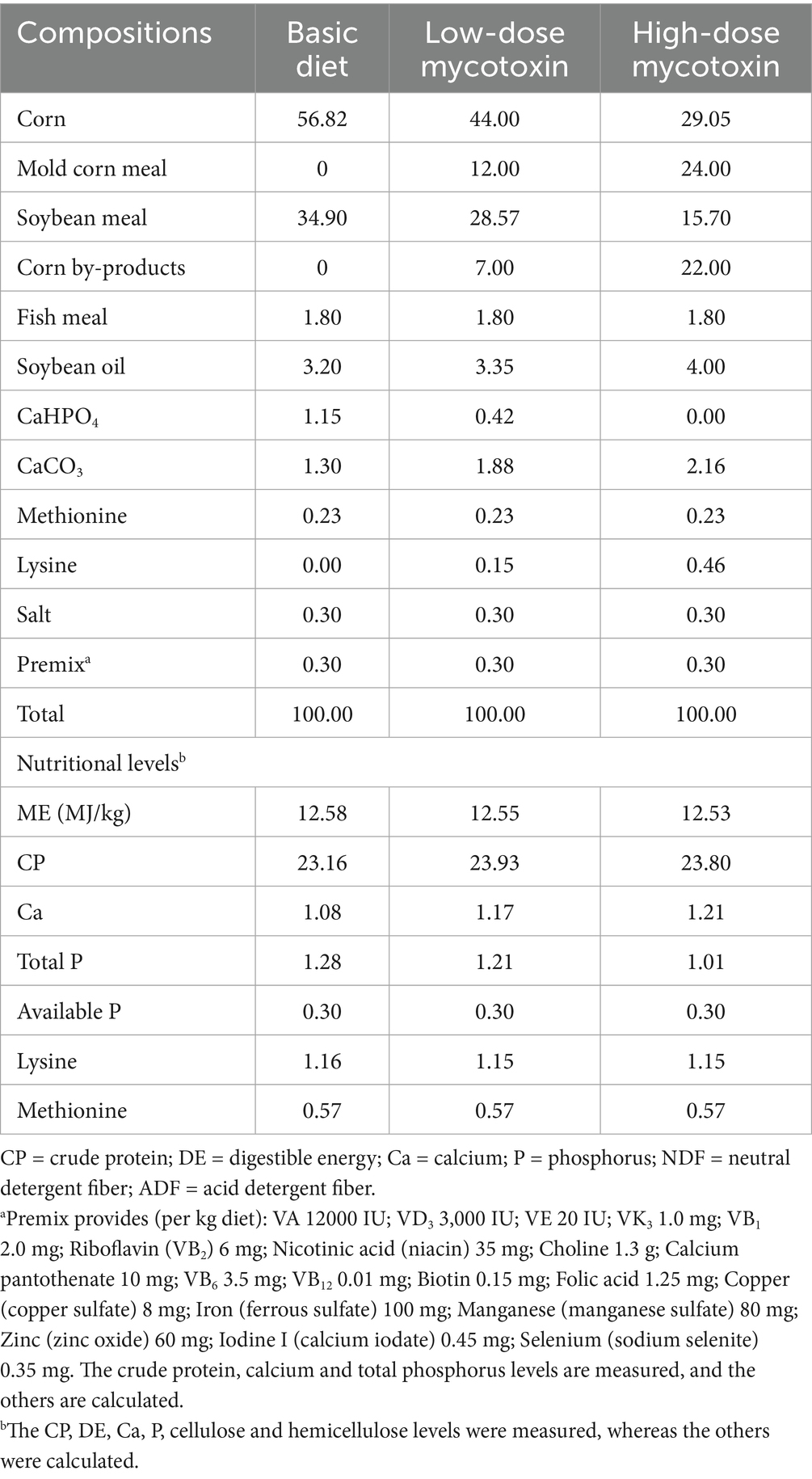
The basal diets were formulated based on the broiler feeding standards outlined in the NRC (1994) guideline. To adjust AFB1 and ZEN contents in diets, normal corn in the basal diet was substituted with moldy corn, while DON content was adjusted with corn by-product. The diet formulation and nutrient levels were listed in Table 1.
2.3 Sample collection and treatment
Excreta collection of broilers in each replication was performed on days 18–20 of the feeding experiment, followed by spraying with 10% sulfuric acid solution for nitrogen fixation, dried at 65°C, and ground for further use. At day 21, blood samples (2 mL) were collected from the wing vein of five broilers in each group and stored in a refrigerator at 4°C until serum precipitation occurred in the collection tube. Subsequently, centrifugation was conducted at 1,520 × g for 10 min to obtain serum, which was then transferred to a sterilized tube and stored at −80°C for future utilization.
2.4 Determinations of nutrient metabolic rates and residues of AFB1, ZEN, and DON
The crude protein (CP), ether extract (EE), calcium (Ca) and phosphorus (P) contents in diets and excreta were determined according to the methods of national standards GB/T 6432–2018, GB/T 6433–2006, GB/T 6436–2002 and GB/T 6437–2018, respectively. The calculation of nutrient metabolic rates was as follows: Nutrient metabolic rate (%) = 100 × (nutrient content in diet−nutrient content in excreta)/nutrient content in diet. Based on the growth performance of broilers in each group at 21 d, broilers in groups A, B, E and H were selected for the determination of excreta toxin residues. The contents of AFB1, ZEN and DON in excreta were detected according to the protocol of Suwei toxin detection kits (Suwei Biological Research Co., Ltd. Jiangsu, China). The calculation of AFB1 degradation rate was as follows: AFB1 degradation rate (%) = (AFB1 content in control group−AFB1 content in test group)/AFB1 content in control group × 100. DON and ZEN degradation rates were calculated in the same way as AFB1.
2.5 Determination of serum biochemical parameters
Using a fully-automated blood biochemistry analyzer to measure the serum contents of glucose (GLU), triglyceride (TG), high-density lipoprotein (HDL), total cholesterol (TC), low-density lipoprotein (LDL), aspartate aminotransferase (AST), lactate dehydrogenase (LDH), alkaline phosphatase (ALP), alanine transaminase (ALT), total protein (TP) and albumin (ALB).
2.6 Serum pretreatment
Based on the growth performance of broilers in each group at 21 d, broilers in groups A, B, E and H were selected for serum metabolomics analysis. Serum sample was thawed at 4°C and vortexed for 1 min using a vortex mixer (BE-2600, Haimen Qilin Bell Instrument Manufacturing Co., Ltd., Nantong, China) to ensure thorough mixing. And then transferred into 2 mL centrifuge tube. Subsequently, 400 μL methanol (stored at −20°C) was added and vortexed for 1 min prior to centrifugation. The sample was centrifuged at 13,680 × g and 4°C for 10 min using a refrigerated centrifuge (H1850-R, Hunan Xiangyi Laboratory Instrument Development Co., Ltd., Changsha, China). The supernatant was carefully transferred to another 2 mL centrifuge tube, then concentrated and dried before being dissolved in 150 μL 4 mg/L 2-chloro-L-phenylalanine (prepared with 80% methanol). The resulting supernatant was filtered through a 0.22 μm filter membrane before being added to the UPLC-MS vial for UPLC-MS detection.
2.7 Analysis of UPLC-MS
MS/MS analysis was performed using the Thermo Vanquish Ultra High-Performance Liquid Chromatography (UHPLC) system (Vanquish, Thermo, Massachusetts, United States) coupled with an ACQUITY UPLC® HSS T3 column (2.1 × 100 mm, 1.8 μm). In positive ion mode, the mobile phases consisted of 0.1% formic acid diluted in water (A1) and 0.1% formic acid diluted in acetonitrile (B1); in negative ion mode, the mobile phases were 5 mM ammonium formate diluted in water (A2) and acetonitrile (B2). Thermo Orbitrap Exploris 120 mass spectrometer (Orbitrap Exploris 120, Thermo, Massachusetts, United States) was used to determine the serum metabolites. Mass spectrometry parameters were as follows: positive ion spray voltage of 3.50 kV, negative ion spray voltage of −2.50 kV.
2.8 Statistical analyses
The animal experimental data were expressed as means ± standard error. The one-way ANOVA test was conducted using SPSS 26.0. Statistical significance was considered at p < 0.05.
The data from metabolomics were processed by XCMS in R package for peak detection, filtering, and alignment of raw mass spectrometry files converted by Proteowizard. Substance identification was conducted by searching and comparison with spectral databases such as HMDB, MassBank, LipidMaps, mzCloud, KEGG, and metabolite standards database of Nomi Metabolism. Differential metabolites were selected based on criteria of p < 0.05 and VIP > 1. Spearman correlation analysis was performed between differential metabolites and production performance, serum biochemistry or three kinds of toxin residues in excreta by using Nomi Metabolism platform.
Multivariate statistical methods including downscaling and categorization such as principal component analysis (PCA) and orthogonal-partial least squares discriminant analysis (OPLS-DA) were employed to analyze the data from serum metabolomics in order to identify the differential metabolites between different groups. The significant differences were determined by t-test and the variable importance projection (VIP) of the first principal component of OPLS-DA. A KEGG pathway enrichment analysis was conducted for the identified differential metabolites to evaluate their potential roles in biological responses.
3 Results
3.1 Effects of CANCGA addition on broiler growth performance
As presented in Table 2, mortality was significantly increased in the low-dose or high-dose mycotoxin groups, compared to the control group. However, supplementation with CANCGA made mortality become zero to remain consistent with the control group, indicating that CANCGA was able to alleviate multi-mycotoxin toxicity for broilers. Moreover, compared to the basal diet, a significant (p < 0.05) improvement of ADG and final body weight were observed when 0.4 g/kg CANCGA was added to the basal diet, while final body weight, ADG, ADFI, and F/G were not significantly different when 0.2 g/kg and 0.6 g/kg CANCGA was added to the basal diet. Notably, final body weight and ADG were significantly higher with the addition of 0.4 g/kg CANCGA to the basal diet than with the 0.2 g/kg addition (p < 0.05). ADG and ADFI were significantly decreased, while F/G was significantly increased for broilers subjected to high-dose mycotoxin diet, compared to both basal and low-dose mycotoxin diets (p < 0.05).
3.2 Effects of CANCGA on nutrient metabolic rates for broilers
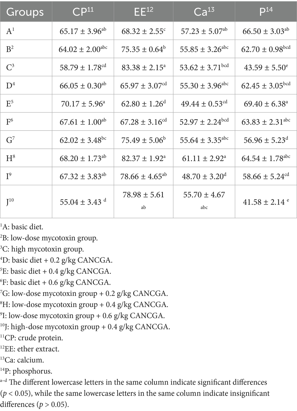
Table 3 showed that high-dose mycotoxin diets without or with CANCGA addition significantly decreased CP and P metabolic rates, while increased EE metabolic rate, compared with the control group (p < 0.05). CANCGA addition in high-dose mycotoxin diet had insignificant effect on nutrient metabolic rates. P metabolic rate was significantly decreased (p < 0.05), while EE metabolic rate was significantly increased with dietary mycotoxin levels increasing (p < 0.05). Compared with the control group, low-dose mycotoxin diet had insignificant effect on other nutrient metabolic rates in spite of increasing EE metabolic rate (p < 0.05). In low-dose mycotoxin diets, 0.4 g/kg CANCGA addition significantly increased EE metabolic rate, 0.6 g/kg CANCGA addition significantly decreased Ca metabolic rate, compared with the low-dose mycotoxin diet without CANCGA addition (p < 0.05). Compared with the control group, 0.4 g/kg CANCGA addition in the basal diet significantly decreased EE and Ca metabolic rates (p < 0.05), while 0.2 g/kg and 0.6 g/kg CANCGA additions in the basal diet had insignificant effect on nutrient metabolic rates. In addition, CANCGA added at 0.4 g/kg in the low mycotoxin group had significantly higher metabolic rates of EE and P than the 0.2 g/kg addition (p < 0.05).
3.3 Effects of CANCGA on mycotoxin contents in excreta of broilers
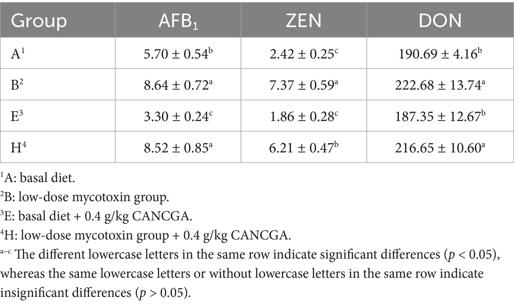
Based on the feeding experiment, the excreta and serum samples from groups A, B, E and H were selected as the representative ones for further analyses. As shown in Table 4, the contents of residual AFB1, ZEN and DON in broiler excreta were significantly increased in the low-dose mycotoxin group, compared to the control group (p < 0.05). Furthermore, compared to the basal diet, residual AFB1 content was significantly decreased when 0.4 g/kg CANCGA was added in the basal diet (p < 0.05); however, CANCGA addition had the tendency to decrease ZEN and ZON contents in excreta. Compared to the low-dose mycotoxin group, ZEN residue was significantly decreased by 0.4 g/kg CANCGA addition (p < 0.05); however, CANCGA addition had the tendency to decrease AFB1 and ZON contents in excreta.
3.4 Effect of CANCGA on serum biochemical parameters of broilers
Figure 1 indicated that serum ALP, LDH, TP, ALB, GLU, TC and TG levels in low-dose mycotoxin group were significantly decreased (p < 0.05), while AST level was significantly increased (p < 0.05), compared with the control group. However, the addition of 0.4 g/kg CANCGA to the basal diet could decrease serum ALP, TG and LDL levels (p < 0.05), while increase HDL level (p < 0.05). Compared with the low-dose mycotoxin group, the addition of 0.4 g/kg CANCGA could decrease serum ALT, AST, ALP, GLU, TG and HDL levels (p < 0.05), while increase ALB, TC levels and ALB/GLB (p < 0.05). It was inferred that CANCGA addition was able to reduce tissue and organ damage and lipid metabolism disorders of broilers induced by multi-mycotoxins.
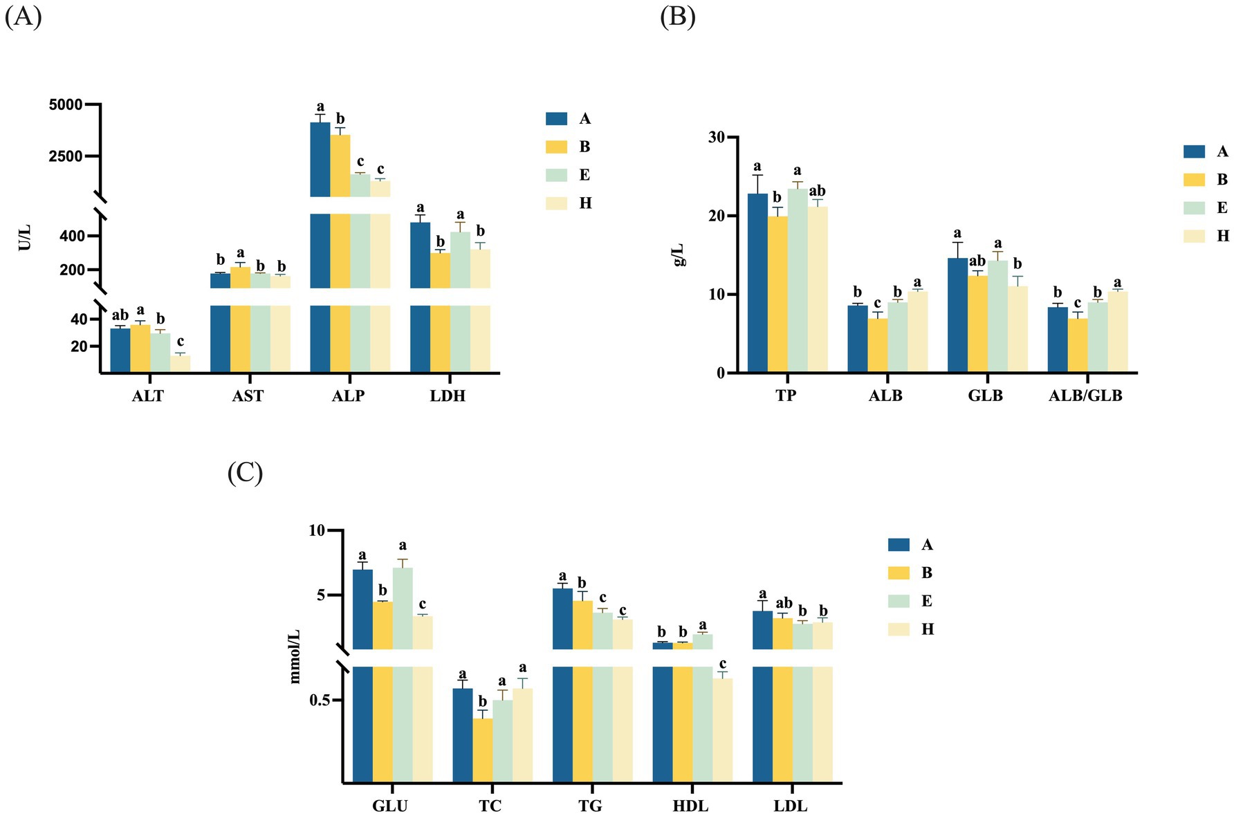
Figure 1. Effect of CANCGA on serum biochemical parameters of broilers (n = 5) (A–C). A: basal diet; B: low-dose mycotoxin group; E: Basal diet + 0.4 g/kg CANCGA; H: Low-dose mycotoxin group + 0.4 g/kg CANCGA. a–c The values with different lowercase letters on each bar indicate significant difference (p < 0.05), whereas the values with the same lowercase letters on each bar indicate insignificant difference (p > 0.05).
3.5 Analysis of serum differential metabolites
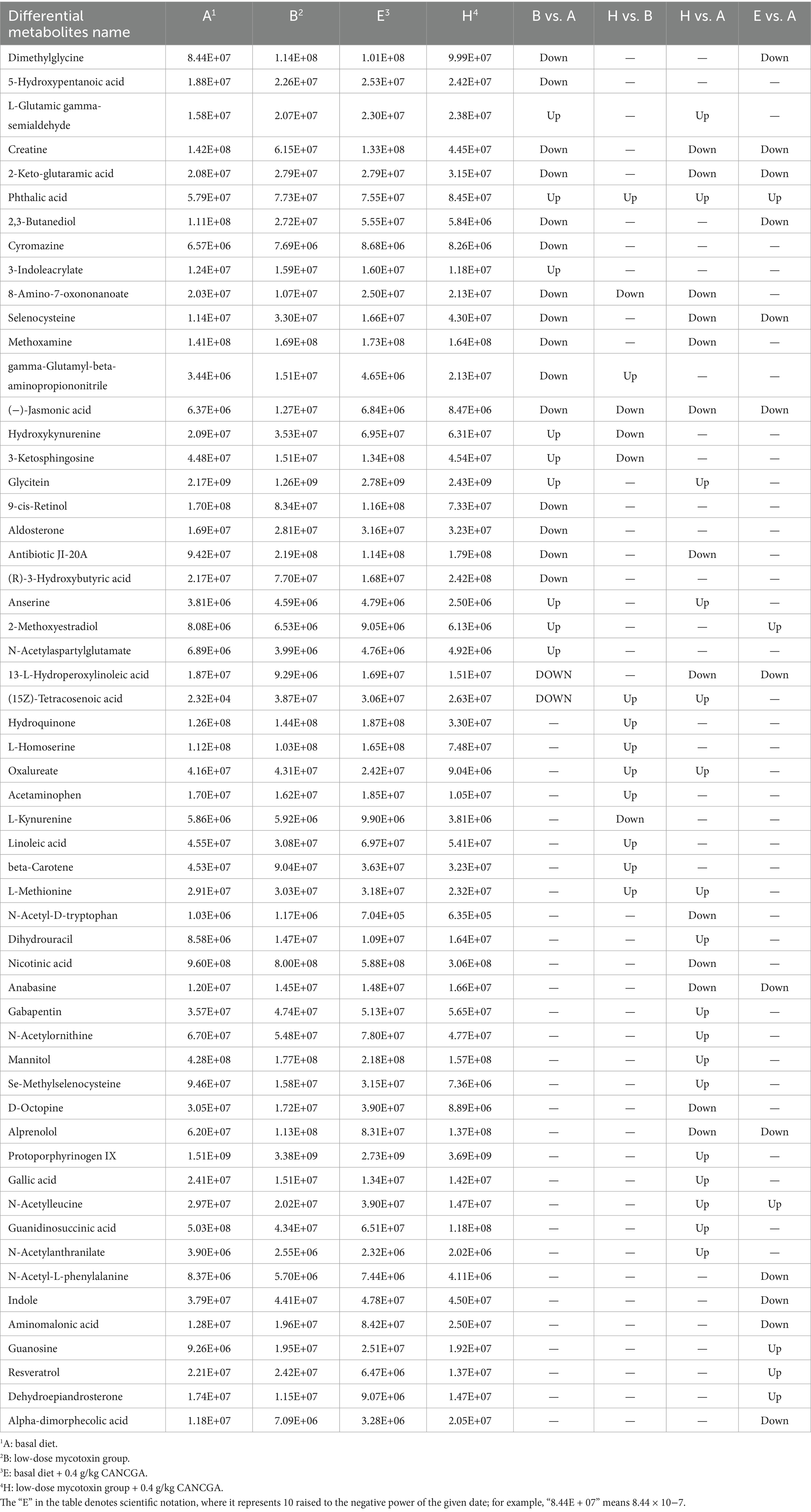
In order to study the mechanism of CANCGA for alleviating multi-mycotoxin toxicity, nutrient metabolism of broilers was analyzed by UPLC-MS spectrometry (Figures 2A,B). In Figure 2C, the horizontal coordinates indicated the similarity between the real grouping of the samples and the 100 random groupings, the vertical coordinates indicated the model evaluation parameters, Q2 and R2 points in the upper right corner indicated the model evaluation parameters of the real grouping. If both Q2 fell below R2, it means that the results are reliable. The results of PCA and OPLS-DA in Figure 2 revealed notable differences of metabolites in broiler serum. In the comparisons of group B vs. group A, group H vs. group B, group H vs. group A, group E vs. group A, about 26, 16, 31 and 18 differential metabolites were identified, respectively (Table 5). Nine metabolites were significantly up-regulated, and seventeen metabolites were down-regulated in the comparison of group B vs. group A (p < 0.05). Seventeen metabolites were significantly up-regulated, and fourteen metabolites were down-regulated in the comparison of group H vs. group A (p < 0.05). Six metabolites were significantly up-regulated, and twelve metabolites were down-regulated in the comparison of group E vs. group A (p < 0.05). Eleven metabolites were significantly up-regulated, and five metabolites were down-regulated in the comparison of group H vs. group B (p < 0.05).
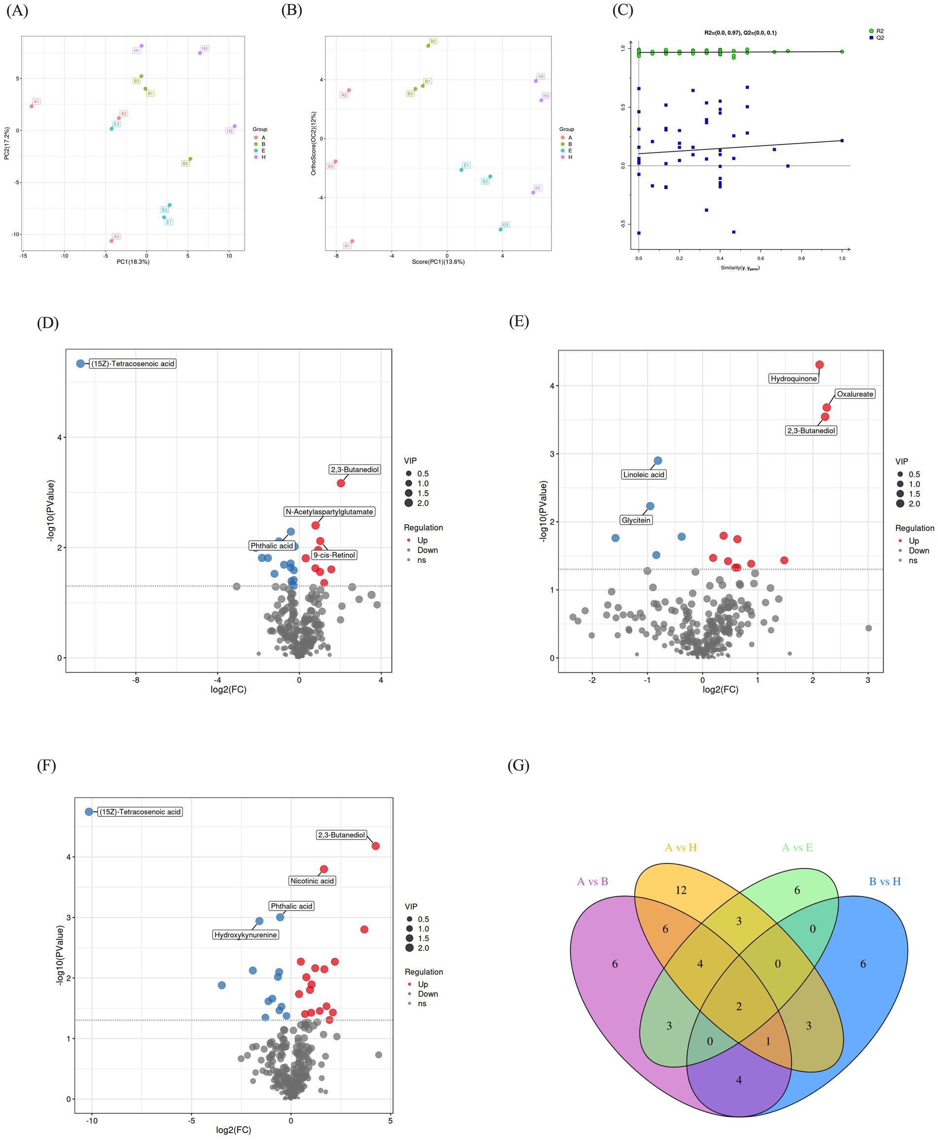
Figure 2. Comprehensive metabolic profiling and comparison of serum metabolites among different groups. Principal component analysis of serum metabolites (A). OPLS-DA model analysis for each group (B,C). Volcanic map of differences in metabolites between two groups. Differential metabolites in the comparisons of group B vs. group A, group H vs. group B, group H vs. group A, respectively (D–F). Venn map of differential metabolites in group A vs. group B, group A vs. group H, group A vs. group E, group B vs. group H (G).
The distribution and alteration of distinct metabolites between the experimental groups and the control group were illustrated in Figure 2D–F. Additionally, the top five metabolites exhibiting statistically significant differences among the groups were highlighted in the volcano plot. Compared to group A, (15Z)-tetracosenoic acid and phthalic acid were downregulated, while 2,3-butanediol, N-acetylaspartylglutamate and 9-cis-retinol were upregulated in group B (Figure 2D). Compared to group B, linoleic acid and glycitein were downregulated, while hydroquinone, oxalureate and 2,3-butanediol were upregulated in group H (Figure 2E). Furthermore, compared to group A, (15Z)-tetracosenoic acid, phthalic acid and hydroxykynurenine were downregulated, while 2,3-butanediol and nicotinic acid were upregulated in group H (Figure 2F). The differential metabolites in each group were analyzed using Venn plots (Figure 2G). In four comparisons such as group B vs. group A, group H vs. group B, group H vs. group A, group E vs. group A, two common differential metabolites (2,3-butanediol and hydroxykynurenine) were identified. Additionally, seven common metabolites (dimethylglycine, 2,3-butanediol, selenocysteine, jasmonic acid, hydroxykynurenine, 3-ketosphingosine and glycitein) were found in the comparisons of group B vs. group A, group H vs. group B.
3.6 KEGG enrichment analysis for serum differential metabolites
To further elucidate the metabolic pathways associated with the differential metabolites in serum, KEGG pathway enrichment analysis was conducted for the following four comparisons: group B vs. group A, group H vs. group B, group H vs. group A, group E vs. group A (Figures 3A–D). Compared with group A, the differential metabolites in group B were primarily enriched in alanine, aspartate and glutamate metabolism pathway; glycine, serine and threonine metabolism pathway; arginine and proline metabolism pathway; steroid hormone biosynthesis pathway (Figure 3A). Compared with group B, the differential metabolites in group H were primarily enriched in the pathways related to glycine, serine and threonine metabolism as well as cysteine and methionine metabolism (Figure 3B). The differential metabolites observed in group H were predominantly associated with selenocompound metabolism, arginine and proline metabolism as well as ABC transporters, compared with group A (Figure 3C).
Further KEGG enrichment analysis was conducted for the common differential metabolites in the comparisons of group B vs. group A, group H vs. group B, revealing the significant associations with three pathways such as selenocompound metabolism, alpha-linolenic acid metabolism as well as glycine, serine and threonine metabolism (Figure 3D). These findings indicated that mycotoxins impacted amino acid synthesis and metabolism as well as steroid hormone biosynthesis pathways to induce organism damage. However, supplementation with CANCGA could mitigate mycotoxin-induced damage by modulating amino acid synthesis and metabolism pathways along with selenium complex metabolism and α-linolenic acid metabolism.
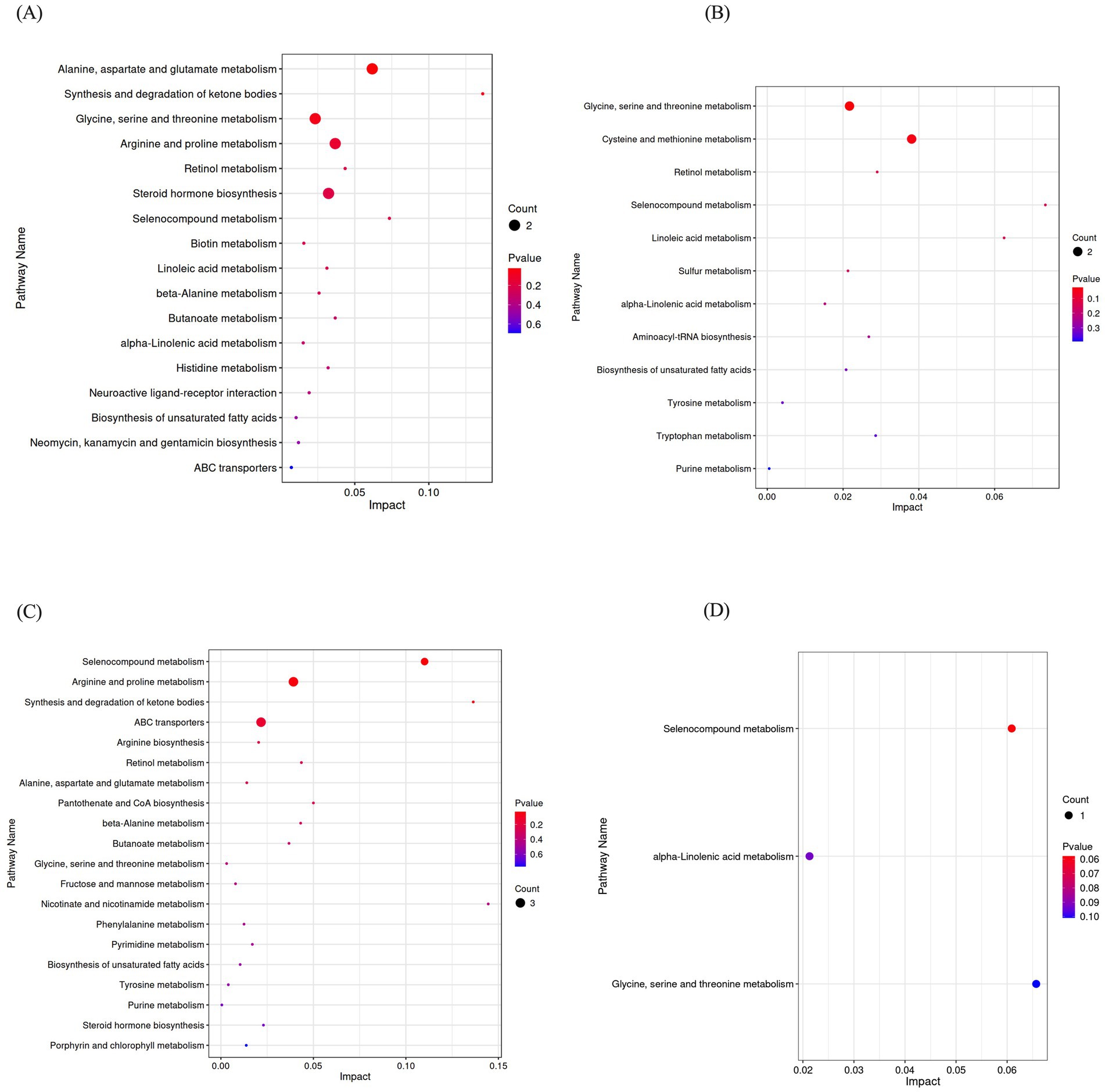
Figure 3. KEGG pathway enrichment of differential metabolites. Differential metabolites were enriched in KEGG pathways in group B vs. group A, group H vs. group B, group H vs. group A (A–C); The differential metabolites were enriched in three pathways by KEGG analysis in group B vs. group A and group H vs. group B (D).
3.7 Correlation between serum differential metabolites and growth performance, serum biochemical parameters or toxin residues in excreta
The relationships between differential metabolites in serum and serum biochemical indices, ADFI, ADG, nutrient metabolic rates or residual levels of mycotoxins in excreta were illustrated in Figure 4. Serum metabolites including 2,3-butanediol, 9-cis-retinol, and Se-methylselenocysteine were significantly positively correlated with serum TG level (p < 0.05). Conversely, selenocysteine, phthalic acid, and 2-keto-glutaramic acid were significantly negatively correlated with serum TG level (p < 0.05). Serum ALP level was positively correlated with the N-acetyl-D-tryptophan (p < 0.05). Serum AST level was significantly positively correlated with oxalureate and nicotinic acid (p < 0.05). Serum N-acetylornithine level was significantly negatively correlated with F/G (p < 0.05). Serum anserine level was significantly negatively correlated with AFB1 residue in excreta, while creatine was significantly negatively correlated with ZEN residue in excreta. Both serum creatine and L-homoserine levels showed significant negative correlations with EE metabolic rate, whereas P metabolic rate had a significant positive correlation with serum 2-methoxyestradiol level. The above results indicated that serum differential metabolites had close correlation with broiler growth performance, serum biochemical parameters or toxin residues in excreta.
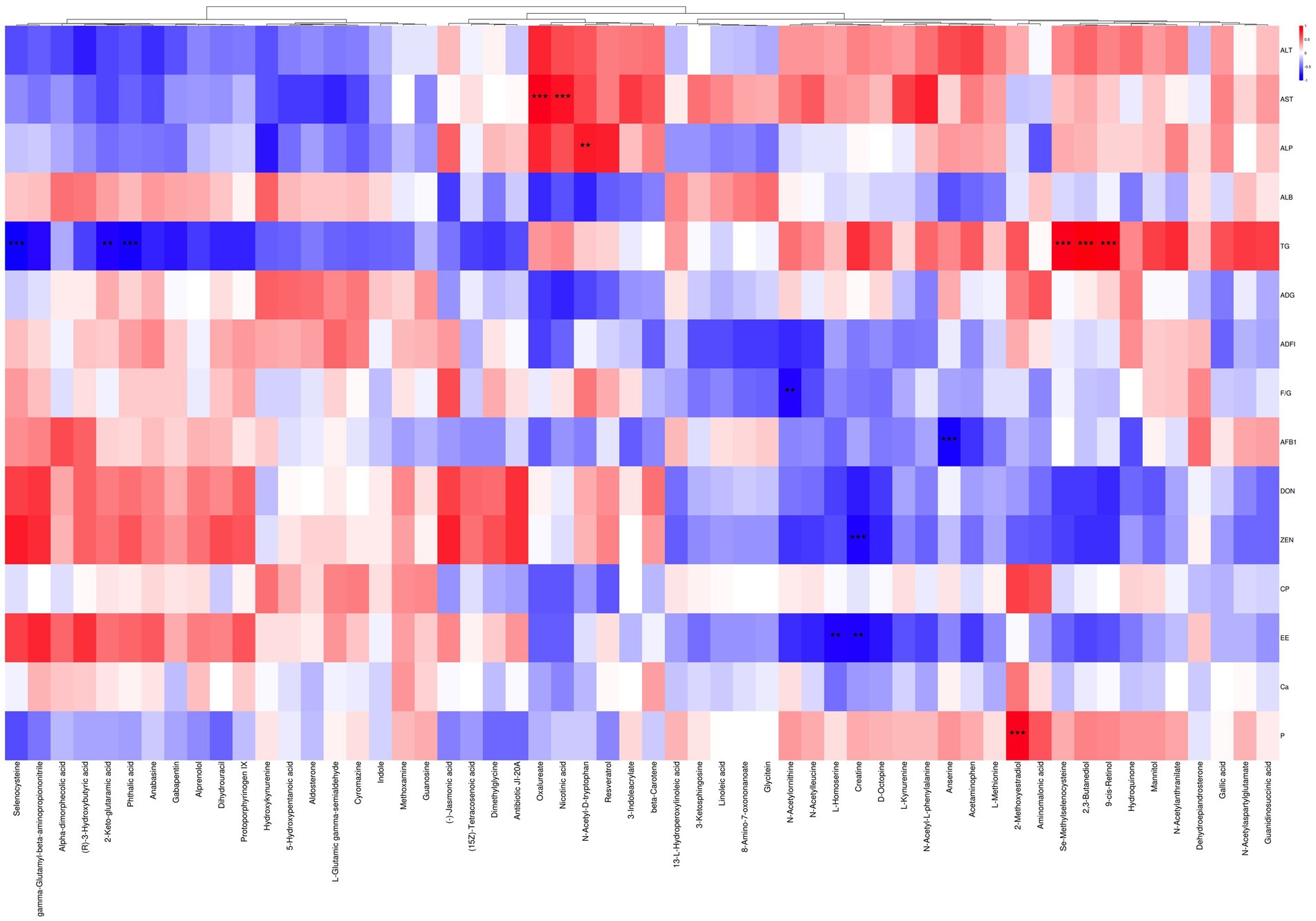
Figure 4. Correlation analysis between serum metabolites and other parameters. Red indicates positive correlation, blue indicates negative correlation; * represents p < 0.05, * * represents p < 0.01, and * * * represents p < 0.001. ALT: alanine aminotransferase; AST: aspartate aminotransferase; ALP: alkaline phosphatase; ALB: albumin; TG: triglycerides; ADG: average daily gain; ADFI: average daily feed intake; F/G: daily feed intake/daily gain; AFB1: aflatoxin B1 residue in excreta; DON: deoxynivalenol residue in excreta; ZEN: zearalenone residue in excreta; CP: crude protein metabolic rate; EE: ether extract metabolic rate; CA: calcium metabolic rate; P: phosphorus metabolic rate.
4 Discussion
About 70% of contaminated feed ingredients contain more than one mycotoxin, posing a serious threat to animal health (1). Previous studies showed the potential of microbial products and plant extracts for this purpose (40). Combining plant extracts with microorganisms may enhance mycotoxin-degradation efficiency because plant extracts contain active compounds capable of binding toxin molecules or promoting their breakdown, thereby mitigating cellular and tissue damage from mycotoxins (41). The research in our laboratory demonstrated that Aspergillus niger solid cultures could degrade AFB1 and ZEN effectively. Another research showed that GA promoted cell proliferation enhanced intestinal barrier function, improved nutrient transport and absorption, and reduced DON-induced damage in piglet intestinal epithelial cells (38). Therefore, the combination of Aspergillus niger solid cultures with GA may be able to alleviate AFB1, ZEN and DON toxicity. This study proved the effectiveness of this combination in alleviating multi-mycotoxins toxicity for broilers. The reason may be due to the ability of Aspergillus niger cultures to degrade AFB1 and ZEN, as well as the GA favoring nutrient transport and absorption in the intestine and alleviating the impairment of nutrient transport by DON.
The consumption of mycotoxin-contaminated feed reduces growth performance and increases pathological traits in broilers (42, 43). High mortality and low ADG of broilers induced by multi-mycotoxins in this study correspond to the above reports. The reasons are that the mycotoxins often cause tissue and organ damage, impair nutrient digestion and utilization, and weaken immune function (44, 45). However, EE metabolic rate was increased with dietary mycotoxin levels increasing, likely due to ZEN similar to endogenous estrogen promoting fat metabolism (46). 0.04 and 0.06% CANCGA supplementation improved growth performance and nutrient metabolisms as well as reduced mortality and mycotoxin levels in feces for broilers exposed to mycotoxins, owing to mycotoxin degradation and detoxification of CANCGA. No significant differences in broiler growth performance were observed with higher or lower CANCGA additions, indicating that the lower cost CANCGA additions were also effective in alleviating the toxic impacts of multi-mycotoxins in production practice.
As mycotoxins are primarily metabolized in the liver, it is the main target for their toxic effects (47). Exposure to mycotoxin-contaminated diets elevated serum AST and ALT levels, leading to hepatic dysfunction, consistent with prior findings (48), and further supporting the link between organ damage and low broiler productivity due to mycotoxins (49). CANCGA addition significantly increased serum TP and ALB levels, while reduced serum AST, ALT and ALP levels, demonstrating the efficacy of CANCGA in mitigating tissue and organ damages induced by multi-mycotoxins, mainly due to the hepatoprotective activity of GA (50) and mycotoxin-degradation ability of Aspergillus niger culture.
To further investigate the impact of mycotoxin exposure and CANCGA supplementation on nutrient metabolisms in broilers, serum metabolomics was conducted in groups A, B, E, and H. The results demonstrated that mycotoxin exposure induced significant alterations in serum metabolites. Specifically, the upregulation of 2,3-butanediol and 9-cis-retinol indicated that mycotoxins disrupt intracellular energy and retinol metabolism, thereby exacerbating oxidative stress and inflammation (51, 52). Compared with the basal diet group, the downregulation of (15Z)-tetracosenoic acid and phthalic acid in the low mycotoxin group reflected the inhibition of normal fatty acid metabolism by mycotoxins, which promoted hepatic lipid accumulation (53) and aggravated liver injury. However, supplementation with CANCGA helped to restore these key metabolites to normal levels. The observed changes in antioxidant metabolites (hydroquinone and glycitein) (54) and lipid metabolites (linoleic acid) (55) suggested that the CANCGA mitigates mycotoxin-induced damage by modulating fatty acid metabolism and enhancing antioxidant defenses, ultimately contributing to the restoration of metabolic balance.
The KEGG pathway enrichment analysis revealed significant metabolic disturbances caused by multi-mycotoxins. These mycotoxins predominantly disrupted normal ketone body metabolism and impaired energy-metabolism stability by interfering with alanine, aspartate, and glutamate metabolisms as well as the synthesis and degradation of ketone bodies. This disruption led to inflammation and lipid metabolism disorders, exacerbating liver damage (56). The enrichments of glycine, serine and threonine metabolisms may indicate the organism’s response to oxidative stress and cellular damage caused by mycotoxins, potentially through the enhancement of antioxidant defense mechanisms. Following the addition of CANCGA, pathway enrichment analysis suggested a partial alleviation of the metabolic disturbances induced by mycotoxins.
This study showed that there were significant increases in metabolites related to glycine, serine and threonine metabolisms as well as cysteine and methionine metabolisms by CANCGA addition. Glycine, serine and threonine are critical amino acids involved in one-carbon metabolism, which is essential for DNA synthesis, repair and cell proliferation (57). Moreover, glycine exhibits detoxifying and hepatoprotective properties, while serine and cysteine are integral to the synthesis of glutathione (GSH), a principal antioxidant in the body that effectively neutralizes free radicals and safeguards cells from oxidative damage (58). Methionine is involved in methylation reactions, which are crucial for maintaining cellular function and stability (59). Consequently, CANCGA addition not only directly degrades mycotoxins but also facilitates the restoration of metabolic pathways, thereby enhancing the synthesis of antioxidants such as GSH and bolstering detoxification processes. It stimulated one-carbon metabolism, promoting cellular repair and regeneration, and mitigating the metabolic disturbances and hepatic damage induced by mycotoxins. Simultaneously, the enrichment of lipid metabolism-related pathways such as alpha-linolenic acid metabolism and unsaturated fatty acid biosynthesis by CANCGA addition indicated a reduction in lipid oxidation, leading to a more balanced fatty acid metabolism, thereby mitigating the impact of mycotoxins on lipid metabolism (60).
Furthermore, the enrichment of selenocompound metabolism by CANCGA addition was intricately linked to the body’s antioxidant capacity, immune regulation and anti-inflammatory responses (61). It is inferred that CANCGA modulated selenium metabolism, enhancing the synthesis and activity of antioxidant enzymes, reducing toxin-induced inflammation, and strengthening the body’s defense against oxidative stress caused by mycotoxins. In conclusion, the mycotoxin group exhibited significant disruptions in amino acid metabolism, lipid metabolism and antioxidant pathways, which were closely associated with oxidative stress, inflammation and hepatocellular damage caused by mycotoxins. After the addition of CANCGA, the disturbed metabolic pathways are restored to the normal levels, especially in anti-oxidative capacity and lipid metabolism regulation.
The differential metabolites common to the four groups were 2,3-butanediol and hydroxylurine. The production of 2,3-butanediol by intestinal microbes (62) suggested that the intestinal microbiome underwent adaptive changes to counteract mycotoxin-induced stress. CANCGA supplementation facilitated adjustments in the gut microflora without altering the protective role of 2,3-butanediol. Hydroxykynurenine, an intermediate in tryptophan metabolism (63), highlighted the tryptophan pathway’s role in sustaining antioxidant capacity and immune function regulated with or without CANCGA intervention.
The level of serum TG was found to be correlated with selenocysteine and methyl selenocysteine, both of which played a role in the selenium metabolism pathway identified through KEGG enrichment analysis. Previous study demonstrated that selenocysteine could reduce TG levels (64). This study showed that CANCGA supplementation increased the level of selenocysteine, resulting in low TG levels. Additionally, a significant positive correlation was observed between TG and 9-cis-retinol, suggesting a link between lipid metabolism and anti-oxidative function. It was reported that 9-cis-retinol regulated the expression of antioxidant genes, thereby enhancing cellular tolerance to oxidative stress (65). Moreover, lipid metabolism provided energy support for sustaining antioxidant and immune functions under mycotoxin stress. The strong positive correlation observed between AST and oxalureate suggested that mycotoxin-induced oxidative stress in the liver promoted purine metabolism, leading to high production of oxalureate (66). Furthermore, the positive association between AST and nicotinic acid implied that mycotoxin-induced stress might augment the demand for nicotinic acid to enhance NAD + synthesis, thereby bolstering cellular anti-oxidative capacity and facilitating liver and tissue resilience against oxidative stress (67).
The significant negative correlation between serum anserine and AFB1 residue in excreta suggested that the anti-oxidative properties of anserine might mitigate oxidative damage induced by AFB1, thereby reducing its presence in the body (68). Additionally, a significant negative correlation was observed between creatine level and ZEN residue in excreta. KEGG enrichment analysis revealed that creatine was involved in metabolic pathways related to glycine, serine, threonine, arginine and proline metabolisms, all of which occur primarily in the liver. Disruption of these pathways due to liver damage led to low creatine levels. CANCGA supplementation could restore the nutrient metabolic disorders induced by multi-mycotoxins.
5 Conclusion
The mortality was increased and growth performance was decreased when broilers were exposed to both low-dose and high-dose multi-mycotoxins. However, dietary CANCGA supplementation could decrease broiler mortality, improve growth performance and nutrient metabolic rates, and alleviate tissue and organ damages caused by mycotoxins. CANCGA ameliorated nutrient metabolic disorders induced by mycotoxins through modulation of pathways involved in glycine, serine and threonine metabolism, cysteine and methionine metabolism, selenium complex metabolism as well as linoleic acid metabolism. This study investigated the role of CANCGA and its mechanism in mitigating the negative effects of mycotoxins on broilers, assessed its value and effectiveness as a feed additive in practical production, and provided an effective mycotoxin mitigation strategy.
Data availability statement
The original contributions presented in the study are included in the article/supplementary material, further inquiries can be directed to the corresponding authors.
Ethics statement
The animal study was approved by the study and included experimental procedures were approved by the guidelines of Animal Care and Use Ethics Committee of Henan Agricultural University (SKLAB-B-2010-003-01). The study was conducted in accordance with the local legislation and institutional requirements.
Author contributions
JT: Formal analysis, Writing – original draft, Conceptualization. MeL: Writing – original draft, Data curation, Methodology. PW: Methodology, Conceptualization, Writing – review & editing. LW: Writing – review & editing, Formal analysis. SJ: Formal analysis, Data curation, Writing – review & editing. XL: Software, Visualization, Investigation, Writing – review & editing. JC: Writing – review & editing, Visualization, Methodology. QY: Project administration, Writing – review & editing, Funding acquisition. CL: Formal analysis, Writing – original draft, Investigation. QZ: Writing – original draft, Validation, Data curation. MaL: Writing – original draft, Visualization, Software. FL: Formal analysis, Writing – original draft, Data curation.
Funding
The author(s) declare that financial support was received for the research and/or publication of this article. This research was funded by the Henan Key Research and Development Project (241111113700), Henan Key Scientific and Technological Projects (242102110058, 242102110011), Xinxiang Key Scientific and Technological Projects (22ZD011).
Conflict of interest
QZ and ML were employed by Henan Delin Biological Product Co. Ltd. FL was employed by Henan Puai Feed Co. Ltd.
The remaining authors declare that the research was conducted in the absence of any commercial or financial relationships that could be construed as a potential conflict of interest.
Generative AI statement
The author(s) declare that no Gen AI was used in the creation of this manuscript.
Publisher’s note
All claims expressed in this article are solely those of the authors and do not necessarily represent those of their affiliated organizations, or those of the publisher, the editors and the reviewers. Any product that may be evaluated in this article, or claim that may be made by its manufacturer, is not guaranteed or endorsed by the publisher.
References
1. Arif, M, Iram, A, Bhutta, MAK, Naiel, MAK, Abd El-Hack, ME, Othman, SI, et al. The biodegradation role of saccharomyces cerevisiae against harmful effects of mycotoxin contaminated diets on broiler performance, immunity status, and carcass characteristics. Animals. (2020) 10:238. doi: 10.3390/ani10020238
2. Alshannaq, A, and Yu, J. Occurrence, toxicity, and analysis of major mycotoxins in food. Int J Environ Res Public Health. (2017) 14:632. doi: 10.3390/ijerph14060632
3. Binder, EM. Managing the risk of mycotoxins in modern feed production. Anim Feed Sci Tech. (2007) 133:149–66. doi: 10.1016/j.anifeedsci.2006.08.008
4. Liu, M, Zhang, L, Mo, Y, Li, J, Yang, J, Wang, J, et al. Ferroptosis is involved in deoxynivalenol-induced intestinal damage in pigs. J Anim Sci Biotechnol. (2023) 14:29. doi: 10.1186/s40104-023-00841-4
5. Wang, J, Qu, J, Liu, S, Xu, Q, Li, X, Zhu, Y, et al. Tannic acid ameliorates systemic glucose and lipid metabolic impairment induced by low-dose t-2 toxin exposure. J Agric Food Chem. (2023) 71:12574–86. doi: 10.1021/acs.jafc.3c02934
6. Juan, C, Covarelli, L, Beccari, G, Colasante, V, and Mañes, J. Simultaneous analysis of twenty-six mycotoxins in durum wheat grain from Italy. Food Control. (2016) 62:322–9. doi: 10.1016/j.foodcont.2015.10.032
7. Svihus, B, Uhlen, AK, and Harstad, OM. Effect of starch granule structure, associated components and processing on nutritive value of cereal starch: a review. Anim Feed Sci Tech. (2005) 122:303–20. doi: 10.1016/j.anifeedsci.2005.02.025
8. Gruber-Dorninger, C, Jenkins, T, and Schatzmayr, G. Global mycotoxin occurrence in feed: a ten-year survey. Toxins. (2019) 11:375. doi: 10.3390/toxins11070375
9. Streit, E, Naehrer, K, Rodrigues, I, and Schatzmayr, G. Mycotoxin occurrence in feed and feed raw materials worldwide: long-term analysis with special focus on Europe and Asia. J Sci Food Agr. (2013) 93:2892–9. doi: 10.1002/jsfa.6225
10. Karsauliya, K, Yahavi, C, Pandey, A, Bhateria, M, Sonker, AK, Pandey, H, et al. Co-occurrence of mycotoxins: a review on bioanalytical methods for simultaneous analysis in human biological samples, mixture toxicity and risk assessment strategies. Toxicon. (2022) 218:25–39. doi: 10.1016/j.toxicon.2022.08.016
11. Lumsangkul, C, Chiang, HI, Lo, NW, Fan, YK, and Ju, JC. Developmental toxicity of mycotoxin fumonisin b₁ in animal embryogenesis: an overview. Toxins. (2019) 11:114. doi: 10.3390/toxins11020114
12. Lumsangkul, C, Tso, KH, Fan, YK, Chiang, HI, and Ju, JC. Mycotoxin fumonisin B (1) interferes sphingolipid metabolisms and neural tube closure during early embryogenesis in brown tsaiya ducks. Toxins. (2021) 13:743. doi: 10.3390/toxins13110743
13. Yang, C, Song, G, and Lim, W. Effects of mycotoxin-contaminated feed on farm animals. J Hazard Mater. (2020) 389:122087. doi: 10.1016/j.jhazmat.2020.122087
14. Eshetu, E, Habtamu, A, and Gebretensa, A. An overview on major mycotoxin in animal: its public health implication, economic impact and control strategies. J Health Med Nur. (2016) 25:64–73. doi: 10.1186/s41043-016-0050-6
15. Holanda, DM, and Kim, SW. Mycotoxin occurrence, toxicity, and detoxifying agents in pig production with an emphasis on deoxynivalenol. Toxins. (2021) 13:171. doi: 10.3390/toxins13020171
16. Dou, X, Wu, G, Ding, Z, and Xie, J. Construction of a nanoscale metal-organic framework aptasensor for fluorescence ratiometric sensing of AFB1 in real samples. Food Chem. (2023) 416:135805. doi: 10.1016/j.foodchem.2023.135805
17. Liu, H, Xie, R, Huang, W, Yang, Y, Zhou, M, Lu, B, et al. Negative effects of aflatoxin B1 (AFB1) in the diet on growth performance, protein and lipid metabolism, and liver health of juvenile hybrid grouper (Epinephelus fuscoguttatus♀×Epinephelus lanceolatus♂). Aquaculture Rep. (2023) 33:101779. doi: 10.1016/j.aqrep.2023.101779
18. Martínez, J, Hernández-Rodríguez, M, Méndez-Albores, A, Téllez-Isaías, G, Mera Jiménez, E, Nicolás-Vázquez, MI, et al. Computational studies of aflatoxin B1 (AFB1): a review. Toxins. (2023) 15:135. doi: 10.3390/toxins15020135
19. Hou, YJ, Zhu, CC, Xu, YX, Cui, XS, Kim, NH, and Sun, SC. Zearalenone exposure affects mouse oocyte meiotic maturation and granulosa cell proliferation. Environ Toxicol. (2015) 30:1226–33. doi: 10.1002/tox.21995
20. Ropejko, K, and Twarużek, M. Zearalenone and its metabolites-general overview, occurrence, and toxicity. Toxins. (2021) 13:35. doi: 10.3390/toxins13010035
21. Tatay, E, Espín, S, García-Fernández, AJ, and Ruiz, MJ. Oxidative damage and disturbance of antioxidant capacity by zearalenone and its metabolites in human cells. Toxicol In Vitro. (2017) 45:334–9. doi: 10.1016/j.tiv.2017.04.026
22. Ghareeb, K, Awad, WA, Böhm, J, and Zebeli, Q. Impacts of the feed contaminant deoxynivalenol on the intestine of monogastric animals: poultry and swine. J Appl Toxicol. (2015) 35:327–37. doi: 10.1002/jat.3083
23. Shen, YB, Weaver, AC, and Kim, SW. Physiological effects of deoxynivalenol from naturally contaminated corn on cerebral tryptophan metabolism, behavioral response, gastrointestinal immune status and health in pigs following a pair-feeding model. Toxins. (2021) 13:393. doi: 10.3390/toxins13060393
24. Vignal, C, Djouina, M, Pichavant, M, Caboche, S, Waxin, C, Beury, D, et al. Chronic ingestion of deoxynivalenol at human dietary levels impairs intestinal homeostasis and gut microbiota in mice. Arch Toxicol. (2018) 92:2327–38. doi: 10.1007/s00204-018-2228-6
25. Peng, Z, Chen, L, Nüssler, AK, Liu, L, and Yang, W. Current sights for mechanisms of deoxynivalenol-induced hepatotoxicity and prospective views for future scientific research: a mini review. J Appl Toxicol. (2017) 37:518–29. doi: 10.1002/jat.3428
26. Wang, X, Chen, X, Cao, L, Zhu, L, Zhang, Y, Chu, X, et al. Mechanism of deoxynivalenol-induced neurotoxicity in weaned piglets is linked to lipid peroxidation, dampened neurotransmitter levels, and interference with calcium signaling. Ecotoxicol Environ Saf. (2020) 194:110382. doi: 10.1016/j.ecoenv.2020.110382
27. Chang, J, Wang, T, Wang, P, Yin, Q, Liu, C, Zhu, Q, et al. Compound probiotics alleviating aflatoxin B1 and zearalenone toxic effects on broiler production performance and gut microbiota. Ecotoxicol Environ Saf. (2020) 194:110420. doi: 10.1016/j.ecoenv.2020.110420
28. Ma, R, Zhang, L, Liu, M, Su, YT, Xie, WM, Zhang, NY, et al. Individual and combined occurrence of mycotoxins in feed ingredients and complete feeds in China. Toxins. (2018) 10:113. doi: 10.3390/toxins10030113
29. Wu, L, Li, J, Li, Y, Li, T, He, Q, Tang, Y, et al. Aflatoxin B (1), zearalenone and deoxynivalenol in feed ingredients and complete feed from different province in China. J Anim Sci Biotechnol. (2016) 7:63–10. doi: 10.1186/s40104-016-0122-8
30. Tsiouris, V, Tassis, P, Raj, J, Mantzios, T, Kiskinis, K, Vasiljević, M, et al. Investigation of a novel multicomponent mycotoxin detoxifying agent in amelioration of mycotoxicosis induced by aflatoxin-b1 and ochratoxina in broiler chicks. Toxins. (2021) 13:367. doi: 10.3390/toxins13060367
31. Wang, Q, Zhang, Y, Zheng, N, Zhao, S, Li, S, and Wang, J. The biochemical and metabolic profiles of dairy cows with mycotoxins-contaminated diets. PeerJ. (2020) 8:e8742. doi: 10.7717/peerj.8742
32. Wu, X, Guo, L, Huang, G, Tang, W, Zhao, S, Wang, J, et al. Effects of dietary natural mycotoxins exposure on performance, biochemical parameters and milk small molecule metabolic pathways of lactating cows. Agric Basel. (2022) 12:420. doi: 10.3390/agriculture12030420
33. Čolović, R, Puvača, N, Cheli, F, Avantaggiato, G, Greco, D, Đuragić, O, et al. Decontamination of mycotoxin-contaminated feedstuffs and compound feed. Toxins. (2019) 11:617. doi: 10.3390/toxins11110617
34. Xing, F, Wang, L, Liu, X, Selvaraj, JN, Wang, Y, Zhao, Y, et al. Aflatoxin B1 inhibition in aspergillus flavus by Aspergillus niger through down-regulating expression of major biosynthetic genes and AFB1 degradation by atoxigenic A. flavus. Int J Mol Sci. (2017) 256:1–10. doi: 10.1016/j.ijfoodmicro.2017.05.013
35. Ji, J, Yu, J, Yang, Y, Yuan, X, Yang, J, Zhang, Y, et al. Exploration on the enhancement of detoxification ability of zearalenone and its degradation products of aspergillus niger fs10 under directional stress of zearalenone. Toxins. (2021) 13:720. doi: 10.3390/toxins13100720
36. Wang, Q, Huang, Y, Li, Y, Zhang, L, Tang, H, Zhang, J, et al. Glycyrrhizic acid mitigates tripterygium-glycoside-tablet-induced acute liver injury via PKM2 regulated oxidative stress. Meta. (2022) 12:1128. doi: 10.3390/metabo12111128
37. Yuan, T, Wang, J, Chen, L, Shan, J, and Di, L. Glycyrrhizic acid improving the liver protective effect by restoring the composition of Lactobacillus. J Funct Foods. (2019) 52:219–27. doi: 10.1016/j.jff.2018.11.001
38. Xu, X, Yan, G, Chang, J, Wang, P, Yin, Q, Liu, C, et al. Comparative transcriptome analysis reveals the protective mechanism of glycyrrhinic acid for deoxynivalenol-induced inflammation and apoptosis in IPEC-J2 cells. Oxidative Med Cell Longev. (2020) 2020:5974157. doi: 10.1155/2020/5974157
39. Huang, W, Chang, J, Wang, P, Liu, C, Yin, Q, Song, A, et al. Effect of compound probiotics and mycotoxin degradation enzymes on alleviating cytotoxicity of swine jejunal epithelial cells induced by aflatoxin b₁ and zearalenone. Toxins. (2019) 11:12. doi: 10.3390/toxins11010012
40. Ndiaye, S, Zhang, M, Fall, M, Ayessou, NM, Zhang, Q, and Li, P. Current review of mycotoxin biodegradation and bioadsorption: microorganisms, mechanisms, and main important applications. Toxins. (2022) 14:729. doi: 10.3390/toxins14110729
41. Arimboor, R. Metabolites and degradation pathways of microbial detoxification of aflatoxins: a review. Mycotoxin Res. (2024) 40:71–83. doi: 10.1007/s12550-023-00515-0
42. Haque, MA, Wang, Y, Shen, Z, Li, X, Saleemi, MK, and He, C. Mycotoxin contamination and control strategy in human, domestic animal and poultry: a review. Microb Pathog. (2020) 142:104095. doi: 10.1016/j.micpath.2020.104095
43. Sun, Y, Huang, K, Long, M, Yang, S, and Zhang, Y. An update on immunotoxicity and mechanisms of action of six environmental mycotoxins. Food Chem Toxicol. (2022) 163:112895. doi: 10.1016/j.fct.2022.112895
44. Magnoli, AP, Poloni, VL, and Cavaglieri, L. Impact of mycotoxin contamination in the animal feed industry. Curr Opin Food Sci. (2019) 29:99–108. doi: 10.1016/j.cofs.2019.08.009
45. Mohaghegh, A, Chamani, M, Shivazad, M, Sadeghi, AA, and Afzali, N. Effect of esterified glucomannan on broilers exposed to natural mycotoxin-contaminated diets. J Appl Anim Res. (2017) 45:285–91. doi: 10.1080/09712119.2016.1174122
46. Torres Irizarry, VC, Jiang, Y, He, Y, and Xu, P. Hypothalamic estrogen signaling and adipose tissue metabolism in energy homeostasis. Front Endocrinol. (2022) 13:898139. doi: 10.3389/fendo.2022.898139
47. Hasuda, AL, Person, E, Khoshal, AK, Bruel, S, Puel, S, Oswald, IP, et al. Deoxynivalenol induces apoptosis and inflammation in the liver: analysis using precision-cut liver slices. Food Chem Toxicol. (2022) 163:112930. doi: 10.1016/j.fct.2022.112930
48. Saminathan, M, Selamat, J, Abbasi Pirouz, A, Abdullah, N, and Zulkifli, I. Effects of nano-composite adsorbents on the growth performance, serum biochemistry, and organ weights of broilers fed with aflatoxin-contaminated feed. Toxins. (2018) 10:345. doi: 10.3390/toxins10090345
49. Lee, J, Jessen, K, Beltran, R, Starkl, V, Schatzmayr, G, Borutova, R, et al. Mycotoxin-contaminated diets and deactivating compound in laying hens: 1. Effects on performance characteristics and relative organ weight. Poultry Sci. (2012) 91:2089–95. doi: 10.3382/ps.2012-02136
50. Yu, JY, Ha, JY, Kim, KM, Jung, YS, Jung, JC, and Oh, S. Anti-inflammatory activities of licorice extract and its active compounds, glycyrrhizic acid, liquiritin and liquiritigenin, in BV2 cells and mice liver. Molecules. (2015) 20:13041–54. doi: 10.3390/molecules200713041
51. Tabbaa, SM, Guilak, F, Lemmerman, LR, Glembotski, N, D'Lima, DD, Wang, T, et al. Elevated lipid metabolites in stored clinical OCA media correlate with chondrocyte death. Am J Sports Med. (2024) 52:2119–28. doi: 10.1177/03635465241252653
52. Wu, G, Bazer, FW, Davis, TA, Kim, SW, Li, P, Marc Rhoads, J, et al. Arginine metabolism and nutrition in growth, health and disease. Amino Acids. (2009) 37:153–68. doi: 10.1007/s00726-008-0210-y
53. Bednarski, T, Mohsin, R, and Jamey, Y. Short-term effect of saturated and monounsaturated fatty acids on hepatic energy metabolism. Diabetes. (2019) 68:285–LB. doi: 10.2337/db19-285-LB
54. Biais, B, Krisa, S, Cluzet, S, Da Costa, G, Waffo-Teguo, P, Mérillon, J-M, et al. Antioxidant and cytoprotective activities of grapevine stilbenes. J Agric Food Chem. (2017) 65:4952–60. doi: 10.1021/acs.jafc.7b01254
55. Huyan, Z, Pellegrini, N, Steegenga, W, and Capuano, E. Insights into gut microbiota metabolism of dietary lipids: the case of linoleic acid. Food Funct. (2022) 13:4513–26. doi: 10.1039/d1fo04254h
56. Zhou, C, Hu, L, Mu, R, Mei, X, Wu, X, Wang, C, et al. Compound green tea (CGT) regulates lipid metabolism in high-fat diet induced mice. RSC Adv. (2022) 12:24301–10. doi: 10.1039/d2ra02831j
57. Pan, S, Fan, M, Liu, Z, Li, X, and Wang, H. Serine, glycine and one-carbon metabolism in cancer (review). Int J Oncol. (2021) 58:158–70. doi: 10.3892/ijo.2020.5158
58. Lv, XC, Wu, Q, Cao, YJ, Lin, YC, Guo, WL, Rao, PF, et al. Ganoderic acid a from ganoderma lucidum protects against alcoholic liver injury through ameliorating the lipid metabolism and modulating the intestinal microbial composition. Food Funct. (2022) 13:5820–37. doi: 10.1039/d1fo03219d
59. Elango, R. Methionine nutrition and metabolism: insights from animal studies to inform human nutrition. J Nutr. (2020) 150:2518s–23s. doi: 10.1093/jn/nxaa155
60. Yi, S, Mai, T, Fang, Y, Tian, Q, and Zhao, S. Repeated injection of xylazine causes liver injury through the PPAR signaling pathway in rats. J Biochem Mol Toxicol. (2025) 39:e70101. doi: 10.1002/jbt.70101
61. Saeedi, M, Soltani, F, Babalar, M, Wiesner-Reinhold, M, Baldermann, S, and Mastinu, A. Selenium enhances growth, phenolic compounds, antioxidant capacity in brassica oleracea var. Chem Biodivers. (2024) 22:e202401731. doi: 10.1002/cbdv.202401731
62. Ujlaki, G, Kovács, T, Vida, A, Kókai, E, Rauch, B, Schwarcz, S, et al. Identification of bacterial metabolites modulating breast cancer cell proliferation and epithelial-mesenchymal transition. Molecules. (2023) 28:5898. doi: 10.3390/molecules28155898
63. Knubel, CP, Insfran, C, Martinez, FF, Diaz Lujan, C, Fretes, RE, Theumer, MG, et al. 3-Hydroxykynurenine, a tryptophan metabolite generated during the infection, is active against trypanosoma cruzi. ACS Med Chem Lett. (2017) 8:757–61. doi: 10.1021/acsmedchemlett.7b00169
64. Sun, X, Yue, SZ, Qiao, YH, Sun, ZJ, Wang, C, and Li, HF. Dietary supplementation with selenium-enriched earthworm powder improves antioxidative ability and immunity of laying hens. Poultry Sci. (2020) 99:5344–9. doi: 10.1016/j.psj.2020.07.030
65. Elomda, AM, Saad, MF, Saeed, AM, Elsayed, A, Abass, AO, Safaa, HM, et al. Antioxidant and developmental capacity of retinol on the in vitro culture of rabbit embryos. Zygote. (2018) 26:326–32. doi: 10.1017/s0967199418000308
66. Rusz, M, Del Favero, G, El Abiead, Y, Gerner, C, Keppler, BK, Jakupec, MA, et al. Morpho-metabotyping the oxidative stress response. Sci Rep. (2021) 11:15471. doi: 10.1038/s41598-021-94585-8
67. Romani, M, Hofer, DC, Katsyuba, E, and Auwerx, J. Niacin: an old lipid drug in a new NAD (+) dress. J Lipid Res. (2019) 60:741–6. doi: 10.1194/jlr.S092007
Keywords: multi-mycotoxin, detoxification, broiler, growth performance, serum metabolomics
Citation: Tu J, Li M, Wang P, Wang L, Jin S, Li X, Chang J, Yin Q, Liu C, Zhu Q, Li M and Lu F (2025) Combination of Aspergillus niger culture and glycyrrhizic acid alleviates the toxic effects of multi-mycotoxins on broiler production performance and nutrient metabolism. Front. Nutr. 12:1628442. doi: 10.3389/fnut.2025.1628442
Edited by:
Laurent Dufossé, Université de la Réunion, FranceReviewed by:
Safiu Suberu, North Carolina Agricultural and Technical State University, United StatesHajrian Rizqi Albarki, Khon Kaen University, Thailand
Copyright © 2025 Tu, Li, Wang, Wang, Jin, Li, Chang, Yin, Liu, Zhu, Li and Lu. This is an open-access article distributed under the terms of the Creative Commons Attribution License (CC BY). The use, distribution or reproduction in other forums is permitted, provided the original author(s) and the copyright owner(s) are credited and that the original publication in this journal is cited, in accordance with accepted academic practice. No use, distribution or reproduction is permitted which does not comply with these terms.
*Correspondence: Chaoqi Liu, bGl1Y3FAaGVuYXUuZWR1LmNu; Qingqiang Yin, cXF5MTk2NEBoZW5hdS5lZHUuY24=
†These authors have contributed equally to this work
 Jinqiu Tu
 Mengke Li1†
 Ping Wang
 Lijun Wang
 Sanjun Jin
 Juan Chang
 Qingqiang Yin
 Chaoqi Liu
 Fushan Lu