- 1Department of Pediatrics, Division of Neonatology, Chang Gung Memorial Hospital, School of Medicine, Chang Gung University, Taoyuan, Taiwan
- 2Department of Pediatrics, Taoyuan Armed Forces General Hospital, Taoyuan, Taiwan
- 3Department of Ophthalmology, Chang Gung Memorial Hospital, School of Medicine, Chang Gung University, Taoyuan, Taiwan
Background: While early-life cytokine profiles have been linked to neurodevelopmental outcomes in preterm infants, their prognostic value is limited by clinical instability and inflammatory comorbidities in the immediate postnatal period. This study explores cytokine levels measured during a more stable developmental window—near term-equivalent age [postmenstrual age (PMA) 34–38 weeks]—and their association with neurodevelopmental outcomes.
Methods: We prospectively enrolled 35 preterm infants (birth weight, 500–1,500 g). Serum cytokine levels were measured at PMA 34, 36, and 38 weeks. Neurodevelopment was assessed at 12 months’ corrected age using standardized tools (BSID-III). Infants were classified into neurodevelopmental impairment (NDI) and non-NDI groups. Cytokine levels and their changes were compared between groups.
Results: Elevated IFN-γ levels at PMA 34 weeks were associated with a higher risk of NDI. Conversely, higher levels of Eotaxin-2, IL-2, IL-11, IL-16, MIP-1δ, PDGF-BB, TIMP-2, and TNF-β at PMA 36–38 weeks were observed more frequently in the non-NDI group. The trends also differed: increased IL-17 and decreased Eotaxin-1, Eotaxin-2, IL-7, IL-16, MIP-1α, MIP-1β, PDGF-BB, and TIMP-2 between PMA 34–36 weeks, and further declines in ICAM-1, IL-7, MIP-1α, and MIP-1β by PMA 38 weeks were associated with adverse outcomes. All identified biomarkers demonstrated good discriminatory ability, particularly changes in Eotaxin-2 between PMA 34 and 36 weeks and PDGF-BB between PMA 34 and 38 weeks.
Conclusions: Serum cytokine levels and their trajectories during PMA 34–38 weeks may serve as potential biomarkers for identifying preterm infants at risk of neurodevelopmental impairment. Further studies with larger cohorts are needed to clarify their interplay with preterm morbidities.
Highlights
• Near-term cytokines help identify preterm infants at neurodevelopment risk.
• Most cytokines predict good neurodevelopment; only IFN-γ and IL-17 predict poor outcomes.
• Both cytokine levels and their changes may predict outcomes in preterm infants.
• Preterm comorbidities impact biomarker levels at near-term age.
Introduction
Premature infants are at an increased risk of developing varying degrees of neurodevelopmental impairment (NDI) (1, 2). In these infants, fetal growth restriction has been identified as an independent risk factor (3). Studies suggest correlations between specific blood biomarkers and neurodevelopmental outcomes in infants. For instance, elevated levels of proinflammatory cytokines, including interleukin (IL)-1β, IL-8, IL-9, tumor necrosis factor (TNF)-α, and regulated upon activation, normal T cell expressed and secreted (RANTES), along with decreased levels of anti-inflammatory cytokines, such as IL-2 and IL-3, within the first day after birth, have shown good sensitivity and specificity in predicting cerebral palsy (CP) in late preterm and term infants (4). Another cohort study conducted on 1,067 extremely low birth weight (≤1,000 g) infants found that IL-8 levels were elevated during the initial four days and remained higher in infants who subsequently developed CP (5).
However, some studies also have provided different insights, suggesting that cytokines in early postnatal age may not effectively predict neurologic outcomes. A study that collected cord blood samples at birth from 400 neonates found no association between elevated levels of cord serum IL-6, C-Reactive Protein (CRP), and Myeloperoxidase (MPO) at birth and poor neurodevelopmental outcomes (6). In a nested case-control study of 615 preterm infants with a gestational age (GA) between 24 and 31 6/7 weeks, cord serum IL-8, IL-1β, and TNF-α levels were not associated with subsequent CP or neurodevelopmental delay at the 2-year follow-up (7). Additionally, a study that measured cytokines on average 2.4 days postnatally in 271 very preterm infants born before 32 weeks GA found no associations between 11 cytokines [IL-1, −2, −4, −5, −6, −8, −10, and −12; granulocyte–macrophage colony-stimulating factor (GMCSF); interferon (IFN)-γ; and TNF-α] and later diagnosis of CP (8).
Conflicting research results may be attributed to the fact that preterm infants often face significant clinical instability during the initial weeks of life. This period may be affected by various pathological insults that can alter their physiological condition. As a result, cytokines in the early postnatal period may not provide strong neurological prognostic indicators. Limited studies have hinted that the relationship between various blood biomarkers and neurodevelopmental outcomes may vary depending on the specific postnatal time points and the types of specimens evaluated. A large cohort study of 1,506 premature infants born at a GA of less than 28 weeks found that serum biomarkers such as CRP, IL-8, and intercellular adhesion molecule-1 (ICAM-1), measured on postnatal days 21 and 28, were associated with mental or psychomotor developmental impairments in these infants (9). In another study of 51 term infants, higher serum levels of IL-1β at six months of age predicted decreased motor skill performance at 30 months of age (10).
Therefore, we investigated cytokine concentrations at postmenstrual ages (PMA) of 34, 36, and 38 weeks—a period considered to represent near term-equivalent age and characterized by relatively stable clinical conditions—to explore potential associations between these biomarkers and neurodevelopmental outcomes.
Methods
Study participants
This prospective cohort study was conducted in the neonatal intensive care units of our hospital between December 1, 2019, and June 30, 2022. Preterm infants with birth weights between 500 g and 1,500 g were enrolled after written informed consent was obtained from their parents. Exclusion criteria included major congenital anomalies, such as chromosomal abnormalities or central nervous system malformations. Infants were also excluded from the final analysis if they died before serum cytokine sampling, had incomplete medical records, or lacked neurodevelopmental assessment data. The study was approved by the Institutional Review Board of our hospital (approval number: 201902088A3).
Data collection
Demographic data were retrospectively collected from electronic medical records, including maternal characteristics [e.g., mother's age, pregnancy complications such as gestational diabetes mellitus (GDM), premature rupture of membranes (PROM), pre-eclampsia/eclampsia], infant baseline information (e.g., GA, birth weight, APGAR scores), and clinical outcomes (e.g., severe intraventricular hemorrhage (IVH), hemodynamically significant patent ductus arteriosus (HsPDA), necrotizing enterocolitis (NEC), Grade II/III bronchopulmonary dysplasia (BPD). Severe IVH was defined as IVH grade ≥3 on intracranial ultrasound. HsPDA was defined as cases requiring medical or surgical ligation. BPD was diagnosed basing on the 2019 Jensen definition (11). Interventions, including postnatal use of intravenous or inhaled steroids and exclusive breast milk feeding, were included in the analysis. Nutritional status was assessed by examining changes in Z-scores from birth to discharge. For infants discharged at a PMA of less than 50 weeks, the Fenton Growth Chart (2013) was used (12), while the 2006 WHO Growth Standards were applied for those discharged at 50 weeks or later (13).
Neurodevelopment assessment
The Bayley Scales of Infant and Toddler Development, Third Edition (BSID-III) were used to assess neurodevelopmental condition at 12 months of corrected age (14). The BSID-III includes domains for cognitive (91 items), language (97 items), motor (138 items), social-emotional (35 items), and adaptive behavior (241 items). Only the first three domains were included in the analysis. Neurodevelopmental impairment (NDI) was defined as a BSID-III score <70 in any of the motor, cognitive, or language domains at 12 months of corrected age. Infants were classified into NDI and non-NDI groups.
Cytokines analysis
Blood samples were collected three times: at 34 weeks PMA, 36 weeks PMA, and 38 weeks PMA, with a 2-week interval between collections. After reviewing the cytokines reported to correlate with neurodevelopment in preterm infants, we selected a commercially available multiplex assay (Quantibody® Human Inflammation Array 3; RayBiotech, Peachtree Corners, GA, USA) to analyze serum cytokine levels at each time point. This assay quantifies 40 cytokines, including B-lymphocyte chemoattractant (BLC), eotaxin-1, eotaxin-2, granulocyte colony-stimulating factor (GCSF), GMCSF, I-309, ICAM-1, IFN-γ, IFN-1α, IFN-1β, IL-1Rα, IL-2, IL-4, IL-5, IL-6, IL-6R, IL-7, IL-8, IL-10, IL-11, IL-12p40, IL-12p70, IL-13, IL-15, IL-16, IL-17, monocyte chemoattractant protein (MCP)-1, macrophage colony-stimulating factor (MCSF), monokine induced by IFN-γ (MIG), macrophage inflammatory protein (MIP)-1α, MIP-1β, MIP-1δ, platelet-derived growth factor-BB (PDGF-BB), RANTES, tissue inhibitor of metalloproteinase (TIMP)-1, TIMP-2, TNF-α, TNF-β, TNF receptor (TNFR)-1, and TNFR-2, including several previously reported to be significant. Serum cytokine levels at each time point, as well as the cytokine changes between each interval, were compared between two groups.
Statistical analysis
In comparing demographic variables, a Chi-square test was used to assess differences between categorical variables, and the independent Student's t-test was applied to analyze continuous, normally distributed variables, which are presented as means and standard deviations (SDs). Serum cytokine levels at each time point and cytokine changes between intervals were compared using the nonparametric Mann–Whitney U-test between two groups. The area under the ROC curve (AUC) was calculated for significant biomarkers. The generalized estimating equations approach was used to identify factors associated with changes in serum cytokine levels. A significant difference was considered for p-values less than 0.05. Statistical analyses were performed using IBM SPSS Statistics (version 27.0, Armonk, NY: IBM Corp).
Results
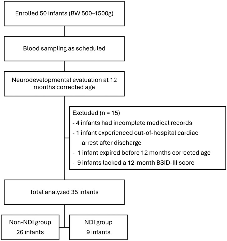
A total of 50 premature infants were enrolled in this study. Fifteen infants were excluded for the following reasons: four had incomplete medical records, one expired before the neurodevelopmental assessment, one experienced an out-of-hospital cardiac arrest after discharge, and nine lacked a 12-month BSID-III score. Thirty-five preterm infants were included in the final analysis, with 26 in the non-NDI group and 9 in the NDI group (Figure 1). The characteristics of these two groups were presented in Table 1. There were no significant differences in maternal and infant characteristics between the two groups.
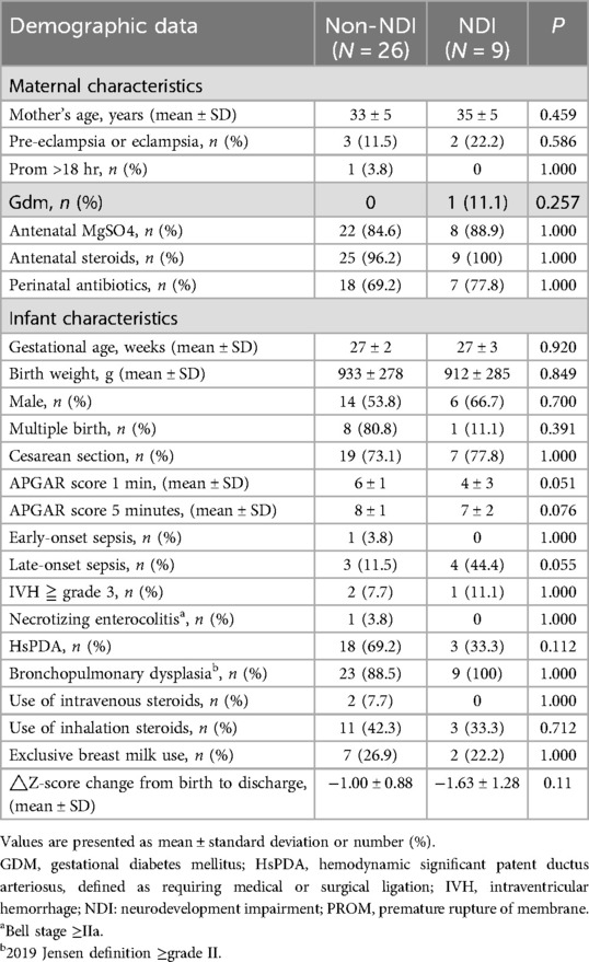
Table 1. Maternal and infant characteristics between infants with and without neurodevelopmental impairment.
When analyzing the serum cytokine levels at each time point, we identified 9 cytokines with significant differences between the two groups, potentially linked to neurodevelopmental outcomes, as shown in Table 2. Higher serum levels of IFN-γ were observed in the NDI group at 34 weeks PMA, suggesting a potentially detrimental effect on neurodevelopment. Conversely, the non-NDI group exhibited higher serum levels of eight other cytokines—Eotaxin-2, IL-2, IL-11, IL-16, MIP-1δ, PDGF-BB, TIMP-2, TNF-β—at 36 weeks PMA or 38 weeks PMA, indicating a potentially favorable effect. The levels of all 40 cytokines at each time point were compared and summarized in Supplementary Table 1. No significant differences were observed for the other 31 cytokines at any time point between the two groups.
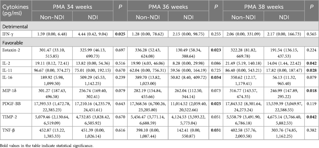
Table 2. Cytokine levels with significant differences at each time point between infants with and without neurodevelopmental impairment.
Subsequently, we investigated the changes in serum cytokine levels between each time point and identified 10 cytokine changes associated with neurodevelopmental outcomes, which were summarized in Table 3. Elevated IL-17 levels, as well as decreased Eotaxin-1, Eotaxin-2, IL-7, IL-16, MIP-1α, MIP-1β, PDGF-BB, and TIMP-2 levels between PMA 34 and PMA 36 weeks, correlated with poor neurodevelopmental outcomes. Besides, decreased in ICAM-1, IL-7, IL-16, MIP-1α, and MIP-1β levels between PMA 34 and PMA 38 weeks was associated with poor neurodevelopmental outcomes. The comparisons of cytokine level changes across all 40 cytokines in each interval were listed in Supplementary Table 2.
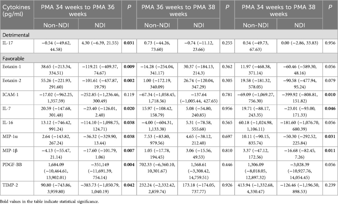
Table 3. Significant changes in cytokine levels at each time interval between infants with and without neurodevelopmental impairment.
The AUC for each significant biomarker was summarized in Table 4. Biomarkers with a detrimental effect indicating NDI showed an AUC of 0.752 for IFN-γ at PMA 34 weeks and an AUC of 0.744 for the change in IL-17 from PMA 34 weeks to 36 weeks. Biomarkers with a favorable effect also demonstrated good discriminatory ability, with all AUC values above 0.7. Notably, the change in Eotaxin-2 between PMA 34 and 36 weeks had an AUC of 0.833, and PDGF-BB between PMA 34 and 38 weeks had an AUC of 0.821.
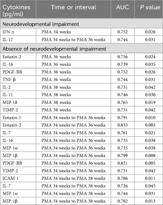
Table 4. Area under the ROC curve (AUC) for detrimental cytokines predicting poor neurodevelopmental outcomes and favorable cytokines predicting absence of neurodevelopmental impairment.
To assess the possible factors contributing to cytokine changes during this period, a generalized estimating equations approach was applied, and the results were summarized in Table 5. From PMA 34 weeks to 36 weeks, infants with severe IVH showed increased levels of IL-2, IL-7, and IL-11, while HsPDA was associated with increased levels of IL-11 and PDGF-BB. On the other hand, NEC was associated with decreased levels of Eotaxin-1, Eotaxin-2, IL-2, IL-16, MIP-1β, and PDGF-BB, and GA was associated with decreased levels of ICAM-1. From PMA 36 weeks to 38 weeks, only HsPDA was associated with increased levels of IL-11, while infants with LOS showed decreased levels of Eotaxin-1, IL-2, and IL-11. Additionally, NEC was associated with decreased levels of Eotaxin-1, Eotaxin-2, IL-2, IL-16, PDGF-BB, and TIMP-2, BPD with decreased MIP-1β levels, and GA with decreased levels of Eotaxin-1, MIP-1β, and ICAM-1. The detailed results of the generalized estimating equations are listed in Supplementary Table 3.
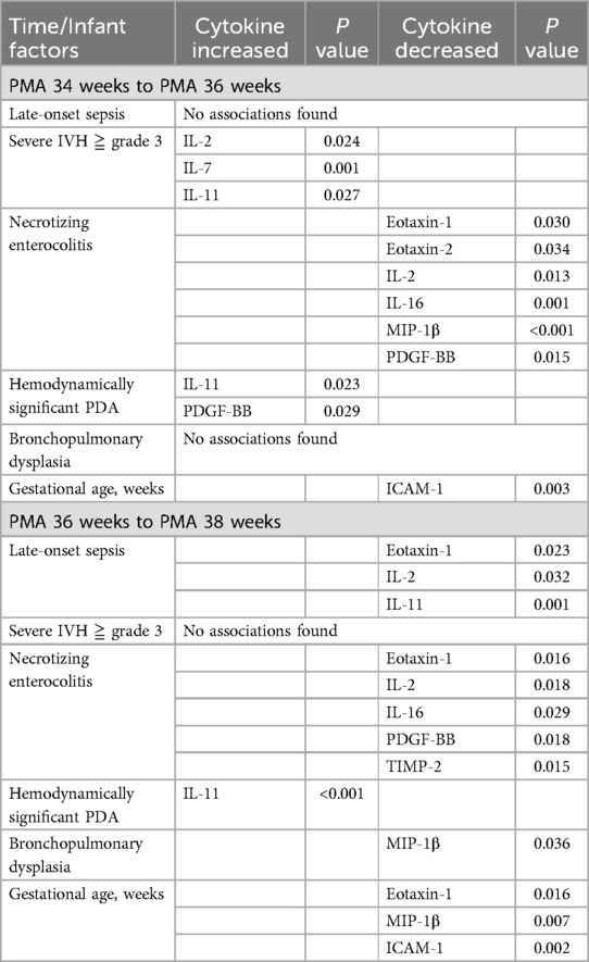
Table 5. Summary of the association between an infant's gestational age and comorbidities with changes in serum cytokine levels between PMA 34 weeks and PMA 38 weeks analyzed using generalized estimating equations.
Discussion
The primary aim of this study was to identify potential biomarkers during a relatively stable period that could predict neurodevelopmental outcomes in preterm infants. Both single time-point cytokine levels and changes over specific intervals may serve as potential predictors. Most biomarkers identified during this phase were associated with favorable outcomes and showed good discriminatory ability, particularly changes in Eotaxin-2 from PMA 34 to 36 weeks (AUC 0.833) and PDGF-BB from PMA 34 to 38 weeks (AUC 0.821). In contrast, higher serum IFN-γ at PMA 34 weeks and greater increases in IL-17 from PMA 34 to 36 weeks were linked to adverse outcomes, with AUCs of 0.752 and 0.744. These findings highlight the potential clinical utility of cytokine profiling during PMA 34–38 weeks for identifying infants at risk of neurodevelopmental impairment.
Biomarkers associated with detrimental effects
IFN-γ is a pro-inflammatory cytokine, and its role in neurodevelopment remains controversial. Some studies suggest that IFN-γ contributes to neuroinflammation and neurodegeneration in mouse models (15), with its absence enhancing cognitive performance through increased hippocampal plasticity (16). Conversely, other research has shown that IFN-γ can promote neurogenesis (17). IL-17, primarily secreted by T helper 17 (Th17) cells, is another inflammatory cytokine implicated in various chronic inflammatory neurological disorders (18). Lu et al. reviewed the role of neuroinflammation in neurological and psychiatric conditions and found IL-17 to be associated with several disorders, including autism spectrum disorder (ASD), Alzheimer's disease (AD), depression, and epilepsy (19). In one study, neonatal mice exposed to sevoflurane showed increased IL-17A expression in the hippocampus; deletion or inhibition of IL-17A attenuated sevoflurane-induced cognitive impairment by reducing hippocampal neuroinflammation (20). Although previous reviews have not reported associations between IFN-γ or IL-17 and neurodevelopmental outcomes in preterm infants (10, 21, 22), the elevated levels observed in our study may influence neurodevelopment based on their known physiological roles.
Biomarkers with favorable effects consistent with previous findings
In our study, higher serum levels of IL-2, IL-7, IL-11, MIP-1α, MIP-1β, MIP-1δ, PDGF-BB, TIMP-2, and TNF-β were positively associated with better neurodevelopmental outcomes. These findings are consistent with previous research on these cytokines in other neurological disorders. IL-2 is known to regulate T-cell proliferation, particularly regulatory T cells (23, 24), thereby modulating inflammatory responses. In a mouse model of traumatic brain injury, IL-2 complex treatment was shown to alleviate inflammation and reduce blood-brain barrier disruption (25). Additionally, elevated levels of TNF-β and IL-2 have been reported at school age in children with a history of neonatal encephalopathy (26). IL-7 is a cytokine involved in B and T cell development (27), and plays a role in restoring immune function following illness (28). Although previous reviews have not established a direct link between IL-7 and neurodevelopment, IL-7 has demonstrated neurotrophic properties and may play an important role in neural development (29). IL-11, an anti-inflammatory cytokine in the IL-6 family (30), has shown neuroprotective effects in animal studies. In rat models, recombinant human IL-11 (rhIL-11) has been found to protect against cerebral ischemia-reperfusion injury (31) and exert anti-apoptotic effects in neonatal hypoxic-ischemic brain injury (32).
MIP-1α, MIP-1β, and MIP-1δ are chemokines that play essential roles in regulating leukocyte trafficking and modulating inflammatory responses (33, 34). MIP-1α, primarily secreted by macrophages, is crucial for recruiting inflammatory cells and has been implicated in various inflammatory diseases (35). In a study analyzing neonatal blood spot samples, lower levels of MIP-1α were associated with both ASD and developmental delays, while reduced MIP-1δ levels were specifically linked to developmental delays (36). However, another study assessing the inflammatory profiles of school-age children born extremely preterm (GA < 28 weeks) found no significant differences in MIP-1α or MIP-1β levels between children with motor, cognitive, or behavioral impairments and their unaffected peers (37).
PDGF-BB is a member of the platelet-derived growth factor (PDGF) family and is known for its roles in neuroprotection and cell survival (38). It has been shown to prevent neuronal apoptosis following ischemic neuronal injury (39, 40) and to promote the proliferation, differentiation, and migration of neuronal progenitor cells (41).
TIMPs are endogenous inhibitors of matrix metalloproteinases. Upregulation of TIMP-2 has been associated with neuronal proliferation and differentiation (42). Research also indicates that TIMP-2 can inhibit microglial activation, suggesting its neuroprotective potential. These findings suggest that TIMP-2 may be a therapeutic target for neuroinflammatory disorders (42). TNF is a family of proinflammatory cytokines involved in immune defense and capable of inducing cell death and neurodegeneration. While TNF-α has been associated with adverse neurodevelopmental outcomes (21), the role of TNF-β is less well documented. Nevertheless, TNF-β may exert protective effects (43) and has been found to be elevated at school age in children with a history of neonatal encephalopathy (26).
Biomarkers with favorable effects different from previous findings
In our study, higher serum levels of Eotaxin-1, Eotaxin-2, ICAM-1, and IL-16 were observed in the non-NDI group, suggesting a potential protective effect on neurodevelopmental outcomes in preterm infants. However, these findings are not consistent with prior reports on these cytokines in the context of other neurological disorders. Eotaxins, part of the C-C motif chemokine family, are potent chemoattractants for eosinophils and play key roles in innate immunity. This group includes Eotaxin-1 [also known as C-C motif chemokine ligand (CCL)11], Eotaxin-2 (CCL24), and Eotaxin-3 (CCL26) (44). Eotaxin-1 and Eotaxin-2 are inflammatory chemokines involved in eosinophil recruitment and the Th2-mediated immune response (45). Elevated serum and cerebrospinal fluid levels of Eotaxin-1 and Eotaxin-2 have been reported in adults with various neurodegenerative diseases (44). ICAM-1 plays a central role in immune responses by facilitating leukocyte trafficking into inflamed tissues and regulating blood-brain barrier integrity (46, 47). Elevated intrathecal, but not serum, levels of soluble ICAM-1 have been reported in inflammatory neurological conditions such as viral meningoencephalitis and bacterial meningitis (46). Additionally, sustained elevation of inflammation-related proteins, including ICAM-1, during the first postnatal month in extremely preterm infants has been associated with increased risk of cognitive impairment at 10 years of age (48). IL-16 is a proinflammatory cytokine expressed in the brain under inflammatory conditions, as shown in rat models (49). Elevated cord blood IL-16 levels have also been linked to severely abnormal neurodevelopmental outcomes at three years of age in infants with perinatal asphyxia and hypoxic-ischemic encephalopathy (50).
We identified several biomarkers associated with either favorable or detrimental effects on the neurodevelopmental outcomes of preterm infants. Some biomarkers had not been previously reported in neonatal groups, and we referred to results from studies conducted in adults or animal models to interpret these findings. However, certain biomarkers with favorable effects were not mechanistically consistent. Although previous studies have linked Eotaxin-1, Eotaxin-2, ICAM-1, and IL-16 to neuroinflammation and adverse neurological outcomes in other populations, our findings suggest a potential protective role in preterm infants. This discrepancy may reflect developmental stage–specific or context-dependent functions of these cytokines. For instance, during early brain development, certain inflammatory mediators may support neurovascular maturation, immune regulation, or tissue remodeling rather than cause injury (51, 52).
Another critical point in explaining the association between these biomarkers and neurodevelopment is that the development of the human immune system and central nervous system is a continuous and dynamic process, characterized by various changes throughout infancy and childhood, and may vary among individuals due to different external stimuli (53, 54). For instance, extremely preterm infants born before 28 weeks, who have not undergone the third-trimester adaptation processes to tolerate maternal and self-antigens, exhibit different responses to inflammatory insults (55). Additionally, at different times after birth, varying cytokine responses to bacterial lipopolysaccharide stimulation were observed (56). This may explain why certain biomarkers previously reported to be significant at different postnatal time periods in other studies did not yield significant results in our research. Similar findings were reported in one review study, which demonstrated that biomarkers significantly associated with neurodevelopmental outcomes varied across different postnatal time periods (21). Furthermore, due to the different developmental statuses of the central nervous and immune systems across infancy, childhood, and adulthood, referencing studies conducted in adults or animal models to explain these associations may not always be suitable.
Associations between preterm comorbidities and biomarkers
In our study, we also found that preterm comorbidities were associated with changes in cytokine levels between PMA 34 and 38 weeks, despite the fact that most of these conditions— such as NEC, HsPDA and severe IVH—typically develop during the first month of life. Indeed, several biomarkers have been reported to be associated with these comorbidities. Cytokines including IL-1β, IL-6, IL-8, IL-18, and TNF-α have been implicated in the pathophysiology of preterm IVH (57, 58). Higher serum levels of IL-6 on day 1 (59) and IL-8 on days 1, 7, and 14 after birth (60) have been linked to an increased risk of IVH. Similarly, elevated levels of inflammatory cytokines such as TNF-α, IL-1, IL-6, IL-8, IL-10, MCP-1/CCL2, and MIP-1α have been associated with the persistence of PDA (61, 62).
NEC is a well-recognized condition marked by a robust inflammatory response and is a significant risk factor for adverse neurodevelopmental outcomes. Infants with NEC exhibit elevated levels of inflammatory cytokines, including IL-1β, IL-6, IL-8, IL-10, MCP-1/CCL2, and MIP-1β/CCL3, along with decreased levels of anti-inflammatory cytokines such as TGF-β and IL-2 (63). Notably, approximately 40% of infants with NEC develop NDI (64), and the severity of intestinal injury has been shown to correlate with an increased risk of NDI (65). The pathogenesis of NDI in the context of NEC is believed to involve gut–brain axis interactions. Specifically, microbial dysbiosis and intestinal injury can trigger systemic inflammation and the release of pro-inflammatory cytokines such as TNF-α, IL-1β, and IL-6, which may cross the immature blood–brain barrier and contribute to neuroinflammation, ultimately disrupting normal brain development (66–68). Neonatal sepsis is another well-established risk factor for adverse neurodevelopmental outcomes (69–72). Potential pathogenic mechanisms include systemic inflammation, cytokine dysregulation, and subsequent white matter injury. Elevated serum levels of several pro-inflammatory cytokines—such as IL-6, IL-8, IL-10, IL-1β, TNF-α, and MCP-1—have been associated with neonatal sepsis (73–75). During the acute phase of sepsis, pro-inflammatory cytokines such as TNF-α and IL-6 predominate, whereas the post-acute phase is characterized by increased levels of anti-inflammatory cytokines, including IL-10 (74).
BPD is a common morbidity in preterm infants. Numerous serum biomarkers have been confirmed to be associated with the subsequent development of BPD (76). Higher levels of proinflammatory, profibrotic and angiogenic cytokines (IL-6, IL-8, IL-10, MCP-1) within the first 5 days after birth have been linked to the later development of moderate to severe BPD (77). Another study analyzed biomarkers before 21 days of age and found that significant biomarkers varied when measured at different time points (78). The study concluded that abnormalities in the transition from innate immune response to adaptive immune response may be related to the occurrence of BPD. Survivors with neonatal BPD are at a higher risk of developing NDI (79, 80). One possible explanation is that preterm infants are generally sicker and more prone to nutritional problems, which may disrupt brain development. Another potential reason is the frequent and prolonged periods of hypoxemia, which can directly cause brain injury (80). Studies investigating the association between BPD, cytokines, and NDI are limited. However, it is theoretically plausible that persistent systemic inflammatory reactions or the effects of inflammatory biomarkers could contribute to NDI. Further research is needed to better understand these relationships.
As discussed above, changes in biomarkers associated with preterm comorbidities have been linked to neurodevelopmental outcomes. However, most existing studies have focused on earlier life stages—primarily within the first month after birth or during the acute phase of illness. In contrast, our study explores the potential long-term effects of comorbidities on biomarkers during a later and more stable period—between 34 and 38 weeks PMA—to assess the relationships among comorbidities, biomarkers, and neurodevelopmental outcomes. Importantly, our analysis focused on identifying biomarkers during this period that may reflect the long-term impact of preterm complications, rather than assessing how a single disease alters biomarker levels. This individual-centered approach emphasizes which biomarkers are associated with neurodevelopmental outcomes and explores their connections to common preterm morbidities, providing new insights into how these conditions may influence brain development.
Another potential confounding factor affecting neurodevelopment is the use of antenatal magnesium sulfate (MgSO₄) and antenatal corticosteroid therapy (ACS). Antenatal MgSO4 is known to provide neuroprotection in preterm births (81–83). Proposed mechanisms include promoting hemodynamic stability, preventing excitotoxic injury, stabilizing neurons, and exerting antioxidant effects (84, 85). Additionally, its anti-inflammatory properties have been reported, helping to reduce proinflammatory cytokines such as IL-1β and TNF-α (86). ACS, administered for threatened preterm birth, accelerates fetal lung maturation and has been proven to reduce mortality, as well as the risk of RDS and IVH (87). However, its impact on long-term neurodevelopmental outcomes remains uncertain. A meta-analysis found that ACS reduced the risk of neurodevelopmental impairment only in extremely preterm births, whereas in late-preterm and full-term births, ACS was associated with an increased risk of neurocognitive disorders (88). Lower cord blood levels of IL-6 have been observed in very low birth weight preterm infants after ACS administration (89). However, its influence on cytokines associated with neurodevelopmental outcomes remains poorly understood. One study reported reduced cord blood levels of neurotrophin-3 (NT-3) in late-preterm infants who received ACS, which could potentially affect neuronal growth, differentiation, and survival (90). Conversely, another study found no significant differences in cord blood concentrations of pro-inflammatory, anti-inflammatory, or neurotrophic cytokines after ACS administration, despite trends toward attenuation of the inflammatory response (91). In our study, however, neither MgSO₄ nor ACS use was significantly associated with neurodevelopmental outcomes.
Limitations
This study has several limitations. First, the relatively small sample size is a key limitation; although 50 cases were initially enrolled, only 35 were included in the final analysis. Although additional details regarding the reasons for exclusion have been provided, the potential selection bias resulting from the exclusion of 15 out of 50 infants (30%) remains a notable concern. Future studies with larger cohorts are needed to validate and extend these findings. Second, although the BSID-III assessment at 24 months of age is most predictive for neurodevelopment, the assessments were only available at 12 months of age due to the study timeline, as most participants had not yet completed the 24-month follow-up. Longitudinal data will be important to evaluate the long-term impact of these biomarkers on neurodevelopmental outcomes. As aforementioned, many studies have focused on cytokines measured at birth or during the early postnatal stage. Cytokines during this period provide valuable insights into the inflammatory processes in preterm infants. However, due to the limitations of our study design, measurements at birth or during the early postnatal period were not included, preventing us from comparing the differences and changes in cytokines between the early stage and the relatively stable later stage. Lastly, although we identified several biomarkers with significant associations, we were unable to fully elucidate the complex network through which these biomarkers influence neurodevelopment. Further research is needed to clarify the mechanistic roles of individual biomarkers and to explore their potential interactions.
Conclusions
Our study suggests that biomarkers measured at PMA 34–38 weeks, including both absolute cytokine levels and their trajectories, may help identify preterm infants at risk for neurodevelopmental impairment. While most biomarkers were associated with favorable outcomes, IFN-γ and IL-17 correlated with adverse neurodevelopmental outcomes. All identified biomarkers demonstrated good discriminatory ability. Additionally, several common neonatal comorbidities significantly influenced cytokine levels, underscoring the complex interplay between systemic inflammation, clinical complications, and neurodevelopment.
Data availability statement
The original contributions presented in the study are included in the article/Supplementary Material, further inquiries can be directed to the corresponding author/s.
Ethics statement
The studies involving humans were approved by the Institutional Review Board of Chang Gung Memorial Hospital (Approval No. 201902088A3. The studies were conducted in accordance with the local legislation and institutional requirements. Written informed consent for participation in this study was provided by the participants legal guardians/next of kin.
Author contributions
YW: Investigation, Data curation, Writing – original draft, Formal analysis. WW: Writing – original draft, Data curation. SC: Writing – original draft, Data curation. RL: Writing – review & editing. MC: Methodology, Conceptualization, Writing – review & editing. CL: Supervision, Investigation, Formal analysis, Writing – review & editing, Conceptualization, Methodology.
Funding
The author(s) declare that no financial support was received for the research and/or publication of this article.
Conflict of interest
The authors declare that the research was conducted in the absence of any commercial or financial relationships that could be construed as a potential conflict of interest.
The author(s) declared that they were an editorial board member of Frontiers, at the time of submission. This had no impact on the peer review process and the final decision.
Generative AI statement
The author(s) declare that no Generative AI was used in the creation of this manuscript.
Any alternative text (alt text) provided alongside figures in this article has been generated by Frontiers with the support of artificial intelligence and reasonable efforts have been made to ensure accuracy, including review by the authors wherever possible. If you identify any issue please contact us.
Publisher's note
All claims expressed in this article are solely those of the authors and do not necessarily represent those of their affiliated organizations, or those of the publisher, the editors and the reviewers. Any product that may be evaluated in this article, or claim that may be made by its manufacturer, is not guaranteed or endorsed by the publisher.
Supplementary material
The Supplementary Material for this article can be found online at: https://www.frontiersin.org/articles/10.3389/fped.2025.1667521/full#supplementary-material
References
1. Ionio C, Riboni E, Confalonieri E, Dallatomasina C, Mascheroni E, Bonanomi A, et al. Paths of cognitive and language development in healthy preterm infants. Infant Behav Dev. (2016) 44:199–207. doi: 10.1016/j.infbeh.2016.07.004
2. Jiang NM, Cowan M, Moonah SN, Petri WA Jr. The impact of systemic inflammation on neurodevelopment. Trends Mol Med. (2018) 24(9):794–804. doi: 10.1016/j.molmed.2018.06.008
3. Kallankari H, Kaukola T, Olsén P, Ojaniemi M, Hallman M. Very preterm birth and foetal growth restriction are associated with specific cognitive deficits in children attending mainstream school. Acta Paediatr. (2015) 104(1):84–90. doi: 10.1111/apa.12811
4. Nelson KB, Dambrosia JM, Grether JK, Phillips TM. Neonatal cytokines and coagulation factors in children with cerebral palsy. Ann Neurol. (1998) 44(4):665–75. doi: 10.1002/ana.410440413
5. Carlo WA, McDonald SA, Tyson JE, Stoll BJ, Ehrenkranz RA, Shankaran S, et al. Cytokines and neurodevelopmental outcomes in extremely low birth weight infants. J Pediatr. (2011) 159(6):919–25.e3. doi: 10.1016/j.jpeds.2011.05.042
6. Sorokin Y, Romero R, Mele L, Iams JD, Peaceman AM, Leveno KJ, et al. Umbilical cord serum interleukin-6, C-reactive protein, and myeloperoxidase concentrations at birth and association with neonatal morbidities and long-term neurodevelopmental outcomes. Am J Perinatol. (2014) 31(8):717–26. doi: 10.1055/s-0033-1359723
7. Varner MW, Marshall NE, Rouse DJ, Jablonski KA, Leveno KJ, Reddy UM, et al. The association of cord serum cytokines with neurodevelopmental outcomes. Am J Perinatol. (2015) 30(2):115–22. doi: 10.1055/s-0034-1376185
8. Nelson KB, Grether JK, Dambrosia JM, Walsh E, Kohler S, Satyanarayana G, et al. Neonatal cytokines and cerebral palsy in very preterm infants. Pediatr Res. (2003) 53(4):600–7. doi: 10.1203/01.Pdr.0000056802.22454.Ab
9. Leviton A, Allred EN, Fichorova RN, Kuban KC, Michael O’Shea T, Dammann O. Systemic inflammation on postnatal days 21 and 28 and indicators of brain dysfunction 2years later among children born before the 28th week of gestation. Early Hum Dev. (2016) 93:25–32. doi: 10.1016/j.earlhumdev.2015.11.004
10. Voltas N, Arija V, Hernández-Martínez C, Jiménez-Feijoo R, Ferré N, Canals J. Are there early inflammatory biomarkers that affect neurodevelopment in infancy? J Neuroimmunol. (2017) 305:42–50. doi: 10.1016/j.jneuroim.2017.01.017
11. Jensen EA, Dysart K, Gantz MG, McDonald S, Bamat NA, Keszler M, et al. The diagnosis of bronchopulmonary dysplasia in very preterm infants. An evidence-based approach. Am J Respir Crit Care Med. (2019) 200(6):751–9. doi: 10.1164/rccm.201812-2348OC
12. Fenton TR, Kim JH. A systematic review and meta-analysis to revise the fenton growth chart for preterm infants. BMC Pediatr. (2013) 13:59. doi: 10.1186/1471-2431-13-59
13. Chou JH, Roumiantsev S, Singh R. Peditools electronic growth chart calculators: applications in clinical care, research, and quality improvement. J Med Internet Res. (2020) 22(1):e16204. doi: 10.2196/16204
14. Albers CA, Grieve AJ. Test review: Bayley, N. (2006). Bayley scales of infant and toddler development– third edition. San Antonio, TX: harcourt assessment. J Psychoeduc Assess. (2007) 25(2):180–90. doi: 10.1177/0734282906297199
15. Corbin-Stein NJ, Childers GM, Webster JM, Zane A, Yang YT, Mudium N, et al. IFNγ drives neuroinflammation, demyelination, and neurodegeneration in a mouse model of multiple system atrophy. Acta Neuropathol Commun. (2024) 12(1):11. doi: 10.1186/s40478-023-01710-x
16. Monteiro S, Ferreira FM, Pinto V, Roque S, Morais M, de Sá-Calçada D, et al. Absence of IFNγ promotes hippocampal plasticity and enhances cognitive performance. Transl Psychiatry. (2016) 6(1):e707. doi: 10.1038/tp.2015.194
17. Yuan X, He F, Zheng F, Xu Y, Zou J. Interferon-gamma facilitates neurogenesis by activating wnt/β-catenin cell signaling pathway via promotion of STAT1 regulation of the β-catenin promoter. Neuroscience. (2020) 448:219–33. doi: 10.1016/j.neuroscience.2020.08.018
18. Milovanovic J, Arsenijevic A, Stojanovic B, Kanjevac T, Arsenijevic D, Radosavljevic G, et al. Interleukin-17 in chronic inflammatory neurological diseases. Front Immunol. (2020) 11:947. doi: 10.3389/fimmu.2020.00947
19. Lu Y, Zhang P, Xu F, Zheng Y, Zhao H. Advances in the study of IL-17 in neurological diseases and mental disorders. Front Neurol. (2023) 14:1284304. doi: 10.3389/fneur.2023.1284304
20. Zhang Q, Li Y, Zhang J, Cui Y, Sun S, Chen W, et al. IL-17A is a key regulator of neuroinflammation and neurodevelopment in cognitive impairment induced by sevoflurane. Free Radic Biol Med. (2025) 227:12–26. doi: 10.1016/j.freeradbiomed.2024.11.039
21. Nist MD, Pickler RH. An integrative review of cytokine/chemokine predictors of neurodevelopment in preterm infants. Biol Res Nurs. (2019) 21(4):366–76. doi: 10.1177/1099800419852766
22. Nist MD, Shoben AB, Pickler RH. Early inflammatory measures and neurodevelopmental outcomes in preterm infants. Nurs Res. (2020) 69(5S Suppl 1):S11–20. doi: 10.1097/nnr.0000000000000448
23. Malek TR. The main function of IL-2 is to promote the development of T regulatory cells. J Leukoc Biol. (2003) 74(6):961–5. doi: 10.1189/jlb.0603272
24. Abbas AK, Trotta E, RS D, Marson A, Bluestone JA. Revisiting IL-2: biology and therapeutic prospects. Sci Immunol. (2018) 3(25):eaat1482. doi: 10.1126/sciimmunol.aat1482
25. Gao W, Li F, Zhou Z, Xu X, Wu Y, Zhou S, et al. IL-2/Anti-IL-2 Complex attenuates inflammation and BBB disruption in mice subjected to traumatic brain injury. Front Neurol. (2017) 8:281. doi: 10.3389/fneur.2017.00281
26. Zareen Z, Strickland T, Eneaney VM, Kelly LA, McDonald D, Sweetman D, et al. Cytokine dysregulation persists in childhood post neonatal encephalopathy. BMC Neurol. (2020) 20(1):115. doi: 10.1186/s12883-020-01656-w
27. Huang J, Long Z, Jia R, Wang M, Zhu D, Liu M, et al. The broad immunomodulatory effects of IL-7 and its application in vaccines. Front Immunol. (2021) 12:680442. doi: 10.3389/fimmu.2021.680442
28. ElKassar N, Gress RE. An overview of IL-7 biology and its use in immunotherapy. J Immunotoxicol. (2010) 7(1):1–7. doi: 10.3109/15476910903453296
29. Michaelson MD, Mehler MF, Xu H, Gross RE, Kessler JA. Interleukin-7 is trophic for embryonic neurons and is expressed in developing brain. Dev Biol. (1996) 179(1):251–63. doi: 10.1006/dbio.1996.0255
30. Rose-John S. Interleukin-6 family cytokines. Cold Spring Harbor Perspect Biol. (2018) 10(2):a028415. doi: 10.1101/cshperspect.a028415
31. Zhang B, Zhang H-X, Shi S-T, Bai Y-L, Zhe X, Zhang S-J, et al. Interleukin-11 treatment protected against cerebral ischemia/reperfusion injury. Biomed Pharmacother. (2019) 115:108816. doi: 10.1016/j.biopha.2019.108816
32. Zuo D, Zheng Q, Xiao M, Wang X, Chen H, Xu J, et al. Anti-apoptosis effect of recombinant human interleukin-11 in neonatal hypoxic-ischemic rats through activating the IL-11Rα/STAT3 signaling pathway. J Stroke Cerebrovasc Dis. (2023) 32(2):106923. doi: 10.1016/j.jstrokecerebrovasdis.2022.106923
33. Zlotnik A, Yoshie O. Chemokines: a new classification system and their role in immunity. Immunity. (2000) 12(2):121–7. doi: 10.1016/s1074-7613(00)80165-x
34. Maurer M, von Stebut E. Macrophage inflammatory protein-1. Int J Biochem Cell Biol. (2004) 36(10):1882–6. doi: 10.1016/j.biocel.2003.10.019
35. Marciniak E, Faivre E, Dutar P, Alves Pires C, Demeyer D, Caillierez R, et al. The chemokine MIP-1α/CCL3 impairs mouse hippocampal synaptic transmission, plasticity and memory. Sci Rep. (2015) 5(1):15862. doi: 10.1038/srep15862
36. Kim DHJ, Krakowiak P, Meltzer A, Hertz-Picciotto I, Van de Water J. Neonatal chemokine markers predict subsequent diagnosis of autism spectrum disorder and delayed development. Brain Behav Immun. (2022) 100:121–33. doi: 10.1016/j.bbi.2021.11.009
37. Van der Zwart S, Knol EF, Gressens P, Koopman C, Benders M, Roze E. Neuroinflammatory markers at school age in preterm born children with neurodevelopmental impairments. Brain Behav Immun Health. (2024) 38:100791. doi: 10.1016/j.bbih.2024.100791
38. Funa K, Sasahara M. The roles of PDGF in development and during neurogenesis in the normal and diseased nervous system. J Neuroimmune Pharmacol. (2014) 9(2):168–81. doi: 10.1007/s11481-013-9479-z
39. Iihara K, Hashimoto N, Tsukahara T, Sakata M, Yanamoto H, Taniguchi T. Platelet-derived growth factor-BB, but not -AA, prevents delayed neuronal death after forebrain ischemia in rats. J Cereb Blood Flow Metab. (1997) 17(10):1097–106. doi: 10.1097/00004647-199710000-00012
40. Xiong L-L, Xue L-L, Jiang Y, Ma Z, Jin Y, Wang Y-C, et al. Suppression of PDGF induces neuronal apoptosis after neonatal cerebral hypoxia and ischemia by inhibiting P-PI3 K and P-AKT signaling pathways. Brain Res. (2019) 1719:77–88. doi: 10.1016/j.brainres.2019.05.012
41. Yang L, Chao J, Kook YH, Gao Y, Yao H, Buch SJ. Involvement of miR-9/MCPIP1 axis in PDGF-BB-mediated neurogenesis in neuronal progenitor cells. Cell Death Dis. (2013) 4(12):e960. doi: 10.1038/cddis.2013.486
42. Jaworski DM, Pérez-Martínez L. Tissue inhibitor of metalloproteinase-2 (TIMP-2) expression is regulated by multiple neural differentiation signals. J Neurochem. (2006) 98(1):234–47. doi: 10.1111/j.1471-4159.2006.03855.x
43. Probert L. TNF And its receptors in the CNS: the essential, the desirable and the deleterious effects. Neuroscience. (2015) 302:2–22. doi: 10.1016/j.neuroscience.2015.06.038
44. Huber AK, Giles DA, Segal BM, Irani DN. An emerging role for eotaxins in neurodegenerative disease. Clin Immunol. (2018) 189:29–33. doi: 10.1016/j.clim.2016.09.010
45. Menzies-Gow A, Ying S, Sabroe I, Stubbs VL, Soler D, Williams TJ, et al. Eotaxin (CCL11) and eotaxin-2 (CCL24) induce recruitment of eosinophils, basophils, neutrophils, and macrophages as well as features of early- and late-phase allergic reactions following cutaneous injection in human atopic and nonatopic volunteers. J Immunol. (2002) 169(5):2712–8. doi: 10.4049/jimmunol.169.5.2712
46. Jander S, Heidenreich F, Stoll G. Serum and CSF levels of soluble intercellular adhesion molecule-1 (ICAM-1) in inflammatory neurologic diseases. Neurology. (1993) 43(9):1809–13. doi: 10.1212/wnl.43.9.1809
47. Dietrich JB. The adhesion molecule ICAM-1 and its regulation in relation with the blood-brain barrier. J Neuroimmunol. (2002) 128(1-2):58–68. doi: 10.1016/s0165-5728(02)00114-5
48. Kuban KC, Joseph RM, O’Shea TM, Heeren T, Fichorova RN, Douglass L, et al. Circulating inflammatory-associated proteins in the first month of life and cognitive impairment at age 10 years in children born extremely preterm. J Pediatr. (2017) 180:116–23.e1. doi: 10.1016/j.jpeds.2016.09.054
49. Hridi SU, Barbour M, Wilson C, Franssen AJ, Harte T, Bushell TJ, et al. Increased levels of IL-16 in the central nervous system during neuroinflammation are associated with infiltrating immune cells and resident glial cells. Biology (Basel). (2021) 10(6):472. doi: 10.3390/biology10060472
50. Ahearne CE, Chang RY, Walsh BH, Boylan GB, Murray DM. Cord blood IL-16 is associated with 3-year neurodevelopmental outcomes in perinatal asphyxia and hypoxic-ischaemic encephalopathy. Dev Neurosci. (2017) 39(1-4):59–65. doi: 10.1159/000471508
51. Bilbo SD, Schwarz JM. The immune system and developmental programming of brain and behavior. Front Neuroendocrinol. (2012) 33(3):267–86. doi: 10.1016/j.yfrne.2012.08.006
52. Hagberg H, Mallard C, Ferriero DM, Vannucci SJ, Levison SW, Vexler ZS, et al. The role of inflammation in perinatal brain injury. Nat Rev Neurol. (2015) 11(4):192–208. doi: 10.1038/nrneurol.2015.13
53. Simon AK, Hollander GA, McMichael A. Evolution of the immune system in humans from infancy to old age. Proc Biol Sci. (2015) 282(1821):20143085. doi: 10.1098/rspb.2014.3085
54. Singh G, Tucker EW, Rohlwink UK. Infection in the developing brain: the role of unique systemic immune vulnerabilities. Front Neurol. (2021) 12:805643. doi: 10.3389/fneur.2021.805643
55. Humberg A, Fortmann I, Siller B, Kopp MV, Herting E, Göpel W, et al. Preterm birth and sustained inflammation: consequences for the neonate. Semin Immunopathol. (2020) 42(4):451–68. doi: 10.1007/s00281-020-00803-2
56. Yerkovich ST, Wikström ME, Suriyaarachchi D, Prescott SL, Upham JW, Holt PG. Postnatal development of monocyte cytokine responses to bacterial lipopolysaccharide. Pediatr Res. (2007) 62(5):547–52. doi: 10.1203/PDR.0b013e3181568105
57. Dammann O, Leviton A. Maternal intrauterine infection, cytokines, and brain damage in the preterm newborn. Pediatr Res. (1997) 42(1):1–8. doi: 10.1203/00006450-199707000-00001
58. Szpecht D, Wiak K, Braszak A, Szymankiewicz M, Gadzinowski J. Role of selected cytokines in the etiopathogenesis of intraventricular hemorrhage in preterm newborns. Childs Nerv Syst. (2016) 32(11):2097–103. doi: 10.1007/s00381-016-3217-9
59. Heep A, Behrendt D, Nitsch P, Fimmers R, Bartmann P, Dembinski J. Increased serum levels of interleukin 6 are associated with severe intraventricular haemorrhage in extremely premature infants. Arch Dis Child Fetal Neonatal Ed. (2003) 88(6):F501–4. doi: 10.1136/fn.88.6.f501
60. Leviton A, Allred EN, Dammann O, Engelke S, Fichorova RN, Hirtz D, et al. Systemic inflammation, intraventricular hemorrhage, and white matter injury. J Child Neurol. (2013) 28(12):1637–45. doi: 10.1177/0883073812463068
61. Wei YJ, Hsu R, Lin YC, Wong TW, Kan CD, Wang JN. The association of patent ductus arteriosus with inflammation: a narrative review of the role of inflammatory biomarkers and treatment strategy in premature infants. Int J Mol Sci. (2022) 23(22):13877. doi: 10.3390/ijms232213877
62. Cucerea M, Marian R, Simon M, Anciuc-Crauciuc M, Racean A, Toth A, et al. Serum biomarkers in patent ductus arteriosus in preterm infants: a narrative review. Biomedicines. (2025) 13(3):670. doi: 10.3390/biomedicines13030670
63. Maheshwari A, Schelonka RL, Dimmitt RA, Carlo WA, Munoz-Hernandez B, Das A, et al. Cytokines associated with necrotizing enterocolitis in extremely-low-birth-weight infants. Pediatr Res. (2014) 76(1):100–8. doi: 10.1038/pr.2014.48
64. Matei A, Montalva L, Goodbaum A, Lauriti G, Zani A. Neurodevelopmental impairment in necrotising enterocolitis survivors: systematic review and meta-analysis. Arch Dis Child Fetal Neonatal Ed. (2020) 105(4):432. doi: 10.1136/archdischild-2019-317830
65. Wang Y, Liu S, Lu M, Huang T, Huang L. Neurodevelopmental outcomes of preterm with necrotizing enterocolitis: a systematic review and meta-analysis. Eur J Pediatr. (2024) 183(8):3147–58. doi: 10.1007/s00431-024-05569-5
66. Lu J, Martin CR, Claud EC. Neurodevelopmental outcome of infants who develop necrotizing enterocolitis: the gut-brain axis. Semin Perinatol. (2023) 47(1):151694. doi: 10.1016/j.semperi.2022.151694
67. Manohar K, Mesfin FM, Liu J, Shelley WC, Brokaw JP, Markel TA. Gut-brain cross talk: the pathogenesis of neurodevelopmental impairment in necrotizing enterocolitis. Front Pediatr. (2023) 11:1104682. doi: 10.3389/fped.2023.1104682
68. Lodha A, Asztalos E, Moore AM. Cytokine levels in neonatal necrotizing enterocolitis and long-term growth and neurodevelopment. Acta Paediatr. (2010) 99(3):338–43. doi: 10.1111/j.1651-2227.2009.01600.x
69. Alshaikh B, Yusuf K, Sauve R. Neurodevelopmental outcomes of very low birth weight infants with neonatal sepsis: systematic review and meta-analysis. J Perinatol. (2013) 33(7):558–64. doi: 10.1038/jp.2012.167
70. Cai S, Thompson DK, Anderson PJ, Yang JY-M. Short- and long-term neurodevelopmental outcomes of very preterm infants with neonatal sepsis: a systematic review and meta-analysis. Children. (2019) 6(12):131. doi: 10.3390/children6120131
71. Sewell E, Roberts J, Mukhopadhyay S. Association of infection in neonates and long-term neurodevelopmental outcome. Clin Perinatol. (2021) 48(2):251–61. doi: 10.1016/j.clp.2021.03.001
72. Ong WJ, Seng JJB, Yap B, He G, Moochhala NA, Ng CL, et al. Impact of neonatal sepsis on neurocognitive outcomes: a systematic review and meta-analysis. BMC Pediatr. (2024) 24(1):505. doi: 10.1186/s12887-024-04977-8
73. Sugitharini V, Prema A, Berla Thangam E. Inflammatory mediators of systemic inflammation in neonatal sepsis. Inflamm Res. (2013) 62(12):1025–34. doi: 10.1007/s00011-013-0661-9
74. Khaertynov KS, Boichuk SV, Khaiboullina SF, Anokhin VA, Andreeva AA, Lombardi VC, et al. Comparative assessment of cytokine pattern in early and late onset of neonatal sepsis. J Immunol Res. (2017) 2017:8601063. doi: 10.1155/2017/8601063
75. Chen S, Kuang M, Qu Y, Huang S, Gong B, Lin S, et al. Expression of Serum cytokines profile in neonatal sepsis. Infect Drug Resist. (2022) 15:3437–45. doi: 10.2147/idr.S368772
76. Gilfillan M, Bhandari A, Bhandari V. Diagnosis and management of bronchopulmonary dysplasia. Br Med J. (2021) 375:n1974. doi: 10.1136/bmj.n1974
77. Vento G, Capoluongo E, Matassa PG, Concolino P, Vendettuoli V, Vaccarella C, et al. Serum levels of seven cytokines in premature ventilated newborns: correlations with old and new forms of bronchopulmonary dysplasia. Intensive Care Med. (2006) 32(5):723–30. doi: 10.1007/s00134-006-0138-1
78. Ambalavanan N, Carlo WA, D'Angio CT, McDonald SA, Das A, Schendel D, et al. Cytokines associated with bronchopulmonary dysplasia or death in extremely low birth weight infants. Pediatrics. (2009) 123(4):1132–41. doi: 10.1542/peds.2008-0526
79. Cheong JLY, Doyle LW. An update on pulmonary and neurodevelopmental outcomes of bronchopulmonary dysplasia. Semin Perinatol. (2018) 42(7):478–84. doi: 10.1053/j.semperi.2018.09.013
80. Anderson PJ, Doyle LW. Neurodevelopmental outcome of bronchopulmonary dysplasia. Semin Perinatol. (2006) 30(4):227–32. doi: 10.1053/j.semperi.2006.05.010
81. Crowther CA, Hiller JE, Doyle LW, Haslam RR. Effect of magnesium sulfate given for neuroprotection before preterm birth: a randomized controlled trial. JAMA. (2003) 290(20):2669–76. doi: 10.1001/jama.290.20.2669
82. Marret S, Marpeau L, Zupan-Simunek V, Eurin D, Lévêque C, Hellot MF, et al. Magnesium sulphate given before very-preterm birth to protect infant brain: the randomised controlled PREMAG trial*. Bjog. (2007) 114(3):310–8. doi: 10.1111/j.1471-0528.2006.01162.x
83. Rouse DJ, Hirtz DG, Thom E, Varner MW, Spong CY, Mercer BM, et al. A randomized, controlled trial of magnesium sulfate for the prevention of cerebral palsy. N Engl J Med. (2008) 359(9):895–905. doi: 10.1056/NEJMoa0801187
84. Marret S, Doyle LW, Crowther CA, Middleton P. Antenatal magnesium sulphate neuroprotection in the preterm infant. Semin Fetal Neonatal Med. (2007) 12(4):311–7. doi: 10.1016/j.siny.2007.04.001
85. Costantine MM, Drever N. Antenatal exposure to magnesium sulfate and neuroprotection in preterm infants. Obstet Gynecol Clin North Am. (2011) 38(2):351–66. doi: 10.1016/j.ogc.2011.02.019
86. Shogi T, Miyamoto A, Ishiguro S, Nishio A. Enhanced release of IL-1beta and TNF-alpha following endotoxin challenge from rat alveolar macrophages cultured in low-mg(2+) medium. Magnes Res. (2003) 16(2):111–9.12892381
87. McGoldrick E, Stewart F, Parker R, Dalziel SR. Antenatal corticosteroids for accelerating fetal lung maturation for women at risk of preterm birth. Cochrane Database Syst Rev. (2020) 12(12):Cd004454. doi: 10.1002/14651858.CD004454.pub4
88. Ninan K, Liyanage SK, Murphy KE, Asztalos EV, McDonald SD. Evaluation of long-term outcomes associated with preterm exposure to antenatal corticosteroids: a systematic review and meta-analysis. JAMA Pediatr. (2022) 176(6):e220483. doi: 10.1001/jamapediatrics.2022.0483
89. Caldas JP, Vilela MM, Braghini CA, Mazzola TN, Marba ST. Antenatal maternal corticosteroid administration and markers of oxidative stress and inflammation in umbilical cord blood from very low birth weight preterm newborn infants. J Pediatr (Rio J). (2012) 88(1):61–6. doi: 10.2223/jped.2158
90. Hodyl NA, Crawford TM, McKerracher L, Lawrence A, Pitcher JB, Stark MJ. Antenatal steroid exposure in the late preterm period is associated with reduced cord blood neurotrophin-3. Early Hum Dev. (2016) 101:57–62. doi: 10.1016/j.earlhumdev.2016.03.016
Keywords: biomarkers, preterm, neurodevelopment, IFN-γ, IL-17
Citation: Wu Y-J, Wu W-C, Chu S-M, Lien R, Chiang M-C and Lee C-C (2025) Serum cytokine profiles at near term-equivalent age and their association with neurodevelopmental outcomes in preterm infants: an exploratory study. Front. Pediatr. 13:1667521. doi: 10.3389/fped.2025.1667521
Received: 16 July 2025; Accepted: 14 August 2025;
Published: 28 August 2025.
Edited by:
Ming-Chih Lin, National Chung Hsing University, TaiwanReviewed by:
Chien-Chou Hsiao, Changhua Christian Children’s Hospital, TaiwanSan-Nan Yang, E-Da Hospital, Taiwan
Copyright: © 2025 Wu, Wu, Chu, Lien, Chiang and Lee. This is an open-access article distributed under the terms of the Creative Commons Attribution License (CC BY). The use, distribution or reproduction in other forums is permitted, provided the original author(s) and the copyright owner(s) are credited and that the original publication in this journal is cited, in accordance with accepted academic practice. No use, distribution or reproduction is permitted which does not comply with these terms.
*Correspondence: Ming-Chou Chiang, bmV3Ym9ybnR3QGdtYWlsLmNvbQ==; Chien-Chung Lee, c2VhODI3QGNnbWgub3JnLnR3
 Yan-Jun Wu
 Wei-Chi Wu
 Shih-Ming Chu1
 Ming-Chou Chiang
 Chien-Chung Lee