Abstract
Heart failure (HF) is one of the main public health problems at present. Although some breakthroughs have been made in the treatment of HF, the mortality rate remains very high. However, we should also pay attention to improving the quality of life of patients with HF. Traditional Chinese medicine (TCM) has a long history of being used to treat HF. To demonstrate the clinical effects and mechanisms of TCM, we searched published clinical trial studies and basic studies. The search results showed that adjuvant therapy with TCM might benefit patients with HF, and its mechanism may be related to microvascular circulation, myocardial energy metabolism, oxidative stress, and inflammation.
Introduction
Heart failure (HF) is a clinical syndrome characterized by with dyspnea, fatigue, and hydroncus, which is caused by decreased cardiac output or elevated endocardial pressure resulting from a change in the cardiac structure or function (McDonagh et al., 2021). As shown in the 2017 Global Burden of Disease Study, the number of patients with HF is about 64.34 million, which makes HF an epidemic public health problem (Disease et al., 2018). The morbidity of HF in adults from developed countries is between 1 and 2% (Groenewegen et al., 2020). Data from the USA in 2019 showed that 6.2 million people aged above 20 years old have HF, and HF morbidity is estimated to increase by 46% from 2012 to 2030 (Benjamin et al., 2019). The 2012–2015 China Hypertension Survey found that the morbidity of HF in adults aged 35 years and over was 1.3% (Wang Z et al., 2018). Moreover, the incidence of heart failure increases with age. Data from the Heart Failure Association of the European Society indicates that 1-year all-cause mortality of acute heart failure (AHF) and chronic heart failure (CHF) are 23.6 and 6.4%, respectively (Crespo-Leiro et al., 2016). The INTERnational Congestive Heart Failure (INTER-CHF) prospective cohort study showed that HF 1-year all-cause mortality was 16.5%, which in Africa was 34%, in Southeast Asia was 15%, in China was 7%, in South America was 9%, and in the Middle East was 9% (Dokainish et al., 2017).
In clinical practice, HF can be divided into HF with reduced ejection fraction [HFrEF, left ventricular ejection fraction (LVEF) ≤ 40%] and HF with preserved ejection fraction (HFpEF, LVEF ≥ 50%) based on the LVEF (Bozkurt et al., 2021). Research into the treatment of HFrEF has formed a new quadruple chemotherapy consisting of angiotensin-converting enzyme inhibitors, beta-blockers, mineralocorticoid receptor antagonists, and sodium-dependent glucose transporters 2 inhibitors in medication (McMurray et al., 2014; McMurray et al., 2019; Packer et al., 2020). Although these treatments show prognostic benefits, the 5-years mortality after hospitalization of patients with HFrEF is still high, even up to 75.3% (Shah et al., 2017). Among all research aimed at HFpEF published to date, only the EMPEROR-Preserved Trial proved that medicine could improve prognosis (Anker et al., 2021). Previous guidelines defined HF (LVEF = 41–49%) as HF with a mid-range ejection fraction (Ponikowski et al., 2016). Meanwhile, the latest European Society of Cardiology guidelines defined it as HF with mildly reduced ejection fraction (HFmrEF); however, we lack prospective experimental evidence for patients with HFmrEF. All analyses and suggestions are based on post hoc analyses of HFrEF and HFpEF (McDonagh et al., 2021). Therefore, the treatment of HF still faces great challenges. One the one hand, we need to focus on improving life qualities of patients with HF, in addition to its notable morbidity and mortality (Fiuzat et al., 2020). On the other hand, medicine exploitation needs new therapeutic targets, such as microvascular circulation, myocardial energy metabolism, inflammation, and mitochondrial function (Reddy et al., 2020; Ghionzoli et al., 2021).
Traditional Chinese Medicine (TCM), based on its own theory, has resulted in Chinese medicinal herbs being used widely in the therapy of HF. For example, many anti-HF prescriptions are recorded in textbooks (such as Zhenwu decoction, Shengmai powder, and Lingguizhugan decoction), while only a few of them have been studied strictly (Fu et al., 2010). Chinese medicinal herbs have been used for a long time to cure heart failure. By contrast, research into the effects and mechanisms of TCM are still in the initial stage. In this article, we summarize recent development in this field, with the aim of clarifying the mechanisms of Chinese medicinal herbs in therapy for HF and to provide new directions for the development and clinical application of HF therapeutic drugs.
Clinical Studies of Traditional Chinese Medicine in Treating Heart Failure
Considering the large number of studies about TCM treatment in HF, we only searched two databases, the Chinese Clinical Trial Registry (http://www.chictr.org.cn) and ClinicalTrials.gov (http://www.clinicaltrials.gov), and then obtained relevant data from the China National Knowledge Infrastructure (https://www.cnki.net/) and PubMed (https://pubmed.ncbi.nlm.nih.gov/), which resulted in 13 studies being selected (Table 1). The search date was August 28, 2021. Two investigators (Anzhu Wang and Pingping Huang) screened the literature separately, and a third investigator (Xiao-Chang Ma) checked the search results. Two investigators (Anzhu Wang and Pingping Huang) used the Cochrane risk of bias assessment tool to assess the risk of bias for the included studies. Disagreements in the process were resolved through discussion and consultation with a third investigator (Xiaochang Ma). The search process and quality assessment were placed in Supplementary Figures S1, S2.
TABLE 1
| Study | Registration number | N | Patient cohort | TCM intervention measures | Length of study | Primary endpoints | Secondary endpoints |
|---|---|---|---|---|---|---|---|
| Li et al. (2013) | ChiCTR-TRC-11001478 | 512 | LVEF ≤ 40%; NT-proBNP ≥450 pg/ml; NYHA II-IV | Qili Qiangxin capsules: Astragulus embranaceus (Fisch.) Bge., Panax ginseng C. A. Mey., Aconitum carmichaelii Debx., Salvia miltiorrhiza Bge., Lepidium apetalum Willd, Alisma orientalis (Sam.)Juzep, Polygonatum odoratum (Mill.) Druce, Cinnamomum cassia Presl, Carthamus tinctorius L., Periploca sepium Bge., and Citrus reticulata Blanco | 12 Weeks | NT-proBNP | CCEs, NYHA, LVEF, LVED, 6MWD, MLHFQ |
| Mao et al. (2020) | NCT01555320 | 640 | Ischemic heart disease; LVEF ≤45% or a history of HF/related clinical symptoms for more than 3 months; NYHA II-IV | Qishen Yiqi dripping pills: Astragulus embranaceus (Fisch.) Bge., Salvia miltiorrhiza Bge., Panax notoginseng (Burk.) F. H. Chen, Dalbergia odorifera T.Chen | 6 Months | 6MWD | Composite endpoints, BNP, LVEF, NYHA, MLHFQ |
| Xian et al. (2016) | ChiCTR-TRC-12003063 | 240 | CHF combined with coronary artery disease; Qi-Yin deficiency; NYHA II-IV | Shenmai Injection: Panax ginseng C.A.Mey., OPhiopogon japonicus (L.f) Ker-Gawl | 7 Days | NYHA | 6MWD, SF-36 hearth survey score, TCM syndrome score, LVEF, BNP |
| Wang et al. (2020) | ChiCTR1800016293 | 120 | Unstable condition requiring further treatment in the hospital; NYHA II–IV | Shenmai Injection: Panax ginseng C.A.Mey., OPhiopogon japonicus (L.f) Ker-Gawl | 7 Days | FFAs, Glucose, LA, PA, BCAAs | NYHA, TCM syndrome score, LVEF, LVIDd, LVIDs, BNP |
| Wang et al. (2019) | ChiCTR-TRC-12002857 | 160 | Coronary heart disease; CHF during acute aggravation; Yang and Qi deficiency; LVEF ≤50%; NYHA III-IV | Shenfu Injection: Panax ginseng C.A. Mey., Aconitum carmichaelii Debx | 7 Days | NYHA, TCM syndrome score | Lee’s CHF score, 6MWD, LVEF, The incidence rate of cardiovascular events and HF emergency/rehospitalization |
| Xu et al. (2011) | ChiCTR-TRC-08000257 | 118 | CHF during acute aggravation Qi deficiency LVEF ≤40%; NYHA III-IV | Huangqi Injection: Astragulus embranaceus (Fisch.)Bge | 7 Days | LVEF | Dyspnea situatio, NYHA, Clinical sign, Tei index |
| Wang et al. (2017b) | ChiCTR-TRC-12002061 | 465 | CHF caused by ischemic heart disease or dilated cardiomyopathy; 35% ≤ LVEF ≤50%; 720/24 h ≤ VPCs ≤ 10,000/24 h; NYHA II-III | Shensong Yangxin capsules: Astragulus embranaceus (Fisch.) Bge., OPhiopogon japonicus (L.f) Ker-Gawl., Cornus officinalis Sieb. et Zucc., Salvia miltiorrhiza Bge., Ziziphus jujuba Mill. var. spinosa (Bunge) Hu ex H.F.Chou, Taxillus chinensis(DC.) Danser, Paeonia lactiflora Pall.; Paeonia veitchii) Lynch, Eupolypha gasinensis Walk, Naradostachys jatamansi DC., Coptis chinensis Franch., Schisandra sphenantheraRehd.et Wils., Fossilia Ossia Mastodi | 12 Weeks | VPCs | LVEF, LVEDD, NT-proBNP, NYHA, 6MWD, MLHFQ. |
| Xian et al. (2015) | ChiCTR-TRC-09000549 | 228 | NYHA II-Ⅲ and Stage C of HF; Qi-Yin deficiency, blood stasis and water stagnation | Yangxinkang Tablets: Panax ginseng C.A.Mey., Astragulus embranaceus (Fisch.) Bge., OPhiopogon japonicus (L.f) Ker-Gawl., Schisandra chinensis (Turcz.) Baill., Ilex pubescens Hook. et Arn., Leonurus japonicus Houtt., Lepidium apetalum Willd | 4 Weeks | NYHA, CM syndromes, Symptom score, Sign Score, MLHFQ | General characteristics |
| Li X. X et al. (2021) | ChiCTR1900022036 | 76 | NYHA Ⅱ-Ⅲ and stage C of HF; Qi deficiency, blood stasis and water stagnation | Qishen Taohong Granule: Astragulus embranaceus (Fisch.) Bge, Codonopsis pilosula (Frnnch.) Nannf, Salvia miltiorrhiza Bge, Prunus persica (L.) Batsch, Carthamus tinctorius L., Morus allba L., Lepidium apetalum Willd, Polyporus umbellatus (Pers) Fries, Lycopur Lucidus Turcz. var. hirtus Regel | 4 Weeks | NYHA, LVEF, CHFQLS | 6MWD, CM syndrome score, Symptom score, Sign score, NT-proBNP. |
| Tang, (2020) | ChiCTR-INR-17010696 | 108 | Insufficient heart blood and heart Yang; LVEF <40% | Yangxinxue granule: Bupleurum chinense DC, Ostrea gigas Thunberg, Ziziphus jujuba Mill. var. spinosa (Bunge) Hu ex H.F. Chou, Angelica sinensis (Oliv.) Diels, Rehmannia glutinosa Libosc, Paeonia lactiflora Pall., Acanthopanax gracilistylus W.W. Smith, Ligusticum chuanxiong Hort., Panax ginseng C. A. Mey., Polygonatum odoratum (Mill.) Druce, Cinnamomum cassia Presl | 6 Months | 1-Year cardiovascular-related mortality | BNP, Length of hospital stay, Number of hospitalization, Rehospitalization frequency and length, 6MWD, MLHFQ |
| Recurrence frequency of AHF | |||||||
| Wang et al. (2017b) | ChiCTR-TRC- 08000059 | 340 | CHF admitted to hospital; LVEF ≤50%; NYHA Ⅱ-Ⅳ | Traditional Chinese Medicine preparations (Shenfu injection, Shenmai injection, Danhong injection, Qili Qiangxin Capsules, Buyiqiangxin tablets): The composition of Buyiqiangxin tablets is as follows: Panax ginseng C. A. Mey., Astragulus embranaceus (Fisch.) Bge., Periploca sepium Bge., Salvia miltiorrhiza Bge, OPhiopogon japonicus (L.f) Ker-Gawl., Lepidium apetalum Willd | 1 ± 2 Weeks and 6 Months | All-cause mortality | BNP, 6MWD, LVEF, MLHFQ |
| Liu, (2020) | ChiCTR2000030921 | 60 | HFpEF; NYHA Ⅰ-Ⅲ | Yangyin Shuxin prescription: Cornus officinalis Sieb. et Zucc., OPhiopogon japonicus (L.f) Ker-Gawl., Poygonautun kingianum Coll. et Hemsl., Coptis chinensis Franch, Trionyx sinensis Wiegmann, Salvia miltiorrhiza Bge., pheretima aspergillum (E.perrier), Pinellia ternata (Thunb.) Breit., Trichosanthes kirilowii Maxim., Citrus aurantium L | 14 ± 3 Days | Cardiac ultrasound index (,two-dimensional ultrasound,pulsed-wave doppler, tissue doppler imaging, speckle tracking imaging), Exercise tolerance (Peak VO₂, minute ventilation/VCO₂ slope) | TCM syndrome score, MLHFQ, BNP |
| Gao et al. (2017) | NCT01939236 | 64 | Coronary heart disease; NYHA II-III; LVEF <40% | Chinese herbal medicine granules: Astragulus embranaceus (Fisch.) Bge, Codonopsis pilosula (Frnnch.) Nannf., Salvia miltiorrhiza Bge, Prunus persica (L.) Batsch, Carthamus tinctorius L., Paeonia veitchii Lynch | 28 Days | Metabolomics analysis | 6MWT, LVEF |
Clinical studies of TCM to treating HF.
6MWD (6-min walking distance), BCAAs (Branched-chain amino acids), BNP (Brain natriuretic peptide), CCEs (Composite cardiac events), CHF (Chronic heart failure), CHFQLS (Quality of Life measured by the CHF Integrated Chinese and Western Medicine Survival Scale), CM (Chinese medicine), ECG (Electrocardiograph), FFAs (Free fatty acids), HF (Heart Failure), LA (Lactic acid), LVED (Left ventricular end-diastolic diameter), LVEDD (Left ventricular end diastolic diameter), LVEF (Left ventricular ejection fraction), LVIDd (Left ventricular internal diastolic diameter), LVIDs (Left ventricular internal dimension systole), MLHFQ (Minnesota Living with Heart Failure Questionnaire), NT-proBNP(N-terminal pro-B-type natriuretic peptide), NYHA (New York Heart Association), PA (Pyroracemic acid), SF-36 (Short-form 36), TCM (Traditional Chinese Medicine), VPCs (Ventricular premature complexes).
Qili Qiangxin Capsules
In 2013, Li et al. (2013) reported a multicenter, randomized, double-blind, parallel-group, placebo-controlled study on the efficacy and safety of Qili Qiangxin capsules in 512 patients with CHF. After 12 weeks of treatment, the level of N-terminal pro-B-type natriuretic peptide (NT-proBNP) in the two groups was significantly lower than baseline. However, reduction in the Qili Qiangxin capsules group was markedly larger than that in placebo group [240.15 pg/ml (−23.15, 1113.85) vs. 0.00 pg/ml (−286.00, 800.00), p = 0.002]. The therapeutic effects of Qili Qiangxin capsules were clearly better than that of the placebo (p < 0.05) in terms of composite cardiac events, New York Heart Association (NYHA) functional classification, LVEF, 6-min walking distance (6MWD), and Quality of life assessment using the Minnesota Living with Heart Failure Questionnaire (MLHFQ). In terms of safety, the differences between two groups for serious adverse events (SAEs) and adverse events (AEs) were not statistically significant (p < 0.05). Elevated levels of circulating NT-proBNP contribute to the diagnosis of HF and are linked to increased mortality and morbidity in HF patients (McDonagh et al., 2021). The findings revealed that Qili Qiangxin capsules can significantly lower NT-proBNP levels, indicating that patients may have a better prognosis with long-term therapy. To test this theory, a large, randomized, controlled trial with all-cause death as the outcome is required.
Qishen Yiqi Dripping Pills
In 2020, Mao et al. (2020) reported a prospective randomized, double-blind, multicenter, placebo-controlled study on the efficacy and safety of Qishen Yiqi dripping pills that enrolled 640 patients with ischemic heart failure (IHF). After 6 months of treatment, the level of 6MWD in the two groups was significantly higher than baseline. However, increase in the Qishen Yiqi dripping pills group was obviously greater than that in the placebo group (336.15 ± 100.84 m to 374.47 ± 103.09 m vs. 334.40 ± 100.27 m to 340.71 ± 104.57 m, p < 0.001). Compared with those in the placebo group, the MLHFQ grades of Qishen Yiqi dripping pills group were better and the NYHA functional classification was ameliorated (p < 0.05). Although Qishen Yiqi dripping pills increased LVEF and BNP during a 6-month period, the effects were not substantially larger than those seen in the placebo group. This was also linked to a lack of meaningful difference in composite clinical events at 6 months and 1 year (follow-up), despite a tendency for decreased HF hospitalizations at 6 months. To thoroughly assess the impact of Qishen Yiqi dripping pills on clinical events in IHF, longer-term medication and follow-up may be required. In terms of safety, AEs in the Qishen Yiqi dripping pills group were less common and milder.
Shenmai Injection
In 2016, Xian et al. (2016) delivered a randomized, double-blind, multicenter, placebo-controlled study on the efficacy and safety of Shenmai injection in 240 patients with CHF. After 7 days of treatment, the levels of the NYHA functional classification in the two groups were lower than baseline. However, the proportion of improved patients in the Shenmai injection group was significantly bigger than placebo group (NYHA Ⅰ: 22.8 vs. 8.8%, NYHA Ⅱ: 12.3 vs. 14.9%, NYHA Ⅲ: 28.1 vs. 19.3%, and NYHA Ⅳ: 7.0 vs. 4.4%, p = 0.001). In 6MWD, the short-form 36 (SF-36) hearth survey score and the TCM syndrome score, Shenmai injection therapy was more effective than the placebo (p < 0.05). For safety, treatment with Shenmai injection within 1 week was well tolerated with no apparent safety concerns (p > 0.05). It is worth noting that, despite indications of improvements in these endpoints, there is no evidence in this trial to demonstrate any benefits in long-term results of Shenmai injection. If a long-term duration is envisaged, more detailed study should be conducted.
In 2020, Wang et al. (2020) initiated a controlled experiment in which 120 patients with HF requiring further treatment in the hospital were randomly assigned to one of three groups: Shenmai injection, trimetazidine, or placebo. After 7 days of treatment, compared with the control group, Shenmai injection could inhibit the production of free fatty acids (FFAs, 452.88 ± 226.62 vs. 571.42 ± 209.40 μmol/L, p < 0.05) and branched-chain amino acids (BCAAs, 0.55 ± 0.17 vs. 0.47 ± 0.17 nmol/μL, p < 0.05), improved the NYHA functional classification, and raised the level of TCM score associated brain natriuretic peptide (BNP, p < 0.05). The advantage of Shenmai injection in HF may be due to an improvement in energy metabolism that was more noticeable than that seen after trimetazidine therapy. However, the number of serological metabolic indexes was limited, and there were no direct associations between metabolic indexes and enhanced cardiac function, nor were there any links between metabolic indexes. As a result, greater study into changes in serological metabolism after Shenmai injection therapy in HF is needed to corroborate these findings.
Shenfu Injection
In 2019, Wang et al. (2019) reported a randomized, double-blinded, multicenter, placebo-controlled trial of Shenfu Injection in 160 patients with CHF during the acute phase of symptom aggravation (Yang and Qi Deficiency Syndrome). After 7 days of treatment, compared with the control group, Shenfu Injection could improve the NYHA classification (78.38 vs. 61.43%, p = 0.0026), and increase the TCM syndrome score (89.19 vs. 60.00%, p < 0.001), Lee’s HF score, and 6MWD (p < 0.05). In terms of safety, treatment with Shenfu Injection within 1 week showed no statistical differences in the occurrence of AEs and adverse drug reactions (ADRs) (p > 0.05). However, this study was limited to a few places, and the observation duration was brief. Furthermore, we found no significant differences in several endpoint markers, such as composite cardiac events (CCEs) or fatalities, across these groups.
Huangqi Injection
In 2011, Xu et al. (2011) reported a randomized controlled trial (RCT) of Huangqi injection in 128 patients with acute decompensated CHF(Qi deficiency syndrome). After 7 days of treatment, compared with the control group, Huangqi injection could improve the LVEF (37.98 ± 12.77 vs. 31.06 ± 10.36, p = 0.003), strengthen dyspnea, and increase the NYHA functional classification and Tei index (p < 0.05). In terms of safety, Huangqi injection was well tolerated, with no AEs. In addition, this trial designed the application dose of Huangqi injection at 40 ml/d, which was double the conventional dosage, suggesting that the application of Huangqi injection for the treatment of AHF can be increased, although fluid intake must be tightly regulated. Currently, there are no thorough pharmacodynamic and pharmacokinetic studies as well as large-scale RCTs on Huangqi injection for the treatment of HF, which need to be examined further.
Shensong Yangxin Capsules
In 2017, Wang et al. (2017b) reported a randomized, double-blind, multicenter placebo-controlled trial of Shensong Yangxin capsules in 465 patients with CHF with frequent ventricular premature complexes (VPCs). After 12 weeks of treatment, compared with the placebo group, the Shensong Yangxin capsules group had significantly fewer total number of VPCs on a 24-h ambulatory electrocardiogram (1538 ± 2187 vs. 2746 ± 3889, p < 0.05). In addition, it also increased NT-proBNP, LVEF, NYHA functional classification, 6MWD, and the MLHFQ score (p < 0.05). In terms of safety, the differences between two groups for SAEs were not statistically significant (p > 0.05). On the basis of conventional CHF therapy, Shensong Yangxin capsules were shown to have the advantages of reducing VPCs and enhancing cardiac function with good compliance in this research. However, because the frequency of VPCs varies daily or on a regular basis, a longer period of ECG, such as 72 h or even 7 days, is seen to be more compelling. Because the 12-weeks timeframe in this study was so short, a bigger size and longer duration of a well-designed clinical trial should be conducted for a more thorough examination of Shensong Yangxin capsules in CHF patients with VPCs.
Yangxinkang Tablets
In 2105, Xian et al. (2015) reported a randomized, double-blind, multicenter, placebo-controlled study of Yangxinkang Tablets in 228 patients with CHF. After 4 weeks of treatment, compared with the placebo group, Yangxinkang Tablets could improve the NYHA functional classification, Chinese medicine (CM) syndromes, the symptom score, the Sign Score, and quality of life measured by the MLHFQ score (p < 0.05). No prominent ADRs were noted during the study. Yangxinkang tablets were shown to relieve clinical symptoms in CHF patients in this trial, although the lack of laboratory markers made the findings less reliable.
Qishen Taohong Granules
In 2021, Li X. X et al. (2021) reported a single-center, prospective, randomized, controlled clinical trial of Qishen Taohong granules in 76 patients with CHF. After 4 weeks of treatment, compared with trimetazidine, the Qishen Taohong granules could improve the NYHA classification (74.29 vs. 54.29%, p < 0.05), quality of life measured using the CHF Integrated Chinese and Western Medicine Survival Scale (CHFQLS; 13.82 ± 6.04 vs. 7.49 ± 2.28, p < 0.05). Treatment with Qishen Taohong granules also showed a superior performance in 6MWT, CM syndrome, shortness of breath, fatigue, gasping, general edema, and NT-proBNP levels (p < 0.05). No remarkable AEs was revealed during the study. The findings of this study should be interpreted with care due to the small sample size and short observation period. Furthermore, because this study lacked quantitative markers directly connected to energy metabolism, it was unable to evaluate if improved qi deficiency symptoms equated to improved energy metabolism.
Yangxinxue Granules
In 2020, Tang (2020) reported a multicenter, randomized, double-blind, placebo-controlled study of Yangxinxue granules in 108 patients with CHF. After 6 months of treatment, as well as 6 months of follow-up, although the differences between two groups for 1-year cardiovascular-related disease mortality were not statistically significant (13.33 vs. 6.52%, p > 0.05), the attack rate and rehospitalization rate were much lower for patients with AHF in the Yangxinxue granule group (p < 0.05). Moreover, the Yangxinxue granules could reduce the BNP level, and improve the 6MWT and MLHFQ scores (p < 0.05). No significant ADRs were noted during the study (p > 0.05). In addition to the standard treatment, Yangxinxue granules could significantly reduce the number of episodes of AHF and BNP levels, indicating that Yangxinxue granules had definite clinical efficacy in the treatment of CHF. The study included four research centers, but all were located in China’s Sichuan province and may not reflect regional differences.
TCM Preparations
In 2017, Wang et al. (2017a) reported a prospective, single-blind, randomized, controlled, and multicenter clinical trial of TCM preparations in 340 patients with CHF. During hospitalization, the control group received polarized solution and the treatment group received Danhong injection with Shenfu injection or Shenmai injection. After discharge, all patients were treated with Qiliqiangxin capsules and Buyiqiangxin tablets or a placebo for 6 months. After 6 months, the Kaplan-Meier curves revealed a significantly improved trend in the treatment group’s cumulative survival rate compared to the control group. Although the treatment group ceased taking TCM and got the same therapy as the control group after 6 months, the treatment group’s cumulative survival rate remained higher than the control group until the 12th month (p = 0.208). More research is needed to identify whether this favorable trend is the result of TCM’s delayed effect or of other reasons. There was no significant difference between the groups in BNP, Lee’s HF scores and LVEF (p > 0.05). The treatment group showed greater improvement in 6MWT and MLHFQ (p < 0.05). No significant ADRs were noted during the study (p > 0.05).
Yangyin Shuxin Prescription
In 2020, Liu (2020) reported a randomized, controlled clinical trial of Yangyin Shuxin prescription in 60 patients with HFpEF. The study evaluated the diastolic function of patients using various methods including two-dimensional ultrasound echocardiography, pulsed-wave doppler, tissue doppler imaging and speckle tracking imaging. After 14 ± 3 days of treatment, the mean difference in untwisting rate was 0.14% (95% CI: 0.07–0.21%, p < 0.001) and the mean difference in left atrial volume index was −4.99 ml/m2 (95% CI: −7.09 to −2.99 ml/m2, p < 0.001) between the two groups on the basis of conventional treatment, suggesting that Yangyin Shuxin prescription could improve diastolic function in patients with HFpEF. This research’s diagnostic criteria correspond to various guidelines, but there were issues such as a limited number of patients, a short observation duration, and insufficient persuasive power of outcome indicators, necessitating a more well-designed clinical investigation.
Chinese Herbal Medicine Granules
In 2017, Gao et al. (2017) reported a randomized, double-blind controlled clinical trial of Chinese herbal medicine granules in 64 patients with HFpEF of Qi deficiency and blood stasis syndrome. After 28 days of treatment, compared with the placebo group, Chinese herbal medicine granules could improve the LVEF and 6MWT (p < 0.05). By comparing metabolic profiles, 9 biomarkers, including 2-arachidonoylglycerophosphocholine, lysophosphatidylethanolamine (LPE) 16:0, phosphatidylserine 21:0, LPE 20:4, LPE 18:0, linoleic acid, LPE 18:2, 4-hydroxybenzenesulfonic acid, and LPE 22:6, may be particularly important for the effect of Chinese herbal medicine granules. This study used a metabolomics approach to validate the efficacy of herbal medicines, which can provide ideas for other studies.
Mechanisms of Traditional Chinese Medicine in Treating Heart Failure
Coronary Microvascular Dysfunction
Ischemic heart disease (IHD) commonly leads to HF (Pagliaro et al., 2020). Besides atherosclerosis and obstructive atherothrombotic events in epicardial coronary arteries, myocardial ischemia also has a close connection with coronary microvascular dysfunction (CMD) (Marzilli et al., 2012; Ford et al., 2018). CMD, characterized by coronary flow reserve (CFR) injury, means that the structure and function of coronary microcirculation are affected (Padro et al., 2020). As for obstructive coronary syndrome, although revascularization could recover the blood flow, CMD in distal coronary vasculature might bring about limited or incomplete recovery of damaged cardiomyocytes. Furthermore, myocardial fibrosis and adverse ventricular remodeling might result in HfrEF (Elgendy et al., 2019). In contrast to HFrEF resulting from myocardial scar and following left ventricular dilatation, the pathophysiological mechanisms of HFpEF are more complicated (Lam et al., 2018). As a result of CMD, heterogeneity in blood flow and oxygen levels would increase. Long term of CFR injury would result in damage to cardiac muscle cells and interstitial fibrosis (Yang et al., 2020). Thus, a wealth of supporting evidence suggests that CMD is the potential mechanism HFpEF and is a therapeutic target (Shah et al., 2018; Taqueti et al., 2018; Ahmad et al., 2021). The endothelium plays an important part in CMD. Endothelial structure and function are distinct in different angiosomes, whereas all of them could secrete and regulate vascular tension and substance permeability, especially in heart muscle capillaries, which are connected directly with endothelial cells and adjacent cardiomyocytes (Brutsaert, 2003; Zuchi et al., 2020). In addition, CMD is also associated with smooth muscle cell dysfunction, microvascular spasm/sympathetic dysfunction and altered microvascular remodeling (Pries and Reglin, 2017).
Astragulus embranaceus (Fisch.) Bge
Many TCM composite preparations include Astragulus embranaceus (Fisch.) Bge., which could affect endothelial function. Further research of the effective ingredient in Astragulus embranaceus (Fisch.) Bge. also proved its function (Tse et al., 2012; Hu et al., 2018). For example, Astragaloside IV (PubChem CID: 13943297) can not only improve vascular endothelial dysfunction induced by hyperglycemia via inhibiting the Toll-like receptor 4 (TLR4)/nuclear factor kappa-B(NF-κB) signaling pathway (Leng et al., 2018), but also inhibits vascular endothelial dysfunction via the phosphatidylinositol 3-kinase (PI3K)/protein kinase B (AKT)/endothelial nitric oxide synthase (eNOS) signaling pathway in rat aortic rings (Lin et al., 2018). Calycosin (PubChem CID: 5280448) can also promote angiogenesis, via a mechanism that involves vascular endothelial growth factor (VEGF)-vascular endothelial growth factor receptor 2 (VEGFR2) and mitogen-activated protein kinase (MAPK) signaling pathways (Tang et al., 2010; Li et al., 2011). Astragalus polysaccharide (PubChem CID: 2782115) can restrain apoptosis of human cardiac microvascular endothelial cells undergoing hypoxia and reoxygenation by activating the PI3K/AKT signaling pathway (Xie et al., 2016), (Table 2).
TABLE 2
| TCM | Component | PubChem CID | Structure | Underlying mechanisms | References |
|---|---|---|---|---|---|
Astragulus embranaceus (Fisch.) Bge, 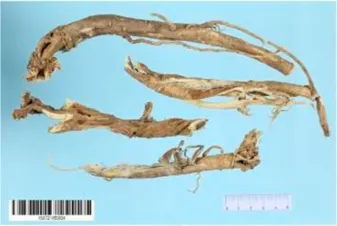 | Astragaloside IV | 13943297 |  | PI3K/AKT pathway↑ | Leng et al. (2018) |
| TLR4/NF-κB pathway↓ | Lin et al. (2018) | ||||
| Calycosin | 5280448 |  | VEGF-VEGFR2 and MAPK pathways↑ | Li et al. (2011) | |
| Tang et al. (2010) | |||||
| Astragalus polysaccharide | 2782115 |  | PI3K/AKT pathway↑ | Xie et al. (2016) | |
Panax ginseng C. A. Mey, 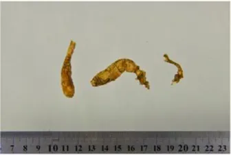 | Ginsenoside Rg3 | 9918693 |  | NF-κB↑ | Kim et al. (2003) |
| Ginsenoside Rg1 | 441923 |  | PI3K/AKT/p70S6K pathway ↑ | Leung et al. (2011) | |
| NRF2-ARE pathway↑ | Wang et al. (2015) | ||||
Salvia miltiorrhiza Bge,  | Tanshinone IIa | 164676 |  | Estrogen receptor pathway↑ | Cui et al. (2016) |
| JAK2/STAT3 pathway↓ | Fan et al. (2011) | ||||
| NRF2 pathway↑ | He L et al. (2021) | ||||
| Ursolic acid | 64945 | 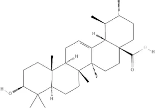 | eNOS↑ and NOX4 ↓ | Steinkamp-Fenske et al. (2007) | |
| Magnesium lithospermate B | 6918234 |  | PI3K/AKT pathway↑ | Liu Y. L et al. (2019) | |
Panax notoginseng (Burk.) F. H. Chen,  | Panax Notoginseng saponins | NA | NA | SIRT1↓ | Bo et al. (2020) |
| AMPK Thr172 and CaMKII Thr287↑, eNOS dependent pathways↑ | Wang et al. (2017a) | ||||
| Wang D et al. (2021) | |||||
| Notoginsenoside R1 | 441934 |  | ANG2/TIE2 pathway↑ | Fan et al. (2016) | |
| PI3K/AKT pathway↑ | Fang et al. (2018) | ||||
| NRF2 pathway↑ | Zhong et al. (2020) | ||||
Ligusticum chuanxiong Hort, 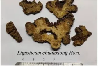 | Tetramethylpyrazine | 14296 | 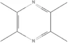 | sEH↓, 14-3-3γ/BCL-2↑ | Mak et al. (2017) |
| Yang et al. (2019) | |||||
Schisandra chinensis (Turcz.) Baill, Schisandra SphenantheraRehd.et Wils,  | Gomisin A | 634470 |  | eNOS↑ | Park et al. (2009) |
Bupleurum chinense DC,  | Saikosaponin C | 131801344 |  | MMP-2↑, VEFG↑, MAPK↑, ERK↑ | Shyu et al. (2004) |
The mechanisms of TCM in improving CMD.
↑ (Upregulation), ↓ (Downregulation), 14-3-3γ (Tyrosine 3-monooxygenase/tryptophan 5-monooxygenase activation protein gamma), AKT (Protein kinase B), AMPK Thr172 (5′-adenosine monophosphat-activated protein kinase), ANG2 (Angiogenin 2), ARE (Antioxidant responsive element), BCL-2 (B-cell lymphoma-2), CaMKII Thr287 (Calcium/calmodulin-dependent protein kinase II), CMD (Coronary microvascular dysfunction), eNOS (Endothelial nitric oxide synthase), ERK (Extracellular-regulated kinase), JAK2 (Janus kinase 2), MAPK (Mitogen-activated protein kinase), MMP (Matrix metalloproteinase), NA (Not applicable), NF-κB (Nuclear factor kappa-B), NOX4 (NADPH oxidase 4), NRF (Nuclear factor-erythroid 2-related factor), p70S6K (Ribosomal protein S6 kinase B1), PI3K (Phosphatidylinositol 3-kinase), sEH (Soluble epoxide hydrolase ), SIRT1 (Sirtuin), STAT3 (Signal transducer and activator of transcription 3), TCM (Traditional Chinese Medicine), TIE2 (Tyrosine kinase with immunoglobin and epidermal growth factor homology domain 2), TLR4 (Toll-like receptor), VEGF (Vascular endothelial growth factor), VEGFR2 (Vascular endothelial growth factor receptor 2).
Panax ginseng C. A. Mey
Ginsenoside Rg3 (PubChem CID: 9918693) isolated from Panax ginseng C. A. Mey. undertakes key roles of relaxing vessels and exerting a cytoprotective effect via induction of inducible nitric oxide synthase (iNOS) by activating NF-κB (Kim et al., 2003). Rg3 can antagonize adriamycin-induced cardiotoxicity by improving endothelial dysfunction via upregulating the nuclear factor-erythroid 2-related factor-2 (NRF2)-antioxidant responsive element (ARE) pathway through activation of AKT (Wang et al., 2015). Under normal cellular oxygen conditions in human umbilical vein endothelial cells, Ginsenoside Rg1 (PubChem CID: 441923) is a valid stimulator of hypoxia-inducible factor, which is an important transcription regulator for numerous angiogenic factors, through activating PI3K/AKT/ribosomal protein S6 kinase B1 (p70S6K) signaling (Leung et al., 2011), (Table 2).
Salvia miltiorrhiza Bge
Studies on effective ingredients of Salvia miltiorrhiza Bge. are numerous. The direct vasorelaxation induced by Tanshinone IIa (PubChem CID: 164676) is mediated by the nongenomic action of the estrogen receptor through endothelial nitric oxide synthase activation and Ca2+ mobilization (Fan et al., 2011). Tanshinone IIa also attenuates hypoxia/reoxygenation (H/R)-induced apoptosis via inhibiting the Janus kinase 2 (JAK2)/signal transducer and activator of transcription 3 (STAT3) signaling pathway (Cui et al., 2016) and protects human coronary artery endothelial cells from ferroptosis by activating the NRF2 pathway (He L et al., 2021). Ursolic acid (PubChem CID: 64945) exerts beneficial effects by upregulation of eNOS and a parallel downregulation of nicotinamide adenine dinucleotide phosphate (NADPH) oxidase 4 (NOX4), leading to an increase in bioactive nitric oxide (NO) (Steinkamp-Fenske et al., 2007). Magnesium lithospermate B (PubChem CID: 6918234) also can activate eNOS, which in turn enhances vascular nitric oxide production, which is medicated via the PI3K/AKT pathway (Liu Y. L et al., 2019), (Table 2).
Panax notoginseng (Burk.) F. H. Chen
Panax notoginseng saponins, which are the major active components of Panax notoginseng (Burk.) F. H. Chen, are a kind of chemical mixture containing different dammarane-type saponins, such as Notoginsenoside R1, and Ginsenosides Rb1, Rd, Re, and Rg1 (Xu et al., 2019). The pro-angiogenic and endothelial protection effects of Panax notoginseng saponins have been demonstrated in vitro and in vivo experimental models by upregulating sirtuin 1 (SIRT1) and antioxidants, and enhancing autophagy through phosphorylation of 5′-adenosine monophosphate-activated protein kinase (AMPK) Thr172 and calcium/calmodulin-dependent protein kinase II (CaMKII) Thr287, and eNOS-dependent pathways (Wang D et al., 2017; Bo et al., 2020; Wang D et al., 2021). Notoginsenoside R1 (PubChem CID: 441934) plays an important role among Panax notoginseng saponin active component by activating the angiogenin 2 (ANG2)/tyrosine kinase with immunoglobin and epidermal growth factor homology domain 2 (TIE2) pathway (Zhong et al., 2020) and PI3K/AKT pathway (Fang et al., 2018) to promote angiogenesis and activating NRF2 in endothelial cells to suppressing monocyte adhesion (Fan et al., 2016), (Table 2).
Others
Tetramethylpyrazine (PubChem CID: 14296), the predominant active ingredient in Ligusticum chuanxiong Hort. or Schisandra sphenantheraRehd.et Wils., can suppress angiotensin II-induced soluble epoxide hydrolase (sEH) expression in the coronary endothelium via an anti-endoplasmic reticulum (ER) stress mechanism (Mak et al., 2017) and attenuates the endotheliotoxicity of doxorubicin via tyrosine 3-monooxygenase/tryptophan 5-monooxygenase activation protein gamma (14-3-3γ)/B-cell lymphoma-2 (BCL-2) (Yang et al., 2019). Gomisin A (PubChem CID: 634470) contained in Schisandra chinensis (Turcz.) Baill. induces Ca2+-dependent activation of eNOS in human coronary artery endothelial cells, events that are linked to NO production and thereby endothelial-dependent vasorelaxation (Park et al., 2009). Saikosaponin C (PubChem CID: 131801344) in Bupleurum chinense DC can induce endothelial cell growth, migration, and capillary tube formation via activating MMP-2, VEGF, MAPK, and ERK (Shyu et al., 2004), (Table 2).
Energy Metabolism
The heart is an organ with high energy demands. The adenosine-triphosphate (ATP) required for cardiac contraction, relaxation, and basal metabolism in healthy adults is mainly provided by mitochondrial oxidative phosphorylation, and a small part comes from glycolysis (Stanley et al., 2005). There are a variety of energy substrates that can be used by cardiomyocytes. Approximately 70–90% of cardiac ATP is a result of the oxidation of fatty acids and the remaining 10–30% comes from the metabolism of glucose, lactate, ketone bodies, and certain amino acids (Doenst et al., 2013). Changes in energy metabolism during HF are mainly related to changes in the metabolic substrates and mitochondrial changes (He Y et al., 2021). The metabolic changes of cardiomyocytes not only depend on the severity and type of HF, but also are related to different underlying diseases (Lopaschuk et al., 2021). Generally, in the early stage of HF, the utilization of fatty acids remains the same or increases slightly as the severity of HF progresses, the rate of myocardial fatty acid oxidation decreases (Bertero and Maack, 2018b). Glucose metabolism in HF is characterized by an increased glucose uptake and glycolysis rate, without accompanying increase in glucose oxidation (De Jong and Lopaschuk, 2017). In the late stage of HF, the reduced sensitivity of the myocardium to insulin can lead to impaired myocardial glucose uptake (Lin et al., 2017). Ketone bodies, as substrates of cardiometabolic metabolism, have received extensive attention in recent years (Selvaraj et al., 2020). The utilization of ketone bodies increases as the use of fatty acid and glucose decreases (Bedi et al., 2016). Clinical studies have shown that exogenous ketone body supplementation can improve heart function in patients with HF (Nielsen et al., 2019). Most of the ATP (about 95%) consumed by the heart comes from oxidative metabolism in the mitochondria (Manolis et al., 2021). Changes in metabolic substrates during HF mean that the function and structure of mitochondria also change (Bayeva et al., 2013; Chaanine et al., 2019). On the one hand, after mitochondrial damage, oxidative phosphorylation is reduced, and the production of high-energy phosphoric acid compounds in the respiratory chain is impeded, resulting in insufficient bioenergy and aggravating the progression of HF (Zhou and Tian, 2018). On the other hand, other changes in mitochondrial function and structure, such as impaired mitochondrial electron transport chain activity, increased formation of reactive oxygen species, aberrant mitochondrial dynamics, and altered ion homeostasis, are also closely related to the occurrence of HF (Brown et al., 2017).
Panax ginseng C. A. Mey
In the Langendorff system, total ginsenosides of Panax ginseng C. A. Mey. may modulate tricarboxylic acid cycle protein expression, such as L-lactate dehydrogenase B chain (LDHB), glycerol-3-phosphate dehydrogenase (G3PD), pyruvate dehydrogenase complex (PDC)and aldose reductase (AR), to improve cardiac energy metabolism in ischemic rat heart tissues (Wang et al., 2012). Panax ginseng polysaccharide protected mitochondria by inhibiting mitochondrial injury and swelling in a concentration-dependent manner, and improved energy metabolism (Li et al., 2009). The SIRT1 signaling pathway could improve glucose aerobic metabolism and mitochondrial biosynthesis and ginsenoside Rc acts as a SIRT1 activator (Huang et al., 2021). Ginsenoside Rg1 adjusts energy metabolism via regulating the expression of associated proteins, thus increasing the activity of mitochondria respiratory complexes and the expression of ATP synthase, H+ transporting, mitochondrial F1 complex, delta subunit (ATP5D), which might be the result of binding to Ras homolog family member A (RHOA) and inactivating RHOA/Rho associated coiled-coil containing protein kinase 1 (ROCK1) signaling (Li et al., 2018). Ginsenoside Rg1 an also promote autophagy, and inhibits apoptosis by weakening the interaction between Beclin1 and BCL-2 (Li et al., 2016). Ginsenoside Rb3(PubChem CID: 12912363) regulates energy metabolism and inhibits myocardial fibrosis by regulating peroxisome proliferator activated receptor a (PPARα) (Chen et al., 2019, Zhang Y et al., 2021). Ginsenoside Rb1(PubChem CID: 9898279) might regulate calcium signaling by alleviating protein O-GlcNAcylation to improve diabetic cardiomyopathy (Qin et al., 2019), (Table 3).
TABLE 3
| TCM | Component | PubChem CID | Structure | Underlying mechanisms | References |
|---|---|---|---|---|---|
Panax ginseng C. A. Mey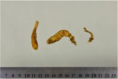 | Total ginsenosides | NA | NA | LDHB↓, G3PD↓, PDC↓, AR↓, ATP↓ | Wang et al. (2012) |
| Panax ginseng polysaccharide | NA | NA | ATP↑, ADP↑, TAP↑, AEC↑ | Li et al. (2009) | |
| Ginsenoside Rc | 12855889 |  | SIRT1 ↑ | Huang et al. (2021) | |
| Ginsenoside Rg1 | 441923 |  | RHOA pathway↓, interaction between Beclin1 and BCL-2↓ | Li et al. (2016) | |
| Li et al. (2018) | |||||
| Ginsenoside Rb3 | 12912363 |  | PPARα pathway↑ | Chen et al. (2019) | |
| Zhang Y et al. (2021) | |||||
| Ginsenoside Rb1 | 9898279 |  | O-GlcNAcylation↓ | Qin et al. (2019) | |
Aconitum carmichaelii Debx,  | Aconitum alkaloids | NA | NA | Glucose↓, and increase AMP↑, GMP↑, ADP↑, ATP↑ | Wu H et al. (2019) |
| Salsolinol | 91588 |  | MCU pathway↓ | Wen et al. (2019) | |
| Aconitine | 245005 |  | SIRT3↑ ,CYPD↓ | Wang N. N et al. (2021) | |
Astragulus embranaceus (Fisch.) Bge,  | Astragaloside IV | 13943297 |  | NF-κB/PGC1α pathway ↑, PPARα↑, MCAD↑, MCPT1↑, GSK-3beta↓, HES1↓, BCL-2-mediated mitochondrial function↑ | Dong et al. (2017) |
| He et al. (2012) | |||||
| Huang et al. (2016) | |||||
| Luo et al. (2019) | |||||
| Tang et al. (2018) | |||||
| Tu et al. (2013) | |||||
| Zhang et al. (2015) | |||||
| Formononetin | 5280378 |  | PPARγ pathway ↑ | Nie et al. (2018) | |
| Astragalus polysaccharide | 2782115 |  | TNF-α/PGC1α pathway ↑ | Luan et al. (2015) | |
Salvia miltiorrhiza Bge, 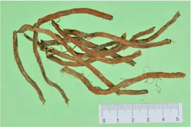 | Tanshinone IIa | 164676 | 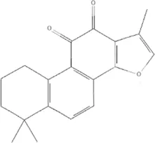 | SIRT1/PGC1α pathway↑ | Zhong et al. (2019) |
| Cryptotanshinone | 160254 | 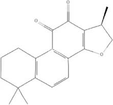 | PGC1α↑, NRF-1↑, TFAM↑ | Zhang et al. (2016) |
The mechanisms of TCM in improving energy metabolism.
↑(Upregulation), ↓(Downregulation), ADP (Adenosine diphosphate), AEC (Adenylate energy charge), AR (Aldose reductase), AMP (Adenosine monophosphate), ATP (Adenosine-triphosphate), BCL-2 (B-cell lymphoma-2), CYPD (Cyclophilin D), G3PD (Glycerol-3-phosphate dehydrogenase), GMP (Guanosine monophosphate), GSK-3beta (Glycogen synthase kinase 3 beta), HES1 (Hairy and enhancer of split-1), LDHB (L-lactate dehydrogenase B chain), MCAD (Medium-chain acyl-CoA dehydrogenase), MCPT1 (Mast cell protease 1), MCU (Mitochondrial calcium uniporter), NA (Not applicable), NF-κB (Nuclear factor kappa-B), NRF-1 (Nuclear factor-erythroid 2-related factor), PDC (Pyruvate dehydrogenase complex), O-GlcNAcylation (O-linked β--acetylglucosamine modification), PGC1α (Peroxisome proliferator-activated receptor gamma coactivator-1 alpha), PPAR (Peroxisome proliferator-activated receptor), RHOA (Ras homolog family member A), SIR (Sirtuin), TAP (Total adenylate pool), TCM (Traditional Chinese Medicine), TFAM (Transcription factor A, mitochondrial), TNF-α (Tumor necrosis factor-α).
Aconitum carmichaelii Debx
Aconitum carmichaelii Debx is a TCM that is, used extensively in HF as the processed product of Aconitum carmichaelii Debeaux tubers (Zhou W et al., 2021). Aconitum alkaloids, which include C19-diterpenoid alkaloids (mainly comprising aconitine, mesaconitine, and hypaconitine) and C20-diterpenoid alkaloids (predominantly benzoylmesaconitine, benzoylaconitine, and benzoylhypacoitine) and other alkaloids (mainly comprising higenamine and salsolinol) have versatile chemical structures (Zhou et al., 2015). Their cardiotoxicity and neurotoxicity hinder the use of Aconitum alkaloids; however, in TCM’s theory that even toxic substances are powerful medicines and can show reduced toxicity by proper methods (Liu et al., 2017; Zong et al., 2019; Mi et al., 2021). Emerging evidence also shows that Aconitum alkaloids could improve energy metabolism and mitochondrial function to generate cardioprotective effects. Aconitum alkaloids could reduce glucose levels, and increase creatine, adenosine monophosphate (AMP), Guanosine monophosphate (GMP), Adenosine diphosphate (ADP), and ATP levels in myocardial infarction rats (Wu H et al., 2019). Salsolinol (PubChem CID: 91588) attenuates doxorubicin-induced CHF in rats via a mechanism that might be related to the mitochondrial calcium uniporter (MCU) pathway (Wen et al., 2019). Aconitine (PubChem CID: 245005) alleviated the energy metabolic dysfunction of a myocardial injury model induced by Angiotensin II in H9c2 cells by activating SIRT3 to deacetylate cyclophilin D (CYPD) (Wang N. N et al., 2021). A combination of Zingiber officinale Rosc. and Aconitum carmichaelii Debx. showed an enhancing effect (Yu et al., 2012; Wen et al., 2020a). The potential mechanism of this effect might be related to mitochondrial energy metabolism, and it can be mediated by MCU in AHF rats induced by propafenone hydrochloride (Zhang et al., 2017) and improved by the liver kinase B1 (LKB1)/AMPKα/SIRT1 signaling pathway in doxorubicin (DOX)-induced CHF rats (Wen et al., 2020b), (Table 3).
Astragulus embranaceus (Fisch.) Bge
Astragaloside IV can increase the ratio of ATP/AMP, and decrease the content of FFAs in heart tissue of rats compared with isoproterenol alone via NF-κB/peroxisome proliferator-activated receptor gamma coactivator-1 alpha (PGC1α) signaling-mediated energy biosynthesis (Zhang et al., 2015). The function of Astragaloside IV in improving fatty acid utilization might be connected with PPARα, medium-chain acyl-CoA dehydrogenase (MCAD), and mast cell protease 1 (MCPT1) (Dong et al., 2017; Tang et al., 2018). Astragaloside IV prevents ischemia/reperfusion (I/R) injury by maintaining the mitochondrial membrane potential, inhibiting mitochondrial permeability transition pore opening, and improving energy metabolism via inactivating glycogen synthase kinase 3 beta (GSK-3beta) and hairy and enhancer of split-1 (HES1), thus improving BCL-2-mediated mitochondrial function (He et al., 2012; Tu et al., 2013; Huang et al., 2016; Luo et al., 2019). Formononetin (PubChem CID: 5280378) has beneficial effects on obesity and insulin resistance by modulating PPARγ activity (Nie et al., 2018). Astragalus polysaccharide attenuates iso-induced cardiac hypertrophy through regulating tumor necrosis factor-α (TNF-α)/PGC1α signaling, resulting in decreased FFA contents (Luan et al., 2015), (Table 3).
Salvia miltiorrhiza Bge
Tanshinone IIa blocks mitochondrial damage via activating the SIRT1/PGC1α pathway in acute cardiac microcirculation I/R injury (Zhong et al., 2019). Cryptotanshinone (PubChem CID: 160254) protects against adriamycin-induced mitochondrial dysfunction in cardiomyocytes by increasing the activities of complexes, except complex II, and promotes mitochondrial biogenesis-relative factors PGC1α, NRF-1, and TFAM (Zhang et al., 2016), (Table 3).
Oxidative Stress
Oxidative stress is defined as an imbalance between the generation of reactive oxygen species (ROS) and the endogenous antioxidant defense mechanisms (Zhou Y et al., 2021). At low concentrations, ROS modulates critical functions in cell homeostasis, whereas an overabundance of ROS plays a crucial role of worsening of the systolic and diastolic function and HF (Munzel et al., 2015). ROS production in the heart is primarily achieved by the mitochondria, NADPH oxidases, xanthine oxidase, and uncoupled nitric oxide synthase (NOS) (Munzel et al., 2017). Although pre-clinical studies have shown the beneficial effects of antioxidant strategies on HF, clinical studies in patients with HF have not achieved ideal results. A meta-analysis of 50 RCTs showed that supplementation with vitamins and antioxidants was not associated with reductions in the risk of major cardiovascular events (Myung et al., 2013). The future of anti-oxidative stress therapies might lie in increasing the endogenous antioxidant capacity or targeting mitochondrial ROS (Bertero and Maack, 2018a; Zhang et al., 2020). Study of TCM in oxidative stress might provide new ideas for the antioxidant treatment of HF (Table 3).
Coptis chinensis Franch
Berberine (PubChem CID: 2353), Palmatine (PubChem CID: 19009), and Coptisine (PubChem CID: 72322) are the main components of Coptis chinensis Franch. (Meng et al., 2018). Berberine protects rat hearts from I/R injury by decreasing the level of ROS and malondialdehyde (MDA), improving mitochondrial dysfunction via increasing MMP and complex I activity, as well as activating the SIRT1/tumor protein p53 (p53) signaling pathway (Liu D. Q et al., 2019). Berberine also ameliorates the doxorubicin-induced oxidative insult and mitochondrial damage as a modulator of SIRT3 or the SIRT1/Src-homology-2-domain-containing transforming protein 1 (p66Shc)-mediated pathway (Coelho et al., 2017; Wu Y. Z et al., 2019). Coptisine reduces oxidative stress by activating AKT and the JNK/NRF2/NAD(P)H quinone dehydrogenase 1 (NQO1) pathway (Hu et al., 2017), and Palmatine can inhibit the increase of lactate dehydrogenase (LDH), Creatine kinase (CK), and MDA and halt the decline of superoxide dismutase (SOD) and catalase (CAT) in I/R cardiac tissues (Kim et al., 2009), (Table 4).
TABLE 4
| TCM | Component | PubChem CID | Structure | Underlying mechanisms | References |
|---|---|---|---|---|---|
Coptis chinensis Franch, 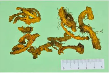 | Berberine | 2353 |  | SIRT1/p53 pathway, SIRT3↑, SIRT1/p66Shc pathway↑ | Coelho et al. (2017) |
| Liu D. Q et al. (2019) | |||||
| Wu Y. Z et al. (2019) | |||||
| Coptisine | 72322 |  | AKT and JNK/NRF2/NQO1 pathway↑ | Hu et al. (2017) | |
| Palmatine | 19009 |  | LDH↓, CK↓, MDA↓, SOD↓, and CAT↓ | Kim et al. (2009) | |
Rhodiola crenulate (Hook .f. et Thoms.) H. Ohba,  | Salidroside | 159278 |  | PI3K-AKT pathway↑, NOX1↓, mTOR↑, AMPK↑, AKT/HO-1↑ | Hao et al. (2021) |
| Ni et al. (2021) | |||||
| Wang et al. (2013) | |||||
| Zhu et al. (2011) | |||||
Polygonum cuspidatum Sieb. et Zucc,  | Polydatin | 445154 |  | NADPH↓, NF-κB↓ | Dong et al. (2015) |
| Notch1/HS1- PTEN/AKT↑,ROCK↓ | Tan et al. (2020) | ||||
| Yu et al. (2018) | |||||
| Resveratrol | 5281718 |  | DJ-1↑, mitochondrial complex I ↑, HMGB1 pathway↓, SIRT1↑, AMPK pathway↑, autophagy by SIRT1/FOXO1/RAB7 axis↑ | Bagul et al. (2015) | |
| Guo et al. (2015) | |||||
| Wang et al. (2014) | |||||
| Wu et al. (2016) | |||||
| Zhang et al. (2018) | |||||
Curcuma Longa L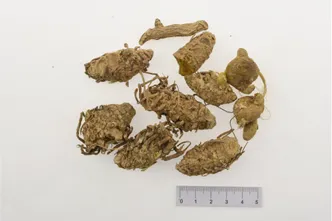 | Curcumin | 969516 | 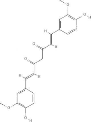 | mTOR or 14-3-3γ pathway↑, SIRT1↑, NRF2↑, NF-κB↓, SIRT1-FOXO1 and PI3K-AKT↑ | He et al. (2018) |
| Liu Q et al. (2019) | |||||
| Ren et al. (2020) | |||||
| Xiao et al. (2016) | |||||
| Yu et al. (2020) | |||||
| Zeng et al. (2015) |
The mechanisms of TCM in improving oxidative stress.
↑ (Upregulation), ↓ (Downregulation), 14-3-3γ (Tyrosine 3-monooxygenase/tryptophan 5-monooxygenase activation protein gamma) AKT (Protein kinase B) AMPK (5′-adenosine monophosphat-activated protein kinase), CAT(Catalase), CK (Creatine kinase), DJ-1 (Parkinsonism associated deglycase), FOXO1 (Forkhead box O1), HMGB1 (High-mobility group box 1), HO-1 (Heme oxygenase-1), HS1 (Heat stable protein 1), JNK (c-Jun N-terminal kinase), LDH (Lactate dehydrogenase), MDA (Malondialdehyde), mTOR (Mechanistic target of rapamycin), NADPH (Nicotinamide Adenine Dinucleotide Phosphate), NF-κB (Nuclear factor kappa-B), Notch1 (Notch receptor 1), NOX (NADPH oxidase), NQO1 [NAD(P)H quinone dehydrogenase 1], NRF2 (Nuclear factor-erythroid 2-related factor), p53 (Tumor protein p53), p66Shc (Src-homology-2-domain-containing transforming protein 1), PI3K (Phosphatidylinositol 3-kinase), PTEN (Phosphatase and tensin homolog), RAB7 (Member RAS oncogene family), ROCK (Rho associated coiled-coil containing protein kinase 1), SIRT(Sirtuin), SOD (Superoxide dismutase), TCM (Traditional Chinese Medicine).
Rhodiola crenulate (Hook. f. et Thoms.) H. Ohba
Salidroside (PubChem CID: 159278) has been identified as one of the most active ingredients isolated from Rhodiola crenulate (Hook.f. et Thoms.) H. Ohba, which reduces oxidative stress to fight against cardiovascular diseases (Tao et al., 2019; Zhao et al., 2021). Salidroside protected cardiomyocytes against hydrogen peroxide-induced injury via an PI3K-AKT dependent pathway and increaed the expression and activities of endogenous PI3K-dependent antioxidant enzymes (Zhu et al., 2011). In doxorubicin-induced cardiotoxicity, salidroside suppressed the excessive oxidative stress by inhibiting NOX1 and augmented the endogenous antioxidative enzymes, catalase, and manganese superoxide dismutase (Wang et al., 2013). Salidroside protects against diabetes-induced cardiac dysfunction by modulating the mechanistic target of rapamycin (mTOR), AMPK, and AKT/heme oxygenase-1(HO-1) signaling pathways (Hao et al., 2021; Ni et al., 2021), (Table 4).
Polygonum cuspidatum Sieb. et Zucc
Over 67 compounds been isolated from Polygonum cuspidatum Sieb. et Zucc. and identified, among them, Polydatin (PubChem CID: 5281718) and Resveratrol (PubChem CID: 445154) have attracted wide attention in the field of oxidative stress (Peng et al., 2013). Polydatin protects myocardial function in diabetic rats via inhibition of NADPH oxidase and NF-κB activities (Tan et al., 2020), and also alleviates oxidative/nitrative stress damage via the Notch receptor 1 (Notch1)/heat stable protein 1(HS1)-phosphatase and tensin homolog (PTEN)/AKT signaling pathway in diabetic myocardial I/R injury (Yu et al., 2018). Polydatin prevents myocardial hypertrophy mediated by inhibition of ROS-dependent ROCK activation (Dong et al., 2015). In models of cardiac I/R injury, pharmacological agent-induced cardiotoxicity, obesity, long-term nicotine intake, and diabetes, Resveratrol activates the antioxidant genes such as those encoding HO-1, SOD, CAT, and glutathione (GSH), which can help to promote the balance between oxidative stress and antioxidants, especially in the mitochondria (Kovacic and Somanathan, 2010; Li et al., 2012; Ahmad and Hoda, 2020; Ramalingam et al., 2021). Parkinsonism associated deglycase (DJ-1) preserves mitochondrial complex I activity, thus playing an important role in Resveratrol-mediated cardioprotective effects against I/R-induced oxidative stress damage (Zhang et al., 2018). For the myocardial damage caused by diabetes, Resveratrol can inhibit the high-mobility group box 1tbox1 (HMGB 1)-mediated signaling pathway (Wu et al., 2016), activate SIRT1 leading to deacetylation of both NF-kB/p65 and histone 3 (H3) (Bagul et al., 2015), improve AMPK-related pathways (Guo et al., 2015), and enhance autophagy via the SIRT1/Forkhead box O1(FOXO1)/member RAS oncogene family (RAB7) axis (Wang et al., 2014), (Table 4).
Curcuma Longa L
Curcumin (PubChem CID: 969516) is an important compound in Curcuma Longa L., which is the golden spice in treating cardiovascular diseases (Li et al., 2020). Chemotherapeutic drugs induce cardiotoxicity, limiting their clinical application, and curcumin rescues DOX-induced cardiac injury by suppressing oxidative stress and improving mitochondrial function via regulation of the mode of cell death (autophagy, apoptosis, and pyroptosis) via an mTOR or 14-3-3γ-dependent pathway (He et al., 2018; Liu Q et al., 2019; Yu et al., 2020). Curcumin acts as an agonist of SIRT1 to protect against myocardial infarction-induced cardiac fibrosis (Xiao et al., 2016). The protection provided by curcumin in myocardial damage induced by metabolic disorders might be associated with activating Nrf2, inactivating NF-κB, and modulating the SIRT1-FOXO1 and PI3K-AKT pathways (Zeng et al., 2015; Ren et al., 2020), (Table 4).
Inflammation
The innate immune system is activated by a variety of cardiac disease states that lead to cardiac injury through the interaction between damage-associated molecular patterns (DAMPs) or pathogen-associated molecular patterns (PAMPs) and pattern-recognition receptors (PRRs), most commonly TLR4 (Dutka et al., 2020). Activation of PRRs induces a variety of non-cellular effectors (pro-inflammatory cytokine, chemokines, and inflammasome assembly) and cellular effectors (neutrophils, monocytes, and macrophages) in the heart, especially NF-kB (Adamo et al., 2020). Adaptive immunity is activated by the innate immune system, resulting in the recruitment of B cells and T cells to injured cardiomyocytes (Rhee and Lavine, 2020). On a myocardial level, inflammation promotes myofibroblast formation and interstitial collagen deposition, and influences multiple peripheral organ systems to exacerbate the development of HF (Murphy et al., 2020). Similar to the clinical studies of oxidative stress, the results of other anti-inflammatory treatments are not satisfactory, except in the CANTOS trial (Everett et al., 2020). The study of inflammation in TCM might provide new ideas for the treatment of HF.
Genus Paeonia
Paeonia lactiflora Pall. and Paeonia veitchii Lynch belong to the genus Paeonia, from which more than 400 compounds have been structurally identified (Yan et al., 2018; Li P et al., 2021). Paeoniflorin (PubChem CID: 442534) is unique to the genus Paeonia and several studies have reported its anti-inflammatory effects (Jiao et al., 2021). Paeoniflorin decreased the levels of tumor necrosis factor-α (TNFα) and interleukin-1β (IL)-1β in a mouse pressure overload-induced cardiac remodeling model by inhibiting NF-κB pathways (Zhou et al., 2013). Paeoniflorin reduces TNFα, IL-6, and monocyte chemoattractant protein (MCP)-1 levels and plays a cardioprotective role in spontaneous hypertensive rats via the MAPK signaling pathway (Liu X et al., 2019). The levels of inflammatory cytokines of TNF-α, IL-1β, IL-6, IL-12, MCP-1, and interferon (IFN)-γ can be decreased by paeoniflorin in endotoxemic mice to improve cardiac function via activation of PI3K/AKT signaling (Zhai and Guo, 2016). Gallic acid is a tannin of the genus Paeonia. Gallic acid (PubChem CID: 370) protects cardiac dysfunction by reducing the level of IL-6 and TNF-α in particulate matter-induced rat model (Radan et al., 2019) and ameliorates angiotensin II-induced atrial fibrillation by inhibiting immunoproteasome-mediated PTEN degradation in mice (Han D et al., 2020). Paeonol (PubChem CID: 11092) is another compound in the genus Paeonia, especially Paeonia suffruticosa Andr., which has a cardioprotective effect in epirubicin-induced cardiotoxicity via increasing MicroRNA-1 (miR-1) to suppress the PI3K/AKT/mTOR and NF-kB signaling pathways (Wu et al., 2018) and reducing inflammatory damage in I/R injury rats (Ma et al., 2016), (Table 5).
TABLE 5
| TCM | Component | PubChem CID | Structure | Underlying mechanisms | References |
|---|---|---|---|---|---|
Paeonia lactiflora Pall,  | Paeoniflorin | 442534 | 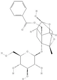 | NF-κB pathway↓ | Liu X et al. (2019) |
| MAPK pathway↓ | , Zhai and Guo, (2016), | ||||
| PI3K/AKT pathway ↑ | Zhou et al. (2013) | ||||
Paeonia veitchii Lynch, 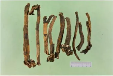 | Gallic acid | 370 | 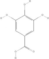 | PTEN↑ | Han D et al. (2020) |
Paeonia suffruticosa Andr, 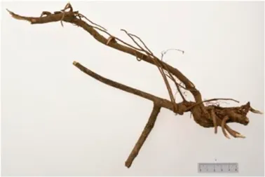 | Paeonol | 11092 | 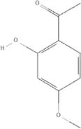 | miR-1↑ | Wu et al. (2018) |
Crataegus pinnatifida Bge. var. major N.E.Br, 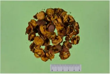 | Hyperoside | 5281643 |  | p38 MAPK, JNK, ERK and NF-κB pathway↓, NLRP1 inflammation pathway↓, AKT pathway ↓, miR-21↓ | Jang et al. (2018) |
| Ku et al. (2014) | |||||
| Wang X et al. (2018) | |||||
| Yang et al. (2021) | |||||
| Zhang J et al. (2021) | |||||
Cinnamomum cassia Presl, 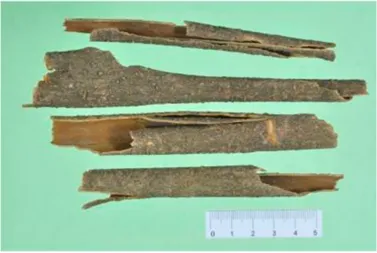 | 2-methoxycinnamaldehyde | 641298 | 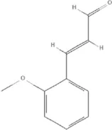 | HO-1↑ | Hwa et al. (2012) |
| Cinnamaldehyde | 637511 |  | TLR4/6-IRAK4/1 signal↓, TLR4-NOX4 pathway↓, TLR4-NF-κB ↓ | Ding et al. (2010) | |
| Kang et al. (2016) | |||||
| Song et al. (2013) | |||||
| Zhao et al. (2016) |
The mechanisms of TCM in improving inflammation.
↑ (Upregulation),↓ (Downregulation), AKT (Protein kinase B), ERK (Extracellular-regulated kinase), HO-1 (Heme oxygenase-1), IRAK (IL-1R-associated kinase), JNK (c-Jun N-terminal kinase), MAPK (Mitogen-activated protein kinase), miR (MicroRNA), NF-κB (Nuclear factor kappa-B),NLRP1 (Nucleotide-binding domain and leucine-rich repeat related family, pyrin domain containing 3), NOX (NADPH oxidase), p38 MAPK (Mitogen-activated protein kinase), PI3K (Phosphatidylinositol 3-kinase), PTEN (Phosphatase and tensin homolog ), TCM (Traditional Chinese Medicine), TLR (Toll-like receptor).
Crataegus pinnatifida Bge. var. Major N.E.Br
Hyperoside (PubChem CID: 5281643), a flavonoid from Crataegus pinnatifida Bge. var. major N.E.Br., suppresses vascular inflammatory responses in diabetic complications and atherosclerosis by downregulating mitogen-activated protein kinases (p38 MAPK, JNK, and ERK) and NF-κB signaling (Ku et al., 2014; Jang et al., 2018). Hyperoside also plays a protective role against heart damage caused by other diseases. Hyperoside ameliorated myocardial hypertrophy, collagen volume fraction, and cardiomyocyte inflammation in the myocardial infarction mice by regulating autophagy via the nucleotide-binding domain and leucine-rich repeat related (NLR) family, pyrin domain containing 1(NLRP1) inflammation pathway (Yang et al., 2021). Hyperoside decreased the levels of inflammatory factors, including IL-1β, IL-6, TNF-α, and MCP-1 in an angiotensin II-induced cardiomyocyte hypertrophy model to improve cardiac function via AKT signaling (Wang X et al., 2018). Hyperoside treated cardiac dysfunction in a mouse model of sepsis by regulating cardiomyocyte viability and inflammation via suppressing miR-21 (Zhang J et al., 2021), (Table 5).
Cinnamomum cassia Presl
The anti-inflammatory effects of Cinnamomum cassia Presl extracts have been used to treat a variety of diseases, and research on its anti-inflammatory mechanism has found that it might be closely related to NF-κB, and Toll-like receptors TLR2 and TLR4 (Reddy et al., 2004; Schink et al., 2018). More than 160 components have been isolated and identified from Cinnamomum cassia Presl. Phenylpropanoids are the main bioactive components, including 2-Methoxycinnamaldehyde (PubChem CID: 641298), Cinnamaldehyde (PubChem CID: 637511) (Zhang et al., 2019). 2-Methoxycinnamaldehyde acid inhibits vascular cell adhesion molecule-1(VCAM-1) and NF-κB expression, which are activated by TNF-α in endothelial cells and inhibited by HO-1 induction, thus the heart function of rats with I/R injury can be protected (Hwa et al., 2012). Cinnamaldehyde inhibits the activation of NLRP3 inflammasomes by attenuating the cluster of differentiation 36 (CD36)-mediated TLR4/6-IRAK (IRAK) 4/1 signaling, thereby reducing fructose-induced cardiac inflammation and fibrosis (Kang et al., 2016). Cinnamaldehyde can also improve lipopolysaccharide-induced cardiac dysfunction through the TLR4-NOX4 pathway (Zhao et al., 2016). Cinnamaldehyde and its derivative cinnamic acid can reduce TNF-α and IL-6 in rats with acute myocardial ischemia induced by isoproterenol (Song et al., 2013), which also directly reduces the inflammation of viral myocarditis induced by coxsackie virus B3 by inhibiting the TLR4-NF-κB signal transduction pathway (Ding et al., 2010), (Table 5).
Discussion
In this review, we retrospectively analyzed clinical studies on the intervention of TCM in HF, and discussed the mechanisms of some commonly used TCMs and their components in the treatment of HF. From the perspective of clinical research, TCM has the advantages of good curative effect and low levels of side effects in the treatment of HF, which can make up for the shortcomings of current treatment methods to a certain extent, and the two sides can produce complementary advantages. Basic research has shown that TCM can play its role in many ways, such as microcirculation improvements, energy metabolism promotion, anti-inflammation, and anti-oxidation. However, it should be noted that the compositions of TCM are complex. On the one hand, a variety of chemical components can affect organisms through many biological reactions. On the other hand, this kind of diversity makes it possible for different active ingredients in TCM to be synergistic, enhancing, and antagonistic. In the basic theories of TCM, the principles of drug application are also mentioned. However, currently, it is unclear which combinations of active ingredients have synergistic and antagonistic effects, or which combinations might increase toxicity, not to mention the effective or optimal dose of each active ingredient in the combination.
HF comprises a group of clinical syndromes with complex pathological mechanisms, involving multiple signaling pathways and targets. Natural medicines or their active ingredients can act on a variety of pathways and targets to effectively treat diseases, which is also the advantage of the application of TCM. To clarify the mechanism of TCM, it is necessary to study the pathways and targets of each active ingredient alone and in different combinations. It is also necessary to find a suitable breakthrough point and establish a reasonable pharmacological model based on genomics, proteomics, functional metabolomics, TCM pharmacology, and other -omics research (Han Y et al., 2020; Ma et al., 2020; Wang T et al., 2021). Determining the mechanism of the effective ingredients can not only explain how the various ingredients in TCM work individually or in combination, but also, and more importantly, it can discover new mechanisms and synergistic effects of the effective ingredients, which is conducive to innovation of TCM and the development of TCM theories.
A large number RCTs of integrated TCM and western medicine are reported every year; however, the quality of these studies is uneven. On the one hand, clinical research in TCM should carry out randomized, double-blind, placebo-controlled large-sample, multi-center RCT studies. During the implementation of RCTs, patients need to be included in strict accordance with the latest diagnostic criteria. The design of the placebo should be completely consistent with TCM in appearance and smell etc. Curative effect indicators should pay more attention on hard endpoints of cardiovascular events or choose internationally recognized intermediate endpoints as the primary endpoints to carry out long-term follow-up. On the other hand, the advantage of TCM treatment lies in individualized therapy based on syndrome differentiation. The direction of our future efforts is to form an RCT research method for TCM treatment and enable TCM with RCT evidence to enter clinical practice.
Conclusion
The results of RCTs indicate that as an adjuvant treatment to conventional drugs, TCM might be beneficial to patients with HF. Recent studies on the mechanism of HF in vitro and in animal models have shown that TCM has microcirculation improvement, energy metabolism promotion, anti-inflammation, and anti-oxidation effects.
Statements
Author contributions
AW: Conceptualization, Methodology, Validation, Formal analysis, Investigation, Data Curation, Writing—original draft, Visualization. WZ: Conceptualization, Methodology, Writing—review and editing. KY: Conceptualization, Methodology, Writing—review and editing. PH: Investigation, Formal analysis, Data Curation. HZ: Investigation, Data Curation. ZZ: Formal analysis, Visualization. DZ: Writing—review and editing, Supervision. XM: Conceptualization, Writing—review and editing, Supervision, Project administration.
Funding
This work was supported by the National Key Research and Development Program of China (No. 2018YFC1707410-02).
Acknowledgments
We would like to acknowledge Hong Liu, Qingzhou Station Primary School, Weifang, China for writing assistance and ETCM (http://www.tcmip.cn/ETCM/index.php/Home/Index/) for providing photographs of TCM decoction pieces.
Conflict of interest
The authors declare that the research was conducted in the absence of any commercial or financial relationships that could be construed as a potential conflict of interest.
Publisher’s note
All claims expressed in this article are solely those of the authors and do not necessarily represent those of their affiliated organizations, or those of the publisher, the editors and the reviewers. Any product that may be evaluated in this article, or claim that may be made by its manufacturer, is not guaranteed or endorsed by the publisher.
Supplementary material
The Supplementary Material for this article can be found online at: https://www.frontiersin.org/articles/10.3389/fphar.2022.810587/full#supplementary-material
Glossary
- 14-3-3γ
Tyrosine 3-monooxygenase/tryptophan 5-monooxygenase activation protein gamma
- 6MWD
6-min walking distance
- ADP
Adenosine diphosphate
- ADRs
Adverse drug reactions
- AEC
Adenylate energy charge
- AEs
Adverse events
- AHF
Acute heart failure
- AKT
Protein kinase B
- AMP
Adenosine monophosphate
- AMPK
5′-adenosine monophosphat-activated protein kinase
- ANG2
Angiogenin 2
- AR
Aldose reductase
- ARE
Antioxidant responsive element
- ATP
Adenosine-triphosphate
- ATP5D
ATP synthase, H+ transporting, mitochondrial F1 complex, delta subunit
- BCAAs
Branched-chain amino acids
- BCL-2
B-cell lymphoma-2
- BNP
Brain natriuretic peptide
- CaMKII
Calcium/calmodulin-dependent protein kinase II
- CAT
Catalase
- CCEs
Composite cardiac events
- CD36
Cluster of differentiation 36
- CFR
Coronary flow reserve
- CHF
Chronic heart failure
- CHFQLS
Quality of Life measured by the CHF Integrated Chinese and Western Medicine Survival Scale
- CK
Creatine kinase
- CM
Chinese medicine
- CMD
Coronary microvascular dysfunction
- CYPD
Cyclophilin D
- DAMPs
Damage-associated molecular patterns
- DJ-1
Parkinsonism associated deglycase
- DOX
Doxorubicin
- ECG
Electrocardiograph
- eNOS
Endothelial nitric oxide synthase
- ERK
Extracellular-regulated kinase
- FFAs
Free fatty acids
- FOXO1
Forkhead box O1
- G3PD
Glycerol-3-phosphate dehydrogenase
- GMP
Guanosine monophosphate
- GSH
Glutathione
- GSK-3beta
Glycogen synthase kinase 3 beta
- H/R
Hypoxia/reoxygenation
- H3
Histone 3
- HES1
Hairy and enhancer of split-1
- HF
Heart Failure
- HFmrEF
HF with mildly reduced ejection fraction
- HFpEF
HF with preserved ejection fraction
- HFrEF
HF with reduced ejection fraction
- HMGB1
High-mobility group box 1
- HO-1
Heme oxygenase-1
- HS1
Heat stable protein 1
- I/R
Ischemia/reperfusion
- IFN
Interferon
- IHD
Ischemic heart disease
- IHF
Ischemic heart failure
- IL
Interleukin
- iNOS
Inducible nitric oxide synthase
- INTER-CHF
INTERnational Congestive Heart Failure
- IRAK
IL-1R-associated kinase
- JAK2
Janus kinase 2
- JNK
c-Jun N-terminal kinase
- LA
Lactic acid
- LDH
Lactate dehydrogenase
- LDHB
L-lactate dehydrogenase B chain
- LKB1
Liver kinase B1
- LPE
Lysophosphatidylethanolamine
- LVED
Left ventricular end-diastolic diameter
- LVEDD
Left ventricular end diastolic diameter
- LVEF
Left ventricular ejection fraction
- LVIDd
Left ventricular internal diastolic diameter
- LVIDs
Left ventricular internal dimension systole
- MAPK
Mitogen-activated protein kinase
- MCAD
Medium-chain acyl-CoA dehydrogenase
- MCP
Monocyte chemoattractant protein
- MCPT1
Mast cell protease 1
- MCU
Mitochondrial calcium uniporter
- MDA
Malondialdehyde
- MI
Myocardial infarction
- miR
MicroRNA
- MLHFQ
Minnesota Living with Heart Failure Questionnaire
- MMP
Matrix metalloproteinase
- mTOR
Mechanistic target of rapamycin
- NADPH
Nicotinamide Adenine Dinucleotide Phosphate
- NF-κB
Nuclear factor kappa-B
- NLRP3
Nucleotide-binding domain and leucine-rich repeat related family, pyrin domain containing 3
- NO
Nitric oxide
- NOS
Nitric oxide synthase
- Notch1
Notch receptor 1
- NOX
NADPH oxidase
- NQO1
NAD(P)H quinone dehydrogenase 1
- NRF
Nuclear factor-erythroid 2-related factor
- NT-proBNP
N-terminal pro-B-type natriuretic peptide
- NYHA
New York Heart Association
- O-GlcNAcylation
O-linked β--acetylglucosamine modification
- p53
Tumor protein p53
- p66Shc
Src-homology-2-domain-containing transforming protein 1
- p70S6K
Ribosomal protein S6 kinase B1
- PA
Pyroracemic acid
- PAMPs
Pathogen-associated molecular patterns
- PDC
Pyruvate dehydrogenase complex
- PGC1α
Peroxisome proliferator-activated receptor gamma coactivator-1 alpha
- PI3K
Phosphatidylinositol 3-kinase
- PPAR
Peroxisome proliferator-activated receptor
- PRRS
Pattern-recognition receptors
- PTEN
Phosphatase and tensin homolog
- RAB7
Member RAS oncogene family
- RCT
Randomized controlled trial
- RHOA
Ras homolog family member A
- ROCK1
Rho associated coiled-coil containing protein kinase 1
- ROS
Reactive oxygen species
- SAEs
Serious adverse events
- sEH
Soluble epoxide hydrolase
- SF-36
Short-form 36
- SIRT
Sirtuin
- SOD
Superoxide dismutase
- STAT3
Signal transducer and activator of transcription 3
- TAP
Total adenylate pool
- TCM
Traditional Chinese Medicine
- TFAM
Transcription factor A, mitochondrial
- TIE2
Tyrosine kinase with immunoglobin and epidermal growth factor homology domain 2
- TLR
Toll-like receptor
- TNF-α
Tumor necrosis factor-α
- VCAM-1
Vascular cell adhesion molecule-1
- VEGF
Vascular endothelial growth factor
- VEGFR2
Vascular endothelial growth factor receptor 2
- VPCs
Ventricular premature complexes
References
1
AdamoL.Rocha-ResendeC.PrabhuS. D.MannD. L. (2020). Reappraising the Role of Inflammation in Heart Failure. Nat. Rev. Cardiol.17 (5), 269–285. 10.1038/s41569-019-0315-x
2
AhmadA.CorbanM. T.ToyaT.VerbruggeF. H.SaraJ. D.LermanL. O.et al (2021). Coronary Microvascular Dysfunction Is Associated with Exertional Haemodynamic Abnormalities in Patients with Heart Failure with Preserved Ejection Fraction. Eur. J. Heart Fail23 (5), 765–772. 10.1002/ejhf.2010
3
AhmadI.HodaM. (2020). Molecular Mechanisms of Action of Resveratrol in Modulation of Diabetic and Non-diabetic Cardiomyopathy. Pharmacol. Res.161 (11), 105112–112. 10.1016/j.phrs.2020.105112
4
AnkerS. D.ButlerJ.FilippatosG.FerreiraJ. P.BocchiE.BöhmM.et al (2021). Empagliflozin in Heart Failure with a Preserved Ejection Fraction. N. Engl. J. Med.385 (16), 1451–1461. 10.1056/NEJMoa2107038
5
BagulP. K.DeepthiN.SultanaR.BanerjeeS. K. (2015). Resveratrol Ameliorates Cardiac Oxidative Stress in Diabetes through Deacetylation of NFkB-P65 and Histone 3. J. Nutr. Biochem.26 (11), 1298–1307. 10.1016/j.jnutbio.2015.06.006
6
BayevaM.GheorghiadeM.ArdehaliH. (2013). Mitochondria as a Therapeutic Target in Heart Failure. J. Am. Coll. Cardiol.61 (6), 599–610. 10.1016/j.jacc.2012.08.1021
7
BediK. C.Jr.SnyderN. W.BrandimartoJ.AzizM.MesarosC.WorthA. J.et al (2016). Evidence for Intramyocardial Disruption of Lipid Metabolism and Increased Myocardial Ketone Utilization in Advanced Human Heart Failure. Circulation133 (8), 706–716. 10.1161/CIRCULATIONAHA.115.017545
8
BenjaminE. J.MuntnerP.AlonsoA.BittencourtM. S.CallawayC. W.CarsonA. P.et al (2019). Heart Disease and Stroke Statistics-2019 Update: A Report from the American Heart Association. Circulation139 (10), e56–528. 10.1161/CIR.0000000000000659
9
BerteroE.MaackC. (2018a). Calcium Signaling and Reactive Oxygen Species in Mitochondria. Circ. Res.122 (10), 1460–1478. 10.1161/CIRCRESAHA.118.310082
10
BerteroE.MaackC. (2018b). Metabolic Remodelling in Heart Failure. Nat. Rev. Cardiol.15 (8), 457–470. 10.1038/s41569-018-0044-6
11
BoY.JianZ.Zhi-JunS.QuingW.HuaZ.Chuan-WeiL.et al (2020). Panax Notoginseng Saponins Alleviates Advanced Glycation End Product-Induced Apoptosis by Upregulating SIRT1 and Antioxidant Expression Levels in HUVECs. Exp. Ther. Med.20 (5), 99. 10.3892/etm.2020.9229
12
BozkurtB.CoatsA. J. S.TsutsuiH.AbdelhamidC. M.AdamopoulosS.AlbertN.et al (2021). Universal Definition and Classification of Heart Failure: a Report of the Heart Failure Society of America, Heart Failure Association of the European Society of Cardiology, Japanese Heart Failure Society and Writing Committee of the Universal Definition of Heart Failure: Endorsed by the Canadian Heart Failure Society, Heart Failure Association of India, Cardiac Society of Australia and New Zealand, and Chinese Heart Failure Association. Eur. J. Heart Fail23 (3), 352–380. 10.1002/ejhf.2115
13
BrownD. A.PerryJ. B.AllenM. E.SabbahH. N.StaufferB. L.ShaikhS. R.et al (2017). Expert Consensus Document: Mitochondrial Function as a Therapeutic Target in Heart Failure. Nat. Rev. Cardiol.14 (4), 238–250. 10.1038/nrcardio.2016.203
14
BrutsaertD. L. (2003). Cardiac Endothelial-Myocardial Signaling: its Role in Cardiac Growth, Contractile Performance, and Rhythmicity. Physiol. Rev.83 (1), 59–115. 10.1152/physrev.00017.2002
15
ChaanineA. H.JoyceL. D.StulakJ. M.MaltaisS.JoyceD. L.DearaniJ. A.et al (2019). Mitochondrial Morphology, Dynamics, and Function in Human Pressure Overload or Ischemic Heart Disease with Preserved or Reduced Ejection Fraction. Circ. Heart Fail12 (2), e005131. 10.1161/CIRCHEARTFAILURE.118.005131
16
ChenX.WangQ.ShaoM.MaL.GuoD.WuY.et al (2019). Ginsenoside Rb3 Regulates Energy Metabolism and Apoptosis in Cardiomyocytes via Activating PPARα Pathway. Biomed. Pharmacother.120, 109487. 10.1016/j.biopha.2019.109487
17
CoelhoA. R.MartinsT. R.CoutoR.DeusC.PereiraC. V.SimõesR. F.et al (2017). Berberine-induced Cardioprotection and Sirt3 Modulation in Doxorubicin-Treated H9c2 Cardiomyoblasts. Biochim. Biophys. Acta Mol. Basis Dis.1863 (11), 2904–2923. 10.1016/j.bbadis.2017.07.030
18
Crespo-LeiroM. G.AnkerS. D.MaggioniA. P.CoatsA. J.FilippatosG.RuschitzkaF.et al (2016). European Society of Cardiology Heart Failure Long-Term Registry (ESC-HF-LT): 1-year Follow-Up Outcomes and Differences across Regions. Eur. J. Heart Fail18 (6), 613–625. 10.1002/ejhf.566
19
CuiZ. T.LiuJ. P.WeiW. L. (2016). The Effects of Tanshinone IIA on Hypoxia/reoxygenation-Induced Myocardial Microvascular Endothelial Cell Apoptosis in Rats via the JAK2/STAT3 Signaling Pathway. Biomed. Pharmacother.83, 1116–1126. 10.1016/j.biopha.2016.07.054
20
De JongK. A.LopaschukG. D. (2017). Complex Energy Metabolic Changes in Heart Failure with Preserved Ejection Fraction and Heart Failure with Reduced Ejection Fraction. Can J. Cardiol.33 (7), 860–871. 10.1016/j.cjca.2017.03.009
21
DingY.QiuL.ZhaoG.XuJ.WangS. (2010). Influence of Cinnamaldehyde on Viral Myocarditis in Mice. Am. J. Med. Sci.340 (2), 114–120. 10.1097/MAJ.0b013e3181dd3b43
22
DiseaseG. B. D.InjuryI.PrevalenceC. (2018). Global, Regional, and National Incidence, Prevalence, and Years Lived with Disability for 354 Diseases and Injuries for 195 Countries and Territories, 1990-2017: a Systematic Analysis for the Global Burden of Disease Study 2017. Lancet392 (10159), 1789–1858. 10.1016/S0140-6736(18)32279-7
23
DoenstT.NguyenT. D.AbelE. D. (2013). Cardiac Metabolism in Heart Failure: Implications beyond ATP Production. Circ. Res.113 (6), 709–724. 10.1161/CIRCRESAHA.113.300376
24
DokainishH.TeoK.ZhuJ.RoyA.AlHabibK. F.ElSayedA.et al (2017). Global Mortality Variations in Patients with Heart Failure: Results from the International Congestive Heart Failure (INTER-CHF) Prospective Cohort Study. Lancet Glob. Health5 (7), e665–e672. 10.1016/S2214-109X(17)30196-1
25
DongM.DingW.LiaoY.LiuY.YanD.ZhangY.et al (2015). Polydatin Prevents Hypertrophy in Phenylephrine Induced Neonatal Mouse Cardiomyocytes and Pressure-Overload Mouse Models. Eur. J. Pharmacol.746, 186–197. 10.1016/j.ejphar.2014.11.012
26
DongZ.ZhaoP.XuM.ZhangC.GuoW.ChenH.et al (2017). Astragaloside IV Alleviates Heart Failure via Activating PPARα to Switch Glycolysis to Fatty Acid β-oxidation. Sci. Rep.7 (1), 2691. 10.1038/s41598-017-02360-5
27
DutkaM.BobińskiR.Ulman-WłodarzI.HajdugaM.BujokJ.PająkC.et al (2020). Various Aspects of Inflammation in Heart Failure. Heart Fail Rev.25 (3), 537–548. 10.1007/s10741-019-09875-1
28
ElgendyI. Y.MahttaD.PepineC. J. (2019). Medical Therapy for Heart Failure Caused by Ischemic Heart Disease. Circ. Res.124 (11), 1520–1535. 10.1161/CIRCRESAHA.118.313568
29
EverettB. M.MacFadyenJ. G.ThurenT.LibbyP.GlynnR. J.RidkerP. M. (2020). Inhibition of Interleukin-1β and Reduction in Atherothrombotic Cardiovascular Events in the CANTOS Trial. J. Am. Coll. Cardiol.76 (14), 1660–1670. 10.1016/j.jacc.2020.08.011
30
FanG.ZhuY.GuoH.WangX.WangH.GaoX. (2011). Direct Vasorelaxation by a Novel Phytoestrogen Tanshinone IIA Is Mediated by Nongenomic Action of Estrogen Receptor through Endothelial Nitric Oxide Synthase Activation and Calcium Mobilization. J. Cardiovasc. Pharmacol.57 (3), 340–347. 10.1097/FJC.0b013e31820a0da1
31
FanJ.LiuD.HeC.LiX.HeF. (2016). Inhibiting Adhesion Events by Panax Notoginseng Saponins and Ginsenoside Rb1 Protecting Arteries via Activation of Nrf2 and Suppression of P38 - VCAM-1 Signal Pathway. J. Ethnopharmacol192, 423–430. 10.1016/j.jep.2016.09.022
32
FangH.YangS.LuoY.ZhangC.RaoY.LiuR.et al (2018). Notoginsenoside R1 Inhibits Vascular Smooth Muscle Cell Proliferation, Migration and Neointimal Hyperplasia through PI3K/Akt Signaling. Sci. Rep.8 (1), 7595. 10.1038/s41598-018-25874-y
33
FiuzatM.LowyN.StockbridgeN.SbolliM.LattaF.LindenfeldJ.et al (2020). Endpoints in Heart Failure Drug Development: History and Future. JACC Heart Fail8 (6), 429–440. 10.1016/j.jchf.2019.12.011
34
FordT. J.CorcoranD.BerryC. (2018). Stable Coronary Syndromes: Pathophysiology, Diagnostic Advances and Therapeutic Need. Heart104 (4), 284–292. 10.1136/heartjnl-2017-311446
35
FuS.ZhangJ.GaoX.XiaY.FerrelliR.FauciA.et al (2010). Clinical Practice of Traditional Chinese Medicines for Chronic Heart Failure. Heart Asia2 (1), 24–27. 10.1136/ha.2009.001123
36
GaoK.ZhaoH.GaoJ.WenB.JiaC.WangZ.et al (2017). Mechanism of Chinese Medicine Herbs Effects on Chronic Heart Failure Based on Metabolic Profiling. Front Pharmacol.8, 864. 10.3389/fphar.2017.00864
37
GhionzoliN.GentileF.Del FrancoA. M.CastiglioneV.AimoA.GiannoniA.et al (2021). Current and Emerging Drug Targets in Heart Failure Treatment. Heart Fail Rev.[online ahead of print]. 10.1007/s10741-021-10137-2
38
GroenewegenA.RuttenF. H.MosterdA.HoesA. W. (2020). Epidemiology of Heart Failure. Eur. J. Heart Fail22 (8), 1342–1356. 10.1002/ejhf.1858
39
GuoS.YaoQ.KeZ.ChenH.WuJ.LiuC. (2015). Resveratrol Attenuates High Glucose-Induced Oxidative Stress and Cardiomyocyte Apoptosis through AMPK. Mol. Cell Endocrinol412, 85–94. 10.1016/j.mce.2015.05.034
40
HanD.ZhangQ. Y.ZhangY. L.HanX.GuoS. B.TengF.et al (2020). Gallic Acid Ameliorates Angiotensin II-Induced Atrial Fibrillation by Inhibiting Immunoproteasome- Mediated PTEN Degradation in Mice. Front Cell Dev Biol8, 594683. 10.3389/fcell.2020.594683
41
HanY.SunH.ZhangA.YanG.WangX. J. (2020). Chinmedomics, a New Strategy for Evaluating the Therapeutic Efficacy of Herbal Medicines. Pharmacol. Ther.216, 107680. 10.1016/j.pharmthera.2020.107680
42
HaoW.LiN.MiC.WangQ.YuY. (2021). Salidroside Attenuates Cardiac Dysfunction in a Rat Model of Diabetes. Diabet Med., e14683. [online ahead of print]. 10.1111/dme.14683
43
HeH.LuoY.QiaoY.ZhangZ.YinD.YaoJ.et al (2018). Curcumin Attenuates Doxorubicin-Induced Cardiotoxicity via Suppressing Oxidative Stress and Preventing Mitochondrial Dysfunction Mediated by 14-3-3γ. Food Funct.9 (8), 4404–4418. 10.1039/c8fo00466h
44
HeL.LiuY. Y.WangK.LiC.ZhangW.LiZ. Z.et al (2021). Tanshinone IIA Protects Human Coronary Artery Endothelial Cells from Ferroptosis by Activating the NRF2 Pathway. Biochem. Biophys. Res. Commun.575, 1–7. 10.1016/j.bbrc.2021.08.067
45
HeY.HuangW.ZhangC.ChenL.XuR.LiN.et al (2021). Energy Metabolism Disorders and Potential Therapeutic Drugs in Heart Failure. Acta Pharm. Sin B11 (5), 1098–1116. 10.1016/j.apsb.2020.10.007
46
HeY.XiJ.ZhengH.ZhangY.JinY.XuZ. (2012). Astragaloside IV Inhibits Oxidative Stress-Induced Mitochondrial Permeability Transition Pore Opening by Inactivating GSK-3β via Nitric Oxide in H9c2 Cardiac Cells. Oxid Med. Cell Longev2012, 935738. 10.1155/2012/935738
47
HuG.YangP.ZengY.ZhangS.SongJ. (2018). Danggui Buxue Decoction Promotes Angiogenesis by Up-Regulation of VEGFR1/2 Expressions and Down-Regulation of sVEGFR1/2 Expression in Myocardial Infarction Rat. J. Chin Med. Assoc.81 (1), 37–46. 10.1016/j.jcma.2017.06.015
48
HuY. R.MaH.ZouZ. Y.HeK.XiaoY. B.WangY.et al (2017). Activation of Akt and JNK/Nrf2/NQO1 Pathway Contributes to the Protective Effect of Coptisine against AAPH-Induced Oxidative Stress. Biomed. Pharmacother.85, 313–322. 10.1016/j.biopha.2016.11.031
49
HuangH.LaiS.WanQ.QiW.LiuJ. (2016). Astragaloside IV Protects Cardiomyocytes from Anoxia/reoxygenation Injury by Upregulating the Expression of Hes1 Protein. Can J. Physiol. Pharmacol.94 (5), 542–553. 10.1139/cjpp-2015-0457
50
HuangQ.SuH.QiB.WangY.YanK.WangX.et al (2021). A SIRT1 Activator, Ginsenoside Rc, Promotes Energy Metabolism in Cardiomyocytes and Neurons. J. Am. Chem. Soc.143 (3), 1416–1427. 10.1021/jacs.0c10836
51
HwaJ. S.JinY. C.LeeY. S.KoY. S.KimY. M.ShiL. Y.et al (2012). 2-methoxycinnamaldehyde from Cinnamomum cassia Reduces Rat Myocardial Ischemia and Reperfusion Injury In Vivo Due to HO-1 Induction. J. Ethnopharmacol139 (2), 605–615. 10.1016/j.jep.2011.12.001
52
JangS. A.ParkD. W.SohnE. H.LeeS. R.KangS. C. (2018). Hyperoside Suppresses Tumor Necrosis Factor α-mediated Vascular Inflammatory Responses by Downregulating Mitogen-Activated Protein Kinases and Nuclear Factor-Κb Signaling. Chem. Biol. Interact294, 48–55. 10.1016/j.cbi.2018.08.013
53
JiaoF.VargheseK.WangS.LiuY.YuH.BoozG. W.et al (2021). Recent Insights into the Protective Mechanisms of Paeoniflorin in Neurological, Cardiovascular, and Renal Diseases. J. Cardiovasc. Pharmacol.77 (6), 728–734. 10.1097/FJC.0000000000001021
54
KangL. L.ZhangD. M.MaC. H.ZhangJ. H.JiaK. K.LiuJ. H.et al (2016). Cinnamaldehyde and Allopurinol Reduce Fructose-Induced Cardiac Inflammation and Fibrosis by Attenuating CD36-Mediated TLR4/6-Irak4/1 Signaling to Suppress NLRP3 Inflammasome Activation. Sci. Rep.6, 27460. 10.1038/srep27460
55
KimN. D.KimE. M.KangK. W.ChoM. K.ChoiS. Y.KimS. G. (2003). Ginsenoside Rg3 Inhibits Phenylephrine-Induced Vascular Contraction through Induction of Nitric Oxide Synthase. Br. J. Pharmacol.140 (4), 661–670. 10.1038/sj.bjp.0705490
56
KimY. M.HaY. M.JinY. C.ShiL. Y.LeeY. S.KimH. J.et al (2009). Palmatine from Coptidis Rhizoma Reduces Ischemia-Reperfusion-Mediated Acute Myocardial Injury in the Rat. Food Chem. Toxicol.47 (8), 2097–2102. 10.1016/j.fct.2009.05.031
57
KovacicP.SomanathanR. (2010). Multifaceted Approach to Resveratrol Bioactivity: Focus on Antioxidant Action, Cell Signaling and Safety. Oxid Med. Cell Longev3 (2), 86–100. 10.4161/oxim.3.2.11147
58
KuS. K.KwakS.KwonO. J.BaeJ. S. (2014). Hyperoside Inhibits High-Glucose-Induced Vascular Inflammation In Vitro and In Vivo. Inflammation37 (5), 1389–1400. 10.1007/s10753-014-9863-8
59
LamC. S. P.VoorsA. A.de BoerR. A.SolomonS. D.van VeldhuisenD. J. (2018). Heart Failure with Preserved Ejection Fraction: from Mechanisms to Therapies. Eur. Heart J.39 (30), 2780–2792. 10.1093/eurheartj/ehy301
60
LengB.TangF.LuM.ZhangZ.WangH.ZhangY. (2018). Astragaloside IV Improves Vascular Endothelial Dysfunction by Inhibiting the TLR4/NF-Κb Signaling Pathway. Life Sci.209, 111–121. 10.1016/j.lfs.2018.07.053
61
LeungK. W.NgH. M.TangM. K.WongC. C.WongR. N.WongA. S. (2011). Ginsenoside-Rg1 Mediates a Hypoxia-independent Upregulation of Hypoxia-Inducible Factor-1α to Promote Angiogenesis. Angiogenesis14 (4), 515–522. 10.1007/s10456-011-9235-z
62
LiD.WangJ.HouJ.FuJ.ChangD.BensoussanA.et al (2016). Ginsenoside Rg1 Protects Starving H9c2 Cells by Dissociation of Bcl-2-Beclin1 Complex. BMC Complement Altern. Med.16, 146. 10.1186/s12906-016-1112-2
63
LiH.SuredaA.DevkotaH. P.PittalàV.BarrecaD.SilvaA. S.et al (2020). Curcumin, the golden Spice in Treating Cardiovascular Diseases. Biotechnol. Adv.38, 107343. 10.1016/j.biotechadv.2019.01.010
64
LiH.XiaN.FörstermannU. (2012). Cardiovascular Effects and Molecular Targets of Resveratrol. Nitric Oxide26 (2), 102–110. 10.1016/j.niox.2011.12.006
65
LiL.PanC. S.YanL.CuiY. C.LiuY. Y.MuH. N.et al (2018). Ginsenoside Rg1 Ameliorates Rat Myocardial Ischemia-Reperfusion Injury by Modulating Energy Metabolism Pathways. Front Physiol.9, 78. 10.3389/fphys.2018.00078
66
LiP.ShenJ.WangZ.LiuS.LiuQ.LiY.et al (2021). Genus Paeonia: A Comprehensive Review on Traditional Uses, Phytochemistry, Pharmacological Activities, Clinical Application, and Toxicology. J. Ethnopharmacol269, 113708. 10.1016/j.jep.2020.113708
67
LiS.LouS.LeiB. U.ChanT. F.KwanY. W.ChanS. W.et al (2011). Transcriptional Profiling of Angiogenesis Activities of Calycosin in Zebrafish. Mol. Biosyst.7 (11), 3112–3121. 10.1039/c1mb05206c
68
LiX.ZhangJ.HuangJ.MaA.YangJ.LiW.et al (2013). A Multicenter, Randomized, Double-Blind, Parallel-Group, Placebo-Controlled Study of the Effects of Qili Qiangxin Capsules in Patients with Chronic Heart Failure. J. Am. Coll. Cardiol.62 (12), 1065–1072. 10.1016/j.jacc.2013.05.035
69
LiX. T.ChenR.JinL. M.ChenH. Y. (2009). Regulation on Energy Metabolism and protection on Mitochondria of Panax Ginseng Polysaccharide. Am. J. Chin Med.37 (6), 1139–1152. 10.1142/S0192415X09007454
70
LiX. X.WuY.FanZ. J.CuiJ.LiD.LinQ.et al (2022). Qishen Taohong Granule () as Adjuvant Therapy for Improving Cardiac Function and Quality of Life in Patients with Chronic Heart Failure: A Randomized Controlled Trial. Chin J. Integr. Med.28 (1), 12–19. 10.1007/s11655-021-2866-z
71
LinC. H.ChengY. C.NicolC. J.LinK. H.YenC. H.ChiangM. C. (2017). Activation of AMPK Is Neuroprotective in the Oxidative Stress by Advanced Glycosylation End Products in Human Neural Stem Cells. Exp. Cell Res359 (2), 367–373. 10.1016/j.yexcr.2017.08.019
72
LinX. P.CuiH. J.YangA. L.LuoJ. K.TangT. (2018). Astragaloside IV Improves Vasodilatation Function by Regulating the PI3K/Akt/eNOS Signaling Pathway in Rat Aorta Endothelial Cells. J. Vasc. Res.55 (3), 169–176. 10.1159/000489958
73
LiuD. Q.ChenS. P.SunJ.WangX. M.ChenN.ZhouY. Q.et al (2019). Berberine Protects against Ischemia-Reperfusion Injury: A Review of Evidence from Animal Models and Clinical Studies. Pharmacol. Res.148, 104385. 10.1016/j.phrs.2019.104385
74
LiuQ.YuanJ. W.ZhangF.QiaoF.SuiX. F.LiuC. H. (2019). Curcumin Protects Rat H9C2 Cardiomyocytes against Doxorubicin Toxicity by Modulating Oxidative Stress and Apoptosis. J. Biol. Regul. Homeost Agents33 (6), 1849–1854. 10.23812/19-365-L10.23812/19-365-L
75
LiuS.LiF.LiY.LiW.XuJ.DuH. (2017). A Review of Traditional and Current Methods Used to Potentially Reduce Toxicity of Aconitum Roots in Traditional Chinese Medicine. J. Ethnopharmacol207, 237–250. 10.1016/j.jep.2017.06.038
76
LiuW. H. (2020). Effectiveness of Yangyin Shuxin Decoction on Diastolic Function and Exercise Capacity of Patients with Heart Failure. Tianjin: Tianjin University of Chinese Medicine.
77
LiuX.ChenK.ZhuangY.HuangY.SuiY.ZhangY.et al (2019). Paeoniflorin Improves Pressure Overload-Induced Cardiac Remodeling by Modulating the MAPK Signaling Pathway in Spontaneously Hypertensive Rats. Biomed. Pharmacother.111, 695–704. 10.1016/j.biopha.2018.12.090
78
LiuY. L.ZhouX. Y.XuanL. J. (2019). Magnesium Lithospermate B Ameliorates Microcirculation Perfusion in Rats by Promoting Vascular NO Production via Activating the PI3K/AKT Pathway. Acta Pharmacol. Sin40 (8), 1010–1018. 10.1038/s41401-018-0203-7
79
LopaschukG. D.KarwiQ. G.TianR.WendeA. R.AbelE. D. (2021). Cardiac Energy Metabolism in Heart Failure. Circ. Res.128 (10), 1487–1513. 10.1161/CIRCRESAHA.121.318241
80
LuanA.TangF.YangY.LuM.WangH.ZhangY. (2015). Astragalus Polysaccharide Attenuates Isoproterenol-Induced Cardiac Hypertrophy by Regulating TNF-Α/pgc-1α Signaling Mediated Energy Biosynthesis. Environ. Toxicol. Pharmacol.39 (3), 1081–1090. 10.1016/j.etap.2015.03.014
81
LuoY.WanQ.XuM.ZhouQ.ChenX.YinD.et al (2019). Nutritional Preconditioning Induced by Astragaloside Ⅳ on Isolated Hearts and Cardiomyocytes against Myocardial Ischemia Injury via Improving Bcl-2-Mediated Mitochondrial Function. Chem. Biol. Interact309, 108723. 10.1016/j.cbi.2019.06.036
82
MaL.ChuangC. C.WengW.ZhaoL.ZhengY.ZhangJ.et al (2016). Paeonol Protects Rat Heart by Improving Regional Blood Perfusion during No-Reflow. Front Physiol.7, 298. 10.3389/fphys.2016.00298
83
MaX.MengY.WangP.TangZ.WangH.XieT. (2020). Bioinformatics-assisted, Integrated Omics Studies on Medicinal Plants. Brief Bioinform21 (6), 1857–1874. 10.1093/bib/bbz132
84
MakS. K.YuC. M.SunW. T.HeG. W.LiuX. C.YangQ. (2017). Tetramethylpyrazine Suppresses Angiotensin II-Induced Soluble Epoxide Hydrolase Expression in Coronary Endothelium via Anti-ER Stress Mechanism. Toxicol. Appl. Pharmacol.336, 84–93. 10.1016/j.taap.2017.10.016
85
ManolisA. S.ManolisA. A.ManolisT. A.ApostolakiN. E.ApostolopoulosE. J.MelitaH.et al (2021). Mitochondrial Dysfunction in Cardiovascular Disease: Current Status of Translational Research/clinical and Therapeutic Implications. Med. Res. Rev.41 (1), 275–313. 10.1002/med.21732
86
MaoJ.ZhangJ.LamC. S. P.ZhuM.YaoC.ChenS.et al (2020). Qishen Yiqi Dripping Pills for Chronic Ischaemic Heart Failure: Results of the CACT-IHF Randomized Clinical Trial. ESC Heart Fail7 (6), 3881–3890. 10.1002/ehf2.12980
87
MarzilliM.MerzC. N.BodenW. E.BonowR. O.CapozzaP. G.ChilianW. M.et al (2012). Obstructive Coronary Atherosclerosis and Ischemic Heart Disease: an Elusive Link!J. Am. Coll. Cardiol.60 (11), 951–956. 10.1016/j.jacc.2012.02.082
88
McDonaghT. A.MetraM.AdamoM.GardnerR. S.BaumbachA.BöhmM.et al (2021). 2021 ESC Guidelines for the Diagnosis and Treatment of Acute and Chronic Heart Failure. Eur. Heart J.42 (36), 3599–3726. 10.1093/eurheartj/ehab368
89
McMurrayJ. J.PackerM.DesaiA. S.GongJ.LefkowitzM. P.RizkalaA. R.et al (2014). Angiotensin-neprilysin Inhibition versus Enalapril in Heart Failure. N. Engl. J. Med.371 (11), 993–1004. 10.1056/NEJMoa1409077
90
McMurrayJ. J. V.SolomonS. D.InzucchiS. E.KøberL.KosiborodM. N.MartinezF. A.et al (2019). Dapagliflozin in Patients with Heart Failure and Reduced Ejection Fraction. N. Engl. J. Med.381 (21), 1995–2008. 10.1056/NEJMoa1911303
91
MengF. C.WuZ. F.YinZ. Q.LinL. G.WangR.ZhangQ. W. (2018). Coptidis Rhizoma and its Main Bioactive Components: Recent Advances in Chemical Investigation, Quality Evaluation and Pharmacological Activity. Chin Med.13, 13. 10.1186/s13020-018-0171-3
92
MiL.LiY.-C.SunM.-R.ZhangP.-L.LiY.YangH. (2021). A Systematic Review of Pharmacological Activities, Toxicological Mechanisms and Pharmacokinetic Studies on Aconitum Alkaloids. Chin. J. Nat. medicines19 (7), 505–520. 10.1016/S1875-5364(21)60050-X
93
MünzelT.CamiciG. G.MaackC.BonettiN. R.FusterV.KovacicJ. C. (2017). Impact of Oxidative Stress on the Heart and Vasculature: Part 2 of a 3-Part Series. J. Am. Coll. Cardiol.70 (2), 212–229. 10.1016/j.jacc.2017.05.035
94
MünzelT.GoriT.KeaneyJ. F.Jr.MaackC.DaiberA. (2015). Pathophysiological Role of Oxidative Stress in Systolic and Diastolic Heart Failure and its Therapeutic Implications. Eur. Heart J.36 (38), 2555–2564. 10.1093/eurheartj/ehv305
95
MurphyS. P.KakkarR.McCarthyC. P.JanuzziJ. L.Jr. (2020). Inflammation in Heart Failure: JACC State-Of-The-Art Review. J. Am. Coll. Cardiol.75 (11), 1324–1340. 10.1016/j.jacc.2020.01.014
96
MyungS. K.JuW.ChoB.OhS. W.ParkS. M.KooB. K.et al (2013). Efficacy of Vitamin and Antioxidant Supplements in Prevention of Cardiovascular Disease: Systematic Review and Meta-Analysis of Randomised Controlled Trials. BMJ346 (18), f10. 10.1136/bmj.f10
97
NiJ.LiY.XuY.GuoR. (2021). Salidroside Protects against Cardiomyocyte Apoptosis and Ventricular Remodeling by AKT/HO-1 Signaling Pathways in a Diabetic Cardiomyopathy Mouse Model. Phytomedicine82, 153406. 10.1016/j.phymed.2020.153406
98
NieT.ZhaoS.MaoL.YangY.SunW.LinX.et al (2018). The Natural Compound, Formononetin, Extracted from Astragalus Membranaceus Increases Adipocyte Thermogenesis by Modulating PPARγ Activity. Br. J. Pharmacol.175 (9), 1439–1450. 10.1111/bph.14139
99
NielsenR.MøllerN.GormsenL. C.TolbodL. P.HanssonN. H.SorensenJ.et al (2019). Cardiovascular Effects of Treatment with the Ketone Body 3-Hydroxybutyrate in Chronic Heart Failure Patients. Circulation139 (18), 2129–2141. 10.1161/CIRCULATIONAHA.118.036459
100
PackerM.AnkerS. D.ButlerJ.FilippatosG.PocockS. J.CarsonP.et al (2020). Cardiovascular and Renal Outcomes with Empagliflozin in Heart Failure. N. Engl. J. Med.383 (15), 1413–1424. 10.1056/NEJMoa2022190
101
PadroT.ManfriniO.BugiardiniR.CantyJ.CenkoE.De LucaG.et al (2020). ESC Working Group on Coronary Pathophysiology and Microcirculation Position Paper on 'coronary Microvascular Dysfunction in Cardiovascular Disease'. Cardiovasc. Res.116 (4), 741–755. 10.1093/cvr/cvaa003
102
PagliaroB. R.CannataF.StefaniniG. G.BologneseL. (2020). Myocardial Ischemia and Coronary Disease in Heart Failure. Heart Fail Rev.25 (1), 53–65. 10.1007/s10741-019-09831-z
103
ParkJ. Y.ShinH. K.ChoiY. W.LeeY. J.BaeS. S.HanJ.et al (2009). Gomisin A Induces Ca2+-dependent Activation of eNOS in Human Coronary Artery Endothelial Cells. J. Ethnopharmacol125 (2), 291–296. 10.1016/j.jep.2009.06.028
104
PengW.QinR.LiX.ZhouH. (2013). Botany, phytochemistry, pharmacology, and potential application of Polygonum cuspidatum Sieb.et Zucc. a review. J. Ethnopharmacol148 (3), 729–745. 10.1016/j.jep.2013.05.007
105
PonikowskiP.VoorsA. A.AnkerS. D.BuenoH.ClelandJ. G.CoatsA. J.et al (2016). 2016 ESC Guidelines for the Diagnosis and Treatment of Acute and Chronic Heart Failure: The Task Force for the Diagnosis and Treatment of Acute and Chronic Heart Failure of the European Society of Cardiology (ESC). Developed with the Special Contribution of the Heart Failure Association (HFA) of the ESC. Eur. J. Heart Fail18 (27), 891–975. 10.1093/eurheartj/ehw12810.1002/ejhf.592
106
PriesA. R.ReglinB. (2017). Coronary Microcirculatory Pathophysiology: Can We Afford it to Remain a Black Box?Eur. Heart J.38 (7), 478–488. 10.1093/eurheartj/ehv760
107
QinL.WangJ.ZhaoR.ZhangX.MeiY. (2019). Ginsenoside-Rb1 Improved Diabetic Cardiomyopathy through Regulating Calcium Signaling by Alleviating Protein O-GlcNAcylation. J. Agric. Food Chem.67 (51), 14074–14085. 10.1021/acs.jafc.9b05706
108
RadanM.DianatM.BadaviM.MardS. A.BayatiV.GoudarziG. (2019). Gallic Acid Protects Particulate Matter (PM10) Triggers Cardiac Oxidative Stress and Inflammation Causing Heart Adverse Events in Rats. Environ. Sci. Pollut. Res. Int.26 (18), 18200–18207. 10.1007/s11356-019-05223-w
109
RamalingamA.BudinS. B.Mohd FauziN.RitchieR. H.ZainalabidinS. (2021). Targeting Mitochondrial Reactive Oxygen Species-Mediated Oxidative Stress Attenuates Nicotine-Induced Cardiac Remodeling and Dysfunction. Sci. Rep.11 (1), 13845. 10.1038/s41598-021-93234-4
110
ReddyA. M.SeoJ. H.RyuS. Y.KimY. S.KimY. S.MinK. R.et al (2004). Cinnamaldehyde and 2-methoxycinnamaldehyde as NF-kappaB Inhibitors from Cinnamomum cassia. Planta Med.70 (9), 823–827. 10.1055/s-2004-827230
111
ReddyY. N. V.BorlaugB. A.O'ConnorC. M.GershB. J. (2020). Novel Approaches to the Management of Chronic Systolic Heart Failure: Future Directions and Unanswered Questions. Eur. Heart J.41 (18), 1764–1774. 10.1093/eurheartj/ehz364
112
RenB. C.ZhangY. F.LiuS. S.ChengX. J.YangX.CuiX. G.et al (2020). Curcumin Alleviates Oxidative Stress and Inhibits Apoptosis in Diabetic Cardiomyopathy via Sirt1-Foxo1 and PI3K-Akt Signalling Pathways. J. Cell Mol Med24 (21), 12355–12367. 10.1111/jcmm.15725
113
RheeA. J.LavineK. J. (2020). New Approaches to Target Inflammation in Heart Failure: Harnessing Insights from Studies of Immune Cell Diversity. Annu. Rev. Physiol.82, 1–20. 10.1146/annurev-physiol-021119-034412
114
SchinkA.NaumoskaK.KitanovskiZ.KampfC. J.Fröhlich-NowoiskyJ.ThinesE.et al (2018). Anti-inflammatory Effects of Cinnamon Extract and Identification of Active Compounds Influencing the TLR2 and TLR4 Signaling Pathways. Food Funct.9 (11), 5950–5964. 10.1039/c8fo01286e
115
SelvarajS.KellyD. P.MarguliesK. B. (2020). Implications of Altered Ketone Metabolism and Therapeutic Ketosis in Heart Failure. Circulation141 (22), 1800–1812. 10.1161/CIRCULATIONAHA.119.045033
116
ShahK. S.XuH.MatsouakaR. A.BhattD. L.HeidenreichP. A.HernandezA. F.et al (2017). Heart Failure with Preserved, Borderline, and Reduced Ejection Fraction: 5-Year Outcomes. J. Am. Coll. Cardiol.70 (20), 2476–2486. 10.1016/j.jacc.2017.08.074
117
ShahS. J.LamC. S. P.SvedlundS.SarasteA.HageC.TanR. S.et al (2018). Prevalence and Correlates of Coronary Microvascular Dysfunction in Heart Failure with Preserved Ejection Fraction: PROMIS-HFpEF. Eur. Heart J.39 (37), 3439–3450. 10.1093/eurheartj/ehy531
118
ShyuK. G.TsaiS. C.WangB. W.LiuY. C.LeeC. C. (2004). Saikosaponin C Induces Endothelial Cells Growth, Migration and Capillary Tube Formation. Life Sci.76 (7), 813–826. 10.1016/j.lfs.2004.10.003
119
SongF.LiH.SunJ.WangS. (2013). Protective Effects of Cinnamic Acid and Cinnamic Aldehyde on Isoproterenol-Induced Acute Myocardial Ischemia in Rats. J. Ethnopharmacol150 (1), 125–130. 10.1016/j.jep.2013.08.019
120
StanleyW. C.RecchiaF. A.LopaschukG. D. (2005). Myocardial Substrate Metabolism in the normal and Failing Heart. Physiol. Rev.85 (3), 1093–1129. 10.1152/physrev.00006.2004
121
Steinkamp-FenskeK.BollingerL.VöllerN.XuH.YaoY.BauerR.et al (2007). Ursolic Acid from the Chinese Herb Danshen (Salvia Miltiorrhiza L.) Upregulates eNOS and Downregulates Nox4 Expression in Human Endothelial Cells. Atherosclerosis195 (1), e104–11. 10.1016/j.atherosclerosis.2007.03.028
122
TanY. Y.ChenL. X.FangL.ZhangQ. (2020). Cardioprotective Effects of Polydatin against Myocardial Injury in Diabetic Rats via Inhibition of NADPH Oxidase and NF-Κb Activities. BMC Complement Med. Ther.20 (1), 378. 10.1186/s12906-020-03177-y
123
TangB.ZhangJ. G.TanH. Y.WeiX. Q. (2018). Astragaloside IV Inhibits Ventricular Remodeling and Improves Fatty Acid Utilization in Rats with Chronic Heart Failure. Biosci. Rep.38 (3). 10.1042/BSR20171036
124
TangD. (2020). A Multicenter,randomized,double-Blind,placebo-Controlled Study of the Efficacy and Safety of Yangxinxue Granule in the Treatment of Chronic Heart Failure. Chengdu: Chengdu University of TCM.
125
TangJ. Y.LiS.LiZ. H.ZhangZ. J.HuG.CheangL. C.et al (2010). Calycosin Promotes Angiogenesis Involving Estrogen Receptor and Mitogen-Activated Protein Kinase (MAPK) Signaling Pathway in Zebrafish and HUVEC. PloS one5 (7), e11822. 10.1371/journal.pone.0011822
126
TaoH.WuX.CaoJ.PengY.WangA.PeiJ.et al (2019). Rhodiola Species: A Comprehensive Review of Traditional Use, Phytochemistry, Pharmacology, Toxicity, and Clinical Study. Med. Res. Rev.39 (5), 1779–1850. 10.1002/med.21564
127
TaquetiV. R.SolomonS. D.ShahA. M.DesaiA. S.GroarkeJ. D.OsborneM. T.et al (2018). Coronary Microvascular Dysfunction and Future Risk of Heart Failure with Preserved Ejection Fraction. Eur. Heart J.39 (10), 840–849. 10.1093/eurheartj/ehx721
128
TseH. Y.HuiM. N.LiL.LeeS. M.LeungA. Y.ChengS. H. (2012). Angiogenic Efficacy of Simplified 2-herb Formula (NF3) in Zebrafish Embryos In Vivo and Rat Aortic Ring In Vitro. J. Ethnopharmacol139 (2), 447–453. 10.1016/j.jep.2011.11.031
129
TuL.PanC. S.WeiX. H.YanL.LiuY. Y.FanJ. Y.et al (2013). Astragaloside IV Protects Heart from Ischemia and Reperfusion Injury via Energy Regulation Mechanisms. Microcirculation20 (8), 736–747. 10.1111/micc.12074
130
WangB.YangQ.SunY. Y.XingY. F.WangY. B.LuX. T.et al (2014). Resveratrol-enhanced Autophagic Flux Ameliorates Myocardial Oxidative Stress Injury in Diabetic Mice. J. Cell Mol Med18 (8), 1599–1611. 10.1111/jcmm.12312
131
WangD.JieQ.LiuB.LiY.DaiL.LuoJ.et al (2017). Saponin Extract from Panax notoginseng Promotesangiogenesis through AMPK- and eNOS-dependent P-athways in HUVECs. Mol. Med. Rep.16 (4), 5211–5218. 10.3892/mmr.2017.7280
132
WangD.LvL.XuY.JiangK.ChenF.QianJ.et al (2021). Cardioprotection of Panax Notoginseng Saponins against Acute Myocardial Infarction and Heart Failure through Inducing Autophagy. Biomed. Pharmacother.136, 111287. 10.1016/j.biopha.2021.111287
133
WangJ. R.ZhouH.YiX. Q.JiangZ. H.LiuL. (2012). Total Ginsenosides of Radix Ginseng Modulates Tricarboxylic Acid Cycle Protein Expression to Enhance Cardiac Energy Metabolism in Ischemic Rat Heart Tissues. Molecules17 (11), 12746–12757. 10.3390/molecules171112746
134
WangN. N.XuH. H.ZhouW.YangH. X.WangJ.MaZ. C.et al (2021). Aconitine Attenuates Mitochondrial Dysfunction of Cardiomyocytes via Promoting Deacetylation of Cyclophilin-D Mediated by Sirtuin-3. J. Ethnopharmacol270, 113765. 10.1016/j.jep.2020.113765
135
WangS. M.YeL. F.WangL. H. (2020). Shenmai Injection Improves Energy Metabolism in Patients with Heart Failure: A Randomized Controlled Trial. Front Pharmacol.11, 459. 10.3389/fphar.2020.00459
136
WangT.LiuJ.LuoX.HuL.LuH. (2021). Functional Metabolomics Innovates Therapeutic Discovery of Traditional Chinese Medicine Derived Functional Compounds. Pharmacol. Ther.224, 107824. 10.1016/j.pharmthera.2021.107824
137
WangX.ChenL.WangT.JiangX.ZhangH.LiP.et al (2015). Ginsenoside Rg3 Antagonizes Adriamycin-Induced Cardiotoxicity by Improving Endothelial Dysfunction from Oxidative Stress via Upregulating the Nrf2-ARE Pathway through the Activation of Akt. Phytomedicine22 (10), 875–884. 10.1016/j.phymed.2015.06.010
138
WangX.HouY.MaoJ.ZhangY.LiZ.ZhaoY.et al (2017a). Western Medication Plus Traditional Chinese Medicine Preparations in Patients with Chronic Heart Failure: a Prospective, Single-Blind, Randomized, Controlled, and Multicenter Clinical Trial. J. Tradit Chin Med.37 (6), 756–766.
139
WangX.HuD.DangS.HuangH.HuangC. X.YuanM. J.et al (2017b). Effects of Traditional Chinese Medicine Shensong Yangxin Capsules on Heart Rhythm and Function in Congestive Heart Failure Patients with Frequent Ventricular Premature Complexes: A Randomized, Double-Blind, Multicenter Clinical Trial. Chin Med. J. (Engl)130 (14), 1639–1647. 10.4103/0366-6999.209906
140
WangX.LiuY.XiaoL.LiL.ZhaoX.YangL.et al (2018). Hyperoside Protects against Pressure Overload-Induced Cardiac Remodeling via the AKT Signaling Pathway. Cell Physiol Biochem51 (2), 827–841. 10.1159/000495368
141
WangX.ZhaoZ.MaoJ.DuT.ChenY.XuH.et al (20192019). Randomized, Double-Blinded, Multicenter, Placebo-Controlled Trial of Shenfu Injection for Treatment of Patients with Chronic Heart Failure during the Acute Phase of Symptom Aggravation (Yang and Qi Deficiency Syndrome). Evid. Based Complement Alternat Med.2019, 9297163. 10.1155/2019/9297163
142
WangX. L.WangX.XiongL. L.ZhuY.ChenH. L.ChenJ. X.et al (2013). Salidroside Improves Doxorubicin-Induced Cardiac Dysfunction by Suppression of Excessive Oxidative Stress and Cardiomyocyte Apoptosis. J. Cardiovasc. Pharmacol.62 (6), 512–523. 10.1097/FJC.0000000000000009
143
WangZ.ChenZ.ZhangL.WangX.HaoG.ZhangZ.et al (2018). Status of Hypertension in China: Results from the China Hypertension Survey, 2012-2015. Circulation137 (22), 2344–2356. 10.1161/CIRCULATIONAHA.117.032380
144
WenJ.MaX.NiuM.HaoJ.HuangY.WangR.et al (2020a). Metabolomics Coupled with Integrated Approaches Reveal the Therapeutic Effects of Higenamine Combined with [6]-gingerol on Doxorubicin-Induced Chronic Heart Failure in Rats. Chin Med.15 (1), 120. 10.1186/s13020-020-00403-0
145
WenJ.ZhangL.LiuH.WangJ.LiJ.YangY.et al (2019). Salsolinol Attenuates Doxorubicin-Induced Chronic Heart Failure in Rats and Improves Mitochondrial Function in H9c2 Cardiomyocytes. Front Pharmacol.10, 1135. 10.3389/fphar.2019.01135
146
WenJ.ZhangL.WangJ.WangJ.WangL.WangR.et al (2020b). Therapeutic Effects of Higenamine Combined with [6]-gingerol on Chronic Heart Failure Induced by Doxorubicin via Ameliorating Mitochondrial Function. J. Cell Mol Med24 (7), 4036–4050. 10.1111/jcmm.15041
147
WuH.LiuX.GaoZ. Y.DaiZ. F.LinM.TianF.et al (2019). Anti-Myocardial Infarction Effects of Radix Aconiti Lateralis Preparata Extracts and Their Influence on Small Molecules in the Heart Using Matrix-Assisted Laser Desorption/Ionization-Mass Spectrometry Imaging. Int. J. Mol. Sci.20 (19). 10.3390/ijms20194837
148
WuH.ShengZ. Q.XieJ.LiR.ChenL.LiG. N.et al (20162016). Reduced HMGB 1-Mediated Pathway and Oxidative Stress in Resveratrol-Treated Diabetic Mice: A Possible Mechanism of Cardioprotection of Resveratrol in Diabetes Mellitus. Oxid Med. Cell Longev2016, 9836860. 10.1155/2016/9836860
149
WuJ.SunC.WangR.LiJ.ZhouM.YanM.et al (2018). Cardioprotective Effect of Paeonol against Epirubicin-Induced Heart Injury via Regulating miR-1 and PI3K/AKT Pathway. Chem. Biol. Interact286, 17–25. 10.1016/j.cbi.2018.02.035
150
WuY. Z.ZhangL.WuZ. X.ShanT. T.XiongC. (2019). Berberine Ameliorates Doxorubicin-Induced Cardiotoxicity via a SIRT1/p66Shc-Mediated Pathway. Oxid Med. Cell Longev2019, 2150394. 10.1155/2019/2150394
151
XianS.YangZ.LeeJ.JiangZ.YeX.LuoL.et al (2016). A Randomized, Double-Blind, Multicenter, Placebo-Controlled Clinical Study on the Efficacy and Safety of Shenmai Injection in Patients with Chronic Heart Failure. J. Ethnopharmacol186, 136–142. 10.1016/j.jep.2016.03.066
152
XianS. X.YangZ. Q.RenP. H.YeX. H.YeS. L.WangQ. H.et al (2015). Effect of Yangxinkang Tablets on Chronic Heart Failure: A Multi-center Randomized Double-Blind Placebo-Controlled Trial. Chin J. Integr. Med.21 (10), 733–742. 10.1007/s11655-015-2170-x
153
XiaoJ.ShengX.ZhangX.GuoM.JiX. (2016). Curcumin Protects against Myocardial Infarction-Induced Cardiac Fibrosis via SIRT1 Activation In Vivo and In Vitro. Drug Des. Devel Ther.10, 1267–1277. 10.2147/DDDT.S104925
154
XieL.WuY.FanZ.LiuY.ZengJ. (2016). Astragalus Polysaccharide Protects Human Cardiac Microvascular Endothelial Cells from Hypoxia/reoxygenation Injury: The Role of PI3K/AKT, Bax/Bcl-2 and Caspase-3. Mol. Med. Rep.14 (1), 904–910. 10.3892/mmr.2016.5296
155
XuC.WangW.WangB.ZhangT.CuiX.PuY.et al (2019). Analytical Methods and Biological Activities of Panax Notoginseng Saponins: Recent Trends. J. Ethnopharmacol236, 443–465. 10.1016/j.jep.2019.02.035
156
XuH. C.CaoA. Q.LiuS. J.ZhouW. B.PanG. M.HuangG. B. (2011). A Randomized Controlled Trial of Astragalus Injection in the Treatment of Patients with Acute Decompensated Chronic Heart Failure and Qi-Deficiecy. J. Liaoning Univ. Traditional Chin. Med.13 (01), 101–105. https://oversea.cnki.net/index/
157
YanB.ShenM.FangJ.WeiD.QinL. (2018). Advancement in the Chemical Analysis of Paeoniae Radix (Shaoyao). J. Pharm. Biomed. Anal160, 276–288. 10.1016/j.jpba.2018.08.009
158
YangB.LiH.QiaoY.ZhouQ.ChenS.YinD.et al (2019). Tetramethylpyrazine Attenuates the Endotheliotoxicity and the Mitochondrial Dysfunction by Doxorubicin via 14-3-3γ/Bcl-2. Oxid Med. Cell Longev2019, 5820415. 10.1155/2019/5820415
159
YangJ. H.ObokataM.ReddyY. N. V.RedfieldM. M.LermanA.BorlaugB. A. (2020). Endothelium-dependent and Independent Coronary Microvascular Dysfunction in Patients with Heart Failure with Preserved Ejection Fraction. Eur. J. Heart Fail22 (3), 432–441. 10.1002/ejhf.1671
160
YangY.LiJ.RaoT.FangZ.ZhangJ. (2021). The Role and Mechanism of Hyperoside against Myocardial Infarction in Mice by Regulating Autophagy via NLRP1 Inflammation Pathway. J. Ethnopharmacol276, 114187. 10.1016/j.jep.2021.114187
161
YuH. Y.WangS. J.TengJ. L.JiX. M.WuZ. C.MaQ. C.et al (2012). Effects of Radix Aconiti Lateralis Preparata and Rhizoma Zingiberis on Energy Metabolism and Expression of the Genes Related to Metabolism in Rats. Chin J. Integr. Med.18 (1), 23–29. 10.1007/s11655-012-0964-7
162
YuL.LiZ.DongX.XueX.LiuY.XuS.et al (20182018). Polydatin Protects Diabetic Heart against Ischemia-Reperfusion Injury via Notch1/Hes1-Mediated Activation of Pten/Akt Signaling. Oxid Med. Cell Longev2018, 2750695. 10.1155/2018/2750695
163
YuW.QinX.ZhangY.QiuP.WangL.ZhaW.et al (2020). Curcumin Suppresses Doxorubicin-Induced Cardiomyocyte Pyroptosis via a PI3K/Akt/mTOR-dependent Manner. Cardiovasc. Diagn. Ther.10 (4), 752–769. 10.21037/cdt-19-707
164
ZengC.ZhongP.ZhaoY.KanchanaK.ZhangY.KhanZ. A.et al (2015). Curcumin Protects Hearts from FFA-Induced Injury by Activating Nrf2 and Inactivating NF-Κb Both In Vitro and In Vivo. J. Mol. Cell Cardiol79, 1–12. 10.1016/j.yjmcc.2014.10.002
165
ZhaiJ.GuoY. (2016). Paeoniflorin Attenuates Cardiac Dysfunction in Endotoxemic Mice via the Inhibition of Nuclear Factor-Κb. Biomed. Pharmacother.80, 200–206. 10.1016/j.biopha.2016.03.032
166
ZhangC.FanL.FanS.WangJ.LuoT.TangY.et al (2019). Cinnamomum cassia Presl: A Review of its Traditional Uses, Phytochemistry, Pharmacology and Toxicology. Molecules24 (19), 3473. 10.3390/molecules24193473
167
ZhangJ.LiuY.LiuL. (2021). Hyperoside Prevents Sepsis-Associated Cardiac Dysfunction through Regulating Cardiomyocyte Viability and Inflammation via Inhibiting miR-21. Biomed. Pharmacother.138, 111524. 10.1016/j.biopha.2021.111524
168
ZhangL.LuX.WangJ.LiP.LiH.WeiS.et al (2017). Zingiberis Rhizoma Mediated Enhancement of the Pharmacological Effect of Aconiti Lateralis Radix Praeparata against Acute Heart Failure and the Underlying Biological Mechanisms. Biomed. Pharmacother.96, 246–255. 10.1016/j.biopha.2017.09.145
169
ZhangS.TangF.YangY.LuM.LuanA.ZhangJ.et al (2015). Astragaloside IV Protects against Isoproterenol-Induced Cardiac Hypertrophy by Regulating NF-Κb/pgc-1α Signaling Mediated Energy Biosynthesis. PloS one10 (3), e0118759. 10.1371/journal.pone.0118759
170
ZhangY.ChenL.LiF.WangH.YaoY.ShuJ.et al (2016). Cryptotanshinone Protects against Adriamycin-Induced Mitochondrial Dysfunction in Cardiomyocytes. Pharm. Biol.54 (2), 237–242. 10.3109/13880209.2015.1029052
171
ZhangY.JiH.QiaoO.LiZ.PecoraroL.ZhangX.et al (2021). Nanoparticle Conjugation of Ginsenoside Rb3 Inhibits Myocardial Fibrosis by Regulating PPARα Pathway. Biomed. Pharmacother.139, 111630. 10.1016/j.biopha.2021.111630
172
ZhangY.LiX. R.ZhaoL.DuanG. L.XiaoL.ChenH. P. (2018). DJ-1 Preserving Mitochondrial Complex I Activity Plays a Critical Role in Resveratrol-Mediated Cardioprotection against Hypoxia/reoxygenation-Induced Oxidative Stress. Biomed. Pharmacother.98, 545–552. 10.1016/j.biopha.2017.12.094
173
ZhangY.MurugesanP.HuangK.CaiH. (2020). NADPH Oxidases and Oxidase Crosstalk in Cardiovascular Diseases: Novel Therapeutic Targets. Nat. Rev. Cardiol.17 (3), 170–194. 10.1038/s41569-019-0260-8
174
ZhaoC. C.WuX. Y.YiH.ChenR.FanG. (2021). The Therapeutic Effects and Mechanisms of Salidroside on Cardiovascular and Metabolic Diseases: An Updated Review. Chem. Biodiversity18 (7), e2100033. 10.1002/cbdv.202100033
175
ZhaoH.ZhangM.ZhouF.CaoW.BiL.XieY.et al (2016). Cinnamaldehyde Ameliorates LPS-Induced Cardiac Dysfunction via TLR4-NOX4 Pathway: The Regulation of Autophagy and ROS Production. J. Mol. Cell Cardiol101, 11–24. 10.1016/j.yjmcc.2016.10.017
176
ZhongJ.LuW.ZhangJ.HuangM.LyuW.YeG.et al (2020). Notoginsenoside R1 Activates the Ang2/Tie2 Pathway to Promote Angiogenesis. Phytomedicine78, 153302. 10.1016/j.phymed.2020.153302
177
ZhongJ.OuyangH.SunM.LuJ.ZhongY.TanY.et al (2019). Tanshinone IIA Attenuates Cardiac Microvascular Ischemia-Reperfusion Injury via Regulating the SIRT1-Pgc1α-Mitochondrial Apoptosis Pathway. Cell Stress Chaperones24 (5), 991–1003. 10.1007/s12192-019-01027-6
178
ZhouB.TianR. (2018). Mitochondrial Dysfunction in Pathophysiology of Heart Failure. J. Clin. Invest128 (9), 3716–3726. 10.1172/JCI120849
179
ZhouG.TangL.ZhouX.WangT.KouZ.WangZ. (2015). A Review on Phytochemistry and Pharmacological Activities of the Processed Lateral Root of Aconitum Carmichaelii Debeaux. J. Ethnopharmacol160, 173–193. 10.1016/j.jep.2014.11.043
180
ZhouH.YangH. X.YuanY.DengW.ZhangJ. Y.BianZ. Y.et al (2013). Paeoniflorin Attenuates Pressure Overload-Induced Cardiac Remodeling via Inhibition of TGFβ/Smads and NF-Κb Pathways. J. Mol. Histol.44 (3), 357–367. 10.1007/s10735-013-9491-x
181
ZhouW.LiuH.QiuL. Z.YueL. X.ZhangG. J.DengH. F.et al (2021). Cardiac Efficacy and Toxicity of Aconitine: A New Frontier for the Ancient Poison. Med. Res. Rev.41 (3), 1798–1811. 10.1002/med.21777
182
ZhouY.MuruganD. D.KhanH.HuangY.CheangW. S. (2021). Roles and Therapeutic Implications of Endoplasmic Reticulum Stress and Oxidative Stress in Cardiovascular Diseases. Antioxidants (Basel)10 (8), 1167. 10.3390/antiox10081167
183
ZhuY.ShiY. P.WuD.JiY. J.WangX.ChenH. L.et al (2011). Salidroside Protects against Hydrogen Peroxide-Induced Injury in Cardiac H9c2 Cells via PI3K-Akt Dependent Pathway. DNA Cell Biol30 (10), 809–819. 10.1089/dna.2010.1183
184
ZongX.YanX.WuJ. L.LiuZ.ZhouH.LiN.et al (2019). Potentially Cardiotoxic Diterpenoid Alkaloids from the Roots of Aconitum Carmichaelii. J. Nat. Prod.82 (4), 980–989. 10.1021/acs.jnatprod.8b01039
185
ZuchiC.TrittoI.CarluccioE.MatteiC.CattadoriG.AmbrosioG. (2020). Role of Endothelial Dysfunction in Heart Failure. Heart Fail Rev.25 (1), 21–30. 10.1007/s10741-019-09881-3
Summary
Keywords
heart failure, traditional Chinese medicine, microvascular circulation, myocardial energy metabolism, inflammation, oxidative stress
Citation
Wang A, Zhao W, Yan K, Huang P, Zhang H, Zhang Z, Zhang D and Ma X (2022) Mechanisms and Efficacy of Traditional Chinese Medicine in Heart Failure. Front. Pharmacol. 13:810587. doi: 10.3389/fphar.2022.810587
Received
07 November 2021
Accepted
24 January 2022
Published
24 February 2022
Volume
13 - 2022
Edited by
Francesco Rossi, University of Campania Luigi Vanvitelli, Italy
Reviewed by
Alexander E. Berezin, Zaporizhia State Medical University, Ukraine
Bin-Nan Wu, Kaohsiung Medical University, Taiwan
Updates
Copyright
© 2022 Wang, Zhao, Yan, Huang, Zhang, Zhang, Zhang and Ma.
This is an open-access article distributed under the terms of the Creative Commons Attribution License (CC BY). The use, distribution or reproduction in other forums is permitted, provided the original author(s) and the copyright owner(s) are credited and that the original publication in this journal is cited, in accordance with accepted academic practice. No use, distribution or reproduction is permitted which does not comply with these terms.
*Correspondence: Xiaochang Ma, maxiaochang@x263
This article was submitted to Cardiovascular and Smooth Muscle Pharmacology, a section of the journal Frontiers in Pharmacology
Disclaimer
All claims expressed in this article are solely those of the authors and do not necessarily represent those of their affiliated organizations, or those of the publisher, the editors and the reviewers. Any product that may be evaluated in this article or claim that may be made by its manufacturer is not guaranteed or endorsed by the publisher.