 Andrew Gorzalski1
 Frank J. Ambrosio2
 Lauryn Massic1
 Michelle R. Scribner2
 Danielle Denise Siao1Chi Hua3Phillip Dykema3
 Emily Schneider3
 Chidinma Njoku4
 Kevin Libuit2Joel R. Sevinsky2Stephanie Van Hooser1
 Mark Pandori1,5,6*
 David Hess1,5*
- 1Nevada State Public Health Laboratory, Reno, NV, United States
- 2Theiagen Consulting LLC, Highlands Ranch, CO, United States
- 3Division of Disease Control and Health Statistics, Washington State Department of Health, Public Health Laboratories, Shoreline, WA, United States
- 4Nevada Department of Health and Human Services, Las Vegas, NV, United States
- 5Department of Pathology and Laboratory Medicine, University of Nevada, Reno School of Medicine, Reno, NV, United States
- 6Department of Microbiology and Immunology, University of Nevada, Reno School of Medicine, Reno, NV, United States
A Candida auris outbreak has been ongoing in Southern Nevada since August 2021. In this manuscript we describe the sequencing of over 200 C. auris isolates from patients at several facilities. Genetically distinct subgroups of C. auris were detected from Clade I (3 distinct lineages) and III (1 lineage). Open-source bioinformatic tools were developed and implemented to aid in the epidemiological investigation. The work herein compares three methods for C. auris whole genome analysis: Nullarbor, MycoSNP and a new pipeline TheiaEuk. We also describe a novel analysis method focused on elucidating phylogenetic linkages between isolates within an ongoing outbreak. Moreover, this study places the ongoing outbreaks in a global context utilizing existing sequences provided worldwide. Lastly, we describe how the generated results were communicated to the epidemiologists and infection control to generate public health interventions.
1. Introduction
Candida auris was first identified in 2009 in Japan and has quickly become an emerging global pathogen (1). Since its discovery it has rapidly spread worldwide (2–4). Genomic analysis of C. auris has identified four main clades in addition to a rarer fifth clade (5). C. auris clades were initially identified by geographical region—Clade 1 (South Asian), Clade 2 (East Asian), Clade 3 (African), Clade 4 (South American) and Clade 5 (Middle Eastern) (2, 5). However, outside of Clade 5, all other clades have escaped their initial geographic boundaries (6).
C. auris presents multiple medical and public health challenges which contribute to its concern as an emerging pathogen. Firstly, C. auris commonly possesses resistance to existing antifungal pharmaceuticals (2). Secondly, C. auris has the ability to colonize hosts both internally and externally and often asymptomatically. This facilitates spread (6, 7) and obfuscates screening strategies. Thirdly, these traits facilitate establishment and spread within health care facilities which has prompted agencies around the world (Centers for Disease Control [CDC], European Centre for Disease Prevention and Control [EDPC] and Public Health England) to release clinical alerts on C. auris (8–10). Fourthly, crude estimates of mortality for hospitalized patients with candidemia is 30–72% in frequently hospitalized indivivduals.
Because of the ability of C. auris to spread and colonize in health care facilities, rapid identification and genomic analysis are necessities in containing outbreaks. In this study we applied robust genomic sequencing analysis to a major outbreak of C. auris in southern Nevada. Analysis revealed two genomically distinct, simultaneous C. auris outbreaks that initiated with chronological proximity. Whole genome sequencing was performed on 208 isolates associated with the outbreak. The sequences generated were utilized both to develop and to assess novel tools. These tools were utilized for identification and for phylogenetic analysis to aid the epidemiologic investigation. Three existing methodologies for analyzing C. auris whole genome sequences were studied and the results are shown: Nullarbor (12, 13), mycosnp (3) and TheiaEuk (14). All methods showed the ability to identify C. auris based on whole genome sequencing and to generate relatedness metrics. Using these tools, we describe the development of a custom, shared single-nucleotide polymorphism (SNP) method that may provide significant aid in the use of C. auris genomic sequences in epidemiologic investigations.
2. Materials and methods
2.1. Collection of specimens
Specimens were isolated from clinical samples collected in Nevada from August 2021 to July 2022. Two additional isolates of interest from Nevada are included in this study from 2022-11-18 and 2023-01-15. C. auris is not a reportable organism in Nevada, so initial clinical samples were obtained in collaboration with our ARLN lab in Washington State and southern Nevada clinical partners. Since, C. auris is not a reportable organism, so it is difficult to estimate the number of cases compared to the number sequenced in this timeframe. However, this study sequenced every C. auris isolate from the time range noted that the Nevada State Public Health Laboratory was able to obtain a cultured isolate. All relevant information for each C. auris isolates including clade designation, Sequence Read Archive identifier at NCBI, antifungal MICs, etc. is included in Supplementary Table S1.
2.2. Whole-genome sequencing of Candida auris
Genomic DNA used for sequencing was extracted using a combination of bead-beating (FastPrep-24, MP Biomedicals, Irvine, CA) and magnetic-bead purification (Maxwell RSC 48, Promega, Madison, WI). First, isolates from Sabouraud Dextrose agar plates were mixed with silica beads (Lysing Matrix C, MP Biomedical) and then mechanically sheared with 2 cycles at 6.0 m/s for 30 s with a 5 min pause between (FastPrep-24, MP Biomedical). Genomic DNA was extracted using PureFood Pathogen Kit (Promega) on a Maxwell RSC 48 (Promega) using manufacturer’s protocol. Genomic DNA was library prepped using DNA Prep Kit (Illumina, San Diego, CA) using manufacturer’s recommended protocol using a STARlet automated liquid handler (Hamilton Company, Reno, NV). Paired-end sequencing (2×151) was performed using Illumina’s MiniSeq and NovaSeq 6,000 to a minimum depth of 35x average coverage.
2.3. Antibiotic susceptibility testing
Candida auris AST was performed using microbroth dilution and predefined gradient of antibiotic concentrations (Etest) methods. A patient isolate was grown on Sabouraud Dextrose agar plate and incubated at 30°C in ambient air for 24 h and used to make 0.5 McFarland inoculum suspension in demineralized sterile water. The 0.5 McFarland suspension was measured by spectrophotometer to verify the 0.5 McFarland (80–82% transmittance). Twenty microliters of 0.5 McFarland suspension were added into 11 mL of RPMI broth tube, and 100 μL of the RPMI diluted sample was distributed to each well of a 96-well plate pre-loaded with antibiotics and incubated along with control plates for 24 h at 35°C. The same 0.5 McFarland inoculum suspension was used to inoculate a RPMI agar plate using a sterile cotton swab. A single Amphotericin B Etest strip was applied to middle of the agar surface using sterile forceps and incubated along with control plates for 24 h at 35°C. The AST of the microbroth dilution panel was read using parabolic magnifying mirror to determine the MIC (lowest concentration where there is ≤50% growth compared to growth control well). For the Amphotericin B Etest, MIC was interpreted at value where there is 100% growth inhibition (number above where ellipse intercepts Etest strip).
2.4. Nullarbor implementation
Nullarbor is a reads-to-report bioinformatics pipeline originally written in Perl. In the Terra workflow version, Nullarbor is implemented as a single task using the Workflow Description Language (WDL). Reads are accepted in two separate arrays for read file one and read file two (n= 16). A tsv input file is generated by iterating through the arrays of read files, and this sample sheet tsv is ultimately passed into the Nullarbor analysis module. Additional inputs include an array of sample names, and a reference genome. The clade specific reference genome should be used, meaning clade must be discerned prior to running this workflow, as there is no clade typing module.
Read cleaning is performed removing sequencing adaptors and low-quality input sequencing data using Trimmomatic (15). Species identification is performed using Kraken 2 with the EuPathDB64 database available here1 (16). De novo assembly is performed using SKESA (17). In addition, sequencing reads are aligned to a user-provided reference genome using Snippy, and the core phylogeny and SNP matrix are produced using snippy-core (18).
2.5. MycoSNP implementation
MycoSNP is an open-source bioinformatics pipeline designed to call variants and construct a phylogeny from mycotic pathogen next generation sequencing data (3). The original version of this tool was written in Nextflow and implemented by the CDC Mycotics Disease Branch2 (19). The components of this tool are wrapped in docker containers. Each of these components is an established bioinformatics method, and output files are in standard format so as to allow compatibility with downstream analytical modules. The inputs to this workflow include the raw read FASTQ files from an Illumina paired end sequencing run and a reference genome in FASTA file format. The reference genomes utilized were the CDC clade 1 reference genome [strain B11205] (GenBank Accession GCA_016772135.1) and the CDC clade 3 reference genome [strain B11221] (GenBank Accession GCA_002775015.1) (5).
MycoSNP was run with default settings as described by Bagal et al (3). The first step of the pipeline is to prepare the reference genome for alignment by masking repeat regions using nucmer3 and generating an index for efficient alignment with the Burrows-Wheeler Aligner (BWA).4 Next, the FASTQ files are processed and checked for quality. For FASTQ processing, SeqKit5 is used to filter unpaired reads, SeqTK6 is used to downsample reads, and FaQCs7 is used to perform quality checks and read trimming. After processing, the reads are aligned to the reference genome using BWA. The resulting binary alignment map (BAM) files are sorted with SAMTools8 and processed to remove duplicates, ensure mate-paired read information is correct, and add read groups with Picard.9 This final step of the alignment process is to perform additional quality checks using FastQC10 and MultiQC.11 Variants are called using GATK.12 The resulting GVCF files from each sample are then combined into a single VCF file, which is then filtered based on normalized variant quality, Phred-scaled probability of strand bias, mapping quality of all reads at the variant site, and the number of filtered reads that support each of the alleles found at the variant site.13 The combined and filtered VCF is then split into individual sample-specific VCF files. Using BCFTools14 and SeqTK, a consensus sequence is generated for each sample, and these sequences are combined into a multi-FASTA to be used as the input to the phylogenetic tree construction tools.
Multiple phylogenies are generated in MycoSNP. The phylogenetic inference tools rapidNJ,15 FastTree2,16 RaxML,17 and IQTree18 are all utilized in this final step of the workflow.
To make this workflow available on the Terra platform, the original pipeline has been split into two separate tools, each wrapped in a WDL workflow. The two new workflows perform variant calling and phylogenetic analysis independently, but the underlying components are the same as the original MycoSNP.
2.6. TheiaEuk implementation
The TheiaEuk_PE workflow performs the assembly, quality assessment, and genomic characterization of fungal genomes (14). This cloud-native workflow is implemented in the Workflow Description Language and has been operationalized on the Terra.bio platform. TheiaEuk_PE has been fashioned to accept Illumina paired-end sequencing data as the primary input but offers many optional inputs to allow the user to specify parameters for all internal components of the workflow. Input reads are preprocessed with a raw-read quality assessment followed by read cleaning (quality trimming and adapter removal), and then an additional quality assessment of the cleaned reads. Subsequently, de novo assembly is performed using the Shovill package with SKESA set as the default assembler. SKESA is implemented using default parameters. Once the assembly has been generated an assembly quality assessment is performed using QUAST. Using the assembly, species taxon identification is performed by GAMBIT (20). The GAMBIT implementation in TheiaEuk_PE uses a custom fungal database containing 5,667 genomes and 245 species. For some taxa identified, taxa-specific sub-workflows will be automatically activated, launching additional taxa-specific characterization tools, including a GAMBIT-based clade-typing tool and antifungal resistance detection performed using Snippy variant calling with a custom query for genes in which there are known antifungal-resistance conferring mutations (14). For C. auris, TheiaEuk queries the Snippy results for strings matching the FKS1, ERG11, and FUR1 genes.
2.7. Benchmarking against other workflows
Three workflows were compared in this study using two sets of C. auris reads. The first set was 60 samples from clade 1 and the second set was 148 samples from clade 3. TheiaEuk was combined with kSNP3 to produce phylogenetic trees and SNP matrices. First, TheiaEuk was used to produce assemblies which were then used as inputs to the kSNP3 workflow to produce a pair of phylogenetic trees and SNP matrices. MycoSNP_Variants was used to produce VCF files which were fed into the MycoSNP_Tree workflow to produce a set of phylogenetic trees and a SNP matrix. Nullarbor was run as a single workflow producing a SNP matrix and a phylogenetic tree. Each VM deployed to run these workflows was given runtime parameters of 32 cpus and 128 GB of memory. These compute resources were allocated to each VM, so workflows that launched several VMs simultaneously took advantage of parallelization.
2.8. Candida auris specific subroutines within TheiaEuk
Upon the taxonomic assignment of C. auris to a sample, TheiaEuk_PE automatically triggers two taxa-specific sub-workflows (14). First, a clade-typing workflow is launched. Clade-typing is performed using a modified version of the GAMBIT module to determine which of the five clade specific references most closely matches the query sequence. The output of the clade-typing module includes the clade assignment as well as a clade-specific annotated genome which is then passed to the antifungal resistance detection module. Snippy is used to align reads to the annotated reference genome and call variants. The variants are annotated with the genes in which they are found because the input reference genome is annotated. The variants are then queried for any that occur within genes known to contain resistance conferring mutations. This method is used rather than reporting only known resistance conferring mutations to ensure that novel resistance conferring mutations are not ignored.
2.9. Shared SNP analysis
This analysis uses the VCF file from kSNP3 which lists each unique SNP in a dataset with the 9 base pairs upstream and downstream of the SNP location (21). The SNP output for clade 1 is made against the CDC clade 1 reference genome [strain B11205] (GenBank Accession GCA_016772135.1) and the SNP output for clade 3 is made against the CDC clade 3 reference genome [strain B11221] (GenBank Accession GCA_002775015.1) (5). The VCF file displays if each SNP is present, absent or unassembled for each input genomes. This analysis focuses only on SNPs that are assembled in each input genome and then filters out from that group SNPs that are in every input genome (save the reference genome) and SNPs that are unique to only one of the input genomes. The SNPs that remain are referred to as “shared SNPs,” falling somewhere between unique and present in all genomes in your query set. These SNPs are manually clustered to form groups that have unique patterns of shared SNPs. These SNPs were not annotated in our analysis.
3. Results
3.1. NV outbreak
An outbreak of C. auris in southern Nevada was first detected in August 2021. As of October 31st, 2022, over 500 cases had been reported with over 200 isolates preserved. We report on 210 isolates including 2 isolates that represent new introductions to Nevada as detected by our analysis pipelines described below. All isolates were subjected to whole genome sequencing.
3.2. Global relatedness of southern Nevada clades to global clades
To initially assess isolates associated with the outbreak genomically, we utilized a phylogenetic tree-based comparison on the entire genome against a subsampling of previously submitted clade 1 or clade 3 strains from organizations around the world (Figures 1A,B; Supplementary Table S2) (21). These trees establish that the southern Nevada strains have a unique phylogenetic signature among all C. auris isolates previously submitted to public repositories. Figure 1A presents C. auris clade 1 samples that have been sequenced and uploaded to public repositories. The major phylogenetic groups are highlighted with different colors and annotated by the region where the C. auris isolates were collected with the Nevada isolates highlighted in purple. The Nevada clade 1 outbreak is genetically distinct from other outbreaks in the U.S. as shown in Figure 1A. The index southern Nevada case for the clade 1 outbreak was SRR23249008 (Supplementary Table S1).
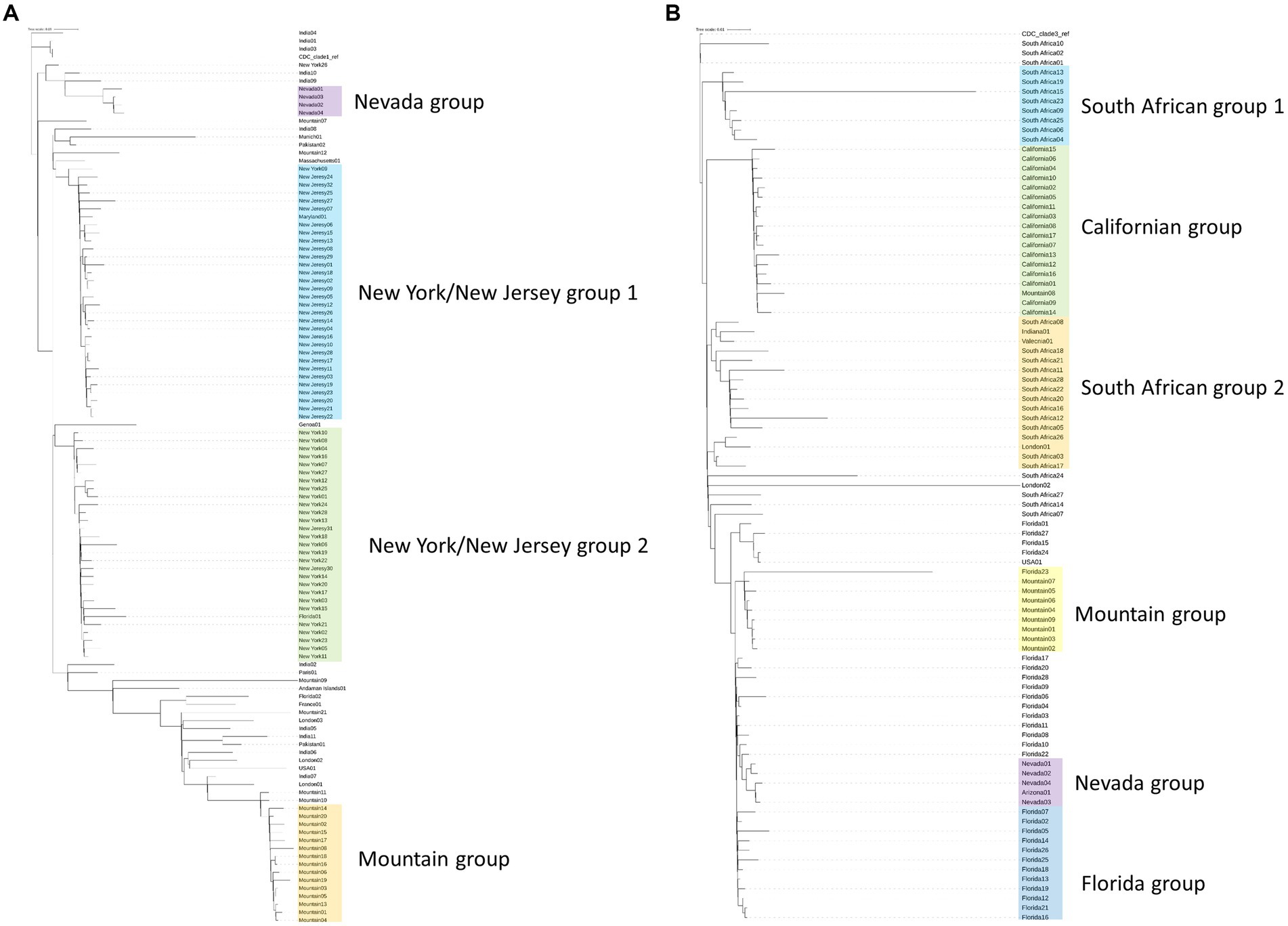
Figure 1. Phylogenetic trees generated by kSNP3 (21) on Candida auris isolates. Isolates are labeled by region from which they were isolated. Samples for each tree are listed with their SRA ID in Supplemental Table 2. Shading on the tree represents highly related branches which clustered by geography. (A) Phylogenetic tree of Clade 1 isolates including 4 isolates from the southern Nevada outbreak (colored in purple). (B) Phylogenetic tree of Clade 3 isolates including 5 isolates from the southern Nevada outbreak (colored in purple).
A similar analysis was performed with clade 3 samples and is shown in Figure 1B. As with clade 1 analysis, it became clear that the Nevada clade 3 samples (highlighted in purple) were genetically distinct from other previously sequenced outbreaks. The index southern Nevada cases for the clade 3 outbreak were SRR19738700 and SRR23109087 which were both collected on 11-02-2021 in the case facility (Supplementary Table S1). Note the one isolate labeled Arizona01 in the shaded purple box was collected in Arizona from a southern Nevadan patient. Epidemiological investigation strongly suggested the patient contracted C. auris in southern Nevada prior to travel to Arizona (data not shown). The information on the case described in the previous sentence was obtained and shared with the Nevada State Public Health Laboratory in collaboration with our public health partners in Utah and Nevada. These data were collected and shared in accordance with IRB protocols. We concluded having a pipeline(s) of rigorous bioinformatic tools capable of handling fungal microbes would be necessary for the public health response to these simultaneous and distinct outbreaks occurring in southern Nevada.
3.3. State of fungal bioinformatic whole-genome sequencing pipelines circa march 2022
After an initial analysis of the outbreak specimens with regard to clade, we sought to further determine the utility of phylogenetic analysis to assist disease control. Upon initiation of sequencing and genomic analysis of the outbreak at the Nevada State Public Health Laboratory, one computational method /pipeline was available for assessments of C. auris (12, 13). Another was completed but unpublished (3). Difficulties with implementation of the former led our group to develop a novel pipeline for identification and phylogenetic analysis of C. auris genomes. This pipeline has been named “TheiaEuk” (14). As we sought to determine the best methods for using sequencing to assist disease control efforts for this outbreak, we sought to compare all three methods in terms of capability and functionality.
3.4. Pipeline comparisons for Candida auris genome assemblies
In testing three methods, we assembled genomes (n = 60) from clade I from patients found infected by C. auris in southern Nevada. Assembly and downstream analyses were completed using each of three workflows: Nullarbor, TheiaEuk and Mycosnp. Upon completion, results from Nullarbor needed no additional analysis. TheiaEuk and Mycosnp required additional step for the generation of SNP matrices and/or phylogenetic trees (Figure 2). Times required for analyses are shown in Table 1. As shown in Table 2, all methods assemble genomes of nearly identical sizes with the average genome lengths of Nullarbor being 12,276,509 bp, TheiaEuk being 12,288,829 bp and MycoSnp being 12,406,106 bp. For assembly, Nullarbor uses Shovill v1.1.0 with SKESA v2.4.0 as the default setting. TheiaEuk uses Shovill v1.1.0 with SKESA v2.4.0 as the default setting. MycoSNP uses a reference guided assembly that produces the same genome length for each sample using method BWA v0.7.17 for read alignment and GATK v4.2.5.0 for variant calling. MycoSNP using reference guided assembly creates a single contig per chromosome where Nullarbor produces an average of 683 contigs from our Clade I samples and TheiaEuk produces an average of 505 contigs from tested clade I samples (Table 2).
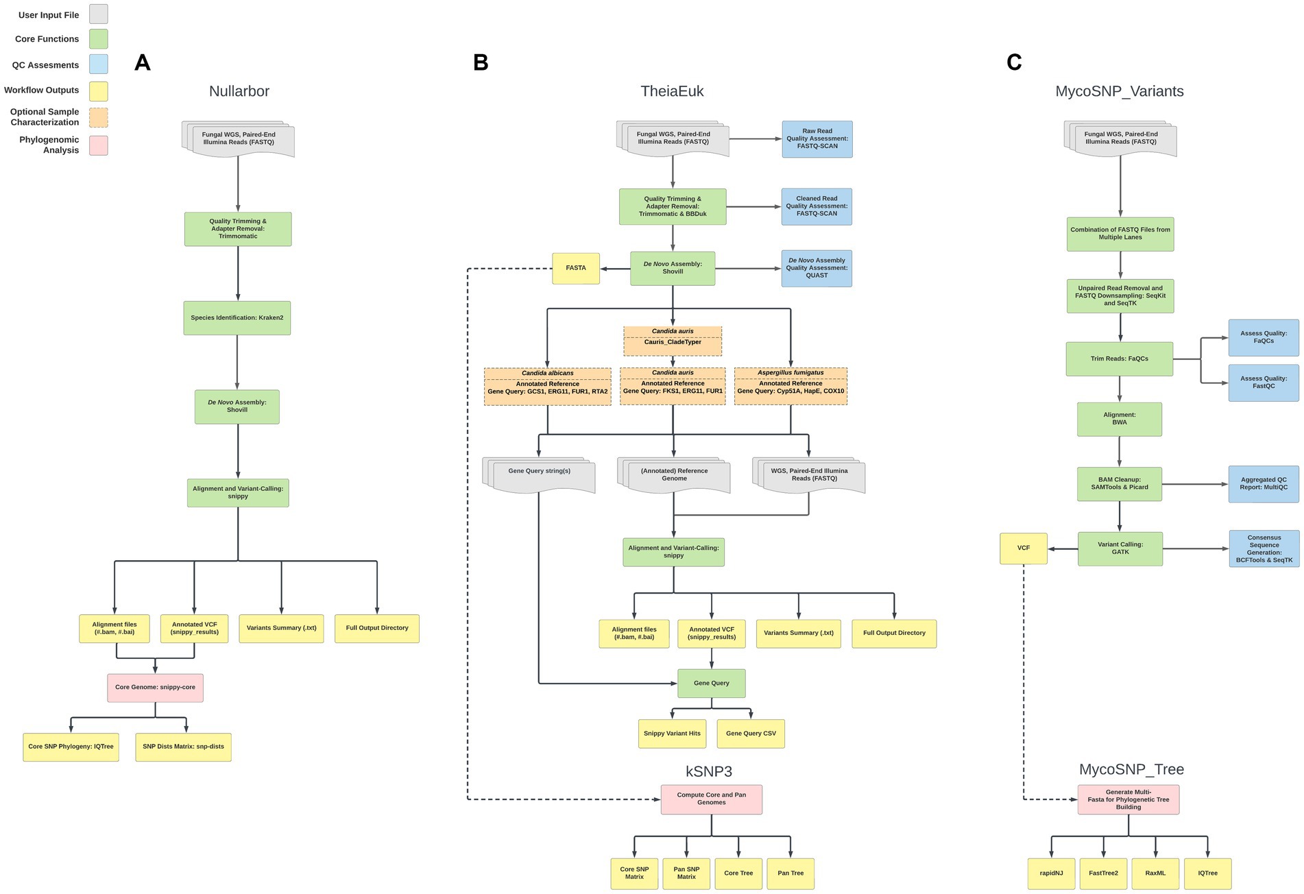
Figure 2. Workflow comparisons of whole-genome sequencing bioinformatic pipelines that analyze C. auris WGS data. The shaded key highlights the major steps performed in analysis for ease of comparison. For details on each workflow see Materials and Methods. (A) Nullarbor (B) TheiaEuk (C) MycoSNP_variants.
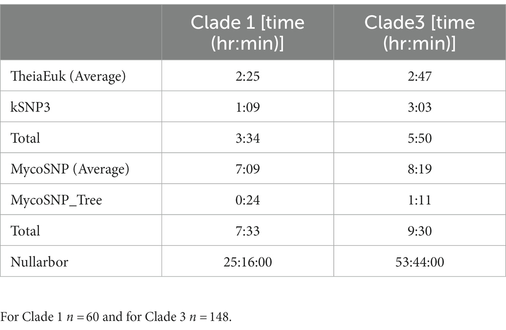
Table 1. Run times for each of the tested WGS pipelines on both the Clade 1 and Clade 3 isolate sets.
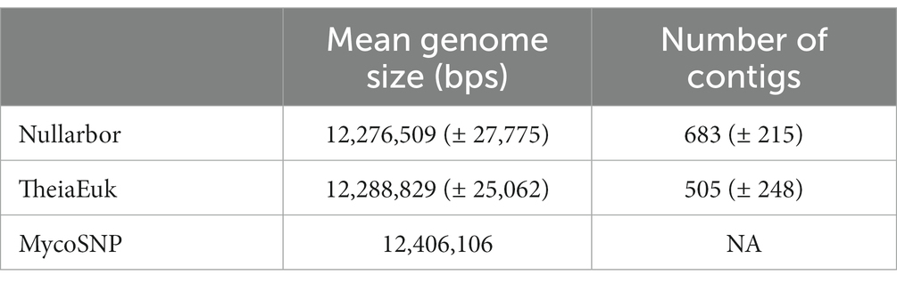
Table 2. For the Clade 1 dataset (n = 60) the comparison of WGS assembly stats (genome length and number of contigs) is reported.
3.5. Benchmarking TheiaEuk, MycoSNP and Nullarbor
Comparison of the three whole-genome sequencing pipelines based on analysis time was performed on the same test set described in the previous section (Table 1). All pipelines were run with the same virtual machines (Materials and Methods). MycoSNP had the fastest report time at 2 h and 5 min, followed by TheiaEuk at 8 h and 10 min with nullarbor requiring 26 h and 12 min.
3.6. Pipeline comparisons for Candida auris SNP matrices
Each method produces SNP matrices which display the calculated number of SNPs between each sample in an analyzed set. We compared the number of SNPs detected by each method compared to the first clade I sample by numerical order based on our internal nomenclature (SRR19664611) to all other samples. We then calculated the absolute differences between each pairwise SNP comparison between two methods. Comparing Nullarbor and TheiaEuk the difference was 1.9 (± 2.1) SNPs with Nullarbor consistently reporting fewer SNPs. Comparing Nullarbor and MycoSNP the difference was 1.9 (± 2.2) SNPs with Nullarbor consistently reported fewer SNPs. Comparison of TheiaEuk and MycoSNP resulted in a difference of 0.57 (± 0.89) SNPs with MycoSNP consistently reporting more SNPs (Table 3).
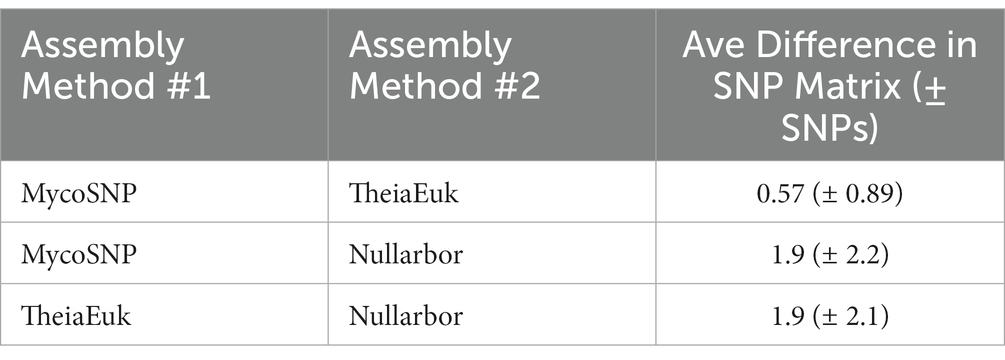
Table 3. Calculated SNP differences between the WGS pipelines for Clade 1 (n = 60).
3.7. Development of a pipeline to distinguish fine grain differences in ongoing outbreaks
Distinguishing genetically related isolates within an outbreak can be challenging for pathogens with low rates of mutation (22). A SNP can be the result not only of biological introduction, but also introduced through sequencing and biocomputational methods. Within the outbreak observed herein, core genome assemblies possess a large number of shared SNPs when compared to the CDC clade references and have relatively smaller number of distinguishing mutations that define subgroups (Figure 3A). For example, all but a single clade 1 isolate share 52 common SNPs (Supplemental data), yet Table 4 shows that the most common clade 1 subclade (Group K) differs by only 3 SNPs from the second most common subgroup (Group B). We propose that the usage of subsets of shared mutations that follow asexual microbial evolution theory would define the most highly related subgroups (Figure 3A). To this end it can be observed that within a clade, most isolates which share a large number of “core” SNPs compared to the CDC reference, show relatedness relevant to epidemiological investigation. Such cases, then with additional SNPs shared, result in cases with a distinct profile of descendancy and thus would be assumed to have the highest level of relatedness (Table 4).
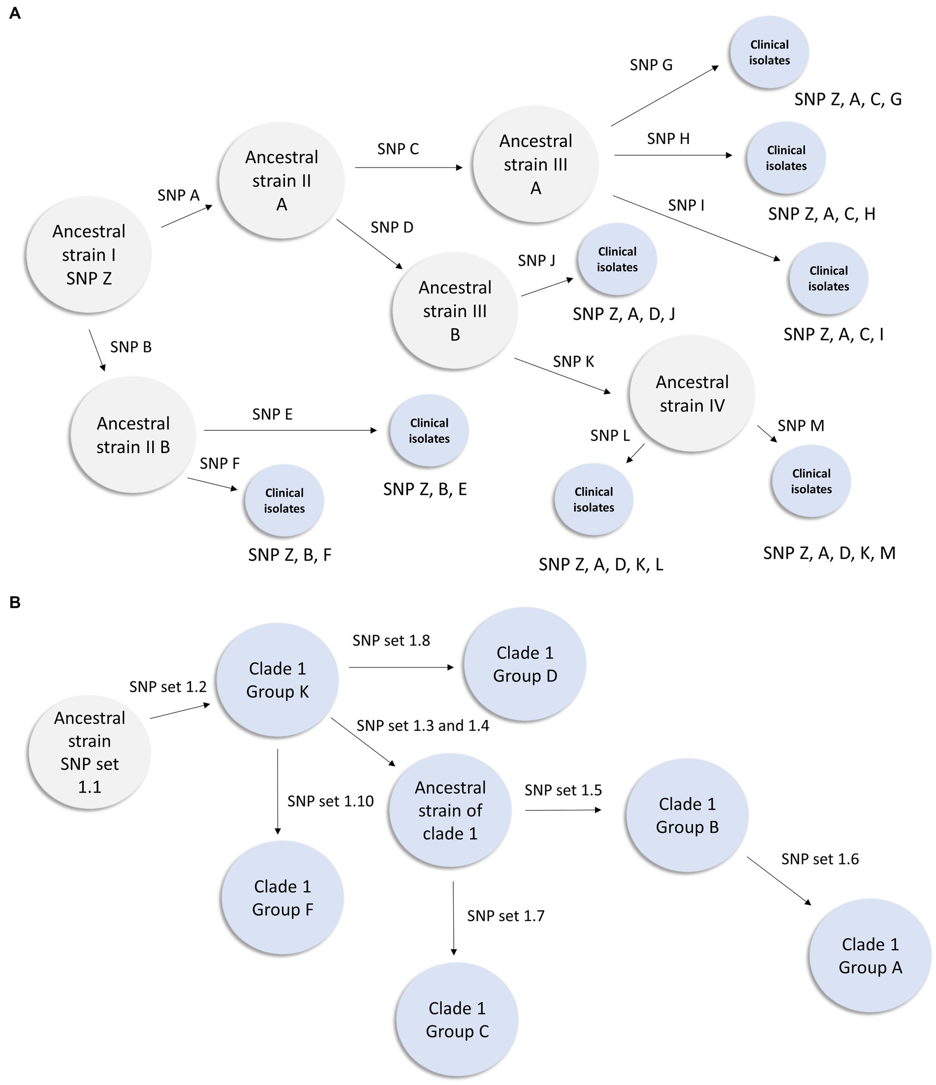
Figure 3. Inferred transmission networks based on shared SNP analysis. (A) Figure illustrates how theoretical Ancestral strain I (with SNP Z) could evolve during an outbreak. When an isolate acquires a novel SNP or SNPs during this hypothetical outbreak, an arrow is displayed and a designation (such as SNP A) is shown above the arrow. Gray circles represent inferred genotypes based on clinical isolates. Blue circles represent clinical isolates and their known genotypic profile. This figure demonstrates how lineage can be inferred from a genetically diverse set of isolates within a known outbreak. (B) Inferred transmission network from the clade 1 southern Nevada outbreak. In this instance we have observed isolates from all intermediates except the most ancestral strain. This representation is derived from the data presented in Table 4.
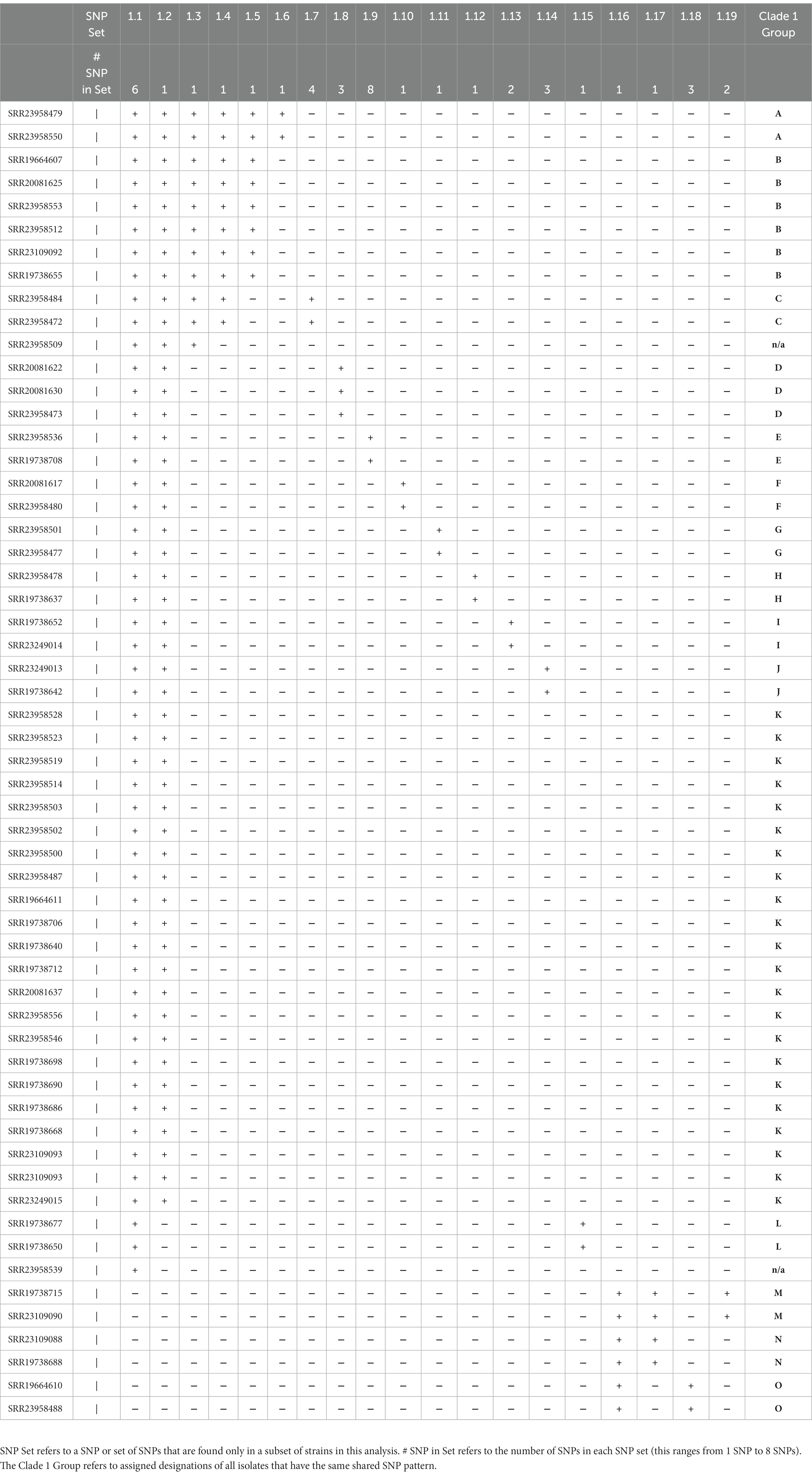
Table 4. Shared SNP analysis of clade 1 isolates.
3.8. Inference of relatedness among Candida auris clade I outbreak samples
In order to utilize whole-genome variation to provide disease control investigators with data regarding the most related sets of isolates, specific SNPs were studied for evidence of inheritance patterns. Clade I (n = 60) isolates were analyzed using kSNP3 using the clade I CDC reference strain [strain B11205] as our reference (21). This analysis generated 208 SNPs whereupon the parent (defined as the reference strain) or variant sequence was detected in all 60 strains. Of these SNPs, 109 were present in only one isolate (aka unique SNPs). Of the 99 SNPs that were shared among two or more Clade I isolates, 54 were present in 57 out of 60 clade I isolates. The remainder of the analysis focused upon these 57 strains which all shared this set of “core NV clade I” SNPs.
Among these 57 strains 19 SNP sets were identified which differentiated this group based on genetics. These SNP sets (Table 4) contained between 1 and 8 SNPs. The presence of the SNP set in an isolate is represented with a “+” in Table Y and its absence is represented with a “-“. We identified 15 groups of isolates that had more than one member and had a unique combination of SNP sets (Table 4)—we designated these as clade 1 Groups A through O. Provision of group designations was to assist disease controllers in Nevada in having a nomenclature to describe cases. It was not an attempt to create a novel general nomenclature field-wide. Lastly, we present a subset of our inferred clade 1 transmission network based on these data in Figure 3B.
3.9. Inference of relatedness among Candida auris clade III outbreak samples
The analysis described above was repeated for the 148 Clade III isolates using the clade III CDC reference strain [strain B11221] as our ancestral outgroup for kSNP3. This analysis generated 401 SNPs where the parent or variant sequence was detected in all 148 strains. Of these SNPs, 280 were present in only one isolate (aka unique SNPs). Of the 121 SNPs that were shared among two or more clade III isolates, 28 were present in 147 out of 148 clade III isolates. Focus was placed upon these 147 strains which all shared this set of “core NV clade III” SNPs.
Among these 147 isolates 42 SNP sets were identified which differentiated this group based on genetics. These SNP sets (Supplementary Table S3) contained between 1 and 5 SNPs. The presence of the SNP set in an isolate is represented with a “+” in Supplementary Table S3 and its absence is represented with a “-“. Groups of isolates (n = 35) were identified that had more than one member and had a unique combination of SNP sets (Supplementary Table S3)—these were designated as Clade 3 Groups A through KK.
3.10. Discovery of new introductions to Candida auris to southern Nevada using shared SNP analysis
The shared SNP analysis described above is performed by the Nevada State Public Health Laboratory on a regular basis since September 2022. During that time, this analysis has identified two novel clade 1 introductions. These novel introductions have a unique set of “core” SNPs that are different from the Southern Nevada “core” SNP signature. To quantify this, we ran kSNP3 with four members of the southern Nevada Clade 1 outbreak with the suspected two novel clade 1 introductions (21). The first novel introduction represented by isolate SRR23137821 has 2,519 SNPs not shared by any of the original clade 1 isolates from this outbreak (Table 5). The second novel introduction represented by isolate SRR23920687 has 87 SNPs not shared by any of the original clade 1 isolates from this outbreak (Table 5). While all three have a small, overlapping set of 10 common SNPs when compared to the CDC clade 1 reference strain, the vast genetic diversity detected by the shared SNP analysis shows that these are new introductions.
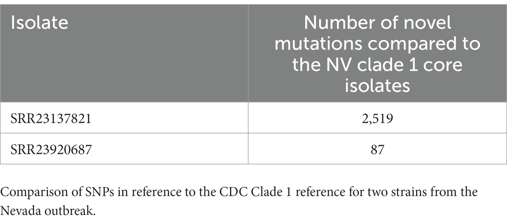
Table 5. Comparison of SNPs in reference to the CDC Clade 1 reference for two strains from the Nevada outbreak.
4. Discussion
Candida auris is among the most challenging of healthcare-associated infections (2, 6–11). It combines the ability to persist environmentally with inherent drug resistance and the ability to cause significant morbidity and mortality. As such, public health must bring every tool at its disposal to bear on this threat. Herein, we assess one possible tool for its ability to assist in combatting C. auris outbreaks: genomics, in response to multiple, complex outbreaks in Nevada, we sought to generate as much genomic intelligence as possible to better understand the spread of the pathogen. The use of genomics to track and to describe pathogens is certainly not novel. However, its application to C. auris outbreaks is relatively new. As a fungal pathogen, C. auris has been shown to have a mutation rate much slower than other, healthcare associated agents (23–26). Slower mutation rates may result in less diversity in outbreak populations, thus limiting the ability to distinguish cases within transmission networks. Confronting this, we assessed the genomic diversity of whole genome sequences from numerous isolates associated with outbreaks of clade I and clade III. The observations that were made led to the use not only of quantification of SNP distances, but also the recognition of the genomic locations of SNPs that were shared among cases. Utilization of “shared” SNPs was found to provide power to the use of whole genomics for studying C. auris during an outbreak. The concept of shared SNPs allowed rational descriptions and delineations of phylogenetic descendancy. The result was the creation of means to more effectively serve disease controllers and epidemiologists in furtherance of their investigations. While this may not make up for a slow mutation rate associated with the pathogen and thus a lower discriminatory capacity, it does create a means to direct investigators to the most related cases in a rational way. The use of shared SNPs has been applied to pathogen genomics in many instances but to our knowledge, this is the first use of the concept to assess case relatedness within a C. auris outbreak (27–29). A previous study applied whole genome sequencing to a small outbreak of C. auris within a hospital (30). Therein it was shown that in fact there was considerable genomic diversity between multiple isolates taken from the same patient, and also taken from different patients who were roomed together and were likely transmission pairs (30).
Sequencing and genomic analysis provided real benefits to disease controllers and epidemiologists who were investigating these outbreaks in Nevada. It became readily possible to distinguish between ongoing transmission within facilities versus novel introductions into facilities on the basis of shared SNP descendancy. This triggered different strategies and tactics on a facility-by-facility basis which were applied based upon phylogenetic information rather than from best guesses. Lastly, we demonstrated the shared SNP analysis detected two novel clade 1 introductions from outside of southern Nevada. This early detection allowed our public health responders to attempt to contain these new introductions and prevent their establishment in southern Nevada. This was critical because the greater the number of overlapping and ongoing outbreaks a region is experiencing, the more complicated the role of disease control investigators and epidemiologists becomes.
Because multiple tools exist to assess whole genome sequencing of C. auris, the work herein rigorously compared and contrasted three such pipelines. Each performed reliably, though specific differences in genome sizes and time-to-answer were found among the three. Notably, pairwise comparisons of SNP distances between fixed sets of isolates across different pipelines revealed that different pipelines will provide different results. This finding indicates that choice and validation of pipelines is not just a matter of formality. As microbiology and bioinformatics continue to merge, it is critical that when new pipelines are constructed that they are validated against existing tools. An ever-increasing number of pipelines does not serve the field of medicine or public health if the pipelines are not clearly assessed from a quality assurance perspective. Much work lies ahead for standards, consultation and accreditation agencies associated with diagnostic science, as emerging bioinformatic tools require rigorous assessment. Even when they are not used as diagnostic tools, their use as aids to epidemiology and disease control will trigger enormous shifts in work-time and resources, which are often limited in the public health realm.
Comprehensive and rapid sequencing of cases as described herein has just begun to impact the public health intervention aspect of the outbreak. Affected sites with continuous transmission have sought additional interventions, including novel means of chemical disinfection and the use of (PCR) screening tests for incoming patients and employees. Unlike the use of sequencing for other hospital acquired infections (e.g., CRE) the comprehensive use of sequencing as shown herein has laid the groundwork for training and familiarization with the use of genomic sequence intelligence. The intense sequencing has additionally led to a highly sophisticated and detailed description of the outbreaks which has gained the attention of elected public servants in the state who have sought additional resources for approaching the outbreak. Additionally, sequencing and analysis have also provided gravitas to the successful actualization of C. auris as a reportable entity in Nevada.
This study possesses unique strengths. It included a large number of isolates, collected prospectively in the course of major outbreaks. The study included analysis of two simultaneous, genomically distinct outbreaks (clade I and clade III), which on the surface resembled a singular outbreak. Additionally, the study compares different tools/pipelines rather than merely showing the construction and functionality of one alone.
This study possesses weaknesses of note. While a high number of isolates associated with a large outbreak were assessed, there are significant gaps in the information that matches epidemiologic data to sequencing data. It is difficult to say with certainty that genomic relatedness ascertained herein is guaranteed to be meaningful from the disease control perspective, without more data. Additionally, not all laboratories or public health jurisdictions could necessarily repeat what was performed herein, as massive resources were necessarily harnessed to generate the granularity of intelligence described.
Data availability statement
The datasets presented in this study can be found in online repositories. The names of the repository/repositories and accession number(s) can be found in the article/Supplementary material.
Author contributions
AG generated data, performed analysis, wrote sections of the manuscript, and helped in revisions. FA and MS created bioinformatic pipelines, performed analysis, wrote sections of the manuscript, and helped in revisions. LM generated a figure and helped in revisions. DS, CH, PD, ES, and CN generated data for the paper and helped in revisions. KL and JS supported and funded the creation of bioinformatic pipelines and helped in revisions. SVH provided administrative supervision of the work. MP conceived of and authored portions of the manuscript and provided review. DH conceived of the projects, generated data, performed analysis, wrote the manuscript, and helped in revisions. All authors contributed to the article and approved the submitted version.
Funding
This research was funded by Centers for Disease Control and Prevention (CDC) Surveillance for Healthcare-Associated and Resistant Pathogens (SHARP); and CDC Epidemiology and Laboratory Capacity (ELC) funding.
Acknowledgments
NB also thanks Celina Morrison and Laurel Stringer for help with data collection. We would like to acknowledge Kimisha Causey for disease control investigation support in the course of this work.
Conflict of interest
Authors FA, MS, KL and JS were employed by Theiagen Consulting LLC.
The remaining authors declare that the research was conducted in the absence of any commercial or financial relationships that could be construed as a potential conflict of interest.
Publisher’s note
All claims expressed in this article are solely those of the authors and do not necessarily represent those of their affiliated organizations, or those of the publisher, the editors and the reviewers. Any product that may be evaluated in this article, or claim that may be made by its manufacturer, is not guaranteed or endorsed by the publisher.
Supplementary material
The Supplementary material for this article can be found online at: https://www.frontiersin.org/articles/10.3389/fpubh.2023.1198189/full#supplementary-material
Footnotes
1. ^https://benlangmead.github.io/aws-indexes/k2
2. ^https://github.com/CDCgov/mycosnp-nf
3. ^https://github.com/garviz/MUMmer/blob/master/nucmer
4. ^https://github.com/lh3/bwa
5. ^https://github.com/shenwei356/seqkit
6. ^https://github.com/lh3/seqtk
7. ^https://github.com/LANL-Bioinformatics/FaQCs
8. ^https://github.com/samtools/
9. ^https://github.com/broadinstitute/picard
10. ^https://github.com/s-andrews/FastQC
11. ^https://github.com/ewels/MultiQC
12. ^https://github.com/broadinstitute/gatk
13. ^https://gatk.broadinstitute.org/hc/en-us/articles/360035890471-Hard-filtering-germline-short-variants
14. ^https://github.com/samtools/bcftools/releases
15. ^https://github.com/johnlees/rapidnj
16. ^https://github.com/citiususc/veryfasttree
References
1. Satoh, K, Makimura, K, Hasumi, Y, Nishiyama, Y, Uchida, K, and Yamaguchi, H. Candida auris sp. nov., a novel ascomycetous yeast isolated from the external ear canal of an inpatient in a Japanese hospital. Microbiol Immunol. (2009) 53:41–4. doi: 10.1111/j.1348-0421.2008.00083.x
2. Lockhart, SR, Etienne, KA, Vallabhaneni, S, Farooqi, J, Chowdhary, A, Govender, NP, et al. Simultaneous emergence of multidrug-re-sistant Candida auris on 3 continents confirmed by whole-genome sequencing and epidemiological analyses. Clin Infect Dis. (2017) 64:134–40. doi: 10.1093/cid/ciw691
3. Bagal, UR, Phan, J, Welsh, RM, Misas, E, Wagner, D, Gade, L, et al. MycoSNP: a portable workflow for performing whole-genome sequencing analysis of Candida auris. Methods Mol Biol. (2022) 2517:215–28. doi: 10.1007/978-1-0716-2417-3_17
4. CDC. Tracking Candida auris (2022) Available at: https://www.cdc.gov/fungal/candida-auris/tracking-c-auris.html
5. Chow, NA, De Groot, T, Badali, H, Abastabar, M, Chiller, TM, and Meis, JF. Potential fifth clade of Candida auris, Iran, 2018. Emerg Infect Dis. (2019) 25:1780–1. doi: 10.3201/eid2509.190686
6. Kenters, N, Kiernan, M, Chowdhary, A, Denning, DW, Pemán, J, Saris, K, et al. Control of Candida auris in healthcare institutions: outcome of an International Society for Antimicrobial Chemotherapy expert meeting. Int J Antimicrob Agents. (2019) 54:400–6. doi: 10.1016/j.ijantimicag.2019.08.013
7. Ong, CW, Chen, SC, Clark, JE, Halliday, CL, Kidd, SE, Marriott, DJ, et al. Australian and New Zealand mycoses interest group (ANZMIG), healthcare infection control special interest group (HICSIG); both of the Australasian Society for Infectious Diseases (ASID). Diagnosis, management and prevention of Candida auris in hospitals: position statement of the Australasian Society for Infectious Diseases. Intern Med J. (2019) 49:1229–43. doi: 10.1111/imj.14612
8. PHE. Candida auris identified in England. Public Health England. (2016) Available at: https://www.gov.uk/government/publications/candida-auris-emergence-in-england/candida-auris-identified-in-england
9. ECDC. Candida auris in healthcare settings – Europe. European Centre for Disease. Prev Control. (2016) Available at: https://www.ecdc.europa.eu/sites/default/files/media/en/publications/Publications/Candida-in-healthcare-settings_19-Dec-2016.pdf
10. CDC. Candida auris clinical update. Centers for Disease Control and Prevention, National Center for Emerging and Zoonotic Infectious Diseases (NCEZID), division of foodborne, waterborne, and environmental diseases (DFWED) (2016). Available at: https://www.cdc.gov/fungal/candida-auris/c-auris-alert-09-17.html?CDC_AA_refVal=https%3A%2F%2Fwww.cdc.gov%2Ffungal%2Fdiseases%2Fcandidiasis%2Fc-auris-alert-09-17.html
11. Cortegiani, A, Misseri, G, Fasciana, T, Giammanco, A, Giarratano, A, and Chowdhary, A. Epidemiology, clinical characteristics, resistance, and treatment of infections by Candida auris. J Intensive Care. (2018) 6:1–13. doi: 10.1186/s40560-018-0342-4
12. Biswas, C, Wang, Q, van Hal, SJ, Eyre, DW, Hudson, B, Halliday, CL, et al. Genetic heterogeneity of Australian Candida auris isolates: insights from a nonoutbreak setting using whole-genome sequencing. Open Forum Infect Dis. (2020) 7:ofaa158,. doi: 10.1093/ofid/ofaa158
13. Available at: https://github.com/tseemann/nullarbor
14. Ambrosio, F, Scribner, MR, Wright, SM, Otieno, J, Gorzalski, A, Siao, DD, et al. TheiaEuk: a species-agnostic bioinformatics workflow for fungal genomic characterization. Front Public Health. (2023)
15. Bolger, AM, Lohse, M, and Usadel, B. Trimmomatic: a flexible trimmer for Illumina sequence data. Bioinformatics. (2014) 30:2114–20. doi: 10.1093/bioinformatics/btu170
16. Wood, DE, Lu, J, and Langmead, B. Improved metagenomic analysis with kraken 2. Genome Biol. (2019) 20:257. doi: 10.1186/s13059-019-1891-0
17. Torsten, S. Prokka: rapid prokaryotic genome annotation. Bioinformatics. (2014) 30:2068–9. doi: 10.1093/bioinformatics/btu153
18. Available at: https://github.com/tseemann/snippy
19. Ewels, P, Peltzer, A, Fillinger, S, Patel, H, Alneberg, J, Wilm, A, et al. The nf-core framework for community-curated bioinformatics pipelines. Nat Biotechnol. (2020) 38:276–8. doi: 10.1038/s41587-020-0439-x
20. Lumpe, J, Gumbleton, L, Gorzalski, A, Libuit, K, Varghese, V, Lloyd, T, et al. GAMBIT (genomic approximation method for bacterial identification and tracking): a methodology to rapidly leverage whole genome sequencing of bacterial isolates for clinical identification. PLoS One. (2023) 18:e0277575. doi: 10.1371/journal.pone.0277575
21. Gardner, SN, Slezak, T, and Hall, BG. kSNP3.0: SNP detection and phylogenetic analysis of genomes without genome alignment or reference genome. Bioinformatics. (2015) 31:2877–8. doi: 10.1093/bioinformatics/btv271
22. Carroll, LM, Wiedmann, M, Mukherjee, M, Nicholas, DC, Mingle, LA, Dumas, NB, et al. Characterization of emetic and diarrheal Bacillus cereus strains from a 2016 foodborne outbreak using whole-genome sequencing: addressing the microbiological, epidemiological, and Bioinformatic challenges. Front Microbiol. (2019) 10:144. doi: 10.3389/fmicb.2019.00144
23. Edwards, HM, and Rhodes, J. Accounting for the biological complexity of pathogenic Fungi in phylogenetic dating. J Fungi (Basel). (2021) 7:661. doi: 10.3390/jof7080661
24. Ene, IV, Farrer, R, Hirakawa, M, Agwamba, K, Cuomo, CA, and Bennett, RJ. Global analysis of mutations driving microevolution of a heterozygous diploid fungal pathogen. Proc Natl Acad Sci U S A. (2018) 115:E8688–97. doi: 10.1073/pnas.1806002115
25. Stoesser, N, Giess, A, Batty, EM, Sheppard, AE, Walker, AS, Wilson, DJ, et al. Genome sequencing of an extended series of NDM-producing Klebsiella pneumoniae isolates from neonatal infections in a Nepali hospital characterizes the extent of community-versus hospital-associated transmission in an endemic setting. Antimicrob Agents Chemother. (2014) 58:7347–57. doi: 10.1128/AAC.03900-14
26. Zhu, YO, Siegal, M, Hall, D, and Petrov, DA. Precise estimates of mutation rate and spectrum in yeast. Proc Natl Acad Sci U S A. (2014) 111:E2310–8. doi: 10.1073/pnas.1323011111
27. Tan, KK, Tan, YC, Chang, LY, Lee, KW, Nore, SS, Yee, WY, et al. Full genome SNP-based phylogenetic analysis reveals the origin and global spread of Brucella melitensis. BMC Genomics. (2015) 16:93. doi: 10.1186/s12864-015-1294-x
28. Engelthaler, DM, Chiller, T, Schupp, JA, Colvin, J, Beckstrom-Sternberg, SM, Driebe, EM, et al. Next-generation sequencing of Coccidioides immitis isolated during cluster investigation. Emerg Infect Dis. (2011) 17:227–32. doi: 10.3201/eid1702.100620
29. Reis, AC, Salvador, LCM, Robbe-Austerman, S, Tenreiro, R, Botelho, A, Albuquerque, T, et al. Whole genome sequencing refines knowledge on the population structure of Mycobacterium bovis from a multi-host tuberculosis system. Microorganisms. (2021) 9:1585. doi: 10.3390/microorganisms9081585
Keywords: Candida auris, epidemiology, whole-genome sequencing, bioinformatics, emerging pathogens
Citation: Gorzalski A, Ambrosio FJ III, Massic L, Scribner MR, Siao DD, Hua C, Dykema P, Schneider E, Njoku C, Libuit K, Sevinsky JR, Van Hooser S, Pandori M and Hess D (2023) The use of whole-genome sequencing and development of bioinformatics to monitor overlapping outbreaks of Candida auris in southern Nevada. Front. Public Health. 11:1198189. doi: 10.3389/fpubh.2023.1198189
Edited by:
Shangxin Yang, University of California, Los Angeles, United StatesReviewed by:
Annabelle De St. Maurice, Los Angeles County Department of Public Health, United StatesKin-Ming (Clement) Tsui, University of British Columbia, Canada
Erin Young, Utah Department of Health and Human Services, United States
Copyright © 2023 Gorzalski, Ambrosio, Massic, Scribner, Siao, Hua, Dykema, Schneider, Njoku, Libuit, Sevinsky, Van Hooser, Pandori and Hess. This is an open-access article distributed under the terms of the Creative Commons Attribution License (CC BY). The use, distribution or reproduction in other forums is permitted, provided the original author(s) and the copyright owner(s) are credited and that the original publication in this journal is cited, in accordance with accepted academic practice. No use, distribution or reproduction is permitted which does not comply with these terms.
*Correspondence: Mark Pandori, bXBhbmRvcmlAbWVkLnVuci5lZHU=; David Hess, ZGhlc3NAdW5yLmVkdQ==