 Frédéric Destaillats
 Manuel Oliveira
 Walter Rakitsky
 Leon Parker
- Checkerspot, Inc., Alameda, CA, United States
Nervonic acid (24:1 n-9, NA) is a monounsaturated very long-chain fatty acid (VLCFA) that plays a fundamental role in brain development, particularly in the biosynthesis of sphingolipids and myelin sheaths. NA is present in minute amounts in human milk and despite its importance in neuronal function and cognitive development, there is currently no ingredient available for the fortification of infant nutrition products. However, recent advances in biotechnology have made it feasible to produce high NA containing oil through fermentation, presenting a significant opportunity to address this nutritional gap. This review explores the potential of NA fortification in infant nutrition products and its impact on neurodevelopment, with a specific focus on two populations: premature infants, who are at higher risk of neurodevelopmental impairments due to incomplete in utero myelination, and healthy term infants, who may experience enhanced cognitive development with improved dietary NA intake when consuming infant formula. By critically examining the scientific basis for NA supplementation, as well as the practical challenges and regulatory considerations associated with its implementation, this review aims at providing a forward-looking perspective on how this emerging ingredient could enhance infant nutrition and improve health outcomes.
Introduction
The first year of life is a critical period of rapid brain growth and neural development, during which fundamental processes such as myelination, synaptogenesis, and neurotransmitter synthesis occur at an accelerated rate, shaping cognitive and sensory functions (1). Proper brain maturation and function during one's lifespan requires a precise balance of nutrients to support essential physiological mechanisms (2–7, 96). Studies have shown that specific nutrients, including phospholipids and sphingomyelin from milk fat globule membrane (MFGM), enhance myelination and cognitive development, underscoring the importance of dietary exposure in early infancy (1, 8). Additionally, brain iron levels play a vital role in key neurodevelopmental processes, as iron is essential for neurotransmitter synthesis and myelination, with research showing a developmental coupling between brain iron and neural activity during the first 150 days of life (9).
Beyond structural development, functional brain connectivity in early infancy has a lasting impact on cognitive abilities in later childhood (10, 11). Research has found that early growth trajectories influence the development of functional connectivity, which is associated with cognitive flexibility at preschool age, highlighting how undernutrition or adverse early environments can negatively impact neural network maturation (12). Furthermore, language acquisition and sensory processing abilities undergo significant refinement in this period, as evidenced by studies demonstrating the emergence of cortical phonetic feature encoding in infants between 4 and 11 months of age, providing neurophysiological proof that early phonetic categorization shapes future language development (13). Similarly, thalamocortical circuit development, which governs sensory integration and higher cognitive functions, follows distinct age-dependent trajectories in infancy, reinforcing the idea of early-life neuroplasticity as a determinant of long-term brain function (14, 15). Collectively, these findings emphasize that the first year of life represents a critical window for neural development, where nutritional, environmental, and genetic factors interact to shape lifelong cognitive and behavioral outcomes.
Lipids are particularly vital during this stage, as they constitute nearly 60% of the brain's dry weight and play fundamental roles in neuronal membrane integrity, fluidity, and function (16, 17). Among these, VLCFAs are of special interest due to their involvement in the synthesis of sphingolipids, which are key structural components of neural membranes and myelin sheaths (18). Myelination, the process of forming the protective sheath around neurons, is essential for efficient brain signaling and cognitive development. One of the most important VLCFA in this context is nervonic acid (NA; 24:1 n-9), a unique fatty acid predominantly found in sphingomyelin. The presence of sphingolipids in human breast milk plays a crucial role in early-life brain development, as they provide essential building blocks for membrane structure and neurodevelopmental functions (19, 20). Studies have shown that infants fed with MFGM containing infant formula, a rich source of phospholipids and sphingomyelin, exhibit enhanced brain myelination and improved cognitive abilities, particularly in non-verbal and fine motor domains (1, 8).
In this review, we explore the potential of NA fortification in infant nutrition products and its impact on neurodevelopment, with a particular focus on two key populations: premature infants, who are at higher risk of neurodevelopmental impairments due to incomplete in utero myelination, and healthy term infants, who may experience enhanced cognitive development with improved dietary NA intake. Additionally, we review the levels of NA in human milk and propose appropriate fortification levels for infant nutrition products to better align with physiological needs and support optimal neurodevelopment. Furthermore, we examine the natural sources of NA and highlight promising research efforts aimed at developing new dietary sources through fermentation of genetic engineered microbes.
The biochemistry and physiological role of nervonic acid in early life
During pregnancy, the placenta plays a critical role in selectively transporting VLCFA such as long-chain polyunsaturated fatty acids (LC-PUFAs), particularly docosahexaenoic acid (DHA) and arachidonic acid (ARA), to the fetus to support neurodevelopment. This selective transfer is facilitated by specialized transport proteins, including plasma membrane fatty acid-binding proteins (p-FABPpm) and fatty acid transport proteins (FATPs), which mediate the uptake of DHA and ARA from maternal circulation (21). Additionally, the major facilitator superfamily domain-containing 2a (MFSD2a) transporter facilitates the transfer of these fatty acids esterified to lysophosphatidylcholine, rather than as free fatty acids (22). While these mechanisms are well-documented for DHA and ARA, current scientific literature does not provide strong evidence that the placenta prioritizes NA and further research is needed to elucidate the placental transport mechanisms of NA and its potential role in fetal neurodevelopment. The study by Bettger and co-workers demonstrated that NA from the maternal diet is efficiently transferred to milk during lactation in a rodent model (23). Dietary supplementation with NA-rich oils led to increased levels of NA in the sphingomyelin fraction of milk, which was subsequently absorbed by suckling rat pups. This transfer resulted in changes in the sphingomyelin composition of the heart and liver in the offsprings, indicating that dietary NA is bioavailable through milk and can be incorporated into developing tissues (23).
During fetal development and infancy, NA supports the rapid expansion of neural networks, the formation of myelin, and the structural integrity of cellular membranes, processes critical for cognitive and motor function maturation (24). NA biosynthesis is particularly active in the liver and brain during early life (see Figure 1), driven by the elongation of oleic (18:1 n-9), gondoic (20:1 n-9) and erucic (22:1 n-9) acids (25). Studies indicate that NA supplementation can enhance myelin synthesis in oligodendrocytes, promoting nerve maturation (26). Once produced, NA is incorporated into sphingomyelin and gangliosides (Figure 2) that are major lipid constituent of the myelin sheath that insulates axons, enabling rapid and efficient nerve impulse conduction (24). NA facilitates proper signal transduction and cell recognition, contributing to optimal neural connectivity and long-term neurodevelopmental outcomes (24). Unlike shorter-chain fatty acids that primarily serve as energy substrates, NA is integral component of cellular membranes, particularly in the nervous system, where they contribute to membrane fluidity, stability, and signaling (24, 27).
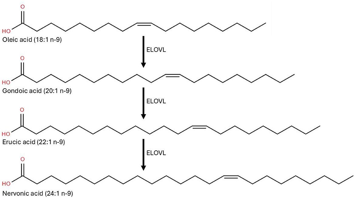
Figure 1. Schematic representation of the elongation of oleic (18:1 n-9) to nervonic (24:1 n-9) by the elongation of very long-chain fatty acid enzymes system [ELOVL; adapted from Garcia Corrales et al. (25)].
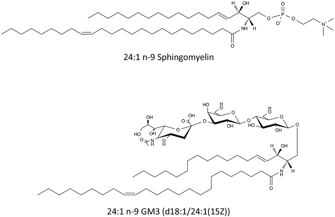
Figure 2. Schematic representation of nervonic acid containing sphingomyelin [N-(15Z-tetracosenoyl)-sphing-4-enine-1-phosphocholine] and ganglioside GM3 [NeuAcα2-3Galβ1-4Glcβ-Cer(d18:1/24:1(15Z))].
The critical role of NA in neurodevelopment is underscored by its association with disorders characterized by defective myelination. Inherited peroxisomal disorders, such as Zellweger syndrome and X-linked adrenoleukodystrophy (X-ALD), involve disruptions in VLCFA metabolism, leading to the accumulation of toxic saturated VLCFA and deficits in myelination (24). These conditions often manifest with severe neurodevelopmental impairments, progressive demyelination, and loss of motor and cognitive function, highlighting the indispensable role of VLCFA like NA in maintaining neuronal structure and function (24).
Evolution of the concentration of nervonic acid in human milk along the lactation stages
In the present review, we evaluate the concentration of NA in human breast milk all along different lactation stages, infant populations (term vs. preterm), and geographic regions based on a comprehensive literature search which has been compiled in Table 1. It is important to note that only a fraction of studies reporting the fatty acid profile of human milk provide data on NA concentrations. The reason for this omission is not entirely clear, especially considering that NA, when analyzed as a methylester derivative, elutes in the same gas-chromatographic region as DHA, a fatty acid that has been reported in >50 studies over the past 30 years. Nevertheless, we were able to gather data from 24 studies, collectively representing 2,404 human milk samples collected across 15 countries (see Table 1). Across all studies, the concentration of NA is highest in colostrum (average: 0.39% of total fatty acids), followed by transitional milk (0.18%) and mature milk (0.11%). The highest NA content in colostrum (0.72%) was observed in samples from Turkey (28), while the lowest (0.19%) was found in samples from Sudan (29).
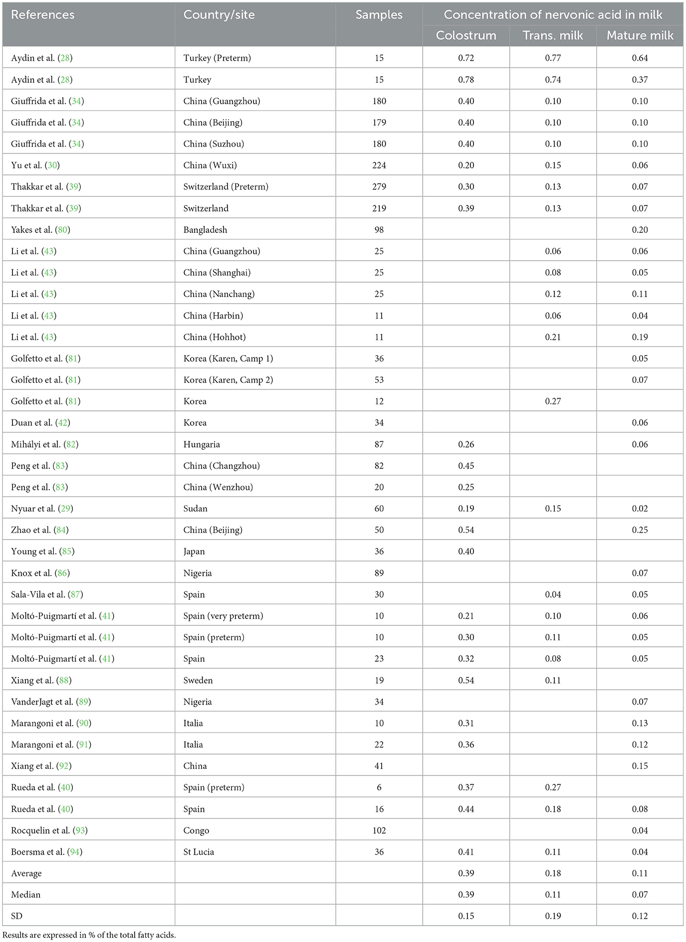
Table 1. Level of nervonic acid in colostrum, transitional milk (trans. Milk) and mature milk reported in 24 published studies, collectively representing 2,404 human milk samples collected across 15 countries.
To further illustrate the decline of NA levels across lactation stages, a model was constructed based on the data from published longitudinal study (30). Figure 3 presents a modeling of the evolution of the NA concentration, expressed in mg of NA per g of fat in human milk, over the first month of lactation (adapted from 28). The model confirms a consistent decline in NA concentration over the first 30 days of lactation (Figure 3). The red curve represents the modeled mean trend, while the black curves indicate confidence intervals (+/- 1 standard deviation, Figure 3). Similar observations has been reported in a collection of studies performed in Hungary (31).
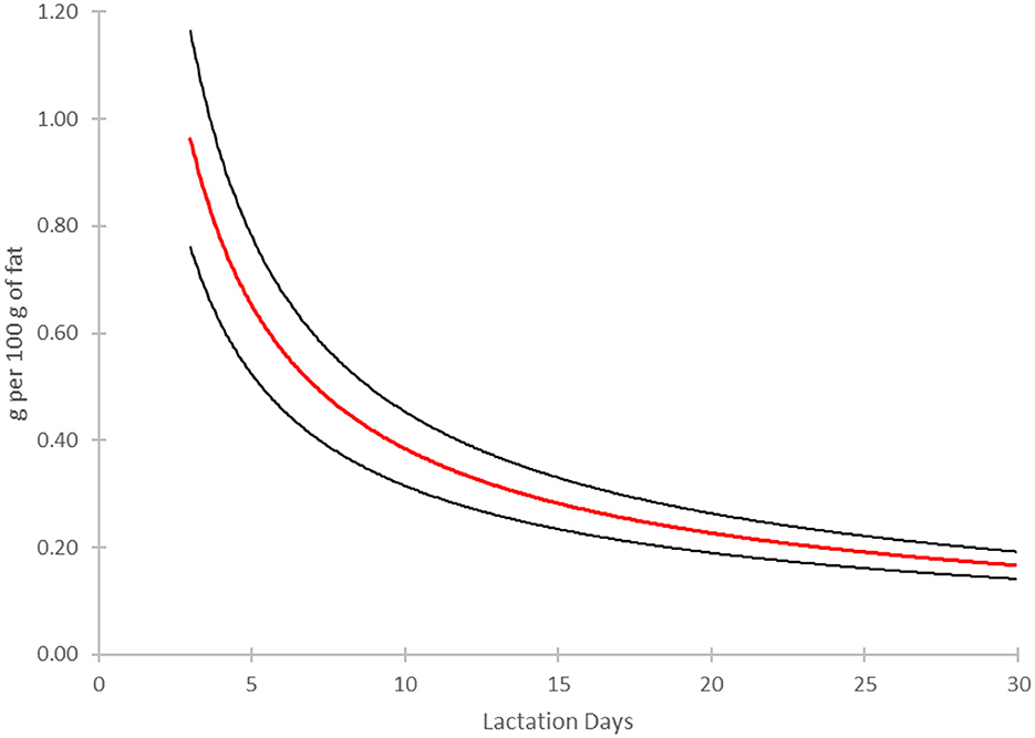
Figure 3. Longitudinal evolution of the concentration of nervonic acid (24:1 n-9, NA) in human milk over the course of the first month of lactation. The model has been built based on the original data (30). The red line represents a power regression (y = 2.2229·x−0.762) fitted to 12 data points describing the concentration of NA (g per 100 g of fat) as a function of lactation days. The regression yielded a high coefficient of determination (R2 = 0.996), indicating an excellent fit. Black lines indicate the mean ± 1 standard deviation of the fitted values.
The decline of NA levels in breast milk observed over time (Figure 3) likely reflects the progressive maturation of the metabolic pathways involved in its de novo synthesis in growing infants, paralleling other developmental shifts in nutrient availability and metabolism. As a key component of myelin, NA plays a crucial role in white matter development during early brain growth (27). At birth, infants rely on maternal milk as a primary source of fatty acids, including NA, to support rapid neural development. However, as enzymatic systems involved in fatty acid elongation and desaturation mature, endogenous synthesis of NA becomes increasingly efficient. This trend mirrors the developmental regulation of enzymes such as very long-chain fatty acid enzymes system type 4 (ELOVL4), which facilitates the elongation of VLCFA, peaking around birth and declining as the brain reaches a steady metabolic state (32). The shift in NA synthesis is analogous to the decline in breast milk protein content over time, which coincides with the slowing of postnatal growth velocity (33). Early in life, higher protein levels in breast milk accommodate the rapid growth demands of the neonate, but as growth rate decreases, so does the protein concentration in milk (33). Similarly, the synthesis of DHA, another critical fatty acid for neural development, follows a comparable trajectory (34), increasing as infants mature and their endogenous enzymatic pathways for fatty acid elongation and desaturation become more active. The competition between substrates for desaturases and elongases, including those shared between NA and omega-3/-6 fatty acid pathways, further suggests that dietary lipid availability influences the timing and efficiency of NA synthesis (35). Additionally, the transition from dependence on breast milk-derived NA to endogenous production aligns with broader metabolic maturation, including improvements in lipid digestion and assimilation mechanisms, such as increased bile salt availability and pancreatic lipase activity (36–38).
A comparison of milk secreted from mother who delivered term and preterm infants revealed no significant differences in NA concentrations at various lactation stages. Thakkar and co-workers reported NA levels of 0.30% in colostrum, 0.13% in transitional milk, and 0.07% in mature milk for preterm infants (39), which closely matched the values observed in term infants (0.39%, 0.13%, and 0.07%, respectively). Similarly, Rueda and co-workers found slightly higher transitional milk NA levels in preterm infants (0.27%) compared to term infants (0.18%), but the overall trend remained consistent, indicating that preterm birth does not significantly impact NA concentrations (40). Same observations have been made in studies conducted in Spain (41), Poland (95), and Turkey (28). In conclusion, NA levels are highest in colostrum and gradually decrease as lactation progresses, with no significant differences observed between term and preterm infants. While regional variations exist, they do not appear to affect the general trend. These findings underscore the importance of early NA provision for neonatal neurodevelopment and highlight the potential for NA supplementation in infant nutrition products.
In contrast, the concentration of NA in infant formula (42, 43) is substantially lower than in human milk, as illustrated in Figure 3. Colostrum exhibits the highest NA content (0.39% of total fatty acids), followed by transitional milk (0.18%) and mature milk (0.11%), while infant formula contains only 0.01% (Figure 4). This considerable difference suggests that formula-fed infants receive markedly lower NA levels than their breastfed counterparts. Given NA's role in neural membrane formation and myelination, its limited presence in infant formula raises concerns regarding potential neurodevelopmental implications. These findings highlight the need for further research into NA fortification in infant nutrition products to better align with the composition of human milk.
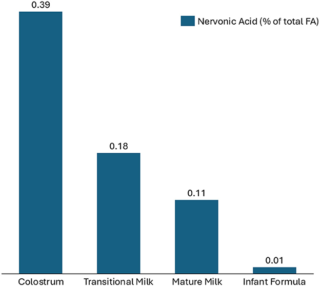
Figure 4. Comparison of the level of nervonic acid (24:1 n-9) in colostrum, transitional, mature human milk (data reported in Table 1) and in infant formula (42, 43). Data are expressed in % of total fatty acids.
Potential benefits of nervonic acid fortification for premature infants
Premature birth, defined as delivery before 37 weeks of gestation, is associated with an increased risk of neurodevelopmental disorders, primarily due to disrupted brain maturation during the third trimester, a critical period for myelination and white matter development (44, 45). During this stage, oligodendrocyte proliferation, differentiation, and myelin deposition occur rapidly, facilitating efficient neural conductivity and cognitive development (46, 47). However, preterm infants miss the crucial placental transfer of DHA and ARA, which play fundamental roles in neuronal membrane integrity, synapse formation, and neural signaling (27, 48).
Clinical studies have demonstrated that low levels of sphingomyelin and VLCFA in preterm infants are associated with cognitive and motor impairments, underscoring the importance of these lipids for early brain development (27, 49). Sphingomyelin, a key component of the myelin sheath, relies on the availability of VLCFA such as NA for its synthesis (27, 50). Insufficient VLCFA supply in preterm infants has been linked to disruptions in myelin formation and white matter injury, increasing the risk of neurodevelopmental conditions, including cerebral palsy, attention-deficit/hyperactivity disorder (ADHD), and global developmental delays (46, 47).
Given these concerns, NA supplementation in preterm infants represents a promising strategy to support the postnatal myelination process. Adequate NA intake may enhance oligodendrocyte function, promoting myelin production and improving neural conductivity, potentially mitigating the long-term neurodevelopmental consequences of premature birth. Current best practices in neonatal intensive care units (NICUs) advocate for feeding premature infants with human milk from either the mother or hospital milk banks, as human milk provides optimal nutrition and immunological benefits essential for infant development (51). However, to meet the specific nutritional needs of preterm infants, human milk supplied to premature infants often requires fortification, particularly with proteins and lipids, to support adequate growth and development (52). When looking at the relatively low level of NA in mature milk (Table 1, Figure 3), it might be beneficial to fortify human milk used with additional level of NA to reach at least the high concentration observed in colostrum which is in the range of 1mg per g of fat in fortified human milk or synthetic formulation (Figure 3).
Potential benefits of nervonic acid fortification for healthy term infants
Numerous studies in pediatric nutrition have demonstrated that early dietary interventions with nutritional lipids, such as DHA and sphingolipids in the form of MFGM, are associated with improved cognitive outcomes, higher IQ scores, and enhanced attention and problem-solving abilities later in childhood (1, 8, 53). Given that NA is a key component of infant neural tissues sphingolipids, its inclusion in infant formulas could provide an additional pool for their biosynthesis promoting neurodevelopmental advantage, potentially enhancing learning, memory retention, and overall brain function. During early life, NA is provided in utero and postnatally through human milk (Table 1, Figure 3). If breastfeeding is not possible, infant formulas are the preferred substitute. However, published studies have shown that current infant formulas do not contain nutritionally relevant amounts of NA (Figure 3), suggesting that fortification with a suitable dietary source of NA at physiological level of ca. 0.10–0.20 mg per g of fat may be a potential solution.
Although direct clinical trials on NA supplementation in infants are currently lacking, growing evidence from studies on sphingomyelin-enriched diets suggests that increasing dietary VLCFA could contribute to improved neurodevelopmental outcomes (8). Dietary SM, a sphingolipid containing VLCFA, has been associated with enhanced white matter maturation, faster information processing speeds, and better motor coordination in infants. For instance, a study by Tanaka and co-workers demonstrated that very low birth weight infants fed SM-fortified milk exhibited improved neurobehavioral development during infancy (54). Similarly, it has been reported that higher dietary SM intake in early life was linked to increased myelination and cognitive performance in children (55). These findings highlight the potential of VLCFA, including NA, to support neurodevelopment and cognitive function in early life, warranting further research to establish optimal dietary strategies for brain health.
Natural sources of nervonic acid and their limitations in food applications
Although NA is naturally present in trace amounts across various plant and animal sources, the concentrations are insufficient for use in the fortification of products. For instance, low-erucic acid (22:1 n-9, EA), rapeseed oil, commercially known as canola oil, typically contains around 0.1% EA and a comparable level of NA. One of the richest known natural sources of NA is the seed oil of Acer truncatum, commonly known as the Shantung maple (56, 57). Native to East Asia, this tree produces seeds with oil containing ~4–8% NA, making it an attractive candidate for industrial extraction (56, 57). Other plant sources include the seeds of the malania (Malania oleifera) tree (58) and the cruciferous plant Lunaria annua (honesty plant), both of which also contain significant levels of NA (57). It has been recently demonstrated that the purification of NA as a methyl ester is possible by combining distillation and crystallization (59). In this study, a four-stage vacuum distillation process was optimized to enrich NA methyl ester from Acer truncatum seed oil, achieving 91% purity, followed by low-temperature crystallization to further purify NA to 98% (59). This complex process may only be cost-effective for the preparation of pure NA as an active pharmaceutical ingredient (API). In addition to plant-based sources, certain fish oils and animal-derived lipids, particularly those from marine organisms and ruminant fats, have been identified as dietary sources of NA. However, the overall dietary contribution of NA from these sources is relatively low, limiting their feasibility for large-scale nutritional applications.
Despite their high NA content, many of these oils also contain excessive levels of EA, a structurally similar VLCFA that has been associated with adverse health effects (see Table 2). Bettger and Blackadar (60) found that dietary EA had minimal impact on the 24:1/24:0 sphingomyelin ratio, unlike NA, which markedly increased it. This suggests EA is more readily degraded than incorporated into structural lipids as confirmed in a later study (61) who reported that 22:1n-9 underwent tissue selective metabolism, with conversion to 18:0 the dominant pathway in the liver presumably for export in the neutral lipids, while in heart it was found primarily as 22:1n-9 in neutral lipids and used for β-oxidation. Furthermore, EA's inefficient oxidation leads to the accumulation of metabolic intermediates, particularly in the heart, where peroxisomal activity is lower (23). Prolonged EA exposure has been associated with myocardial lipidosis, characterized by triglyceride accumulation within cardiac muscle fibers, impairing mitochondrial oxidative metabolism (62). Cardiac lesions, including necrosis and fibrosis, have been observed in male Sprague-Dawley rats fed high-EA rapeseed oil (63). Additionally, EA suppresses long-chain fatty acid oxidation, exacerbating lipid accumulation and interfering with normal cardiac energy metabolism (62). Structural alterations, including intracellular lipid droplet accumulation and lysosomal involvement in triglyceride hydrolysis, may further contribute to its cardiotoxic effects (64). While these toxic effects are well-documented in animal models, human evidence remains inconclusive, necessitating further research. In contrast, NA is efficiently utilized for sphingolipid synthesis in the heart, reducing the risk of excessive accumulation and associated toxicity (23). As a result, stringent regulatory limits have been imposed on its presence in food products. The U.S. Food and Drug Administration (FDA) have established strict guidelines on a maximum of 2% of EA in edible oils (Code of Federal Regulation: 21 CFR 184.1555). The authors did not find any studies reporting harmful effects of high doses of nervonic acid (NA) in either preclinical models or clinical trials.
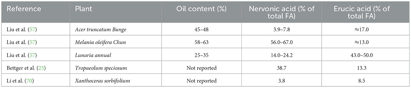
Table 2. Oil, nervonic (24:1 n-9) and erucic (22:1 n-9) acids content in some selected seed oil.
Due to these safety concerns, traditional plant sources of NA are generally unsuitable for direct inclusion in food formulations. This challenge has led to increasing interest in biotechnological alternatives, such as fermentation technologies, which offer a scalable, sustainable solution for the nutrition and health industries (65).
Production of nervonic acid in transgenic plants and microorganisms
Biosynthesis of NA has emerged as a promising alternative to conventional extraction methods, providing a sustainable and potentially high-yield approach to meet growing market demands. It has been recently reviewed in great details (65–67) and this section critically examines advancements and challenges associated with NA biosynthesis through transgenic plants and microorganisms.
Transgenic plants have long been studied for NA production due to their naturally high oil content and well-established genetic modification protocols. Initial efforts in the late 20th century were aimed at inserting elongase genes from various species into oilseed crops to boost NA synthesis. One pioneering study demonstrated that introducing an elongase gene into rapeseed significantly enhanced NA yields, establishing proof-of-concept for NA biosynthesis via transgenic approaches (68). Subsequent research successfully transferred the KCS gene from Lunaria annua (69) and Xanthoceras sorbifolium (70) to Arabidopsis thaliana, resulting in an increase in EA and NA content. Another notable study involved the introduction of the KCS gene from Cardamine graeca into Brassica species, which substantially boosted NA production and emphasized the critical importance of selecting suitable donor genes and recipient plant species (71). In this study, the highest NA level in transgenic B. carinata lines reached 44%, with only 6% of residual of EA (71). Recent research confirmed the high elongation activity of the MoKCS11 gene from Malania oleifera when expressed in Arabidopsis thaliana and Camelina sativa, thereby demonstrating its versatility and significant potential for broader application in agricultural biotechnology (72).
Microorganisms, particularly yeasts, have been engineered for NA production due to their rapid growth rates, scalability, and ease of cultivation under controlled conditions. Initial success in microbial NA and EA synthesis was reported in Saccharomyces cerevisiae by overexpressing genes encoding KCS and elongases, marking a significant milestone in microbial production capabilities (70, 73). The potential for producing NA using genetically engineered Yarrowia lipolytica has been previously investigated and reviewed (74). Further advancements have led to the development of Y. lipolytica strain capable of producing NA at 22–23% and gondoic acid (20:1 n-9, GA) at 8–10% of total fatty acids, providing a strong foundation for future optimization efforts (75). Further work on Y. lipolytica strains has explored multiple strategies in parallel, including the overexpression of genes involved in the desaturation and TAG assembly, leading to several noteworthy findings (76, 77). Additional work involved coexpression of the KCS genes from Crambe abyssinica and C. graeca in Rhodosporidium toruloides, significantly enhancing NA and EA concentrations, 7.9% and 5.8% of total FA, respectively in a 7L fermentation trial (78). To our knowledge, the best composition that has been achieved to date in a yeast framework has been obtained in R. toruloides by integrating KCS gene and optimization of the metabolic pathway (79) allowing to reach a concentration of NA of 46.3% and of EA of 2.8% of total fatty acids. While the composition obtained in the study is extremely interesting, the productivity (44.2 g/L in a 5 L bioreactor.) obtained remains a concern for commercial production.
Significant advancements have been made in biosynthesis across transgenic plants, microorganisms, and microalgae; however, considerable challenges such as improving yield and productivity in fermentation operations and reducing cultivation costs remain. Future research should emphasize optimizing genetic engineering, fermentation conditions, and cultivation practices to enhance NA production efficiency, yield, and sustainability, thus addressing critical market demands and supporting sustainable nutritional strategies.
Challenges and considerations in implementing nervonic acid fortification
Despite the promising potential of NA in infant nutrition, several challenges must be addressed before its widespread implementation. One major hurdle is regulatory approval, as novel food ingredients require extensive clinical validation to ensure safety and efficacy. Additionally, the stability of NA in infant formula formulations must be optimized to prevent oxidation and maintain bioavailability. Furthermore, ethical and economic considerations must be considered to ensure equitable access to NA-fortified products, preventing disparities in infant nutrition. As research progresses, collaborations between the scientific community, regulatory agencies, and industry stakeholders will be crucial in overcoming these barriers and bringing NA-fortified formulas to market.
Conclusion
The integration of NA into infant nutrition products represents an exciting opportunity for pediatric nutrition, with the potential to enhance neurodevelopmental outcomes in both premature and term infants. As biotechnology continues to progress, the sustainable microbial production of NA-rich oils will facilitate its inclusion in infant formulas, bridging a critical gap in lipid nutrition. Future research should focus on establishing optimal NA dosage, assessing long-term neurodevelopmental benefits, and addressing regulatory challenges to ensure the safe and effective implementation of NA fortification in infant nutrition.
Author contributions
FD: Writing – review & editing, Writing – original draft. MO: Writing – original draft, Writing – review & editing. WR: Writing – review & editing, Resources. XZ: Resources, Writing – review & editing. LP: Writing – review & editing, Resources.
Funding
The author(s) declare that no financial support was received for the research and/or publication of this article.
Conflict of interest
FD, MO, WR, XZ, and LP were employed by Checkerspot, Inc.
Generative AI statement
The author(s) declare that no Gen AI was used in the creation of this manuscript.
Publisher's note
All claims expressed in this article are solely those of the authors and do not necessarily represent those of their affiliated organizations, or those of the publisher, the editors and the reviewers. Any product that may be evaluated in this article, or claim that may be made by its manufacturer, is not guaranteed or endorsed by the publisher.
References
1. Deoni SCL, Beauchemin J, D'Sa V, Bonham K, Klepac-Ceraj V. Enhanced brain myelination and cognitive development in young children associated with milk fat globule membrane (MFGM) intake: a temporal cohort study. Preprint on Research Square. (2024). doi: 10.21203/rs.3.rs-4999582/v1
2. Zamroziewicz MK, Barbey AK. Nutritional cognitive neuroscience: innovations for healthy brain aging. Front Neurosci. (2016) 10:240. doi: 10.3389/fnins.2016.00240
3. Georgieff MK. Early life nutrition and brain development: breakthroughs, challenges and new horizons. Proc Nutr Soc. (2023) 82:104–12. doi: 10.1017/S0029665122002774
4. Gómez-Pinilla F. Brain foods: the effects of nutrients on brain function. Nat Rev Neurosci. (2008) 9:568–78. doi: 10.1038/nrn2421
5. Benton D. The influence of dietary status on the cognitive performance of children. Mol Nutr Food Res. (2010) 54:457–70. doi: 10.1002/mnfr.200900158
6. Bourre JM. Effects of nutrients (in food) on the structure and function of the nervous system: update on dietary requirements for brain. Part 1: micronutrients. J Nutr Health Aging. (2006) 10:377–385.
7. Bourre JM. Effects of nutrients (in food) on the structure and function of the nervous system: update on dietary requirements for brain. Part 2: macronutrients. J Nutr Health Aging 10. (2006) 386–99.
8. Hernell O, Domellöf M, Grip T, Lönnerdal B, Timby N. Physiological effects of feeding infants and young children formula supplemented with milk fat globule membranes. Nestlé Nutr Inst Workshop Ser. (2019) 90:35–42. doi: 10.1159/000490291
9. Ji L, Yoon YB, Hendrix CL, Kennelly EC, Majbri A, Bhatia T, et al. Developmental coupling of brain iron and intrinsic activity in infants during the first 150 days. Dev Cogn Neurosci. (2023) 64:101326. doi: 10.1016/j.dcn.2023.101326
10. Woodburn M, Bricken CL, Wu Z, Li G, Wang L, Lin W, et al. The maturation and cognitive relevance of structural brain network organization from early infancy to childhood. Neuroimage. (2021) 238:118232. doi: 10.1016/j.neuroimage.2021.118232
11. Yin W, Li T, Wu Z, et al. Charting brain functional development from birth to 6 years of age. Nat Hum Behav. (2025). doi: 10.1038/s41562-025-02160-2
12. Bulgarelli C, Blasi A, McCann S, Milosavljevic B, Ghillia G, Mbye E, et al. Growth in early infancy drives optimal brain functional connectivity which predicts cognitive flexibility in later childhood. bioRxiv. (2024). doi: 10.7554/eLife.94194.1
13. Di Liberto GM, Attaheri A, Cantisani G, Reilly RB, Ní Choisdealbha Á, Rocha S, et al. Emergence of the cortical encoding of phonetic features in the first year of life. Nat Commun. (2023) 14:7789. doi: 10.1038/s41467-023-43490-x
14. Alcauter S, Lin W, Smith JK, Short SJ, Goldman BD, Reznick JS, et al. Development of thalamocortical connectivity during infancy and its cognitive correlations. J Neurosci. (2014) 34:9067–75. doi: 10.1523/JNEUROSCI.0796-14.2014
15. Wilkinson C, Yankowitz L, Chao J, Gutierrez R, Rhoades J, Shinnar S, et al. Developmental trajectories of EEG aperiodic and periodic power: implications for understanding the timing of thalamocortical development during infancy. Preprint on Research Square. (2023). doi: 10.21203/rs.3.rs-3215728/v1
16. Cermenati G, Mitro N, Audano M, Melcangi RC, Crestani M, De Fabiani E, et al. Lipids in the nervous system: from biochemistry and molecular biology to pathophysiology. Biochim Biophys Acta Lipids Lipid Metab. (2015) 1851:51–60. doi: 10.1016/j.bbalip.2014.08.011
17. Yoon JH, Seo Y, Jo YS, Lee S, Cho E, Cazenave-Gassiot A, et al. Brain lipidomics: From functional landscape to clinical significance. Sci Adv. (2022) 8:eadc9317. doi: 10.1126/sciadv.adc9317
18. Sassa T, Kihara A. Metabolism of very long-chain fatty acids: genes and pathophysiology. Biomol Ther. (2014) 22:83–92. doi: 10.4062/biomolther.2014.017
20. Zheng L, Fleith M, Giuffrida F, O'Neill BV, Schneider N. Dietary polar lipids and cognitive development: a narrative review. Adv Nutr. (2019) 10:1163–76. doi: 10.1093/advances/nmz051
21. Dutta-Roy AK. Transport of fatty acids across the human placenta: a review. Prog Lipid Res. (2009) 48:52–61. doi: 10.1016/j.plipres.2008.11.001
22. Blades F, Yazici AT, Cater RJ, Mancia F. MFSD2A in focus: the molecular mechanism of omega-3 fatty acid transport. Physiology. (2025). doi: 10.1152/physiol.00068.2024
23. Bettger WJ, DiMichelle-Ranalli E, Dillingham B, Blackadar CB. Nervonic acid is transferred from the maternal diet to milk and tissues of suckling rat pups. J Nutr Biochem. (2003) 14:61–7. doi: 10.1016/S0955-2863(02)00280-2
24. Phung NV, Rong F, Xia WY, Fan Y, Li XY, Wang SA, et al. Nervonic acid and its sphingolipids: biological functions and potential food applications. Crit Rev Food Sci Nutr. (2024) 64:8766–85. doi: 10.1080/10408398.2023.2203753
25. Garcia Corrales AV, Haidar M, Bogie JFJ, Hendriks JJA. Fatty acid synthesis in glial cells of the CNS. Int J Mol Sci. (2021) 22:8159. doi: 10.3390/ijms22158159
26. Lewkowicz N, Piatek P, Namiecińska M, Domowicz M, Bonikowski R, Szemraj J, et al. Naturally occurring nervonic acid ester improves myelin synthesis by human oligodendrocytes. Cells. (2019) 8:786. doi: 10.3390/cells8080786
27. Martinez M, Mougan I. Fatty acid composition of human brain phospholipids during normal development. J Neurochem. (1998) 71:2528–33. doi: 10.1046/j.1471-4159.1998.71062528.x
28. Aydin I, Turan Ö, Aydin FN, Koc E, Hirfanoglu IM, Akyol, et al. Comparing the fatty acid levels of preterm and term breast milk in Turkish women. Turk. J. Med. Sci. (2014) 44:305–10. doi: 10.3906/sag-1302-131
29. Nyuar KB, Min Y, Ghebremeskel K, Khalil AKH, Elbashir MI, Crawford MA. Milk of northern Sudanese mothers whose traditional diet is high in carbohydrate contains low docosahexaenoic acid. Acta Paediatr. (2010) 99:1400–5. doi: 10.1111/j.1651-2227.2010.01940.x
30. Yu J, Yuan T, Zhang X, Jin Q, Wei W, Wang X. Quantification of nervonic acid in human milk in the first 30 days of lactation: influence of lactation stages and comparison with infant formulae. Nutrients. (2019) 11:1892. doi: 10.3390/nu11081892
31. Marosvölgyi T, Dergez T, Szentpéteri JL, Szabó É, Decsi T. Higher availability of long-chain monounsaturated fatty acids in preterm than in full-term human milk. Life. (2023) 13:1205. doi: 10.3390/life13051205
32. Mandal MN, Ambasudhan R, Wong PW, Gage PJ, Sieving PA, Ayyagari R. Characterization of mouse orthologue of ELOVL4: genomic organization and spatial and temporal expression. Genomics. (2004) 83:626–35. doi: 10.1016/j.ygeno.2003.09.020
33. Lönnerdal B, Erdmann P, Thakkar SK, Sauser J, Destaillats F. Longitudinal evolution of true protein, amino acids and bioactive proteins in breast milk: a developmental perspective. J Nutr Biochem. (2017) 41:1–11. doi: 10.1016/j.jnutbio.2016.06.001
34. Giuffrida F, Cruz-Hernandez C, Bertschy E, Fontannaz P, Masserey Elmelegy I, Tavazzi I, et al. Temporal changes of human breast milk lipids of Chinese mothers. Nutrients. (2016) 8:715. doi: 10.3390/nu8110715
35. Deák F, Anderson RE, Fessler JL, Sherry DM. Novel cellular functions of very long chain-fatty acids: insight from ELOVL4 mutations. Front Cell Neurosci. (2019) 13:428. doi: 10.3389/fncel.2019.00428
36. Sari RN, Jing L, Jiaping L. The change of bile salt stimulated lipase during 6 months and the correlation with macronutrients in Chinese human milk. Canrea J. (2023) 6:119–28. doi: 10.20956/canrea.v6i2.1033
37. Li X, Lindquist S, Lowe M, Noppa L, Hernell O. Bile salt-stimulated lipase and pancreatic lipase-related protein 2 are the dominating lipases in neonatal fat digestion in mice and rats. Pediatr Res. (2007) 62:537–41. doi: 10.1203/PDR.0b013e3181559e75
38. Lindquist S, Hernell O. Lipid digestion and absorption in early life: an update. Curr Opin Clin Nutr Metab Care. (2010) 13:314–20. doi: 10.1097/MCO.0b013e328337bbf0
39. Thakkar SK, De Castro CA, Beauport L, Tolsa JF, Fischer Fumeaux CJ, Affolter M, et al. Temporal progression of fatty acids in preterm and term human milk of mothers from Switzerland. Nutrients. (2019) 11:112. doi: 10.3390/nu11010112
40. Rueda R, Ramírez M, García-Salmerón JL, Maldonado J, Gil A. Gestational age and origin of human milk influence total lipid and fatty acid contents. Ann Nutr Metab. (1998) 42:12–22. doi: 10.1159/000012713
41. Moltó-Puigmartí C, Castellote AI, Carbonell-Estrany X, López-Sabater MC. Differences in fat content and fatty acid proportions among colostrum, transitional, and mature milk from women delivering very preterm, preterm, term infants. Clini Nutr. (2011) 30:116–23. doi: 10.1016/j.clnu.2010.07.013
42. Duan B, Shin JA, Lee KT. Quantitative analysis of nervonic and erucic acids in human milk: comparison with infant formula with different fat sources and nutritional stages. J Oleo Sci. (2024) 73:333–40. doi: 10.5650/jos.ess23146
43. Li J, Fan Y, Zhang Z, Yu H, An Y, Kramer JKG, et al. Evaluating the trans fatty acid, CLA, PUFA and erucic acid diversity in human milk from five regions in China. Lipids. (2009) 44:257–71. doi: 10.1007/s11745-009-3282-x
44. Volpe JJ. Brain injury in premature infants: a complex amalgam of destructive and developmental disturbances. Lancet Neurol. (2009) 8:110–24. doi: 10.1016/S1474-4422(08)70294-1
45. Ream MA, Lehwald L. Neurologic consequences of preterm birth. Curr Neurol Neurosci Rep. (2018) 18:48. doi: 10.1007/s11910-018-0862-2
46. Back SA, Miller SP. Brain injury in premature neonates: a primary cerebral dysmaturation disorder? Ann. Neurol. (2014) 75:469–86. doi: 10.1002/ana.24132
47. Lammertink F, Vinkers CH, Tataranno ML, Benders MJNL. Premature birth and developmental programming: mechanisms of resilience and vulnerability. Front Psychiatry. (2021) 11:531571. doi: 10.3389/fpsyt.2020.531571
48. Koletzko B, Boey CCM, Campoy C, Carlson SE, Chang N, Guillermo-Tuazon MA, et al. Current information and Asian perspectives on long-chain polyunsaturated fatty acids in pregnancy, lactation, and infancy: systematic review and practice recommendations from an Early Nutrition Academy Workshop. Ann Nutr Metab. (2014) 65:49–80. doi: 10.1159/000365767
49. Gould JF, Colombo J, Collins CT, Makrides M, Hewawasam E, Smithers LG. Assessing whether early attention of very preterm infants can be improved by an omega-3 long-chain polyunsaturated fatty acid intervention: a follow-up of a randomized controlled trial. BMJ Open. (2018) 8:e020043. doi: 10.1136/bmjopen-2017-020043
50. Hussain G, Wang J, Rasul A, Anwar H, Imran A, Qasim M, et al. Role of cholesterol and sphingolipids in brain development and neurological diseases. Lipids Health Dis. (2019) 18:26. doi: 10.1186/s12944-019-0965-z
51. Tomlinson C Haiek L Breastfeeding N and human milk in the NICU: from birth to discharge. Paediatr Child Health. (2023) 28:510–7. doi: 10.1093/pch/pxad034
52. Arslanoglu S, Boquien C-Y, King C, Lamireau D, Tonetto P, Barnett D, et al. Fortification of human milk for preterm infants: update and recommendations of the European Milk Bank Association (EMBA) Working Group on Human Milk Fortification. Front Pediatr. (2019) 7:76. doi: 10.3389/fped.2019.00076
53. Timby N, Domellöf E, Hernell O, Lönnerdal B, Domellöf M. Neurodevelopment, nutrition, and growth until 12 months of age in infants fed a low-energy, low-protein formula supplemented with bovine milk fat globule membranes: a randomized controlled trial. Am J Clin Nutr. (2014) 99:860–8. doi: 10.3945/ajcn.113.064295
54. Tanaka K, Hosozawa M, Kudo N, Yoshikawa N, Hisata K, Shoji H, et al. The pilot study: sphingomyelin-fortified milk has a positive association with the neurobehavioural development of very low birth weight infants during infancy, randomized control trial. Brain Dev. (2013) 35:45–52. doi: 10.1016/j.braindev.2012.03.004
55. Schneider N, Hauser J, Oliveira M, Cazaubon E, Colombo Mottaz S, O'Neill BV, et al. Sphingomyelin in brain and cognitive development: preliminary data. eNeuro. (2019) 6:ENEURO.0421-18.2019. doi: 10.1523/ENEURO.0421-18.2019
56. Ma Q, Wang Y, Zhu L, Bi C, Li S, Li S, et al. Characterization of the complete chloroplast genome of Acer truncatum Bunge (Sapindales: Aceraceae): A new woody oil tree species producing nervonic acid. Biomed Res Int. (2019) 2019:7417239. doi: 10.1155/2019/7417239
57. Liu F, Wang P, Xiong X, Zeng X, Zhang X, Wu G. A review of nervonic acid production in plants: prospects for the genetic engineering of high nervonic acid cultivars. Front Plant Sci. (2021) 12:626625. doi: 10.3389/fpls.2021.626625
58. Xu CQ, Liu H, Zhou SS, Zhang DX, Zhao W, Wang S, et al. Genome sequence of Malania oleifera, a tree with great value for nervonic acid production. GigaScience. (2019) 8:giy164. doi: 10.1093/gigascience/giy164
59. Gao Y, Wei T, Wang J, Tu Y, Zhou Z, Du C, Ren Z. Enrichment and purification of nervonic acid from Acer truncatum seed oil by combining vacuum distillation and low-temperature crystallization: experiments and process modeling. Chin J Chem Eng. (2024). doi: 10.1016/j.cjche.2024.09.035
60. Bettger WJ, Blackadar CB. Dietary very long-chain fatty acids directly influence the ratio of tetracosenoic (24:1) to tetracosanoic (24:0) acids of sphingomyelin in rat liver. Lipids. (1997) 32:51–5. doi: 10.1007/s11745-997-0008-1
61. Murphy CC, Murphy EJ, Golovko MY. Erucic acid is differentially taken up and metabolized in rat liver and heart. Lipids. (2008) 43:391–400. doi: 10.1007/s11745-008-3168-3
62. Stam H, Geelhoed-Mieras T, Hulsmann WC. Erucic acid-induced alteration of cardiac triglyceride hydrolysis. Biochim Biophys Acta. (1979) 574:258–69.
63. Charlton KM, Corner AH, Davey K, Kramer JKG, Mahadevan S, Sauer FD. Cardiac lesions in rats fed rapeseed oils. Can J Comp Med. (1975) 39:261–9.
64. Galanty A, Grudzińska M, Pazdziora W, Pasko P. Erucic acid—both sides of the story: a concise review on its beneficial and toxic properties. Molecules. (2023) 28:1924. doi: 10.3390/molecules28041924
65. Ling C, Li F, Zhao J, Wen M, Han X. Research progress of nervonic acid biosynthesis. J Oleo Sci. (2023) 72:889–900. doi: 10.5650/jos.ess23039
66. Fan Y, Meng HM, Hu GR, Li FL. Biosynthesis of nervonic acid and perspectives for its production by microalgae and other microorganisms. Appl Microbiol Biotechnol. (2018) 102:3027–35. doi: 10.1007/s00253-018-8859-y
67. Liu F, Wu R, Ma X, Su E. The advancements and prospects of Nervonic Acid production. J Agric Food Chem. (2022) 70:12772–83. doi: 10.1021/acs.jafc.2c05770
68. Yi S. The new target of Calgi's biotechnology: rapeseed with high nervonic acid content. Cereal Oil. (1997) 02:51.
69. Guo Y, Mietkiewska E, Francis T, Katavic V, Brost JM, et al. Increase in nervonic acid content in transformed yeast and transgenic plants by introduction of a Lunaria annua L. 3-ketoacyl-CoA synthase (KCS) gene. Plant Mol Biol 69. (2009) 565–75. doi: 10.1007/s11103-008-9439-9
70. Li L, Liang C, Zhang W, Zhang X, Yu H, Liu X, et al. 3-ketoacyl-CoA synthase 7 from Xanthoceras sorbifolium seeds is a crucial regulatory enzyme for nervonic acid biosynthesis. Plant Sci. (2024) 347:112184. doi: 10.1016/j.plantsci.2024.112184
71. Taylor DC, Francis T, Guo Y, Brost JM, Katavic V, et al. Molecular cloning and characterization of a KCS gene from Cardamine graeca and its heterologous expression in Brassica oilseeds to engineer high nervonic acid oils for potential medical and industrial use. Plant Biotechnol J. (2009) 7:925–38. doi: 10.1111/j.1467-7652.2009.00454.x
72. Li Z, Ma S, Song H, Yang Z, Zhao C, et al. A 3-ketoacyl-CoA synthase 11 (KCS11) homolog from Malania oleifera synthesizes nervonic acid in plants rich in 11Z-eicosenoic acid. Tree Physiol. (2021) 41:331–42. doi: 10.1093/treephys/tpaa125
73. Zhang Y, Yang C, Xia J, Deng L, Wang F, Liu J. Overproducing nervonic acid by synergism of fatty acid elongases in engineered Saccharomyces cerevisiae. Process Biochem. (2022) 122:341–6. doi: 10.1016/j.procbio.2022.09.013
74. Giwa AS, Ali N. Perspectives of nervonic acid production by Yarrowia lipolytica. Biotechnol Lett. (2022) 44:193–202. doi: 10.1007/s10529-022-03231-4
75. Wang K, Lin L, Wei P, Ledesma-Amaro R, Ji XJ. Combining orthogonal plant and non-plant fatty acid biosynthesis pathways for efficient production of microbial oil enriched in nervonic acid in Yarrowia lipolytica. Bioresour Technol. (2023) 378:129012. doi: 10.1016/j.biortech.2023.129012
76. Zhao XR, Chen XL, Yang JL, Gao Q, Shi JT, Hua Q, et al. De novo synthesis of nervonic acid and optimization of metabolic regulation by Yarrowia lipolytica. Bioresour Bioprocess. (2023) 10:70. doi: 10.1186/s40643-023-00689-6
77. Su H, Shi P, Shen Z, Meng H, Meng Z, Han X, et al. High-level production of nervonic acid in the oleaginous yeast Yarrowia lipolytica by systematic metabolic engineering. Commun Biol. (2023) 6:1125. doi: 10.1038/s42003-023-05502-w
78. Fillet S, Ronchel C, Callejo C, Fajardo MJ, Moralejo H, et al. Engineering Rhodosporidium toruloides for the production of very long-chain monounsaturated fatty acid-rich oils. Appl Microbiol Biotechnol. (2017) 101:7271–80. doi: 10.1007/s00253-017-8461-8
79. Liu F, Lu Z, Lu T, Shi M, Wang H, Wu R, et al. Metabolic engineering of oleaginous yeast in the lipogenic phase enhances production of nervonic acid. Metab Eng. (2023) 80:193–206. doi: 10.1016/j.ymben.2023.10.001
80. Yakes EA, Arsenault JE, Islam MM, Hossain MB, Ahmed T, German JB, et al. Intakes and breast-milk concentrations of essential fatty acids are low among Bangladeshi women with 24–48-month-old children. Br J Nutr. (2011) 105:1660–70. doi: 10.1017/S0007114510004964
81. Golfetto I, McGready R, Ghebremeskel K, Min Y, Dubowitz L, Nosten F, et al. Fatty acid composition of milk of refugee Karen and urban Korean mothers: is the level of DHA in breast milk of Western women compromised by high intake of saturated fat and linoleic acid? Nutr. Health. (2007) 18:319–32. doi: 10.1177/026010600701800402
82. Mihályi K, Györei E, Szabó É, Marosvölgyi T, Lohner S, Decsi T. Contribution of n-3 long-chain polyunsaturated fatty acids to human milk is still low in Hungarian mothers. Eur J Pediat. (2015) 174:393–8. doi: 10.1007/s00431-014-2411-6
83. Peng Y, Zhou T, Wang Q, Liu P, Zhang T, Zetterström R, et al. Fatty acid composition of diet, cord blood, and breast milk in Chinese mothers with different dietary habits. Prostaglandins Leukot Essent Fatty Acids. (2009) 81:325–30. doi: 10.1016/j.plefa.2009.07.004
84. Zhao P, Zhang S, Liu L, Pang X, Yang Y, Lu J, et al. Differences in the triacylglycerol and fatty acid compositions of human colostrum and mature milk. J Agric Food Chem. (2018) 66:4571–9. doi: 10.1021/acs.jafc.8b00868
85. Young C, Hikita T, Kaneko S, Shimizu Y, Hanaka S, Abe T, et al. Fatty acid compositions of colostrum, cord blood, maternal blood and major infant formulas in Japan. Acta Paediatr Jpn. (1997) 39:299–304. doi: 10.1111/j.1442-200X.1997.tb03740.x
86. Knox E, VanderJagt DJ, Shatima D, Huang YS, Chuang LT, Glew RH. Nutritional status and intermediate chain-length fatty acids influence the conservation of essential fatty acids in the milk of northern Nigerian women. Prostaglandins Leukot Essent Fatty Acids. (2000) 63:195–202. doi: 10.1054/plef.2000.0206
87. Sala-Vila A, Campoy C, Castellote AI, Garrido FJ, Rivero M, Rodríguez-Palmero M, et al. Influence of dietary source of docosahexaenoic and arachidonic acids on their incorporation into membrane phospholipids of red blood cells in term infants. Prostaglandins Leukot Essent Fatty Acids. (2006) 74:143–8. doi: 10.1016/j.plefa.2005.10.003
88. Xiang M, Alfvén G, Blennow M, Trygg M, Zetterström R. Long-chain polyunsaturated fatty acids in human milk and brain growth during early infancy. Acta Paediatr. (2000) 89:142–7. doi: 10.1080/080352500750028735
89. VanderJagt DJ, Arndt CD, Okolo SN, Huang YS, Chuang LT, Glew RH. Fatty acid composition of the milk lipids of Fulani women and the serum phospholipids of their exclusively breast-fed infants. Early Hum Dev. (2000) 60:73–87. doi: 10.1016/S0378-3782(00)00111-0
90. Marangoni F, Agostoni C, Lammardo AM, Giovannini M, Galli C, Riva E. Polyunsaturated fatty acid concentrations in human hindmilk are stable throughout 12-months of lactation and provide a sustained intake to the infant during exclusive breastfeeding: an Italian study. Br J Nutr. (2000) 84:103–9. doi: 10.1017/S0007114500001288
91. Marangoni F, Agostoni C, Lammardo AM, Bonvissuto M, Giovannini M, Galli C, et al. Polyunsaturated fatty acids in maternal plasma and in breast milk. Prostaglandins Leukot Essent Fatty Acids. (2002) 66:535–40. doi: 10.1054/plef.2002.0396
92. Xiang M, Lei S, Li T, Zetterström R. Composition of long-chain polyunsaturated fatty acids in human milk and growth of young infants in rural areas of northern China. Acta Paediatr. (1999) 88:126–31. doi: 10.1080/08035259950170268
93. Rocquelin G, Tapsoba S, Dop MC, Mbemba F, Traissac P, Martin-Prével Y. Lipid content and essential fatty acid composition of mature Congolese breast milk are influenced by mothers' nutritional status: impact on infants' essential fatty acid supply. Eur J Clin Nutr. (1997) 52:164–71. doi: 10.1038/sj.ejcn.1600529
94. Boersma ER, Offringa PJ, Muskiet FAJ, Chase WM, Simmons IJ. Vitamin E, lipid fractions, and fatty acid composition of colostrum, transitional milk, and mature milk: an international comparative study. Am J Clin Nutr. (1991) 53:1197–204. doi: 10.1093/ajcn/53.5.1197
95. Bobinski R, Bobinska J. Fatty acids of human milk – a review. Int J Vitam Nutr Res. (2022) 92:280–91. doi: 10.1024/0300-9831/a000651
Keywords: nervonic acid, infant nutrition, myelination, milk-fat globule membranes, premature infants, fermentation, brain development, sphingolipids
Citation: Destaillats F, Oliveira M, Rakitsky W, Zhou X and Parker L (2025) Nervonic acid in infant nutrition: a forward-looking approach to enhancing neurodevelopmental outcomes. Front. Nutr. 12:1635266. doi: 10.3389/fnut.2025.1635266
Received: 26 May 2025; Accepted: 12 June 2025;
Published: 26 June 2025.
Edited by:
Claude Billeaud, Independent Paediatrics Nutrition Expert, FranceReviewed by:
Eva Szabo, University of Pécs, HungaryFrancesca Giuffrida, Société des Produits Nestlé S.A., Switzerland
Copyright © 2025 Destaillats, Oliveira, Rakitsky, Zhou and Parker. This is an open-access article distributed under the terms of the Creative Commons Attribution License (CC BY). The use, distribution or reproduction in other forums is permitted, provided the original author(s) and the copyright owner(s) are credited and that the original publication in this journal is cited, in accordance with accepted academic practice. No use, distribution or reproduction is permitted which does not comply with these terms.
*Correspondence: Frédéric Destaillats, ZmRlc3RhaWxsYXRzQGNoZWNrZXJzcG90LmNvbQ==