- Department of Clinical Pharmacy, Xiangtan Central Hospital, The Affiliated Hospital of Hunan University, Xiangtan, China
Neurodegenerative diseases (NDDs) are a group of disorders characterized by the progressive loss of neurons in specific areas of the central nervous system. In recent years, more and more research has focused on the influence of diet on NDDs. As a common food, processed red meat is widely consumed worldwide. Many studies have shown that processed red meat may increase the risk of cancer, diabetes and cardiovascular disease. Unfortunately, it is unclear whether processed red meat affects NDDs. Therefore, we reviewed the existing literature on the role of processed meats in NDDs. We concluded that intake of processed meat may have an adverse effect on NDDs.
1 Introduction
The brain and spinal cord are composed of neurons. Neurons have different functions that affect human motor perception and memory cognition (1). Since neurons usually cannot be regenerated, excessive damage can severely impair brain and nerve function (2). NDDs are a group of disorders characterized by the progressive death of neurons, and they include Alzheimer’s disease (AD), Parkinson’s disease (PD), Huntington’s disease (HD), and Amyotrophic lateral sclerosis (ALS) (3).
Previous studies had focused on drugs and traditional plants in the treatment of NDDs (4, 5). With the development of society and medical technology, a large number of new treatment methods such as gene therapy, aquatherapy, brain energy rescue, nanoparticle therapy, and regenerative stem cell therapy have appeared (6–8). In addition to treatment, diet also plays an important role in NDDs. The mediterranean diet, the DASH (Dietary Approaches to Stop Hypertension) diet, and the MIND (Mediterranean-DASH Intervention for Neurodegenerative Delay) diet have been documented to protect against NDDs (9). Furthermore, some nutrients, such as vitamin B6, vitamin B12, folate, caffeine, and lecithin, have beneficial effects on NDDs (10).
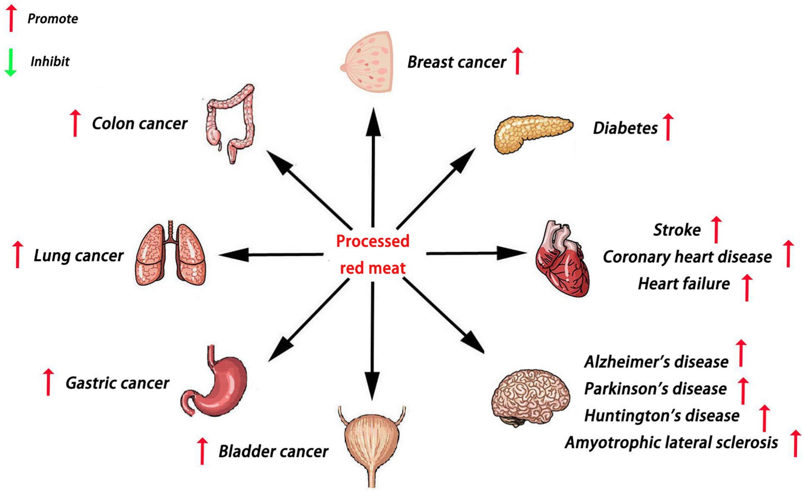
Red meat is a type of meat that appears red before cooking, mainly including pork, beef, lamb, and other mammalian meat (11). As a popular food, processed red meat is consumed globally (12). However, many studies have reported that processed red meat may increase the risk of cancer, diabetes, and cardiovascular disease (13) (Figure 1). Regrettably, it is still unclear whether processed red meat influences NDDs. Therefore, we reviewed the current literature on the role of processed red meat in NDDs. We speculate that excessive intake of processed red meat may promote the development of NDDs.
As a motor neuron disease, ALS is primarily characterized by the loss of motor neurons in the brain and spinal cord (14). Pupillo et al. (15) surveyed 212 patients with newly diagnosed ALS from three Italian administrative regions. They found that processed red meat may be a risk factor for ALS. As the most common NDD, AD was first proposed by Alois Alzheimer in 1907 (16). Aggregation of Amyloid-β (Aβ) peptide is linked to the pathophysiology of AD (17). Numerous epidemiological studies have examined the relationship between the dietary heme intake (processed red meat) with the risk of AD (18, 19). Heme prevents the formation of Aβ peptide aggregates by binding with Aβ peptide (18). It may lead to dysfunctional mitochondria and altered metabolic activity in the brains of AD patients (20). Another study suggests that excessive consumption of processed red meat might correlate with the risk of mild cognitive impairment patients to develop AD (21). As the second most common NDD, PD has long been characterized by the loss of dopamine (DA) in the substantia nigra. Processed red meat may be one of the critical factors associated with an increased risk of PD (22). Zapała et al. (23) compared diet preferences in PD patients and healthy controls. They found that the consumption of processed red meat in PD patients was significantly higher than healthy controls (23). Coimbra CG’s study suggests that the elimination of red meat promotes the recovery of some motor functions in PD patients (24). Neuroinflammation and DNA damage are major mechanisms in PD pathogenesis (25). Several studies have shown that processed red meat consumption is positively linked to PD. Processed red meat consumption may promote inflammation (26). The chemical N-methyl-phenyl-tetrahydropyridine (MPTP) and 2-amino-1-methyl-6-phenylimidazo(4,5-b)pyridine (PhIP) are neurotoxicants formed in processed red meat (27). MPTP can cause a parkinsonian syndrome in men (28). PhIP can induce DNA damage in galactose-dependent SH-SY5Y cells (27).
Excessive consumption of processed red meat may increase the risk of diabetes, Alzheimer’s disease, Parkinson’s disease, stroke, coronary heart disease, heart failure, Huntington’s disease, amyotrophic lateral sclerosis, colon cancer, breast cancer, lung cancer, gastric cancer, and bladder cancer.
2 Mechanisms of NDDs
Over the past few decades, more and more NDDs have resulted in premature death or disability as the population ages (29). Understanding the pathogenesis of NDDs is important to explore the role of processed red meat in NDDs. Current studies have found that the pathogenesis of NDDs is related to oxidative stress, mitochondrial dysfunction, inflammation, and the disturbance of Ca2+ (30).
Oxidative stress is a state caused by the imbalance between reactive oxygen species production and antioxidant defense. It is characterized by excessive production of free radicals and reactive oxygen species (31). Studies have indicated that oxidative stress plays an important role in the pathogenesis of NDDs (32). Oxidative damage of nerve tissue has been found in NDDs such as AD, PD, HD, and ALS. On the one hand, a high concentration of ROS will damage the DNA, proteins, lipids, and other macromolecules in nerve cells, and eventually lead to neuronal necrosis and apoptosis. On the other hand, the use of free radical scavengers or antioxidants can significantly improve these NDDs (33).
Mitochondria are tiny structures in the cytoplasm that are involved in the production and metabolism of energy (34). Earlier studies have found that mitochondria are not only the main sources of ROS, but also the main “tools” for clearance (34). Mitochondrial dysfunction plays an important role in the pathogenesis of NDDs (35). Mitochondrial dysfunction can lead to the imbalance between ROS production and elimination, and ultimately lead to neuronal damage and apoptosis (35). The abundant vascular system in the brain guarantees the huge blood supply, and also provides sufficient glucose and oxygen for brain energy metabolism (36). Mitochondrial dysfunction can also contribute to the progression of NDDs by affecting energy metabolism (37).
It is well known that inflammation plays an important role in NDDs (38). The main sign of brain inflammation is the activation of glial cells. Under normal physiological conditions, microglia maintain the homeostasis of the central nervous system by engulfing pathogens and apoptotic cells (39). When microglia are repeatedly activated by inflammation, neuroinflammation is transformed into chronic inflammation and accompanied by the release of inflammatory factors such as IL-6, TNF-α, and IL-1. These inflammatory factors accelerate the production and aggregation of neurotoxic proteins, resulting in neuronal damage and death (39). Therefore, neuroinflammation is also a key mechanism in the pathogenesis of NDDs. As an important messenger in brain neurons, Ca2+ plays an important role in the development of neurons, the growth of axons, and the formation of synapses (40). When Ca2+ balance is disrupted, the growth and development of nerve cells are affected. On the one hand, excessive concentration of Ca2+ will increase the aggregation of Aβ protein and the over-phosphorylation of Tau protein, resulting in the impairment of patients’ learning and memory ability (40). On the other hand, excessive concentration of Ca2+ can also activate apoptosis pathways, aggravate oxidative stress, and cause apoptosis (40). Meanwhile, more and more studies have pointed out that the disturbance of zinc, iron, and copper can also lead to the occurrence of many NDDs (41).
In addition to the above mechanisms, pathogenic mechanisms of NDDs include the misfolding and aggregation of proteins, abnormal repair of DNA, excitatory toxins (glutamate), autophagy, pyroptosis, and ferroptosis (42–44).
3 Risks associated with components of processed red meat
3.1 Methionine
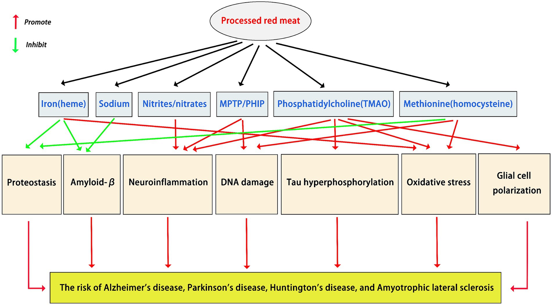
Processed red meat is a methionine-rich food (45). As an essential sulfur-containing amino acid, methionine is involved in various biochemical processes (46). Epidemiological studies have indicated that high methionine consumption has a negative effect on NDDs (47). Firstly, Methionine metabolism can produce toxic byproducts (homocysteine), which contribute to oxidative damage (48). Moreover, methionine-rich diet (processed red meat) can lead to mitochondrial dysfunction by impairing mitochondrial DNA integrity and affecting mitochondrial dynamics (47). Mitochondrial oxidative stress and dysfunction can contribute to NDD pathogenesis (35). Secondly, methionine-rich diet (processed red meat) has been shown to induce inflammation by activating pro-inflammatory signaling pathways and generating inflammatory mediators (47). Studies have shown that inflammation can contribute to NDD pathogenesis (49). Thirdly, the health of the microvasculature, blood-brain barrier, proteostasis, and functional connectivity are essential for efficient cognitive function (50). Methionine-rich diet (processed red meat) can lead to cognitive impairments and neuronal damage by disrupting these normal processes (47).
3.2 Iron
Processed red meat is rich in heme iron (51). As an important cofactor, iron is essential for neuronal development, synaptic plasticity, and myelination. However, excessive intake of iron can be harmful to health (52). Studies have shown that high consumption of processed red meat and its products, and thereby iron, particularly in the form of heme, increases the risk of many diseases (53). For decades, deposits of iron have been detected in patients with AD, PD, ALS, and HD (54). Excessive iron accumulation is harmful because it can promote the formation of free radicals resulting in oxidative stress, lipid peroxidation, protein aggregation, and eventually cell/neuronal death (55).
3.3 Sodium
Processed red meat is also a high-sodium food (56). However, the relationship between the high-sodium diet and AD is not clear. The alteration of sodium homeostasis significantly contributes to synaptic dysfunction and neuronal loss in AD (57). Attenuation of hippocampal hyperactivity, an earliest neuronal abnormality observed in AD brains, has been attributed in part to the dysfunction of sodium channels (58). Unusual cerebrovascular morphology and structure may contribute to cerebral hypoperfusion in AD. Baumgartner et al. (58) found that a high-sodium diet reduced vascular density (59). These results suggest that a high-sodium diet can induce cerebrovascular morphology changes in AD mouse models (59). In addition, Taheri et al. (60) found that a high-sodium diet influences the accumulation of Aβ peptide, exacerbates cognitive decline, and increases the propensity to AD. It is well known that the pathophysiology of HD is very complex. Intracerebral sodium accumulation has a crucial role in the pathophysiology of HD (61). Interestingly, Reetz et al. (62) found an increase in sodium concentration of the entire brain in HD patients.
3.4 Nitrite and nitrate
Boll et al. (63) recorded the sum of nitrites and nitrates from patients with any of the four NDDs (PD/AD/HD/ALS), and they found it increased in all of them. As a metabolite of nitric oxide (NO), nitrite can lead to nitrosative stress in the nigrostriatal system (64). There is evidence that nitrosative stress is an important factor promoting degeneration in PD (65). 3-nitropropionic acid (3-NP), a hemotoxin of fungal origin, has been used in rodents to model HD (66). A large number of studies have shown that 3-NP can significantly increase the levels of nitrite in HD models (67, 68). Many natural drugs such as rutin, lycopene, resveratrol, lutein, and safranal can prevent 3-NP-induced HD by decreasing the levels of nitrite (69–73). Previous studies have demonstrated that the nitrite and nitrate levels were significantly increased in the cerebrospinal fluid (CSF) and serum from ALS patients (74). Interestingly, motoneuron survival was inversely correlated with nitrate/nitrite concentrations in the ALS mouse model (75). A systemic pro-inflammatory state plays a central role in ALS pathogenesis (76). As the macrophages of the central nervous system, microglia are responsible for the inflammatory component of ALS (77). Meanwhile, microglia also contribute to motoneuron injury in ALS. In the pathogenesis of ALS, microglia can induce more neuronal death by producing and releasing more nitrite and nitrate (78).
3.5 Phosphatidylcholine
Extensive data demonstrate that lipids play a crucial role in NDD pathogenesis. For example, the accumulation of lipids is a risk factor for PD (79). Lipid dysregulation is a feature of ALS (80). Aberrant lipid metabolism is linked to the pathophysiology of AD (81). Phosphatidylcholine is one of the most common fats found in processed red meat (82). Some studies suggest that the consumption of phosphatidylcholine may have adverse effects on NDDs. As an early marker of neurodegeneration, phosphatidylcholine may promote tau hyperphosphorylation (83). Trimethylamine n-oxide (TMAO) is a gut microbiota metabolite derived from phosphatidylcholine (84). There is evidence that TMAO is associated with the pathogenesis of various NDDs (85, 86). On the one hand, TMAO levels increase with age-related cognitive dysfunction (86). On the other hand, TMAO also induces mitochondrial dysfunction, oxidative stress, neuroinflammation, and glial cell polarization in the brain (85). The mechanisms of processed red meat components in NDDs are shown in Figure 2.
4 Discussion
With the development of society and the economy, more and more unhealthy dietary patterns have harmful effects on people’s brains (85). Among these dietary patterns, processed red meat consumption is an interesting potential factor. Accumulating evidence suggests that processed red meat intake might be associated with NDD pathogenesis. The potential adverse effects on NDDs of processed red meat have been attributed to its ingredients such as methionine (47), heme iron (54), sodium (57), nitrite/nitrate (63), and phosphatidylcholine (83). Many studies have revealed the harmful effects of these ingredients on brain health (23, 87–89). Nevertheless, these studies have several limitations (23, 87–89). Firstly, the sample size of many studies was not large enough to ensure sufficient statistical power (88). Meanwhile, some cases may not have been classified by type of disease, which may attenuate association between processed red meat intake and risk of NDD development (89). Secondly, harmful substances of processed red meat may also be produced during the cooking of other foods (83). Meanwhile, some components of processed red meat play a protective role in NDDs (83). Therefore, it is difficult to conclude that processed red meat is the main cause of NDDs. There may be an intricate influence of multiple factors, including alcohol consumption, smoking, obesity, and stress (83). Thirdly, the conflicting results of some studies may be due to the fact that the dose of processed red meat is not enough (87). In the future, different dosage standards will need to be used when we study the relationship between processed red meat and NDDs. Meanwhile, it would be of interest to go beyond the use of questionnaires by also including biomarkers or metabolomics to study the associations between processed red meat consumption and NDDs. In addition, some of the observed results in previous studies may be limited by inadequate adjustment for potential confounders (23). More complete adjustment for a broad spectrum of potential confounders in future studies could help to address this potential limitation. Nevertheless, we believe that the consumption of high red meat may have adverse effects on NDDs. In conclusion, it is interesting to explore the relationship between the processed red meat and NDDs. Understanding the mechanism and role of processed red meat in NDDs has broad prospects for the prevention and treatment of NDDs.
Author contributions
K-qC: Writing – original draft. W-jC: Writing – review & editing. ZL: Writing – review & editing. R-zL: Writing – review & editing.
Funding
The author(s) declare that no financial support was received for the research and/or publication of this article.
Conflict of interest
The authors declare that the research was conducted in the absence of any commercial or financial relationships that could be construed as a potential conflict of interest.
Generative AI statement
The authors declare that no Gen AI was used in the creation of this manuscript.
Any alternative text (alt text) provided alongside figures in this article has been generated by Frontiers with the support of artificial intelligence and reasonable efforts have been made to ensure accuracy, including review by the authors wherever possible. If you identify any issues, please contact us.
Publisher’s note
All claims expressed in this article are solely those of the authors and do not necessarily represent those of their affiliated organizations, or those of the publisher, the editors and the reviewers. Any product that may be evaluated in this article, or claim that may be made by its manufacturer, is not guaranteed or endorsed by the publisher.
References
1. Dejanovic, B, Sheng, M, and Hanson, JE. Targeting synapse function and loss for treatment of neurodegenerative diseases. Nat Rev Drug Discov. (2024) 23:23–42. doi: 10.1038/s41573-023-00823-1
2. Zhou, ZD, Yi, LX, and Tan, EK. Targeting gasdermin E in neurodegenerative diseases. Cell Rep Med. (2023) 4:101075. doi: 10.1016/j.xcrm.2023.101075
3. Menéndez-González, M. Toward a new nosology of neurodegenerative diseases. Alzheimers Dement. (2023) 19:3731–7. doi: 10.1002/alz.13041
4. Chen, W, Hu, Y, and Ju, D. Gene therapy for neurodegenerative disorders: advances, insights and prospects. Acta Pharm Sin B. (2020) 10:1347–59. doi: 10.1016/j.apsb.2020.01.015
5. Wahid, M, Ali, A, Saqib, F, Aleem, A, Bibi, S, Afzal, K, et al. Pharmacological exploration of traditional plants for the treatment of neurodegenerative disorders. Phytother Res. (2020) 34:3089–112. doi: 10.1002/ptr.6742
6. Cunnane, SC, Trushina, E, Morland, C, Prigione, A, Casadesus, G, Andrews, ZB, et al. Brain energy rescue: an emerging therapeutic concept for neurodegenerative disorders of ageing. Nat Rev Drug Discov. (2020) 19:609–33. doi: 10.1038/s41573-020-0072-x
7. Sivandzade, F, and Cucullo, L. Regenerative stem cell therapy for neurodegenerative diseases: an overview. Int J Mol Sci. (2021) 22:2153. doi: 10.3390/ijms22042153
8. Sun, J, and Roy, S. Gene-based therapies for neurodegenerative diseases. Nat Neurosci. (2021) 24:297–311. doi: 10.1038/s41593-020-00778-1
9. Tao, Y, Leng, SX, and Zhang, H. Ketogenic diet: an effective treatment approach for neurodegenerative diseases. Curr Neuropharmacol. (2022) 20:2303–19. doi: 10.2174/1570159x20666220830102628
10. Stefaniak, O, Dobrzyńska, M, Drzymała-Czyż, S, and Przysławski, J. Diet in the prevention of Alzheimer's disease: current knowledge and future research requirements. Nutrients. (2022) 14:4564. doi: 10.3390/nu14214564
11. Yun, Z, Nan, M, Li, X, Liu, Z, Xu, J, Du, X, et al. Processed meat, red meat, white meat, and digestive tract cancers: a two-sample Mendelian randomization study. Front Nutr. (2023) 10:1078963. doi: 10.3389/fnut.2023.1078963
12. Lescinsky, H, Afshin, A, Ashbaugh, C, Bisignano, C, Brauer, M, Ferrara, G, et al. Health effects associated with consumption of unprocessed red meat: a burden of proof study. Nat Med. (2022) 28:2075–82. doi: 10.1038/s41591-022-01968-z
13. Kennedy, J, Alexander, P, Taillie, LS, and Jaacks, LM. Estimated effects of reductions in processed meat consumption and unprocessed red meat consumption on occurrences of type 2 diabetes, cardiovascular disease, colorectal cancer, and mortality in the USA: a microsimulation study. Lancet Planet Health. (2024) 8:e441–51. doi: 10.1016/s2542-5196(24)00118-9
14. Feldman, EL, Goutman, SA, Petri, S, Mazzini, L, Savelieff, MG, Shaw, PJ, et al. Amyotrophic lateral sclerosis. Lancet. (2022) 400:1363–80. doi: 10.1016/s0140-6736(22)01272-7
15. Pupillo, E, Bianchi, E, Chiò, A, Casale, F, Zecca, C, Tortelli, R, et al. Amyotrophic lateral sclerosis and food intake. Amyotroph Lateral Scler Frontotemporal Degener. (2018) 19:267–74. doi: 10.1080/21678421.2017.1418002
16. Trejo-Lopez, JA, Yachnis, AT, and Prokop, S. Neuropathology of Alzheimer's disease. Neurotherapeutics. (2022) 19:173–85. doi: 10.1007/s13311-021-01146-y
17. Yarns, BC, Holiday, KA, Carlson, DM, Cosgrove, CK, and Melrose, RJ. Pathophysiology of Alzheimer's disease. Psychiatr Clin North Am. (2022) 45:663–76. doi: 10.1016/j.psc.2022.07.003
18. Atamna, H. Heme binding to amyloid-beta peptide: mechanistic role in Alzheimer's disease. J Alzheimer's Dis. (2006) 10:255–66. doi: 10.3233/jad-2006-102-310
19. Ghosh, C, Seal, M, Mukherjee, S, and Ghosh Dey, S. Alzheimer's disease: a Heme-aβ perspective. Acc Chem Res. (2015) 48:2556–64. doi: 10.1021/acs.accounts.5b00102
20. Hooda, J, Shah, A, and Zhang, L. Heme, an essential nutrient from dietary proteins, critically impacts diverse physiological and pathological processes. Nutrients. (2014) 6:1080–102. doi: 10.3390/nu6031080
21. Yuan, L, Liu, J, Ma, W, Dong, L, Wang, W, Che, R, et al. Dietary pattern and antioxidants in plasma and erythrocyte in patients with mild cognitive impairment from China. Nutrition. (2016) 32:193–8. doi: 10.1016/j.nut.2015.08.004
22. Anwar, L, Ahmad, E, Imtiaz, M, Ahmad, M, Aziz, MF, and Ibad, T. The impact of diet on Parkinson's disease: a systematic review. Cureus. (2024) 16:e70337. doi: 10.7759/cureus.70337
23. Zapała, B, Stefura, T, Milewicz, T, Wątor, J, Piwowar, M, Wójcik-Pędziwiatr, M, et al. The role of the Western diet and oral microbiota in Parkinson's disease. Nutrients. (2022) 14:355. doi: 10.3390/nu14020355
24. Coimbra, CG, and Junqueira, VB. High doses of riboflavin and the elimination of dietary red meat promote the recovery of some motor functions in Parkinson's disease patients. Braz J Med Biol Res. (2003) 36:1409–17. doi: 10.1590/s0100-879x2003001000019
25. Picca, A, Calvani, R, Coelho-Junior, HJ, Landi, F, Bernabei, R, and Marzetti, E. Mitochondrial dysfunction, oxidative stress, and Neuroinflammation: intertwined roads to neurodegeneration. Antioxidants. (2020) 9:647. doi: 10.3390/antiox9080647
26. Shermon, S, Goldfinger, M, Morris, A, Harper, B, Leder, A, Santella, AJ, et al. Effect of modifiable risk factors in Parkinson's disease: a case-control study looking at common dietary factors, toxicants, and anti-inflammatory medications. Chronic Illn. (2022) 18:849–59. doi: 10.1177/17423953211039789
27. Bellamri, M, Brandt, K, Cammerrer, K, Syeda, T, Turesky, RJ, and Cannon, JR. Nuclear DNA and mitochondrial damage of the cooked meat carcinogen 2-Amino-1-methyl-6-phenylimidazo[4,5-b]pyridine in human neuroblastoma cells. Chem Res Toxicol. (2023) 36:1361–73. doi: 10.1021/acs.chemrestox.3c00109
28. Williams, AC, and Ramsden, DB. Nicotinamide homeostasis: a xenobiotic pathway that is key to development and degenerative diseases. Med Hypotheses. (2005) 65:353–62. doi: 10.1016/j.mehy.2005.01.042
29. Jiang, Y, Wang, H, He, X, Fu, R, Jin, Z, Fu, Q, et al. The evolving global burden of young-onset Parkinson's disease (1990-2021): regional, gender, and age disparities in the context of rising incidence and declining mortality. Brain Behav. (2025) 15:e70659. doi: 10.1002/brb3.70659
30. Moujalled, D, Strasser, A, and Liddell, JR. Molecular mechanisms of cell death in neurological diseases. Cell Death Differ. (2021) 28:2029–44. doi: 10.1038/s41418-021-00814-y
31. Yoshikawa, T, and You, F. Oxidative stress and bio-regulation. Int J Mol Sci. (2024) 25:3360. doi: 10.3390/ijms25063360
32. Teleanu, DM, Niculescu, AG, Lungu, II, Radu, CI, Vladâcenco, O, Roza, E, et al. An overview of oxidative stress, neuroinflammation, and neurodegenerative diseases. Int J Mol Sci. (2022) 23:5938. doi: 10.3390/ijms23115938
33. Bhandari, UR, Danish, SM, Ahmad, S, Ikram, M, Nadaf, A, Hasan, N, et al. New opportunities for antioxidants in amelioration of neurodegenerative diseases. Mech Ageing Dev. (2024) 221:111961. doi: 10.1016/j.mad.2024.111961
34. Suomalainen, A, and Nunnari, J. Mitochondria at the crossroads of health and disease. Cell. (2024) 187:2601–27. doi: 10.1016/j.cell.2024.04.037
35. Mahadevan, MH, Hashemiaghdam, A, Ashrafi, G, and Harbauer, AB. Mitochondria in neuronal health: from energy metabolism to Parkinson's disease. Adv Biol. (2021) 5:e2100663. doi: 10.1002/adbi.202100663
36. Chen, CLH, and Rundek, T. Vascular brain health. Stroke. (2021) 52:3700–5. doi: 10.1161/strokeaha.121.033450
37. Trigo, D, Avelar, C, Fernandes, M, Sá, J, and da Cruz, ESO. Mitochondria, energy, and metabolism in neuronal health and disease. FEBS Lett. (2022) 596:1095–110. doi: 10.1002/1873-3468.14298
38. Rauf, A, Badoni, H, Abu-Izneid, T, Olatunde, A, Rahman, MM, Painuli, S, et al. Neuroinflammatory markers: key indicators in the pathology of neurodegenerative diseases. Molecules. (2022) 27:3194. doi: 10.3390/molecules27103194
39. Gao, C, Jiang, J, Tan, Y, and Chen, S. Microglia in neurodegenerative diseases: mechanism and potential therapeutic targets. Signal Transduct Target Ther. (2023) 8:359. doi: 10.1038/s41392-023-01588-0
40. Abeti, R, and Abramov, AY. Mitochondrial Ca(2+) in neurodegenerative disorders. Pharmacol Res. (2015) 99:377–81. doi: 10.1016/j.phrs.2015.05.007
41. Górska, A, Markiewicz-Gospodarek, A, Markiewicz, R, Chilimoniuk, Z, Borowski, B, Trubalski, M, et al. Distribution of Iron, copper, zinc and cadmium in glia, their influence on glial cells and relationship with neurodegenerative diseases. Brain Sci. (2023) 13:911. doi: 10.3390/brainsci13060911
42. Shadfar, S, Brocardo, M, and Atkin, JD. The complex mechanisms by which neurons die following DNA damage in neurodegenerative diseases. Int J Mol Sci. (2022) 23:2484. doi: 10.3390/ijms23052484
43. Verma, M, Lizama, BN, and Chu, CT. Excitotoxicity, calcium and mitochondria: a triad in synaptic neurodegeneration. Transl Neurodegener. (2022) 11:3. doi: 10.1186/s40035-021-00278-7
44. Vidal, RL, Matus, S, Bargsted, L, and Hetz, C. Targeting autophagy in neurodegenerative diseases. Trends Pharmacol Sci. (2014) 35:583–91. doi: 10.1016/j.tips.2014.09.002
45. Ungvari, A, Gulej, R, Csik, B, Mukli, P, Negri, S, Tarantini, S, et al. The role of methionine-rich diet in unhealthy cerebrovascular and brain aging: mechanisms and implications for cognitive impairment. Nutrients. (2023) 15:4662. doi: 10.3390/nu15214662
46. Sanderson, SM, Gao, X, Dai, Z, and Locasale, JW. Methionine metabolism in health and cancer: a nexus of diet and precision medicine. Nat Rev Cancer. (2019) 19:625–37. doi: 10.1038/s41568-019-0187-8
47. Martínez, Y, Li, X, Liu, G, Bin, P, Yan, W, Más, D, et al. The role of methionine on metabolism, oxidative stress, and diseases. Amino Acids. (2017) 49:2091–8. doi: 10.1007/s00726-017-2494-2
48. Derouiche, F, Djemil, R, Sebihi, FZ, Douaouya, L, Maamar, H, and Benjemana, K. High methionine diet mediated oxidative stress and proteasome impairment causes toxicity in liver. Sci Rep. (2024) 14:5555. doi: 10.1038/s41598-024-55857-1
49. Stephenson, J, Nutma, E, van der Valk, P, and Amor, S. Inflammation in CNS neurodegenerative diseases. Immunology. (2018) 154:204–19. doi: 10.1111/imm.12922
50. Ahmad, A, Patel, V, Xiao, J, and Khan, MM. The role of neurovascular system in neurodegenerative diseases. Mol Neurobiol. (2020) 57:4373–93. doi: 10.1007/s12035-020-02023-z
51. Gamage, SMK, Dissabandara, L, Lam, AK, and Gopalan, V. The role of heme iron molecules derived from red and processed meat in the pathogenesis of colorectal carcinoma. Crit Rev Oncol Hematol. (2018) 126:121–8. doi: 10.1016/j.critrevonc.2018.03.025
52. Borowska, S, and Brzóska, MM. Metals in cosmetics: implications for human health. J Appl Toxicol. (2015) 35:551–72. doi: 10.1002/jat.3129
53. White, DL, and Collinson, A. Red meat, dietary heme iron, and risk of type 2 diabetes: the involvement of advanced lipoxidation endproducts. Adv Nutr. (2013) 4:403–11. doi: 10.3945/an.113.003681
54. Daglas, M, and Adlard, PA. The involvement of Iron in traumatic brain injury and neurodegenerative disease. Front Neurosci. (2018) 12:981. doi: 10.3389/fnins.2018.00981
55. Czerwonka, M, and Tokarz, A. Iron in red meat-friend or foe. Meat Sci. (2017) 123:157–65. doi: 10.1016/j.meatsci.2016.09.012
56. Lajous, M, Bijon, A, Fagherazzi, G, Rossignol, E, Boutron-Ruault, MC, and Clavel-Chapelon, F. Processed and unprocessed red meat consumption and hypertension in women. Am J Clin Nutr. (2014) 100:948–52. doi: 10.3945/ajcn.113.080598
57. Pannaccione, A, Piccialli, I, Secondo, A, Ciccone, R, Molinaro, P, Boscia, F, et al. The Na(+)/ca(2+) exchanger in Alzheimer's disease. Cell Calcium. (2020) 87:102190. doi: 10.1016/j.ceca.2020.102190
58. Baumgartner, TJ, Haghighijoo, Z, Goode, NA, Dvorak, NM, Arman, P, and Laezza, F. Voltage-gated Na(+) channels in Alzheimer's disease: physiological roles and therapeutic potential. Life. (2023) 13:1655. doi: 10.3390/life13081655
59. Yu, J, Zhu, H, Kindy, MS, and Taheri, S. The impact of a high-sodium diet regimen on cerebrovascular morphology and cerebral perfusion in Alzheimer's disease. Cereb Circ Cogn Behav. (2023) 4:100161. doi: 10.1016/j.cccb.2023.100161
60. Taheri, S, Yu, J, Zhu, H, and Kindy, MS. High-sodium diet has opposing effects on mean arterial blood pressure and cerebral perfusion in a transgenic mouse model of Alzheimer's disease. J Alzheimer's Dis. (2016) 54:1061–72. doi: 10.3233/jad-160331
61. Grimaldi, S, El Mendili, MM, Zaaraoui, W, Ranjeva, JP, Azulay, JP, Eusebio, A, et al. Increased sodium concentration in substantia Nigra in early Parkinson's disease: a preliminary study with ultra-high field (7T) MRI. Front Neurol. (2021) 12:715618. doi: 10.3389/fneur.2021.715618
62. Reetz, K, Romanzetti, S, Dogan, I, Saß, C, Werner, CJ, Schiefer, J, et al. Increased brain tissue sodium concentration in Huntington's disease – a sodium imaging study at 4 T. NeuroImage. (2012) 63:517–24. doi: 10.1016/j.neuroimage.2012.07.009
63. Boll, MC, Alcaraz-Zubeldia, M, Montes, S, and Rios, C. Free copper, ferroxidase and SOD1 activities, lipid peroxidation and NO(x) content in the CSF. A different marker profile in four neurodegenerative diseases. Neurochem Res. (2008) 33:1717–23. doi: 10.1007/s11064-008-9610-3
64. Gupta, SP, Yadav, S, Singhal, NK, Tiwari, MN, Mishra, SK, and Singh, MP. Does restraining nitric oxide biosynthesis rescue from toxins-induced parkinsonism and sporadic Parkinson's disease? Mol Neurobiol. (2014) 49:262–75. doi: 10.1007/s12035-013-8517-4
65. Stykel, MG, and Ryan, SD. Nitrosative stress in Parkinson's disease. NPJ Parkinsons Dis. (2022) 8:104. doi: 10.1038/s41531-022-00370-3
66. Mehan, S, Parveen, S, and Kalra, S. Adenyl cyclase activator forskolin protects against Huntington's disease-like neurodegenerative disorders. Neural Regen Res. (2017) 12:290–300. doi: 10.4103/1673-5374.200812
67. Ahuja, M, Chopra, K, and Bishnoi, M. Inflammatory and neurochemical changes associated with 3-nitropropionic acid neurotoxicity. Toxicol Mech Methods. (2008) 18:335–9. doi: 10.1080/15376510701563738
68. Rahmani, H, Moloudi, MR, Hashemi, P, Hassanzadeh, K, and Izadpanah, E. Alpha-Pinene alleviates motor activity in animal model of Huntington's disease via enhancing antioxidant capacity. Neurochem Res. (2023) 48:1775–82. doi: 10.1007/s11064-023-03860-9
69. Binawade, Y, and Jagtap, A. Neuroprotective effect of lutein against 3-nitropropionic acid-induced Huntington's disease-like symptoms: possible behavioral, biochemical, and cellular alterations. J Med Food. (2013) 16:934–43. doi: 10.1089/jmf.2012.2698
70. Fotoohi, A, Moloudi, MR, Hosseini, S, Hassanzadeh, K, Feligioni, M, and Izadpanah, E. A novel pharmacological protective role for Safranal in an animal model of Huntington's disease. Neurochem Res. (2021) 46:1372–9. doi: 10.1007/s11064-021-03271-8
71. Kumar, P, Kalonia, H, and Kumar, A. Lycopene modulates nitric oxide pathways against 3-nitropropionic acid-induced neurotoxicity. Life Sci. (2009) 85:711–8. doi: 10.1016/j.lfs.2009.10.001
72. Kumar, P, Padi, SS, Naidu, PS, and Kumar, A. Effect of resveratrol on 3-nitropropionic acid-induced biochemical and behavioural changes: possible neuroprotective mechanisms. Behav Pharmacol. (2006) 17:485–92. doi: 10.1097/00008877-200609000-00014
73. Suganya, SN, and Sumathi, T. Effect of rutin against a mitochondrial toxin, 3-nitropropionicacid induced biochemical, behavioral and histological alterations-a pilot study on Huntington's disease model in rats. Metab Brain Dis. (2017) 32:471–81. doi: 10.1007/s11011-016-9929-4
74. Kokić, AN, Stević, Z, Stojanović, S, Blagojević, DP, Jones, DR, Pavlović, S, et al. Biotransformation of nitric oxide in the cerebrospinal fluid of amyotrophic lateral sclerosis patients. Redox Rep. (2005) 10:265–70. doi: 10.1179/135100005x70242
75. Xiao, Q, Zhao, W, Beers, DR, Yen, AA, Xie, W, Henkel, JS, et al. Mutant SOD1(G93A) microglia are more neurotoxic relative to wild-type microglia. J Neurochem. (2007) 102:2008–19. doi: 10.1111/j.1471-4159.2007.04677.x
76. Ehrhart, J, Smith, AJ, Kuzmin-Nichols, N, Zesiewicz, TA, Jahan, I, Shytle, RD, et al. Humoral factors in ALS patients during disease progression. J Neuroinflammation. (2015) 12:127. doi: 10.1186/s12974-015-0350-4
77. Bond, S, Saxena, S, and Sierra-Delgado, JA. Microglia in ALS: insights into mechanisms and therapeutic potential. Cells. (2025) 14:421. doi: 10.3390/cells14060421
78. Beers, DR, Henkel, JS, Xiao, Q, Zhao, W, Wang, J, Yen, AA, et al. Wild-type microglia extend survival in PU.1 knockout mice with familial amyotrophic lateral sclerosis. Proc Natl Acad Sci USA. (2006) 103:16021–6. doi: 10.1073/pnas.0607423103
79. Qiu, J, Wei, L, Su, Y, Tang, Y, Peng, G, Wu, Y, et al. Lipid metabolism disorder in cerebrospinal fluid related to Parkinson's disease. Brain Sci. (2023) 13:1166. doi: 10.3390/brainsci13081166
80. Dodge, JC, Jensen, EH, Yu, J, Sardi, SP, Bialas, AR, Taksir, TV, et al. Neutral lipid cacostasis contributes to disease pathogenesis in amyotrophic lateral sclerosis. J Neurosci. (2020) 40:9137–47. doi: 10.1523/jneurosci.1388-20.2020
81. Whiley, L, Sen, A, Heaton, J, Proitsi, P, García-Gómez, D, Leung, R, et al. Evidence of altered phosphatidylcholine metabolism in Alzheimer's disease. Neurobiol Aging. (2014) 35:271–8. doi: 10.1016/j.neurobiolaging.2013.08.001
82. Zheng, Y, Li, Y, Rimm, EB, Hu, FB, Albert, CM, Rexrode, KM, et al. Dietary phosphatidylcholine and risk of all-cause and cardiovascular-specific mortality among US women and men. Am J Clin Nutr. (2016) 104:173–80. doi: 10.3945/ajcn.116.131771
83. Solomon, V, Hafez, M, Xian, H, Harrington, MG, Fonteh, A, and Yassine, HN. An association between saturated fatty acid-containing phosphatidylcholine in cerebrospinal fluid with tau phosphorylation. J Alzheimers Dis. (2022) 87:609–17. doi: 10.3233/jad-215643
84. Buawangpong, N, Pinyopornpanish, K, Siri-Angkul, N, Chattipakorn, N, and Chattipakorn, SC. The role of trimethylamine-N-oxide in the development of Alzheimer's disease. J Cell Physiol. (2022) 237:1661–85. doi: 10.1002/jcp.30646
85. Praveenraj, SS, Sonali, S, Anand, N, Tousif, HA, Vichitra, C, Kalyan, M, et al. The role of a gut microbial-derived metabolite, trimethylamine N-oxide (TMAO), in neurological disorders. Mol Neurobiol. (2022) 59:6684–700. doi: 10.1007/s12035-022-02990-5
86. Qiao, CM, Quan, W, Zhou, Y, Niu, GY, Hong, H, Wu, J, et al. Orally induced high serum level of trimethylamine N-oxide worsened glial reaction and neuroinflammation on MPTP-induced acute Parkinson's disease model mice. Mol Neurobiol. (2023) 60:5137–54. doi: 10.1007/s12035-023-03392-x
87. Levy, R, Little, A, Chuaqui, P, and Reith, M. Early results from double-blind, placebo controlled trial of high dose phosphatidylcholine in Alzheimer's disease. Lancet. (1983) 1:987–8. doi: 10.1016/s0140-6736(83)92108-6
88. Mao, CJ, Zhong, CK, Yang, Y, Yang, YP, Wang, F, Chen, J, et al. Serum sodium and chloride are inversely associated with dyskinesia in Parkinson's disease patients. Brain Behav. (2017) 7:e00867. doi: 10.1002/brb3.867
Keywords: processed red meat, neurodegenerative diseases, Alzheimer’s disease, Parkinson’s disease, Huntington’s disease, amyotrophic lateral sclerosis
Citation: Chen K-q, Cao W-j, Liu Z and Liu R-z (2025) Mini-review: Processed red meat intake and risk of neurodegenerative diseases. Front. Nutr. 12:1663647. doi: 10.3389/fnut.2025.1663647
Edited by:
Nour S. Erekat, Jordan University of Science and Technology, JordanReviewed by:
Zhaojun Wang, Sun Yat-sen University, ChinaCopyright © 2025 Chen, Cao, Liu and Liu. This is an open-access article distributed under the terms of the Creative Commons Attribution License (CC BY). The use, distribution or reproduction in other forums is permitted, provided the original author(s) and the copyright owner(s) are credited and that the original publication in this journal is cited, in accordance with accepted academic practice. No use, distribution or reproduction is permitted which does not comply with these terms.
*Correspondence: Wen-jin Cao, YmVzc2llamluZ0AxNjMuY29t; Zheng Liu, NTI0MTI5NjkzQHFxLmNvbQ==; Ren-zhu Liu, bGF2ZW5kZXIxNjlAMTYzLmNvbQ==
 Ke-qian Chen
 Wen-jin Cao*
