 Ionel Şerban
 Alexandru Enesca*
- Product Design, Mechatronics and Environmental Department, Transilvania University of Brasov, Brasov, Romania
The present mini review contains a concessive overview on the recent achievement regarding the implementation of a metal oxide semiconductor (MOS) in biosensors used in biological and environmental systems. The paper explores the pathway of enhancing the sensing characteristics of metal oxides by optimizing various parameters such as synthesis methods, morphology, composition, and structure. Four representative metal oxides (TiO2, ZnO, SnO2, and WO3) are presented based on several aspects: synthesis method, morphology, functionalizing molecules, detection target, and limit of detection (LOD).
Introduction
Biosensors represent key components in medical care, environmental processes, energy efficient systems, food safety, chemical, and agricultural industries. The necessity of using continuous onsite monitoring with flexible and reliable characteristics have recommended biosensors as an efficient tool for rapid measurement and analysis. Adapting the biosensors materials to various applications (quality control, screening methods, safety equipment, environmental evaluation) represents an important research topic with difficult challenges to overcome. In the last decade there were many papers presenting materials such as photonic crystals (Hocini et al., 2019), polymers (Gupta et al., 2020), graphene (Yuan et al., 2019), metals (Rezaei et al., 2019), transition metal dichalcogenties (Wang et al., 2017), and metal organic frameworks (Osman et al., 2019) as suitable for biosensors applications. Some of these materials require significant improvement regarding morphologic optimization, chemical stability, compatibility with different biomolecules, and increase of LOD.
A particular case is represented by MOS materials. These have a high potential to become highly competitive materials in the biosensors market, based on their morphologic versatility (Song et al., 2020), chemical stability (Hernández-Cancel et al., 2015), physicochemical interfacial properties (Scognamiglio et al., 2019), and their ability to combine in composite structures (Zheng et al., 2020). Among others, TiO2 (Wang M. et al., 2019), WO3 (Liu et al., 2015), SnO2 (Dong and Zheng, 2014), and ZnO (Zhang et al., 2019) have attracted considerable attention due to their electrochemical sensitive properties (Enesca et al., 2012a) and energy band alignment (Enesca et al., 2012b) suitable for enzyme based biosensors. Another advantage of these materials is represented by a large number of cost effective synthesis methods such as co-precipitation (Dong and Zheng, 2014), sonochemical precipitation (Zhou et al., 2013), thermal oxidation (Li et al., 2010), chemical etching (Liu et al., 2010), polyol (Elahi et al., 2019), hydrothermal (Zhou et al., 2017), or sol-gel (Rathinamala et al., 2019) allowing the formation of various morphologies such as porous quasi-nanospheres (Liu H. et al., 2017), hollow nano-spheres (Santos et al., 2016), nanorods (Dong et al., 2017), nanosheets (Zhang et al., 2020), or flower-like particles (Feng et al., 2018). Additionally, these materials can be combined between them or with others to form tandem heterostructures (Enesca et al., 2015), hybrid structures (Mihaly et al., 2008), or composite structures (Visa et al., 2016) with advanced electrochemical properties which can be adapted to a specific biosensor application.
MOS with multifunctional properties able to monitor molecules from biological systems represent a step forward in the development of more complex autonomous medical decision-making systems. Enzyme-based biosensors containing MOS have several advantages such as: (1) chemical stability in various environments (Zheng et al., 2020), (2) high energy efficiency (Solaimuthu et al., 2020), (3) good sensitivity (Yi et al., 2020), and (4) adaptability to specific working conditions (Han et al., 2019). There are several issues to overcome in order to implement MOS in biosensing applications: organic/inorganic interface compatibility, increasing the carrier charge mobility, decreasing electron-hole recombinations, and finding facile synthesis techniques.
The present mini review represents a synthesis of the recent achievement of the implementation and optimization of MOS used as biosensor components in biological and environmental systems. The paper is focused on various methods of enhancing the metal oxides' sensing characteristics by optimizing parameters such as synthesis methods, morphology, composition, and structure.
The Mechanism of Enzyme-Based Biosensors
A biosensor structure (see Figure 1) can be broken down in a biotransducer and its auxiliary signal processing elements (Yin et al., 2018). The biotransducer is made up of a biocompatible layer that has biological recognition entities (enzymes, probe molecules, proteins, etc.) attached to the transducer surface. These entities induce a physicochemical interaction between the target analyte and the transducer, sending signal impulses to the signal processor. An important challenge represents the compatibility between metal oxide inorganic materials with the organic material Wang Q. et al., 2019; Yilmaz et al., 2020. In this sense MOS (i.e., TiO2, WO3, SnO2, ZnO) functionalization in order to increase the compatibility with the organic materials has attracted much interest. The metal oxide semiconductors present some advantages regarding biomolecule immobilizations, such as: (a) high isoelectric point (IEP) which induces electrostatic attraction forces with many lower electrostatic point biomolecules (Ramon-Marquez et al., 2018; Zhao et al., 2019) and (b) morphological versatility exhibited by a high surface area-to-volume ratio characteristic for nanomaterials and favorable for enzyme immobilization, (Fiorani et al., 2019).
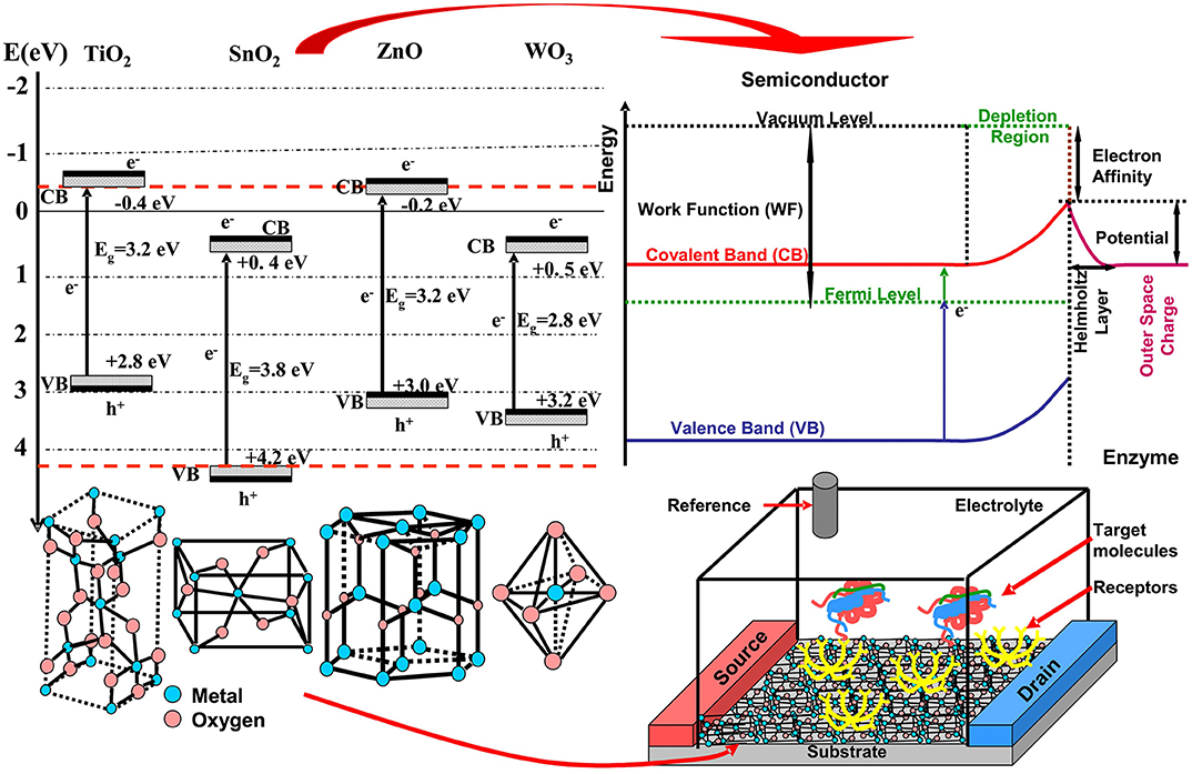
Figure 1. MOS used in biosensors: band energies, crystalline structure, and biosensor configuration.
Most of the metal oxides materials used in biosensors are wide band gap semiconductors (see Figure 1) consisting of various crystalline structures, unique electrochemical, optical, electronic, gravimetric, pyroelectric, and piezoelectric properties (Huang et al., 2016). Surface potential represents an important property in the biosensor application. The space charge effect is a result of native and imposed semiconductor potential. The bulk chemical composition, crystallization degree, and chemical interaction between semiconductor surface and the analyte will influence the displacement of Fermi energy and induced depletion (Cao et al., 2020). Consequently, the surface potential inducing the space charge double layer is directly dependent on the adsorbed layer characteristics of the electrode surface (Chen et al., 2019). Additionally, some of these materials may exhibit super hydro –phobicity/philicity, self-cleaning and antimicrobial activity. Other factors such as light exposure at certain wavelengths (~380 nm for TiO2 and ZnO, ~320 nm for SnO2 and ~443 nm for WO3) induce an increase of charge carrier mobility. During the light irradiation, oxygen vacancies are formed at the semiconductor surface, which can forward develop oxidative species (Ge et al., 2019).
An important advantage in the metal oxides semiconductor functionalization for biosensor application is represented by the low toxicity and low probability of negative interference with the common pharmaceutical compounds (Soldatkina et al., 2018). The major functionalization methodologies are based on covalent interactions (Feizabadi et al., 2019) but non-covalent interactions have been used as well (Ortiz et al., 2019). The covalent conjugation can be done using different molecules such as dimercaptosuccinic acid (DMSA), 1-ethyl-3-(3-dimethylaminopropyl) carbodiimide), (EDC), N-(15-carboxypentadecanoyloxy) succinimide, 16-(2-pyridyldithiol) hexadecanoic acid, etc. During the functionalization the MOS became more stable and reduced the nanoparticles aggregation, (Xu et al., 2020). After functionalization many analytes can be used as detecting materials for: urea, immunoglobulin, DNA, RNA, dopamine, cancer cells, viruses, etc.
The interactions between the bio-transducer and the analytes will alter the physico-chemical surface semiconductor characteristics. The surface potential, impedance or current characteristics can be correlated with the specific chemical stimuli induced by the analytes (Yoo et al., 2019). Various techniques are based on these characteristics, such as cyclic voltametry, impedimentric, differential pulse voltametry, etc.
The fidelity of the results from such a biosensor are however heavily influenced by the environmental factors of the experiment, such as temperature, humidity, pH, presence of oxygen, and foreign organic compounds, all of which can affect the stability of the interface. The applications of such biosensors spread across all domains, depending on the possible interactions between the immobilized biomolecules (enzymes, antibodies, DNA) and the analytes of interest.
Metal Oxide Semiconductors for Enzyme-Based Biosensors
There are various MOS's used in biosensor applications. The majority part use mono-component semiconductors but there are many papers (Oh et al., 2013; Kao et al., 2015) presenting multi-component semiconductors or coupled semiconductors (composite, tandem, heterostructures, etc.). Additionally, in order to enhance certain properties these materials have been coupled with metals nanoparticles or doped with other metal ions. The MOS exhibit a multitude of morphologies such as: rods, stars, flowers, cone, porous or dense films, etc. This mini review will consider only four metal oxides (TiO2, SnO2, ZnO, and WO3) as representative for biosensor applications. Many other papers which are not included here have the potential to contain highly innovative work. A summarized data collection containing the four metal oxides is presented in Table 1.
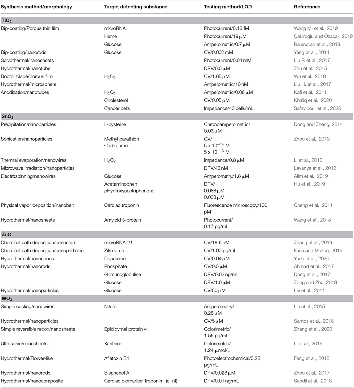
Table 1. Representative studies on metal oxide semiconductors used in biosensors.
TiO2-based Biosensors
TiO2 is an n-type semiconductor considered as a key material in many applications like photocatalysis, biosensors, photovoltaics, or energy storage due to his properties such as high chemical stability, biocompatibility, morphological versatility, etc.
Dip-coating technique was employed to obtain TiO2 films serving as sensors for microRNA (Wang M. et al., 2019), heme (Çakiroglu and Özacar, 2019), or glucose (Rajendran et al., 2018). The microRNA sensor is based on black TiO2 deposed on indium tin oxide (ITO) substrate and improved with Au nanopoarticles. The semiconductor was functionalized with histostar antibodies and based on photocurrent measurements the LOD was established at 0.13 fM. Photocurrent was used for LOD evaluation of heme using TiO2/ITO sensitized with CdS quantum dots, and the result was 19 μM. The glucose detector based on TiO2 film was functionalized with glucose oxidase (GOx) and the LOD was 0.7 μM. A better LOD value (0.5 μM) in glucose detection was obtained by replacing the TiO2 films with TiO2 nanotubes (Zhu et al., 2015). The nanotubes were developed using the hydrothermal method and functionalized with GOx. TiO2 nanorods (Yang et al., 2014) and nanosheets (Liu P. et al., 2017) were tested as glucose sensors after functionalizing with GO. Better LOD was obtained for nanorods morphology (0.002 mM) comparing with nanosheets (0.01 mM) mostly due to higher surface coverage, which was 3.32 × 10⁻¹¹ mol/cm. Concluding, the LOD depends mostly on the active surface, which explains why nanotubes give better results compared with TiO2 films.
TiO2 was used for H2O2 detection in the form of microspheres (Liu H. et al., 2017) or macro-mesoporous film (Wu et al., 2018). The TiO2 microspheres with a bag-like structure were hydrothermally obtained and functionalized with hemoglobin (Hem). The amperometric measurements indicate a LOD of 10 nM. This value is considerably lower compared with TiO2 mesoporous films (1.65 μM LOD) obtained by doctor blade technique and functionalized with horseradish peroxidase (HPOx).
Anodization of titanium was intensively used to obtain TiO2 nanotubes for H2O2 (Kafi et al., 2011), cholesterol (Khaliq et al., 2020) and breast cancer cell (Safavipour et al., 2020) detection. When the TiO2 nanotubes were functionalized with Hem the LOD evaluated by amperometry was 0.08 μM. Better LOD value was obtained for cholesterol detection (0.05 μM) based on a non-enzymatic approach to the oxidation process. Finally, TiO2 nanotubes were functionalized with human mucin-1 aptamers, inducing sensitive electrochemical detection of breast cancer cells (MCF-7). In this case the 40 cells/mL LOD represent an encouraging result for future biomedical TiO2 application in breast cancer detection.
SnO2-based Biosensors
Due to features such as high surface area, good biocompatibility, nontoxicity, excellent chemical stability, and catalytic activity, SnO2 was used in many applications such as light energy conversion, biosensors, smart windows, and electrochemistry. Tin oxide is an n-type semiconductor with a wide band gap of 3.8 eV and rutile structure (see Figure 1).
SnO2 nanoparticles were synthesized by precipitation (Dong and Zheng, 2014), sonication (Zhou et al., 2013) and microwave irradiation (Lavanya et al., 2012). By using precipitation method, the SnO2 nanoparticles have an average diameter of 4 nm and were used for L-cysteine detection. In order to employ the chronoamperometric detection method, the SnO2 surface was coated with multiwall carbon nanotubes giving a LOD of 0.03 μM. The SnO2 nanoparticles obtained by sonication methods were used for pesticide detection based on acetylcholinesterase as a functionalize agent. The LOD evaluated by cyclic voltametry (CV) was 5 × 10⁻¹⁴ for methyl parathion and 5 × 10⁻¹³ for carbofuran. The microwave irradiation method was employed to obtain SnO2 nanoparticles with application for H2O2 detection. SnO2 surface was functionalized with HPOx and, based on differential pulse voltammetry (DPV), a LOD of 43 nM was obtained.
An H2O2 sensor was developed using SnO2 nanowires synthesized by the thermal evaporation method (Li et al., 2010). Using the same functionalizing molecule as SnO2 nanoparticles, the LOD measured by CV was 0.8 μM. SnO2 nanowires were also obtained by the electrospinning method (Alim et al., 2019) for glucose amperometric detection. In this case the functionalizing procedure was done with both HPOx and GOx, giving a LOD of 1.8 μM. SnO2 nanobelts (Cheng et al., 2011) and nanosheets (Wang et al., 2018) were obtained by physical evaporation deposition, respectively using hydrothermal methods. The nanobelts functionalized with D-biotin molecules were successfully used as a troponin I detector, a protein marker for myocardial infarction (100 pM LOD). The nanosheets were used for amyloid β-protein (Aβ) detection after a previous functionalizing procedure with anti-Aβ antibody. Based on photocurrent measurements the LOD value was 0.17 pg/mL, considered as promising for applications in the detection of disease-related biomarkers.
ZnO-based Biosensors
ZnO is a direct wide band gap semiconductor which under UV radiation exhibits n-type conductivity. During the crystallization forms a hexagonal wurtzite structure (see Figure 1) which has particular piezoelectric properties based on noncentrosymmetric crystal structures. The major part of the ZnO synthesis procedures are wet techniques. Compared with tin oxide, ZnO has a better binding ability with biological entities, which is a prerequisite for future biosensor applications in medicine. Due to its nontoxicity and compatibility with human skin, ZnO can be adapted as a permanent human sensor in chronic diseases such as diabetes.
Chemical bath deposition (CBD) has been used (Zhang et al., 2019) to obtain ZnO nanostars for detecting microRNA-21 in cancer cells. Previously, the surface was functionalized using thiol-modified hairpin and hybridization chain reactions, considering the development of electrochemiluminescence (ECL) biosensors. The LOD was evaluated at 18.6 aM, which makes this material a good candidate for clinical bioassay. The same technique was also employed (Faria and Mazon, 2019) to develop ZnO nanoparticles for detection of Zika virus in undiluted urine. The Zika virus is transmitted through mosquito bites and gives symptoms such as headaches, arthralgia, myalgia, or conjunctivitis (Faria and Mazon, 2019). The ZIKV-NS1 antibody was immobilized using cystamine and glutaraldehyde on the ZnO nanoparticles. The LOD was evaluated using CV and the result was 1.00 pg/mL. This MOS biosensor can be used in early detection of the Zika virus.
Another technique that has been extensively used for ZnO synthesis with biosensing application is the hydrothermal procedure. Both ZnO nanorods (Zong and Zhu, 2018) and nanoparticles (Lei et al., 2011) hydrothermally obtained were used in biosensors for glucose detection. The ZnO nanorods where functionalized with GOx by simple immersion and the LOD via DPV was 1.0 μM. These results are significantly better compared with ZnO nanopowder functionalized with GOx, where LOD was 50 μM. ZnO nanorods where hydrothermally obtained and used as sensors for phosphate (Ahmad et al., 2017) and G Imunoglobuline (Dong et al., 2017) detection. For phosphate detection the ZnO was functionalized with pyruvate oxidase by immersion, and the LOD was 0.5 μM. In order to develop a G Imunoglobuline sensor with 0.03 ng/mL LOD, the ZnO surface was functionalized with myoglobin by immersion and cold drying. ZnO nanocone arrays were developed using the hydrothermal technique (Yuea et al., 2020) for dopamine detection. The nanocones were functionalized using Au nanoparticles with carboxyl groups obtaining a sensor with high sensitivity (4.36 μA/μM) and low LOD (0.04 μM).
WO3-based Biosensors
WO3 is an n-type semiconductor with a band gap of 2.8 eV and a versatile crystalline structure varying from cubic to octahedral, depending on the synthesis temperature. High surface to volume ratio WO3-based materials can be developed using physical and chemical techniques with well-controlled dimensionality, sizes, and crystal structure for sensors research.
A WO3 nitrite-based sensor was prepared by a simple casting (Liu et al., 2015) and hydrothermal (Santos et al., 2016) methods. In the first case WO3 has nanowire morphology and was functionalized with hemoglobin, while WO3 nanoparticles were obtained and functionalized with cytochrome c nitrite reductase using the hydrothermal procedure. The LOD value in the case of WO3 nanowire is significantly lower (0.28 μM) compared with WO3 nanopowder (5 μM) underlining the significance of semiconductor morphology, synthesis and functionalizing procedures. The hydrothermal method has been used to produce WO3 sensors with different morphologies: flower-like for aflatoxin B1 (Feng et al., 2018), nanorods for bisphenol A (Zhou et al., 2017), and nanosheets for cardiac biomarker Troponin I (Sandil et al., 2018). The WO3 flower-like morphology was functionalized with bovine serum albumin and the LOD corresponding to aflatoxin B1 was very low (0.28 pg/mL). The nanorods were doped with Na+ ions, giving a LOD value of 0.028 μM. Finally, the nanosheets were functionalized with 3-aminopropyl tri-ethoxy saline for the activation of amino groups and the LOD value was 0.01 ng/mL. In the case of WO3-based biosensors the nanosheets morphology gives a better LOD for cardiac biomarker Troponin I compared with the flower-like morphology used for alfatoxin B1 detection.
Other techniques were used to obtain WO3 nanosheets, such as simple reversible redox (Zhang et al., 2020) and ultrasonic exfoliation (Li et al., 2019) processes. Using the reversible redox process makes it possible to develop WO3 nanosheets with an average width of 150 nm, and LOD for human epididymal protein 4 detection of 1.56 pg/mL. Ultrasonic exfoliation of bulk WO3 in water allows the preparation of WO3 nanosheets in the range of 20 to 40 nm. These nanosheets were functionalized with 3, 3′, 5, 5′-tetramethylbenzidine and the LOD for xanthine in urine (based on colorimetric evaluation) was 1.24 μmol/L.
Conclusions
Metal oxides are considered as versatile materials that can be successfully integrated in biosensor technology. Based on features such as chemical stability, light excitation/light conversion, and high surface-to-volume ratio, these materials are highly competitive in the biosensors market. This mini review has outlined that the biosensors' LOD depends on many parameters such as: morphology (active surface), functionalizing molecule, evaluation procedure, and detecting target. The same material with similar morphology and detecting targets can give different LOD depending on surface functionalization and evaluation procedures. Even if most of the biosensors are used for glucose (H2O2, uric acid) there are encouraging results for cancer cell or virus detection. Recent advancements indicate a promising future for MOS in applications such as skin bioelectronics, neural interfaces, and smart biosensing devices. However, much effort is required to overcome important issues related to optimizing organic/inorganic interface compatibility, the enzyme electrochemistry at the MOS interface, and LOD improvement. To achieve this goal, facile MOS synthesis technologies allowing good interface control must be implemented.
Author Contributions
IŞ was responsible for article database regarding TiO2, SnO2, and WO3, including Table 1. AE has coordinated the writing, introduction, biosensors mechanism (including Figure 1), and article database for ZnO.
Conflict of Interest
The authors declare that the research was conducted in the absence of any commercial or financial relationships that could be construed as a potential conflict of interest.
References
Ahmad, R., Ahn, M. S., and Hahn, Y. B. (2017). ZnO nanorods array based field-effect transistor biosensor for phosphate detection. J. Colloid Interf. Sci. 498, 292–297. doi: 10.1016/j.jcis.2017.03.069
Alim, S., Kafi, A. K. M., Rajan, J., and Yusoff, M. M. (2019). Application of polymerized multiporous nanofiber of SnO2 for designing a bienzyme glucose biosensor based on HRP/Gox. Int. J. Biol. Macromol. 123, 1028–1034. doi: 10.1016/j.ijbiomac.2018.11.171
Çakiroglu, B., and Özacar, M. (2019). A self-powered photoelectrochemical biosensor for H2O2, and xanthine oxidase activity based on enhanced chemiluminescence resonance energy transfer through slow light effect in inverse opal TiO2. Biosens. Bioelectron. 141:111385. doi: 10.1016/j.bios.2019.111385
Cao, S. P., Hu, H. M., Liang, R. P., and Qiu, J. D. (2020). An ultrasensitive electrochemiluminescence resonance energy transfer biosensor for divalent mercury monitoring. J. Electroanal. Chem. 856:113494. doi: 10.1016/j.jelechem.2019.113494
Chen, D., Lv, L., Peng, L., Peng, J., and Tu, J. (2019). Controlled synthesis of mesoporous zinc oxide containing oxygen vacancies in low annealing temperature for photoelectrochemical biosensor. Ceram. Int. 45, 18044–18051. doi: 10.1016/j.ceramint.2019.06.024
Cheng, Y., Chen, K. S., Meyer, N. L., Yuan, J., Hirst, L. S., Chase, P. B., et al. (2011). Functionalized SnO2 nanobelt field-effect transistor sensors for label-free detection of cardiac troponin. Biosens. Bioelectron. 26, 4538–4544. doi: 10.1016/j.bios.2011.05.019
Dong, S., Tong, M., Zhang, D., and Huang, T. (2017). The strategy of nitrite and immunoassay human IgG biosensors basedon ZnO@ZIF-8 and ionic liquid composite film. Sens. Actuat. B-Chem. 251, 650–657. doi: 10.1016/j.snb.2017.05.047
Dong, Y., and Zheng, J. (2014). A nonenzymatic L-cysteine sensor based on SnO2-MWCNTs nanocomposites. J. Mol. Liq. 196, 280–284. doi: 10.1016/j.molliq.2014.03.046
Elahi, N., Kamali, M., Baghersad, M. H., and Amini, B. (2019). A fluorescence nano-biosensors immobilization on iron (MNPs) and gold (AuNPs) nanoparticles for detection of Shigella spp. Mater. Sci. Eng. C 105:110113. doi: 10.1016/j.msec.2019.110113
Enesca, A., Andronic, L., and Duta, A. (2012a). Optimization of opto-electrical and photocatalytic properties of SnO2 thin films using Zn2+ and W6+ dopant ions. Catal. Lett. 142, 224–230. doi: 10.1007/s10562-011-0762-4
Enesca, A., Andronic, L., and Duta, A. (2012b). The influence of surfactants on the crystalline structure, electrical and photocatalytic properties of hybrid multi-structured (SnO2, TiO2 and WO3) thin films. Appl. Surf. Sci. 258, 4339–4346. doi: 10.1016/j.apsusc.2011.12.110
Enesca, A., Isac, L., and Duta, A. (2015). Charge carriers injection in tandem semiconductors for dyes mineralization. Appl. Catal. B Environ. 162, 352–363. doi: 10.1016/j.apcatb.2014.06.059
Faria, A. M., and Mazon, T. (2019). Early diagnosis of zika infection using a ZnO nanostructures-based rapid electrochemical biosensor. Talanta 203, 153–160. doi: 10.1016/j.talanta.2019.04.080
Feizabadi, M., Soleymanpour, A., Faridnouri, H., and Ajloo, D. (2019). Improving stability of biosensor based on covalent immobilization of horseradish peroxidase by γ-aminobutyric acid and application in detection of H2O2. Int. J. Biol. Macromol. 136, 597–606. doi: 10.1016/j.ijbiomac.2019.06.103
Feng, J., Li, Y., Gao, Z., Lv, H., Zhang, H., Dong, Y., et al. (2018). A competitive-type photoelectrochemical immunosensor for aflatox in B1 detection based on flower-like WO3 as matrix and Ag2S-enhanced BiVO4 for signal amplification. Sens. Actuat. B-Chem. 270, 104–111. doi: 10.1016/j.snb.2018.05.015
Fiorani, A., Merino, J. P., Zanut, A., Criado, A., and Paolucci, F. (2019). Advanced carbon nanomaterials for electrochemiluminescent biosensor applications. Curr. Opin. Electrochem. 16, 66–74. doi: 10.1016/j.coelec.2019.04.018
Ge, L., Liu, Q., Jiang, D., Ding, L., and Wang, K. (2019). Oxygen vacancy enhanced photoelectrochemical performance of Bi2MoO6/B, N co-doped graphene for fabricating lincomycin aptasensor. Biosens. Bioelectron. 135, 145–152. doi: 10.1016/j.bios.2019.04.022
Gupta, S., Sharma, A., and Verma, R. S. (2020). Polymers in biosensor devices for cardiovascular applications. Curr. Opin. Biomed. Eng. 13, 69–75. doi: 10.1016/j.cobme.2019.10.002
Han, S., Zhang, Q., Zhang, X., Liu, X., and Zheng, G. (2019). A digital microfluidic diluter-based microalgal motion biosensor for marine pollution monitoring. Biosens. Bioelectron. 143:111597. doi: 10.1016/j.bios.2019.111597
Hernández-Cancel, G., Suazo-Dávila, D., Medina-Guzmán, J., Rosado-González, M., and Griebenow, K. (2015). Chemically glycosylation improves the stability of an amperometric horseradish peroxidase biosensor. Anal. Chim. Acta 854, 129–139. doi: 10.1016/j.aca.2014.11.008
Hocini, A., Temmar, M. N., Khedrouche, D., and Zamani, M. (2019). Novel approach for the design and analysis of a terahertz microstrip patch antenna based on photonic crystals. Photonics Nanostruct. 36:100723. doi: 10.1016/j.photonics.2019.100723
Hu, W., Zhang, Z., Li, L., Ding, Y., and An, J. (2019). Preparation of electrospun SnO2 carbon nanofiber composite for ultrasensitive detection of APAP and p-Hydroxyacetophenone. Sens. Actuat. B. 299:127003. doi: 10.1016/j.snb.2019.127003
Huang, Q., Wang, Y., Lei, L., Xu, Z., and Zhang, W. (2016). Photoelectrochemical biosensor for acetylcholinesterase activity study based on metal oxide semiconductor nanocomposites. J. Electroanal. Chem.781, 377–382. doi: 10.1016/j.jelechem.2016.07.007
Kafi, A. K. M., Wu, G., Benvenuto, P., and Chen, A. (2011). Highly sensitive amperometric H2O2 biosensor based on hemoglobin modified TiO2 nanotubes. J. Electroanal. Chem. 662, 64–69. doi: 10.1016/j.jelechem.2011.03.021
Kao, C. H., Chen, H., Yao, F., Hou, S., Chang, S. W., and Lin, M. H. (2015). Fabrication of multianalyte CeO2 nanograin electrolyte–insulator–semiconductor biosensors by using CF4 plasma treatment, sensing and bio-sensing Res. 5, 71–77. doi: 10.1016/j.sbsr.2015.07.001
Khaliq, N., Rasheed, M. A., Cha, G., Khan, M., Karim, S., Schmuki, P., et al. (2020). Development of non-enzymatic cholesterol bio-sensor based on TiO2 nanotubes decorated with Cu2O nanoparticles. Sens. Actuat. B 302:127200. doi: 10.1016/j.snb.2019.127200
Lavanya, N., Radhakrishnan, S., and Sekar, C. (2012). Fabrication of hydrogen peroxide biosensor based on Ni doped SnO2 nanoparticles. Biosens. Bioelectron. 36, 41–47. doi: 10.1016/j.bios.2012.03.035
Lei, Y., Yan, X., Zhao, J., Liu, X., Song, Y., Luo, N., et al. (2011). Improved glucose electrochemical biosensor by appropriate immobilization of nano-ZnO, Colloid. Surface. B 82, 168–172. doi: 10.1016/j.colsurfb.2010.08.034
Li, L., Huang, J., Wang, Y., Zhang, H., Liu, Y., and Li, J. (2010). An excellent enzyme biosensor based on Sb-doped SnO2 nanowires. Biosens. Bioelectron. 25, 2436–2441. doi: 10.1016/j.bios.2010.03.037
Li, Z., Liu, X., Liang, X. H., Zhong, J., Guo, L., and Fu, F. (2019). Colorimetric determination of xanthine in urine based on peroxidase-like activity of WO3 nanosheets. Talanta 204, 278–284. doi: 10.1016/j.talanta.2019.06.003
Liu, C. H., Hong, M. H., Lum, M. C., Flotow, H., Ghadessy, F, and Zhang, J. B. (2010). Large-area micro/nanostructures fabrication in quartz by laser interference lithography and dry etching. Appl. Phy. A. 101, 237–241. doi: 10.1007/s00339-010-5807-9
Liu, H., Duan, C., Yang, C., Chen, X., Shen, W., and Zhu, Z. (2015). A novel nitrite biosensor based on the direct electron transfer hemoglobin immobilized in the WO3 nanowires with high length–diameter ratio. Mater. Sci. Eng. C 53, 43–49. doi: 10.1016/j.msec.2015.04.016
Liu, H., Guo, K., Duan, C., Dong, X., and Gao, J. (2017). Hollow TiO2 modified reduced graphene oxide microspheres encapsulating hemoglobin for a mediator-free biosensor. Biosens. Bioelectron. 87, 473–479. doi: 10.1016/j.bios.2016.08.089
Liu, P., Huo, X., Tang, Y., Xu, J., Liu, X., and Wong, K. Y. (2017). A TiO2 nanosheet-g-C3N4 composite photoelectrochemical enzyme biosensor excitable by visible irradiation. Anal. Chim. Acta 984, 86–95. doi: 10.1016/j.aca.2017.06.043
Mihaly, M., Lacatusu, I., Enesca, A., and Meghea, A. (2008). Hybride nanomaterials based on silica coated C-60 clusters obtained by microemulsion technique. Mol. Cryst. Liq. Cryst. 483, 205–215. doi: 10.1080/15421400801906885
Oh, J., Yoo, G., Chang, Y. W., Kim, H. J., and Yoo, K. H. (2013). A carbon nanotube metal semiconductor field effect transistor-based biosensor for detection of amyloid-beta in human serum. Biosens. Bioelectron. 50, 345–350. doi: 10.1016/j.bios.2013.07.004
Ortiz, E., Gallay, P., Galicia, L., Eguílaz, M., and Rivas, G. (2019). Nanoarchitectures based on multi-walled carbon nanotubes non-covalently functionalized with Concanavalin A: a new building-block with supramolecular recognition properties for the development of electrochemical biosensors. Sens. Actuat. B-Chem. 292, 254–262. doi: 10.1016/j.snb.2019.04.114
Osman, D. I., El-Sheikh, S. M., Sheta, S. M., Ali, O. I., and Shawky, S. M. (2019). Nucleic acids biosensors based on metal-organic framework (MOF): paving the way to clinical laboratory diagnosis. Biosens. Bioelectron. 141:111451. doi: 10.1016/j.bios.2019.111451
Rajendran, S., Manoj, D., Raju, K., Dionysiou, D. D., Naushad, M., Gracia, F., et al. (2018). Influence of mesoporous defect induced mixed-valent NiO (Ni2+/Ni3+)-TiO2 nanocomposite for non-enzymatic glucose biosensors. Sens. Actuat. B 264, 27–37. doi: 10.1016/j.snb.2018.02.165
Ramon-Marquez, T., Medina-Castillo, A. L., Fernandez-Gutierrez, A., and Fernandez-Sanchez, J. F. (2018). Evaluation of two sterically directed attachments of biomolecules on a coaxial nanofibre membrane to improve the development of optical biosensors. Talanta 187, 83–90. doi: 10.1016/j.talanta.2018.05.004
Rathinamala, I., Jeyakumaran, N., and Prithivikumaran, N. (2019). Sol-gel assisted spin coated CdS/PS electrode based glucose biosensor. Vacuum 161, 291–296. doi: 10.1016/j.vacuum.2018.12.045
Rezaei, H., Motovali-bashi, M., and Radfar, S. (2019). An enzyme-free electrochemical biosensor for simultaneous detection of two hemophilia A biomarkers: Combining target recycling with quantum dots-encapsulated metal-organic frameworks for signal amplification. Anal. Chim. Acta 1092, 66–74. doi: 10.1016/j.aca.2019.09.037
Safavipour, M., Kharaziha, M., Amjadi, E., Karimzadeh, F., and Allafchian, A. (2020). TiO2 nanotubes/reduced GO nanoparticles for sensitive detection of breast cancer cells and photothermal performance, Talanta 208:120369. doi: 10.1016/j.talanta.2019.120369
Sandil, D., Srivastava, S., Malhotra, B. D., Sharma, S. C., and Puri, N. K. (2018). Biofunctionalized tungsten trioxide-reduced graphene oxide nanocomposites for sensitive electrochemical immunosensing of cardiac biomarker. J. Alloy. Compd. 763, 102–110. doi: 10.1016/j.jallcom.2018.04.293
Santos, L., Silveira, C. M., Elangovan, E., Neto, J. P., Nunes, D., Pereira, L., et al. (2016). Synthesis of WO3 nanoparticles for biosensing applications. Sens. Actuat. B-Chem. 223, 186–194. doi: 10.1016/j.snb.2015.09.046
Scognamiglio, V., Antonacci, A., Arduini, F., Moscone, D., and Palleschi, G. (2019). An eco-designed paper-based algal biosensor for nanoformulated herbicide optical detection. J. Hazard. Mater. 373, 483–492. doi: 10.1016/j.jhazmat.2019.03.082
Solaimuthu, A., Vijayan, A. N., Murali, P., and Korrapati, P. S. (2020). Nano-biosensors and their relevance in tissue engineering, Curr. Opin. Biomed. Eng. 13, 84–93. doi: 10.1016/j.cobme.2019.12.005
Soldatkina, O. V., Soldatkin, O. O., Velychko, T. P., Prilipko, V. O., and Dzyadevych, S. V. (2018). Conductometric biosensor for arginine determination in pharmaceutics. Bioelectrochemistry 124, 40–46. doi: 10.1016/j.bioelechem.2018.07.002
Song, H., Zhang, Y., Wang, S., Huang, K., and Xu, W. (2020). Label-free polygonal-plate fluorescent-hydrogel biosensor for ultrasensitive microRNA detection. Sensor. Actuat. B-Chem. 306:27554. doi: 10.1016/j.snb.2019.127554
Visa, M., Andronic, L., and Enesca, A. (2016). Behavior of the new composites obtained from fly ash and titanium dioxide in removing of the pollutants from wastewater. Appl. Surf. Sci. 388, 359–369. doi: 10.1016/j.apsusc.2015.12.154
Wang, M., Yin, H., Zhou, Y., Sui, C., Wang, Y., Meng, X., et al. (2019). Photoelectrochemical biosensor for microRNA detection based on a MoS2/g-C3N4/black TiO2 heterojunction with Histostar@AuNPs for signal Amplification. Biosens. Bioelectron. 128, 137–143. doi: 10.1016/j.bios.2018.12.048
Wang, Q., Zhao, Y., Yang, Q., Du, D., and Lin, Y. (2019). Amperometric sarcosine biosensor with strong anti-interference capabilities based on mesoporous organic-inorganic hybrid materials. Biosens. Bioelectron. 141:111431. doi: 10.1016/j.bios.2019.111431
Wang, Y., Fan, D., Zhao, G., Feng, J., Wei, D., Zhang, N., et al. (2018). Ultrasensitive photoelectrochemical immunosensor for the detection of amyloid β-protein based on SnO2/SnS2/Ag2S nanocomposites. Biosens. Bioelectron. 120, 1–7. doi: 10.1016/j.bios.2018.08.026
Wang, Y. H., Huang, K. J., and Wu, X. (2017). Recent advances in transition-metal dichalcogenides based electrochemical biosensors: a review. Biosens. Bioelectron. 97, 305–316. doi: 10.1016/j.bios.2017.06.011
Wu, X., Zhang, H., Huang, K., Zeng, Y., and Zhu, Z. (2018). Rose petal and P123 dual-templated macro-mesoporous TiO2 for a hydrogen peroxide biosensor. Bioelectrochemistry 120, 150–156. doi: 10.1016/j.bioelechem.2017.12.009
Xu, B., Huang, J., Ding, L., and Cai, J. (2020). Graphene oxide-functionalized long period fiber grating for ultrafast label-free glucose biosensor. Mater. Sci. Eng. C 107:110329. doi: 10.1016/j.msec.2019.110329
Yang, Z., Tang, Y., Li, J., Zhang, Y., and Hu, X. (2014). Facile synthesis of tetragonal columnar-shaped TiO2 nanorods for the construction of sensitive electrochemical glucose biosensor. Biosens. Bioelectron. 54, 528–533. doi: 10.1016/j.bios.2013.11.043
Yi, Y., Xie, B., Zhao, T., Qian, Z., and Liu, H. (2020). The effect of anode hydrodynamics on the sensitivity of microbial fuel cell based biosensors and the biological mechanism. Bioelectrochemistry 132:107351. doi: 10.1016/j.bioelechem.2019.107351
Yilmaz, Y. Y., Yalcinkaya, E. E., Demirkol, D. O., and Timur, S. (2020). 4-aminothiophenol-intercalated montmorillonite: Organic-inorganic hybrid material as an immobilization support for biosensors. Sens. Actuat. B-Chem. 307:127665. doi: 10.1016/j.snb.2020.127665
Yin, M.-J., Gu, B., An, Q.-F., Yang, C., and Yong, K.-T. (2018). Recent development of fiber-optic chemical sensors and biosensors: Mechanisms, materials, micro/nano-fabrications and applications. Coordin. Chem. Rev. 376, 348–392. doi: 10.1016/j.ccr.2018.08.001
Yoo, T., Lim, K., Sultan, M. T., Lee, J. S., and Jang, M. (2019). The real-time monitoring of drug reaction in HeLa cancer cell using temperature/impedance integrated biosensors, Sens. Actuat. B-Chem. 291, 17–24. doi: 10.1016/j.snb.2019.03.145
Yuan, Y., Wang, Y., Wang, H., and Hou, S. (2019). Gold nanoparticles decorated on single layer graphene applied for electrochemical ultrasensitive glucose biosensor. J. Electroanal. Chem. 855:113495. doi: 10.1016/j.jelechem.2019.113495
Yuea, H. Y., Zhanga, H. J., Huanga, S., Lua, X. X., Gaoa, X., Songa, S. S., et al. (2020). Highly sensitive and selective dopamine biosensor using Au nanoparticles-ZnO nanocone arrays/graphene foam electrode. Mater. Sci. Eng. C 108:110490. doi: 10.1016/j.msec.2019.110490
Zhang, B., Wang, H., Xi, J., Zhao, F., and Zeng, B. (2020). In situ formation of inorganic/organic heterojunction photocatalyst of WO3/Au/ polydopamine for immunoassay of human epididymal protein 4. Electrochim. Acta 331:135350. doi: 10.1016/j.electacta.2019.135350
Zhang, X., Li, W., Zhou, Y., Chai, Y., and Yuan, R. (2019). An ultrasensitive electrochemiluminescence biosensor for MicroRNA detection based on luminol-functionalized Au NPs@ZnO nanomaterials as signal probe and dissolved O2 as coreactant. Biosens. Bioelectron.135, 8–13. doi: 10.1016/j.bios.2019.04.004
Zhao, D., Liu, Y., Zhang, Q., Zhang, Y., and Sang, S. (2019). Surface stress-based biosensor with stable conductive AuNPs network for biomolecules detection. Appl. Surf. Sci. 491, 443–450. doi: 10.1016/j.apsusc.2019.06.178
Zheng, H., Liu, M., Yan, Z., and Chen, J. (2020). Highly selective and stable glucose biosensor based on incorporation of platinum nanoparticles into polyaniline-montmorillonite hybrid composites. Microchem. J. 152:104266. doi: 10.1016/j.microc.2019.104266
Zhou, Q., Yang, L., Wang, G., and Yang, Y. (2013). Acetylcholinesterase biosensor based on SnO2 nanoparticles–carboxylic graphene–nafion modified electrode for detection of pesticides. Biosens. Bioelectron. 49, 25–31. doi: 10.1016/j.bios.2013.04.037
Zhou, Y., Yang, L., Li, S., and Dang, Y. (2017). A novel electrochemical sensor for highly sensitive detection of bisphenol A based on the hydrothermal synthesized Na-doped WO3 nanorods. Sens. Actuat. B-Chem. 245, 238–246. doi: 10.1016/j.snb.2017.01.034
Zhu, J., Liu, X., Wang, X., Huo, X., and Yan, R. (2015). Preparation of polyaniline–TiO2 nanotube composite for the development of electrochemical biosensors. Sens. Actuat. B 221, 450–457. doi: 10.1016/j.snb.2015.06.131
Keywords: metal oxides, semiconductors, nanostructures, energy bands, biosensors
Citation: Şerban I and Enesca A (2020) Metal Oxides-Based Semiconductors for Biosensors Applications. Front. Chem. 8:354. doi: 10.3389/fchem.2020.00354
Received: 04 February 2020; Accepted: 06 April 2020;
Published: 19 May 2020.
Edited by:
Liwei Wang, Guangxi University, ChinaReviewed by:
Zhongqiu Hua, Hebei University of Technology, ChinaLiping Gao, Chuzhou University, China
Copyright © 2020 Şerban and Enesca. This is an open-access article distributed under the terms of the Creative Commons Attribution License (CC BY). The use, distribution or reproduction in other forums is permitted, provided the original author(s) and the copyright owner(s) are credited and that the original publication in this journal is cited, in accordance with accepted academic practice. No use, distribution or reproduction is permitted which does not comply with these terms.
*Correspondence: Alexandru Enesca, YWVuZXNjYUB1bml0YnYucm8=