- 1Department of Intensive Care Medicine, The First Affiliated Hospital of Soochow University, Suzhou, China
- 2Department of Nursing, The First Affiliated Hospital of Soochow University, Suzhou, China
Purpose: This study systematically reviewed and elucidated the current status and key determinants of enteral nutrition interruption (ENI) in critically ill patients. By shedding light on these factors, we aimed to furnish compelling evidence to mitigate the occurrence of ENI in this critical setting.
Methods: We embarked on a comprehensive search across seven prominent databases, PubMed, Embase, Web of Science, Cochrane Library, Scopus, EBSCO, and Ovid Medline, spanning from their inception to 27 May 2024. Two independent researchers meticulously screened and assessed the quality of the literature, extracting data on the current status and influencing factors of ENI. This rigorous approach culminated in a descriptive systematic review and analysis.
Results: From an initial pool of 2,984 studies, 28 were deemed suitable for inclusion in this review, comprising 20 cross-sectional and eight cohort studies. Moreover, 16 studies highlighted ENI incidence rates ranging from 4.7% to a staggering 100%, with an overall average of 48.3%. Among 17 studies, a total of 4,890 ENI episodes were reported involving 2,008 critically ill patients, translating to an average of 2–3 episodes per patient. Four studies detailed the cumulative ENI duration in 327 critically ill patients, totaling 11037.2 h, with an individual average of 33.8 h per patient. The analysis revealed four primary factors influencing ENI: procedures, gastrointestinal events, feeding tube problems, and hemodynamic instability. Procedures accounted for 29.8%–85.0% of ENI frequency and 34.6%–81.2% of duration, with averages of 63.4% and 52.1%, respectively. Gastrointestinal events contributed to 9.4%–59.7% of ENI frequency and 11.5%–21.4% of duration, averaging 19.2% and 18.1%. Feeding tube problems ranged from 0.9% to 29.3% in frequency and 1.3%–25.6% in duration, with averages of 9.3% and 11.6%. Hemodynamic instability was responsible for 0.9%–20.0% of ENI frequency and 1.1%–5.1% of duration, averaging 3.9% and 2.6%.
Conclusion: The incidence and frequency of ENI in critically ill patients are notably high, with interruptions lasting for extended durations. The primary culprits, procedures, gastrointestinal events, feeding tube problems, and hemodynamic instability, influenced ENI occurrence.
Systematic review registration: https://www.crd.york.ac.uk/prospero/display_recordphp?ID=CRD42024554417, identifier CRD42024554417.
1 Introduction
Nutritional status is recognized as a nursing-sensitive outcome and plays critical role in patient recovery and overall wellbeing in critically ill patients. Adequate nutrition is of vital importance in critically ill patients (1). It plays a key role in modulating inflammatory responses, maintaining immune function, slowing skeletal muscle catabolism, promoting tissue repair, and maintaining the gastrointestinal and pulmonary mucosal barrier (2, 3). Meanwhile, adequate nutrition has been shown to reduce infection complications (P < 0.03) (4), shorten intensive care unit (ICU) stays (P < 0.01) (5), reduce mortality (P < 0.01) (6), and enhance long-term recovery (7, 8). However, several studies have shown that critically ill patients receive only around 60% of their targeted nutritional goal (9). That insufficient caloric and protein intake increases the risk of malnutrition (10). Lew et al. (11) reported that the prevalence of malnutrition in the ICU ranges from 38% to 78%. Malnourished patients are at higher risk of complications, including infections, pressure ulcers, impaired wound healing, and prolonged hospital stays, which ultimately result in higher mortality and increased healthcare costs (2, 12).
A common reason for inadequate intake of calories and protein in critically ill patients is an enteral nutrition interruption (ENI) (13). However, there is currently no universal consensus on the definition of ENI. Through a systematic review of the current literature, we have identified notable discrepancies in the criteria used to define ENI across various studies. Some studies define ENI based on its frequency, considering any single interruption in enteral nutrition (EN) support for critically ill patients as an ENI event (14). Others define ENI by its duration, though there is no universal agreement on the precise time threshold. A commonly used criterion is an interruption lasting 1 h or more during continuous EN infusion. For intermittent infusion, ENI is defined as administering EN three times daily for 30 min each, with the patient failing to receive the expected nutrition within that time frame (15, 16). A recent study showed that 68% of patients had a period of ENI during their stay in the ICU (17). ENI impact clinical outcomes and prognosis in critically ill patients. Compared to patients without ENI, the occurrence of ENI is associated with a higher rate of inadequate feeding (54.0% vs. 15.0%) (17) and an increased mortality rate (46.0% vs. 21.7%) (18). Additionally, having three or more interruptions during the ICU stay is associated with a higher risk of mortality (19). The increased mortality may be associated with various factors, which not only contribute to the mortality rate of critically ill patients but could also be one of the causes leading to the occurrence of ENI. For example, frequent diagnostic or therapeutic procedures are one such factor, as they not only interrupt EN delivery but also increase metabolic stress and energy expenditure. Similarly, gastrointestinal intolerance often leads to ENI, which may reflect underlying systemic inflammation or organ dysfunction, affecting the patient’s poor prognosis.
In conclusion, given the role of adequate nutrition in modulating clinical outcomes in critically ill patients, a comprehensive assessment of the current status of ENI and its influencing factors is necessary. Currently, there has not yet been a systematic review published on the status and influencing factors of ENI. Thus, the systematic review aims to explore the status and influencing factors of ENI in critically ill patients including the status of ENI incidence, frequency and duration.
2 Methods
This review was pre-registered in PROSPERO (CRD42024554417) and adhered to the Preferred Reporting Items for Systematic Reviews and Meta-analyses (PRISMA) guidelines (20). PRISMA 2020 checklist are provided in Supplementary Tables 1, 2.
2.1 Aim
The aim of this study was to investigate the incidence, frequency, and duration of ENl, with the ultimate goal of providing valuable insights to inform and improve patient care strategies in critical care settings.
2.2 Search strategy
We systematically searched the electronic databases PubMed, Embase, Web of Science, Cochrane Library, Scopus, EBSCO, and Ovid Medline for eligible studies from their inception to 27 May 2024. The search was limited to full-text articles available in English. Initially, the inclusion of the qualifier “ICU” resulted in a limited amount of literature. Consequently, the search strategy was refined by reducing the emphasis on “ICU” to enhance the retrieval of relevant studies. Following a series of preliminary searches, the final search strategy was determined. This involved combining subject terms and free terms and employing Boolean logic operators to optimize retrieval accuracy. Additionally, the reference lists of identified articles were manually reviewed to uncover any further relevant publications. Detailed search strategies are provided in Supplementary Table 3.
2.3 Inclusion and exclusion criteria
Inclusion criteria were as follows: (1) critically ill patients aged 18 years or older receiving EN support; (2) observational studies, including cross-sectional, cohort, and case-control studies; (3) primary or secondary outcome measures included the current status or influencing factors of ENI.
Exclusion criteria were as follows: (1) reviews, conference abstracts, lectures, animal experiments, reader letters, and research protocols; (2) studies with unavailable data extraction; (3) duplicate publications; (4) literature without full-text access; (5) articles with quality assessment scores below five points.
2.4 Study selection and quality assessment
Based on the search results, the literature was first imported into the reference manager EndNote X9 for deduplication. Two researchers then independently screened the literature according to the inclusion and exclusion criteria. The screening process involved initially reviewing the titles and abstracts, followed by reading the full texts to determine eligibility. Additionally, the references of included articles were manually searched to identify further relevant studies. In cases where consensus could not be reached, a third researcher was consulted. The quality of the included studies was independently evaluated by two researchers. Cross-sectional studies were assessed using the scale recommended by the Agency for Healthcare Research and Quality (AHRQ), which includes 11 items evaluated with “yes,” “no,” or “unsure.” Each “yes” answer earns one point, resulting in a total score ranging from 0 to 11. Studies scoring 0–3 points were classified as low quality, 4–7 points as medium quality, and 8–11 points as high quality. The quality evaluation of cohort studies was conducted using the Newcastle-Ottawa Scale (NOS). The NOS comprises eight items categorized into three dimensions: selection, comparability, and outcome. Comparability can score up to two points, while each of the other items can score up to one point, with a total score ranging from 0 to 9. Based on the score, literature quality was categorized as low quality (1–3 points), medium quality (4–6 points), or high quality (7–9 points) (21).
2.5 Data extraction
Relevant information was extracted from the included literature using a standardized data collection form. The extracted data included: (1) basic information: first author, year of publication, country, department; (2) study type; (3) main inclusion criteria; (4) follow-up duration; (5) outcome indicators; (6) factors influencing ENI; (7) Acute Physiology and Chronic Health Evaluation II (APACHE II) score; (8) number of patients on mechanical ventilation and duration of mechanical ventilation; (9) start time of EN, feeding route, and infusion method of EN after ICU admission; (10) number of patients receiving EN; (11) number of patients with ENI, frequency of ENI, and duration of ENI; (12) main findings.
2.6 Data analysis
This study described the current status of ENI in critically ill patients by calculating the average incidence, average frequency, and average duration of ENI. Additionally, data on relevant influencing factors reported in two or more studies were combined by summing up the frequency and duration of ENI and then calculating the average proportion based on factor classification. Factors that could not be combined were subjected to descriptive analysis only.
3 Results
3.1 Selection process and quality assessment
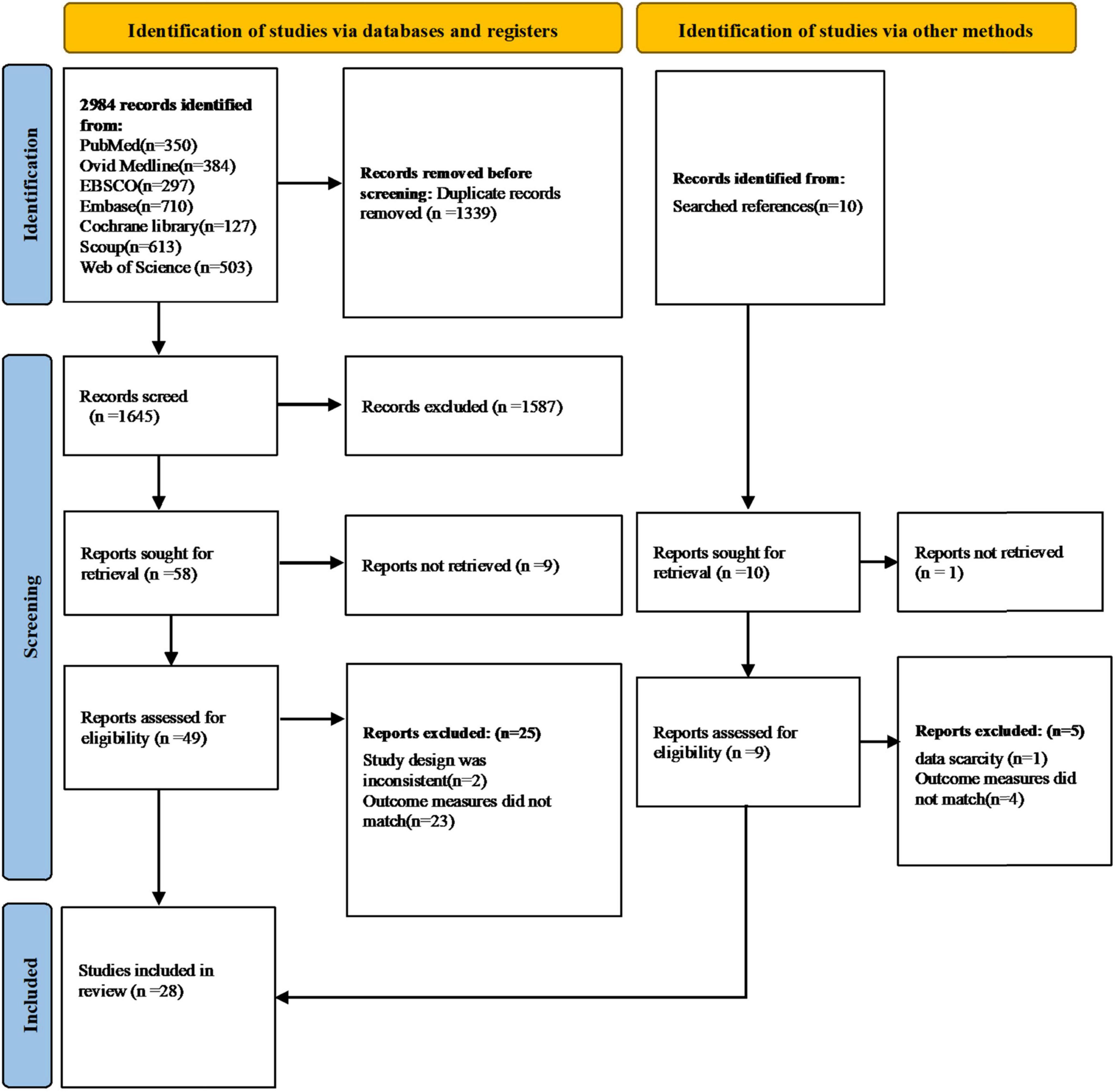
Figure 1 illustrates the selection process, detailing the number of studies at each review stage. The database search initially identified 2,984 relevant articles. After removing 1,339 duplicates, 1,645 articles were screened. From these, 1,587 were excluded based on titles and abstracts, and nine articles were inaccessible in full text, leaving 49 articles for full-text screening. Ultimately, 24 articles met the inclusion criteria, all of which were met the criteria of quality assessment. A supplementary manual search of the reference lists of these articles yielded an additional four relevant studies, resulting in a total of 28 articles included in the review (13, 14, 17, 19, 22–45). The characteristics of the included studies are detailed in Table 1.
Out of the 28 studies, 20 were cross-sectional, and eight were cohort studies. Among the cross-sectional studies, nine were of high quality, and 11 were of medium quality. For the cohort studies, five were of high quality, and three were of medium quality. The detailed quality assessment results are presented in Tables 2, 3.
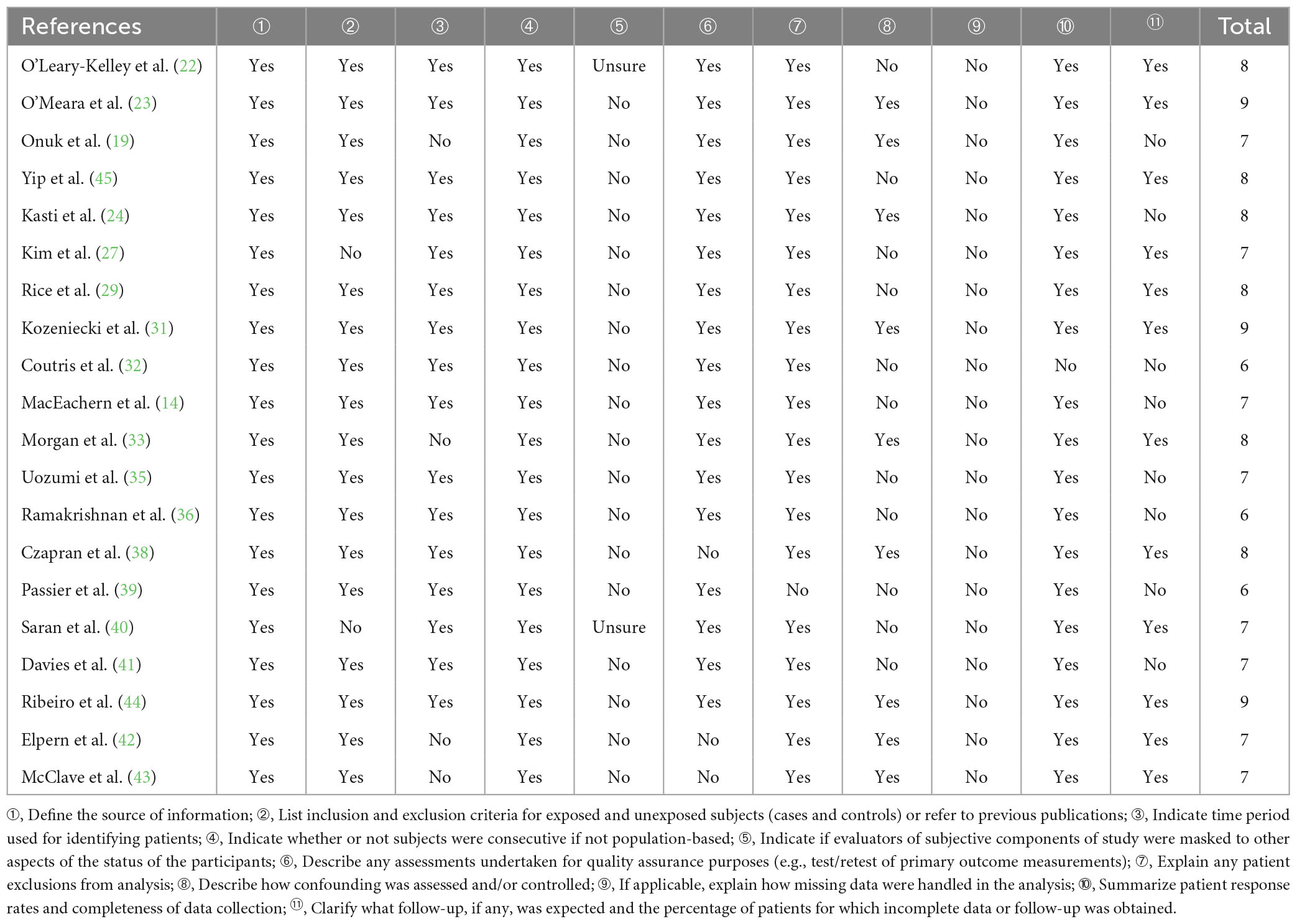
Table 2. Agency for Healthcare Research and Quality (AHRQ) scale to assess the quality of cross-sectional studies.
3.2 The current status of ENI
Given the heterogeneity of EN feeding routes (gastric and post-pyloric), infusion methods (continuous and intermittent), patient characteristics (illness severity, mechanical ventilation status, and EN contraindications), and outcome metrics (ENI incidence, frequency, and duration), a meta-analysis could not be conducted to consolidate the data. Thus, a descriptive analysis of ENI in critically ill patients was performed.
Among the 28 studies included, 16 studies (13, 17, 22, 24–29, 36–41, 45) reported ENI incidence, 17 studies (14, 17, 19, 23, 25–36, 45) reported ENI frequency, and four studies (23, 30, 32, 44) reported ENI duration. The incidence of ENI was reported in 16 studies (13, 17, 22, 24–29, 36–41, 45), of which 2,412 critically ill patients received EN, 1,165 developed ENI, with incidence ranging from 4.7% to 100.0%, and an average rate of 48.3%. Subgroup analyses were conducted based on ICU type, geographical region and publication year. The incidence of ENI were 64.2% in mixed/general ICU, 55.3% in surgical ICU, 25.3% in medical ICU, Studies published in 2014 or earlier reported an incidence of 79.8%, while those published in 2015 or later reported 42.1%. By geographic region, the incidence was 66.5% in Europe, 63.1% in Asia, 31.5% in the Americas and 76.4% in Oceania.
Seventeen studies reported ENI frequency (14, 17, 19, 23, 25–36, 45), with the number of episodes ranging from 1 to 7 in those studies, and a total of 4,890 episodes occurring in 2,008 patients, averaging 2–3 episodes per patient during their ICU stay. Four studies reported the duration of ENI in 327 patients (23, 30, 32, 44), with ranging from 24.3 to 46.6 h in those studies, totaling 11037.2 h during the ICU stay, an average of 33.8 h per patients.
3.3 Influencing factors of ENI
All 28 studies (13, 14, 17, 19, 22–45) included in this systematic review reported factors influencing ENI. Due to the heterogeneity of the studies, a descriptive analysis was conducted. Factors mentioned in at least two studies were combined by summing the frequency and duration of ENI and calculating the average proportion for each category.
The factors were grouped into four categories: procedures, gastrointestinal events, feeding tube problems, and hemodynamic instability. Procedural factors included airway procedures (e.g., intubation, extubation, and tracheostomy), therapeutic procedures (e.g., surgery, dialysis, and drainage), diagnostic procedures (e.g., imaging and endoscopy), and nursing procedures (e.g., bathing and dressing changes). Gastrointestinal events include intolerance [e.g., high gastric residual volume (GRV > 250 m) (46), abdominal distension, diarrhea, abdominal pain, nausea, vomiting, and reflux aspiration] along with other complications (e.g., gastrointestinal bleeding, ileus, and anastomotic leaks). Feeding tube problems cover problems such as blockage, displacement, and dislodgement. Hemodynamic instability [mean arterial pressure (MAP) < 65 mmHg] (46) is characterized by symptoms including shock, weakness, instability, and discomfort. A detailed descriptive analysis of these four categories and the combined results is presented in Figures 2, 3.
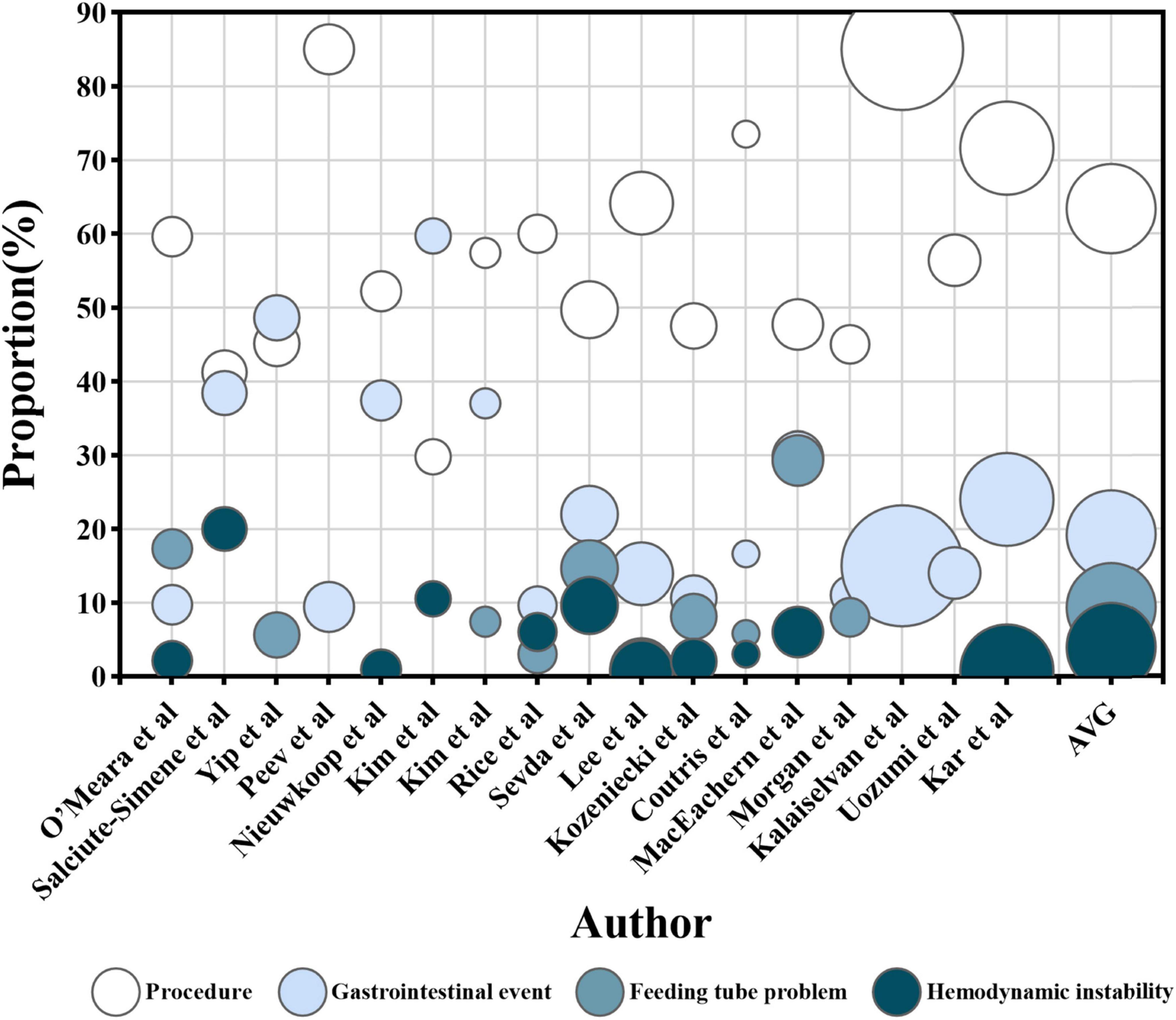
Figure 2. The proportion of enteral nutrition interruption (ENI) frequency. The horizontal axis represents each research, while the vertical axis represents the proportion of ENI frequency caused by each influencing factor out of the total frequency of ENI in single research. The color of the circles represents four influencing factors, and the size of the circles represents the number of participants receiving EN in each research. The AVG represents the average proportion of ENI frequency caused by four different factors, relative to the total ENI frequency, with the circle’s area being meaningless.
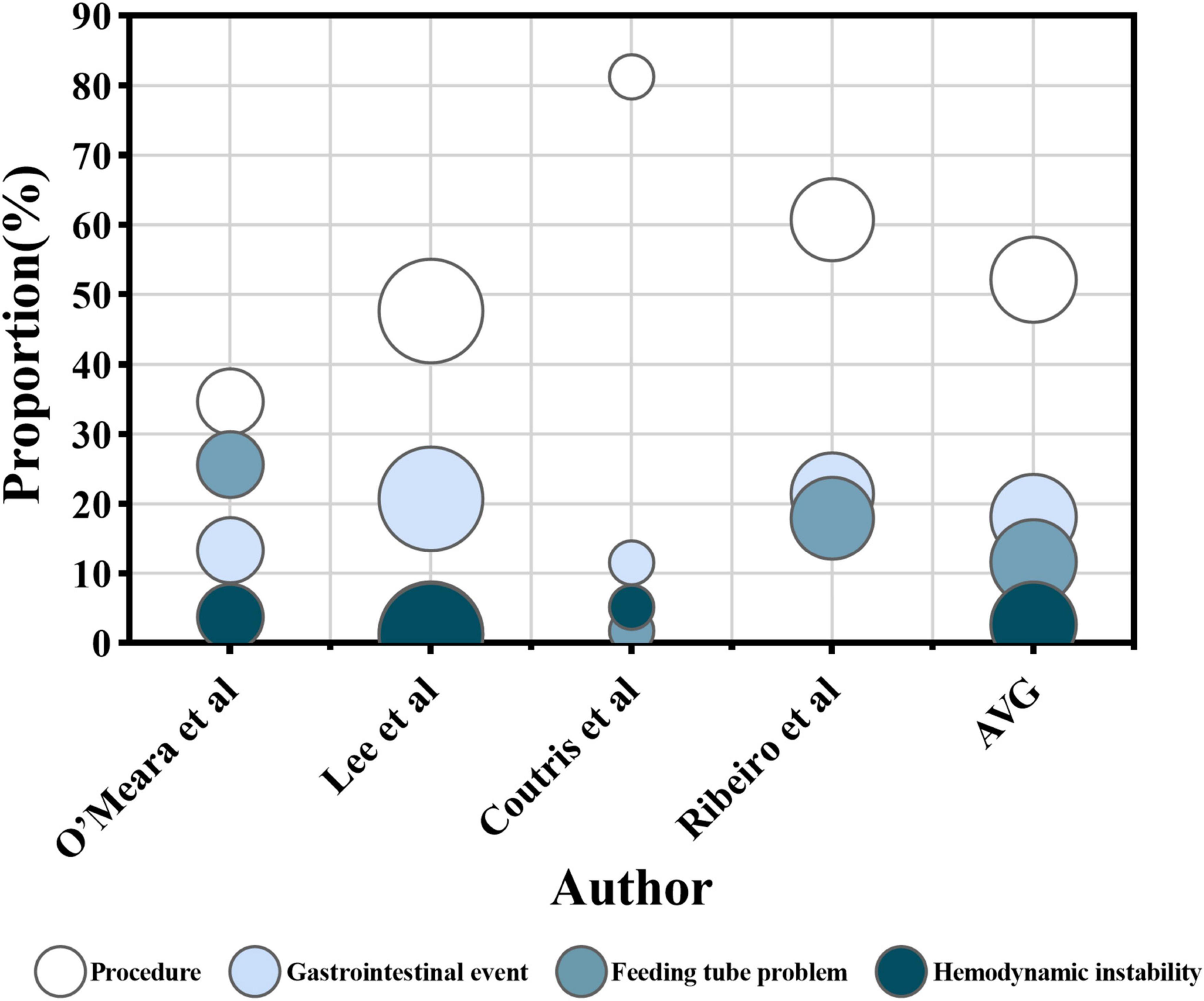
Figure 3. The proportion of enteral nutrition interruption (ENI) duration. The horizontal axis represents each research, while the vertical axis represents the proportion of ENI duration caused by each influencing factor out of the total duration of ENI in single research. The color of the circles represents four influencing factors, and the size of the circles represents the number of participants receiving EN in each research. The AVG represents the average proportion of ENI duration caused by four different factors, relative to the total ENI duration, with the circle’s area being meaningless.
3.3.1 Procedural factors
Among the 28 studies analyzed, 27 identified procedure as an important factor influencing ENI (13, 14, 17, 19, 22–39, 41–45). A total of 17 identified a total of 4,890 interruptions (14, 17, 19, 23, 25–36, 45), with 63.4% on average attributed to procedural factors. The proportion of ENI frequency due to procedures ranged from 29.8% to 85.0% in those studies (Figure 2). Subgroup analyses were conducted based on ICU type, geographical region and publication year. The proportion of ENI frequency due to procedures was 67.8% in mixed/general ICU, 54.5% in surgical ICU and 53.5% in medical ICU. Studies published in 2014 or earlier reported the proportion of 51.7%, while those published in 2015 or later reported 66.7%. By geographic region, the proportion was 48.3% in Europe, 69.3% in Asia and 57.1% in the Americas. Four studies reported a total interruption time of 11037.2 h (23, 30, 32, 44), of which procedural factors contributed to 5746.5 h with an average of 52.1%). The proportion of ENI duration due to procedures ranged from 34.6% to 81.2% in those studies (Figure 3).
Procedural types contributed to ENI with varying frequency (Figure 4): airway procedures (14, 17, 19, 23, 25–32, 34, 35) were the most frequent, accounting for an average of 29.1% of ENI (1,089 of 3,739). Therapeutic (17, 19, 23, 25–27, 30–35) and diagnostic (17, 19, 23, 25–27, 30–34) procedures accounted for 15.0% (537 of 3,577) and 12.0% (360 out of 3,010) of ENI, respectively. Nursing procedures (23, 28, 29, 33, 35, 36) contributed 7.5% (173 of 2,302).
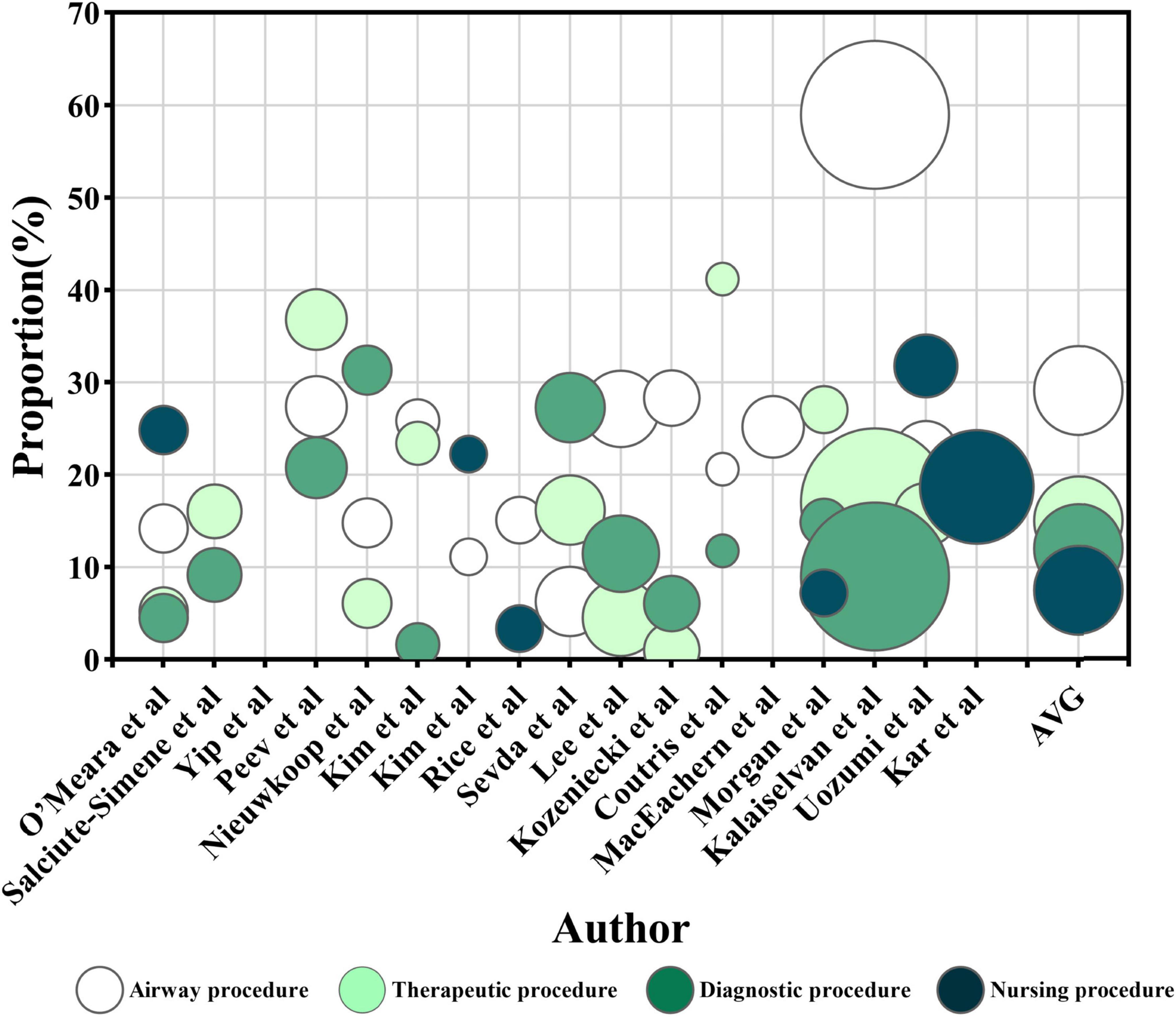
Figure 4. The proportion of enteral nutrition interruption (ENI) frequency caused by different categories of procedures. The horizontal axis represents each research, while the vertical axis represents the proportion of ENI frequency caused by various procedure categories out of the total frequency of ENI in a single article. The color of the circles represents the category of the procedure, and the size of the circles represents the number of participants receiving EN in each research. The AVG represents the average proportion of ENI frequency caused by different procedure categories relative to the total ENI frequency, with the circle’s area being meaningless.
Regarding ENI duration (Figure 5), airway procedures (23, 30, 32, 44) accounted for 27.1% (2986.4 h) of the total ENI time, while therapeutic (23, 30, 32) procedures accounted for 12.1% (964.1 h) and diagnostic procedures (30, 32, 44) for 11.7% (993 h). Nursing procedures (23, 44) caused 5.9% (330 h) of ENI.
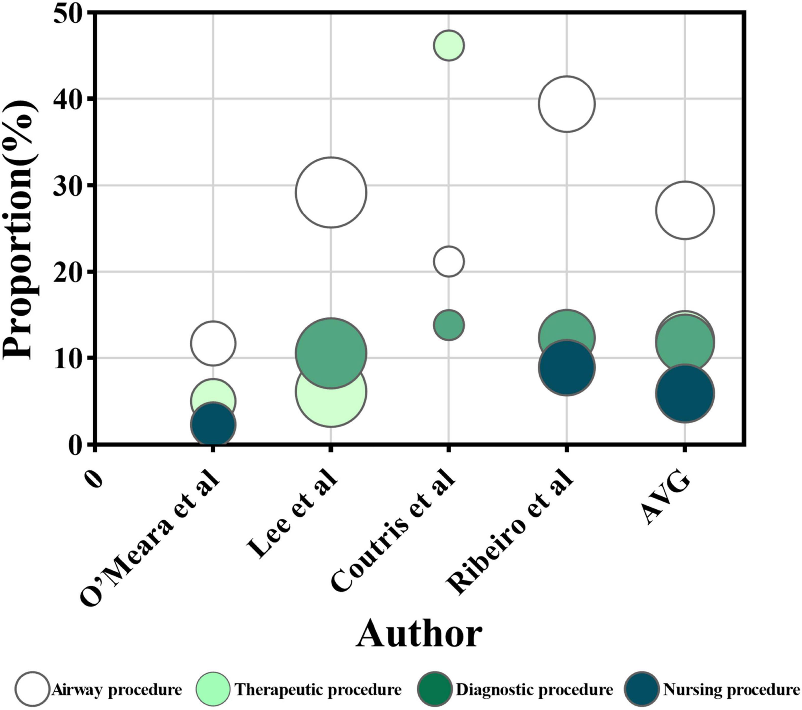
Figure 5. The proportion of enteral nutrition interruption (ENI) duration caused by different categories of procedures. The horizontal axis represents each research, while the vertical axis represents the proportion of ENI duration caused by various procedure categories out of the total duration of ENI in a single article. The color of the circles represents the category of the procedure, and the size of the circles represents the number of participants receiving EN in each research. The AVG represents the average proportion of ENI duration caused by different procedure categories relative to the total ENI duration, with the circle’s area being meaningless.
3.3.2 Gastrointestinal events
Among the 28 studies reviewed, 26 identified gastrointestinal events as a key factor influencing ENI (13, 14, 17, 19, 22–36, 38, 40–45). Seventeen studies (14, 17, 19, 23, 25–36, 45) reported a total ENI frequency of 4,890 episodes, with 940 attributed to gastrointestinal events, accounting for an average of 19.2% of ENI frequency. The proportion of ENI frequency due to gastrointestinal events ranged from 9.4% to 59.7% in those studies (Figure 2). Subgroup analyses were conducted based on ICU type, geographical region, and publication year. The proportion of ENI frequency due to gastrointestinal events was 19.8% in mixed/general ICU, 22.6% in surgical ICU and 15.4% in medical ICU. Studies published in 2014 or earlier reported the proportion of 19.7%, while those published in 2015 or later reported 19.1%. By geographic region, the proportion was 28.6% in Europe, 20.4% in Asia and 12.7% in the Americas. Four studies (23, 30, 32, 44) reported a total ENI duration of 11037.2 h, with 2002.8 h due to gastrointestinal events representing 18.1% of the total ENI duration. The proportion of ENI duration due to gastrointestinal events ranged from 11.5% to 21.4% in those studies (Figure 3).
Gastrointestinal events were further categorized to assess their contribution to ENI frequency (Figure 6). High GRV was identified as a significant factor in 13 studies (14, 17, 19, 23, 25–32, 45), causing 230 interruptions out of 2,321, averaging 9.9% of the total ENI. Nausea, vomiting, and reflux aspiration accounted for 7.4% of ENIs (191 of 2,595) (14, 19, 23, 26, 27, 29–32, 36, 45), while abdominal pain, diarrhea, and bloating accounted for 6.1% (126 of 2,075) (14, 17, 19, 30, 31, 36, 45). Gastrointestinal bleeding caused 4.5% of ENIs (82 of 1,803) (17, 26), while anastomotic leaks and ileus accounted for 2.4% and 1.4% (17, 19, 26, 27), respectively.
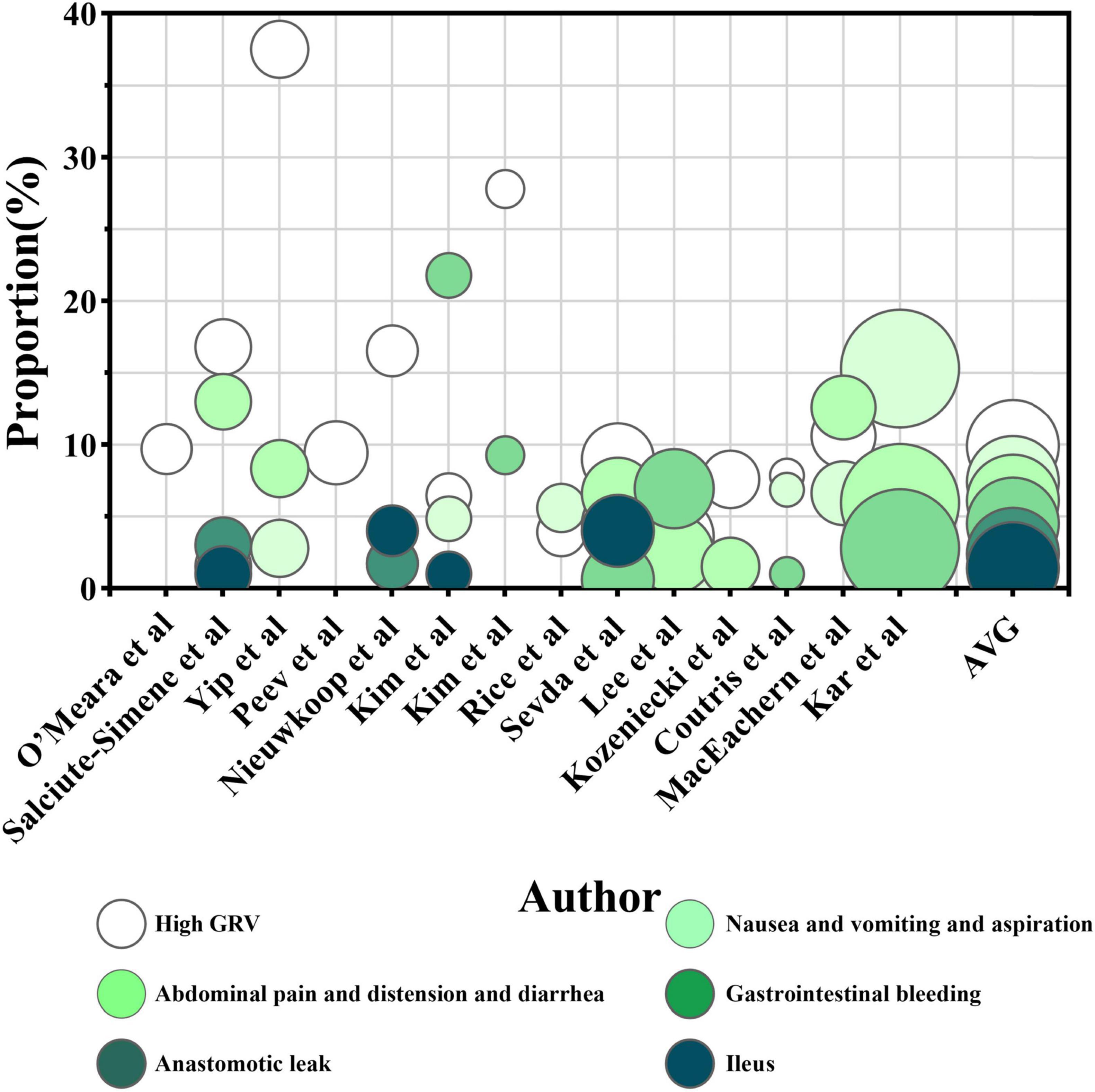
Figure 6. The proportion of enteral nutrition interruption (ENI) frequency caused by different categories of gastrointestinal events. The horizontal axis represents each research, while the vertical axis represents the proportion of ENI frequency caused by various gastrointestinal event categories out of the total frequency of ENI in a single article. The color of the circles represents the category of the gastrointestinal event, and the size of the circles represents the number of participants receiving EN in each research. The AVG represents the average proportion of ENI frequency caused by different gastrointestinal event categories relative to the total ENI frequency, with the circle’s area being irrelevant.
In terms of ENI duration (Figure 7), high GRV was the primary contributor, causing 521.8 h of ENI across three studies, averaging 6.5% of the total ENI time (23, 30, 32). Nausea, vomiting, and reflux aspiration contributed 46 h, accounting for 0.8% of the total ENI duration (30, 32). The studies conducted by Ribeiro et al. (44), O’Leary-Kelley et al. (22), Elpern et al. (42) did not categorize gastrointestinal intolerance but reported it as a significant factor contributing to 21.4%, 19.8%, and 22.8% of ENI duration, respectively.
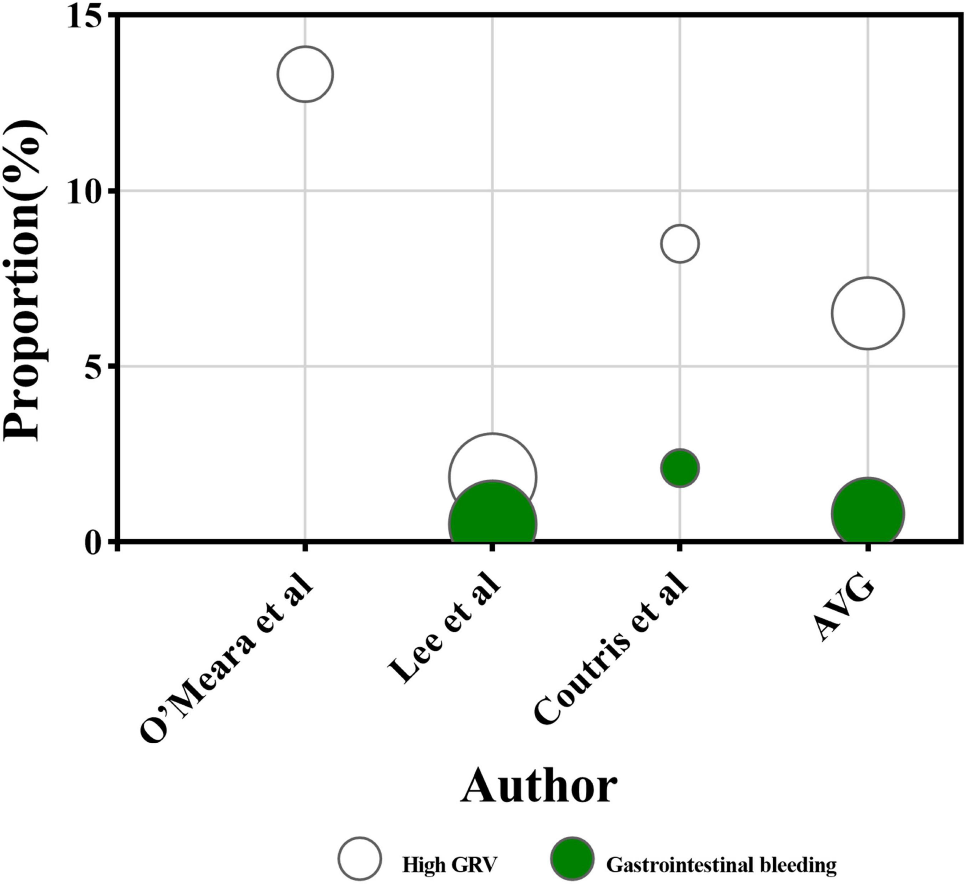
Figure 7. The proportion of ENI duration caused by different categories of gastrointestinal events. The horizontal axis represents each research, while the vertical axis represents the proportion of ENI duration caused by various gastrointestinal event categories out of the total duration of ENI in a single article. The color of the circles represents the category of the gastrointestinal event, and the size of the circles represents the number of participants receiving EN in each research. The AVG represents the average proportion of ENI duration caused by different gastrointestinal event categories relative to the total ENI duration, with the circle’s area being irrelevant.
3.3.3 Feeding tube problems
Among the 28 studies reviewed, 16 identified (14, 19, 22, 23, 28–33, 38, 41–45) feeding tube problems as a significant factor influencing ENI. Ten studies (14, 19, 23, 28–33, 45) reported a total ENI frequency of 2,067 episodes, with 193 attributed specifically to feeding tube problems, accounting for 9.3% of ENI frequency on average. The proportion of ENI frequency due to feeding tube problems ranged from 0.9% to 29.3% in those studies (Figure 2). Subgroup analyses were conducted based on ICU type, geographical region, and publication year. The proportion of ENI frequency due to feeding tube problems was 3.7% in mixed/general ICU, 7.4% in surgical ICU and 14.1% in medical ICU. Studies published in 2014 or earlier reported the proportion of 11.1%, while those published in 2015 or later reported 7.9%. By geographic region, the proportion was 14.6% in Europe, 2.6% in Asia and 10.4% in the Americas. Additionally, four studies (23, 30, 32, 44) documented a total interruption duration of 11037.2 h, with 1276.0 h caused by feeding tube problems, representing 11.6% of the total ENI duration. The proportion of ENI duration due to feeding tube problems ranged from 1.3% to 25.6% in those studies (Figure 3).
In the study by Onuk et al. (19), the median ENI duration was 960 min during the ICU stay, with feeding tube problems resulting in the longest interruption, lasting 1,230 min. Similarly, O’Meara et al. (23) found that feeding tube problems accounted for 17.3% of the total ENI frequency and 25.6% of the total ENI duration, respectively.
3.3.4 Hemodynamic instability
Among the 28 studies reviewed, 12 identified hemodynamic instability as a factor influencing ENI (14, 17, 19, 23, 26, 27, 29–32, 36, 42). Eleven studies (14, 17, 19, 23, 26, 27, 29–32, 36) reported 2,946 interruptions, with 116 episodes attributed specifically to hemodynamic instability, accounting for 3.9% of the total ENI frequency on average. The proportion of ENI frequency due to hemodynamic instability ranged from 0.9% to 20.0% in those studies (Figure 2). Subgroup analyses were conducted based on ICU type, geographical region, and publication year. The proportion of ENI frequency due to hemodynamic instability was 3.1% in mixed/general ICU, 7.1% in surgical ICU and 4.7% in medical ICU. Studies published in 2014 or earlier reported the proportion of 2.5%, while those published in 2015 or later reported 5.6%. By geographic region, the proportion was 10.2% in Europe, 1.6% in Asia and 3.4% in the Americas. Additionally, three studies (23, 30, 32) documented a total interruption duration of 7988.2 h, with 204.9 h attributed to hemodynamic instability, representing 2.6% of the total ENI duration. The proportion of ENI duration due to hemodynamic instability ranged from 1.1% to 5.1% in those studies (Figure 3).
In a prospective study by Salciute-Simene et al. (17), 73 critically ill patients experienced a total of 26 episodes of ENI (20.0%) due to hemodynamic instability. Similarly, Elpern et al. (42) reported that hemodynamic instability accounted for 13.5% of the total ENI duration.
4 Discussion
4.1 The current status of ENI
The review showed that the incidence and frequency of ENI was high, and ENI duration was prolonged in critically ill patients. The result was consistent with the studies by Liu et al. (48), indicating that approximately half of the patients experienced ENI, and EN was interrupted on 35% of the trial days.
However, the incidence (4.7%–100.0%), frequency (1–7 episodes during ICU stay), and duration (24.3–46.6 h during ICU stay) of ENI varied widely in critically ill patients. This variability can be attributed to inconsistencies in how ENI is defined across studies, particularly regarding interruption duration, which affects the comparability of results. For instance, O’Leary-Kelley et al. (22) define ENI as interruptions lasting over 15 min, while Salciute-Simene et al. (17) set the threshold at 1 h. A 15 min threshold captures shorter ENI events, such as repositioning or suctioning, that may not significantly impact nutrition intake, thereby increasing ENI incidence, frequency, and duration. Conversely, a 1 h threshold focuses on interruptions that disrupt nutritional delivery, such as those caused by surgeries, while overlooking brief ENI, resulting in lower reported incidence, frequency, and duration. Additionally, the clinical status of critically ill patients and the feeding route used can influence ENI. Severely ill patients, due to their condition or acute changes, often undergo procedures that extend ENI (47). Moreover, gastric feeding is more prone to complications such as gastric residuals, reflux, and vomiting, resulting in longer and more frequent ENI compared to post-pyloric feeding (48). To reduce this heterogeneity, future studies should standardize the definition of ENI, focusing on a patient-centered approach and establishing a consistent threshold for ENI duration and frequency. Additionally, uniform inclusion and exclusion criteria should be adopted across studies to ensure a more accurate assessment of ENI’s true impact on critically ill patients.
Subgroup analysis revealed that the incidence of ENI was higher in mixed/general ICU, studies published in 2014 or earlier and Oceania compared to other subgroups. This disparity may stem from the integration of medical and surgical ENI factors in mixed/general ICU. Meanwhile, the continuous advancement of medical technology and the growing emphasis placed by healthcare professionals on the management of ENI in critically ill patients may potentially reduce the incidence risk of ENI (49, 50). Notably, the high incidence in Oceania might be attributed to limited sample size (only one study included), which could introduce statistical power insufficiency as a potential bias.
4.2 Influencing factors of ENI
4.2.1 Procedures
The results showed that procedures were the primary factor influencing ENI among critically ill patients, with airway procedures being the most common cause. Furthermore, procedural factors consistently accounted for a relatively large proportion across all subgroup analyses, which may be due to the advancements in medical technology since 2015 have increased the frequency of examinations and invasive procedures, further elevating ENI risks. Consistent with this finding, a prospective observational study by Lee et al. (30) had reported that airway procedures accounted for the highest frequency and duration of ENI.
Current clinical practices for pre-procedural feeding interruption in critically ill patients are primarily extrapolated from perioperative fasting guidelines designed for elective surgical patients, with no unified practice standard. The 2017 Practice Guidelines for Preoperative Fasting by the American Society of Anesthesiologists (ASA) recommend discontinuing liquid intake 6 h prior to procedures to reduce the risk of pulmonary aspiration in critically ill patients (51). In addition, in critically ill patients undergoing endotracheal intubation, diagnostic, or therapeutic procedures, anesthetic agents reduce respiratory muscle strength, inducing respiratory depression (52). Concurrently, certain pneumoperitoneum surgery may elevate intra-abdominal pressure (IAP), then transmitting through the diaphragm to increase intrathoracic pressure, which reduces pulmonary compliance and exacerbates respiratory depression (53, 54). In this context, continuous EN may elevates gastric pressure, which is associated with elevated IAP. Elevated IAP may impair respiratory mechanics (55, 56). Therefore, interrupting EN may improving respiratory depression. Moreover, the updated 2023 ASA guidelines further specify that clear liquids should be withheld for at least 2 h before anesthesia or procedural sedation (57). In light of these recommendations, ICU staff could carefully coordinate medical procedures to minimize unnecessary enteral nutrition interruptions and ensure both nutritional adequacy and patient safety.
4.2.2 Gastrointestinal events
The result showed that gastrointestinal events had a slightly lower impact on the occurrence of ENI in critically ill patients compared to procedural factors, with GRV being the most common factor. Kim et al. (28) showed that gastrointestinal events were the main contributors to ENI, accounting for about 60% of all ENI frequency. That may be attributed to the inclusion of critically ill patients with contraindications to enteral nutrition, many of whom had pre-existing gastrointestinal symptoms, such as intestinal obstruction or gastrointestinal bleeding, at the time of enrollment.
According to the 2023 international guidelines (4) and supporting evidence from clinical studies such as Salciute-Simene et al. (17), temporary ENI is recommended for critically ill patients experiencing gastrointestinal intolerance, including high GRV, nausea/vomiting, or diarrhea. The reason for ENI caused by high GRV lies in its association with delayed gastric emptying and increased risk of aspiration. However, discrepancies exist in GRV thresholds for ENI across studies: while the guidelines define GRV ≥ 500 mL/6 h as the critical threshold (4), study has shown that higher GRV ranges (250–500 mL) do not statistically correlate with adverse outcomes such as aspiration pneumonia (58, 59). Hence, for critically ill patients with GRV < 500 mL, post-pyloric feeding may be prioritized. If symptoms persist, prokinetic agents could be considered. For issues that cannot be resolved by prokinetic agents or repositioning the feeding tube, short-term ENI may be used as an emergency measure (59). For diarrhea management, the guidelines advocate initial etiological evaluation and EN regimen optimization, Persistent diarrhea may benefit from probiotic-supplemented formulas to restore gut microbiota balance (59, 60). For high-aspiration-risk patients, post-pyloric feeding via nasojejunal tubes may be preferred, reducing reflux episodes compared to gastric feeding (4, 61).
4.2.3 Feeding tube problems
The impact of feeding tube problems on ENI was slightly less significant than that of procedural and gastrointestinal event factors. Similarly, Sevda et al. (19), Nieuwkoop et al. (26) also reported that the proportion of ENI caused by feeding tube problems was relatively low compared to those caused by procedures and gastrointestinal events.
Feeding tube problems such as tube blockage, dislodgement or kinking may hinder nutrient delivery. In such situations, temporarily ENI allows for tube replacement or repositioning, which may prevent further gastrointestinal injury. To minimize ENI caused by feeding tube problems, regular monitoring, proper fixation and routine flushing protocols are essential. These measures may help maintain tube patency and ensure uninterrupted, effective nutritional support.
4.2.4 Hemodynamic instability
Although the frequency and duration of ENI caused by hemodynamic instability was relatively low, its impact should not be underestimated. Salciute-Simene et al. (17) Showed that hemodynamic instability was a major factor in ENI, contributing the highest proportion. This is likely due to the severe nature of the patients’ conditions in this trial, with many suffering from comorbidities including 72.0% critically ill patients having septic shock and 36.6% requiring vasopressors.
In critically ill patients with hemodynamic instability, particularly those in shock or receiving vasopressors (NE ≥ 0.1 μg/kg/min) (62), patients with NE ≥ 0.1 μg/kg/min exhibit significantly lower citrulline levels (< 10 μmol/L) and elevated intestinal fatty acid-binding protein (I-FABP > 150 pg/mL), indicating mucosal ischemia (63, 64). Continuing EN may further compromise gastrointestinal blood flow, worsen ischemia, and increase the risk of complications such as non-occlusive mesenteric ischemia or intestinal necrosis (65). Therefore, to minimize further gastrointestinal damage with hemodynamic instability, it is advisable to temporarily interrupt EN when vasopressors (NE ≥ 0.1 μg/kg/min) are used, as this can exacerbate mucosal ischemia. Once hemodynamic stability is restored or the vasopressor dose reduced (NE < 0.05 μg/kg/min), EN may be cautiously reintroduced, starting with a low dose to ensure safe and effective delivery.
4.3 Implications
This study systematically evaluated 28 studies to identify four influencing factors of ENI: procedures, gastrointestinal events, feeding tube problems and hemodynamic instability. Early recognition of these factors enables targeted interventions to minimize unnecessary ENI. Specifically, ICU staff should adhere to evidence-based guidelines to minimize preoperative fasting durations. When gastrointestinal events occur, a tiered management approach should be implemented (e.g., adjusting infusion rates, modifying formulas and adding prokinetic agents). For feeding tube problems, establish protocols for regular monitoring, secure fixation and routine flushing. Additionally, critically ill patients with hemodynamic instability initiate low dose EN only after hemodynamic stabilization (e.g., NE ≤ 0.05 μg/kg/min). These measures may prevent ENI or reduce unnecessary ENI duration.
4.4 Limitations
The systematic review also has several limitations. This study only included English-language literature. This study was dedicated to conducting a comprehensive systematic review, meticulously examining the occurrence and determinants of ENI in ICU patients to provide valuable insights and foster improved patient care strategies. Furthermore, due to the heterogeneity in definitions across the included studies, we did not conduct a meta-analysis. The variations in how ENI was defined and measured across studies precluded a quantitative synthesis of the data. Instead, we conducted a descriptive systematic review, which limits the ability to draw definitive conclusions. In addition, the lack of a standardized definition for high volume of gastric residue, reflux and diarrhea across the included studies further constrained the comparability of this outcome. Therefore, future studies should standardize those definitions and include research from a broader range of languages to improve the generalizability and robustness of the findings.
5 Conclusion
The incidence and frequency of ENI in critically ill patients are notably high, with interruptions often lasting for extended durations. Various factors, such as procedures, gastrointestinal events, feeding tube problems, and hemodynamic instability, significantly influence ENI occurrence.
Data availability statement
The raw data supporting the conclusions of this article will be made available by the authors, without undue reservation.
Author contributions
XL: Writing – original draft, Writing – review and editing. XW: Writing – original draft, Writing – review and editing. WY: Writing – review and editing, Writing – original draft. JC: Writing –original draft, Writing – review and editing. YW: Writing – review and editing, Writing – original draft. YC: Writing – original-draft. LD: Writing – review and editing. QW: Writing – review and editing.
Funding
The author(s) declare that no financial support was received for the research and/or publication of this article.
Conflict of interest
The authors declare that the research was conducted in the absence of any commercial or financial relationships that could be construed as a potential conflict of interest.
Publisher’s note
All claims expressed in this article are solely those of the authors and do not necessarily represent those of their affiliated organizations, or those of the publisher, the editors and the reviewers. Any product that may be evaluated in this article, or claim that may be made by its manufacturer, is not guaranteed or endorsed by the publisher.
Supplementary material
The Supplementary Material for this article can be found online at: https://www.frontiersin.org/articles/10.3389/fnut.2025.1462131/full#supplementary-material
References
1. Al-Dorzi HM, Arabi YM. Nutrition support for critically ill patients. JPEN J Parenter Enteral Nutr. (2021) 45:47–59. doi: 10.1002/jpen.2228
2. Sharma K, Mogensen KM, Robinson MK. Pathophysiology of critical Illness and role of nutrition. Nutr Clin Pract. (2019) 34:12–22. doi: 10.1002/ncp.10232
3. Stumpf F, Keller B, Gressies C, Schuetz P. Inflammation and nutrition: Friend or foe? Nutrients. (2023) 15:1159. doi: 10.3390/nu15051159
4. Singer P, Blaser AR, Berger MM, Calder PC, Casaer M, Hiesmayr M, et al. ESPEN practical and partially revised guideline: Clinical nutrition in the intensive care unit. Clin Nutr. (2023) 42:1671–89. doi: 10.1016/j.clnu.2023.07.011
5. Labeau SO, Afonso E, Benbenishty J, Blackwood B, Boulanger C, Brett SJ, et al. Prevalence, associated factors and outcomes of pressure injuries in adult intensive care unit patients: The DecubICUs study. Intensive Care Med. (2021) 47:160–9. doi: 10.1007/s00134-020-06234-9
6. Docking RI. Nutritional support in the critically ill. Anaesthesia Intensive Care Med. (2024) 25:63–5. doi: 10.1016/j.mpaic.2023.10.004
7. Reignier J, Rice TW, Arabi YM, Casaer M. Nutritional support in the ICU. Bmj. (2025) 388:e077979. doi: 10.1136/bmj-2023-077979
8. Boeykens K. Nutritional support in the intensive care unit: Implications for nursing care from evidence-based guidelines and supporting literature. Dimens Crit Care Nurs. (2021) 40:14–20. doi: 10.1097/dcc.0000000000000448
9. Heyland DK, Dhaliwal R, Wang M, Day AG. The prevalence of iatrogenic underfeeding in the nutritionally ‘at-risk’ critically ill patient: Results of an international, multicenter, prospective study. Clin Nutr. (2015) 34:659–66. doi: 10.1016/j.clnu.2014.07.008
10. Cederholm T, Barazzoni R, Austin P, Ballmer P, Biolo G, Bischoff SC, et al. ESPEN guidelines on definitions and terminology of clinical nutrition. Clin Nutr. (2017) 36:49–64. doi: 10.1016/j.clnu.2016.09.004
11. Lew CC, Yandell R, Fraser RJ, Chua AP, Chong MF, Miller M. Association between malnutrition and clinical outcomes in the intensive care unit: A systematic review. JPEN J Parenter Enteral Nutr. (2017) 41:744–58. doi: 10.1177/0148607115625638
12. Pohlenz-Saw JA, Merriweather JL, Wandrag L. (Mal)nutrition in critical illness and beyond: A narrative review. Anaesthesia. (2023) 78:770–8. doi: 10.1111/anae.15951
13. Ritter CG, Medeiros IM, Pádua CS, Gimenes FR, Prado PR. Risk factors for protein-caloric inadequacy in patients in an intensive care unit. Rev Bras Ter Intensiva. (2019) 31:504–10. doi: 10.5935/0103-507x.20190067
14. Maceachern KN, Kraguljac AP, Mehta S. Nutrition care of critically ill patients with leukemia: A retrospective study. Can J Diet Pract Res. (2019) 80:34–8. doi: 10.3148/cjdpr-2018-033
15. Kim H, Stotts NA, Froelicher ES, Engler MM, Porter C, Kwak H. Adequacy of early enteral nutrition in adult patients in the intensive care unit. J Clin Nurs. (2012) 21:2860–9. doi: 10.1111/j.1365-2702.2012.04218.x
16. Stechmiller J, Treloar DM, Derrico D, Yarandi H, Guin P. Interruption of enteral feedings in head injured patients. J Neurosci Nurs. (1994) 26:224–9. doi: 10.1097/01376517-199408000-00006
17. Salciute-Simene E, Stasiunaitis R, Ambrasas E, Tutkus J, Milkevicius I, Sostakaite G, et al. Impact of enteral nutrition interruptions on underfeeding in intensive care unit. Clin Nutr. (2021) 40:1310–7. doi: 10.1016/j.clnu.2020.08.014
18. Martins JR, Shiroma GM, Horie LM, Logullo L, Silva Mde L, Waitzberg DL. Factors leading to discrepancies between prescription and intake of enteral nutrition therapy in hospitalized patients. Nutrition. (2012) 28:864–7. doi: 10.1016/j.nut.2011.07.025
19. Onuk S, Ozer NT, Savas N, Sipahioglu H, Temel S, Ergul SS, et al. Enteral nutrition interruptions in critically ill patients: A prospective study on reasons, frequency and duration of interruptions of nutritional support during ICU stay. Clin Nutr ESPEN. (2022) 52:178–83. doi: 10.1016/j.clnesp.2022.10.019
20. Page MJ, Mckenzie JE, Bossuyt PM, Boutron I, Hoffmann TC, Mulrow CD, et al. The PRISMA 2020 statement: An updated guideline for reporting systematic reviews. Syst Rev. (2021) 10:89. doi: 10.1186/s13643-021-01626-4
21. Ma LL, Wang YY, Yang ZH, Huang D, Weng H, Zeng XT. Methodological quality (risk of bias) assessment tools for primary and secondary medical studies: What are they and which is better? Mil Med Res. (2020) 7:7. doi: 10.1186/s40779-020-00238-8
22. O’Leary-Kelley CM, Puntillo KA, Barr J, Stotts N, Douglas MK. Nutritional adequacy in patients receiving mechanical ventilation who are fed enterally. Am J Crit Care. (2005) 14:222–31. doi: 10.4037/ajcc2005.14.3.222
23. O’Meara D, Mireles-Cabodevila E, Frame F, Hummell AC, Hammel J, Dweik RA, et al. Evaluation of delivery of enteral nutrition in critically ill patients receiving mechanical ventilation. Am J Crit Care. (2008) 17:53–61.
24. Kasti AN, Theodorakopoulou M, Katsas K, Synodinou KD, Nikolaki MD, Zouridaki AE, et al. Factors associated with interruptions of enteral nutrition and the impact on macro- and micronutrient deficits in ICU patients. Nutrients. (2023) 15:917. doi: 10.3390/nu15040917
25. Peev MP, Yeh DD, Quraishi SA, Osler P, Chang Y, Gillis E, et al. Causes and consequences of interrupted enteral nutrition: A prospective observational study in critically ill surgical patients. JPEN J Parenter Enteral Nutr. (2015) 39:21–7. doi: 10.1177/0148607114526887
26. Van Nieuwkoop MM, Ramnarain D, Pouwels S. Enteral nutrition interruptions in the intensive care unit: A prospective study. Nutrition. (2022) 96:111580. doi: 10.1016/j.nut.2021.111580
27. Kim H, Shin JA, Shin JY, Cho OM. Adequacy of nutritional support and reasons for underfeeding in neurosurgical intensive care unit patients. Asian Nurs Res (Korean Soc Nurs Sci). (2010) 4:102–10. doi: 10.1016/s1976-1317(10)60010-2
28. Kim H, Stotts NA, Froelicher ES, Engler MM, Porter C. Enteral nutritional intake in adult korean intensive care patients. Am J Crit Care. (2013) 22:126–35. doi: 10.4037/ajcc2013629
29. Rice TW, Swope T, Bozeman S, Wheeler AP. Variation in enteral nutrition delivery in mechanically ventilated patients. Nutrition. (2005) 21:786–92. doi: 10.1016/j.nut.2004.11.014
30. Lee ZY, Ibrahim NA, Mohd-Yusof BN. Prevalence and duration of reasons for enteral nutrition feeding interruption in a tertiary intensive care unit. Nutrition. (2018) 53:26–33. doi: 10.1016/j.nut.2017.11.014
31. Kozeniecki M, Mcandrew N, Patel JJ. Process-related barriers to optimizing enteral nutrition in a tertiary medical intensive care unit. Nutr Clin Pract. (2016) 31:80–5. doi: 10.1177/0884533615611845
32. Coutris N, Gawaziuk JP, Cristall N, Logsetty S. Interrupted nutrition support in patients with burn injuries: A single-centre observational study. Plast Surg (Oakv). (2019) 27:334–9. doi: 10.1177/2292550319880917
33. Morgan LM, Dickerson RN, Alexander KH, Brown RO, Minard G. Factors causing interrupted delivery of enteral nutrition in trauma intensive care unit patients. Nutr Clin Pract. (2004) 19:511–7. doi: 10.1177/0115426504019005511
34. Kalaiselvan MS, Arunkumar AS, Renuka MK, Sivakumar RL. Nutritional adequacy in mechanically ventilated patient: Are we doing enough? Indian J Crit Care Med. (2021) 25:166–71. doi: 10.5005/jp-journals-10071-23717
35. Uozumi M, Sanui M, Komuro T, Iizuka Y, Kamio T, Koyama H, et al. Interruption of enteral nutrition in the intensive care unit: A single-center survey. J Intensive Care. (2017) 5:52. doi: 10.1186/s40560-017-0245-9
36. Ramakrishnan N, Daphnee DK, Ranganathan L, Bhuvaneshwari S. Critical care 24 × 7: But, why is critical nutrition interrupted? Indian J Crit Care Med. (2014) 18:144–8. doi: 10.4103/0972-5229.128704
37. Shankar B, Daphnee DK, Ramakrishnan N, Venkataraman R. Feasibility, safety, and outcome of very early enteral nutrition in critically ill patients: Results of an observational study. J Crit Care. (2015) 30:473–5. doi: 10.1016/j.jcrc.2015.02.009
38. Czapran A, Headdon W, Deane AM, Lange K, Chapman MJ, Heyland DK. International observational study of nutritional support in mechanically ventilated patients following burn injury. Burns. (2015) 41:510–8. doi: 10.1016/j.burns.2014.09.013
39. Passier RH, Davies AR, Ridley E, Mcclure J, Murphy D, Scheinkestel CD. Periprocedural cessation of nutrition in the intensive care unit: Opportunities for improvement. Intensive Care Med. (2013) 39:1221–6. doi: 10.1007/s00134-013-2934-8
40. Saran D, Brody RA, Stankorb SM, Parrott SJ, Heyland DK. Gastric vs small bowel feeding in critically ill neurologically injured patients: Results of a multicenter observational study. JPEN J Parenter Enteral Nutr. (2015) 39:910–6. doi: 10.1177/0148607114540003
41. Davies AR, Morrison SS, Ridley EJ, Bailey M, Banks MD, Cooper DJ, et al. Nutritional therapy in patients with acute pancreatitis requiring critical care unit management: A prospective observational study in Australia and New Zealand. Crit Care Med. (2011) 39:462–8. doi: 10.1097/ccm.0b013e318205df6d
42. Elpern EH, Stutz L, Peterson S, Gurka DP, Skipper A. Outcomes associated with enteral tube feedings in a medical intensive care unit. Am J Crit Care. (2004) 13:221–7. doi: 10.4037/ajcc2004.13.3.221
43. Mcclave SA, Sexton LK, Spain DA, Adams JL, Owens NA, Sullins MB, et al. Enteral tube feeding in the intensive care unit: Factors impeding adequate delivery. Crit Care Med. (1999) 27:1252–6. doi: 10.1097/00003246-199907000-00003
44. Ribeiro LM, Oliveira Filho RS, Caruso L, Lima PA, Damasceno NR, Soriano FG. Adequacy of energy and protein balance of enteral nutrition in intensive care: What are the limiting factors? Rev Bras Ter Intensiva. (2014) 26:155–62. doi: 10.5935/0103-507x.20140023
45. Yip KF, Rai V, Wong KK. Evaluation of delivery of enteral nutrition in mechanically ventilated Malaysian ICU patients. BMC Anesthesiol. (2014) 14:127. doi: 10.1186/1471-2253-14-127
46. Singer M, Deutschman CS, Seymour CW, Shankar-Hari M, Annane D, Bauer M, et al. The third international consensus definitions for sepsis and septic shock (Sepsis-3). JAMA. (2016) 315:801–10. doi: 10.1001/jama.2016.0287
47. Xie J, Wu W, Li S, Hu Y, Hu M, Li J, et al. Clinical characteristics and outcomes of critically ill patients with novel coronavirus infectious disease (COVID-19) in China: A retrospective multicenter study. Intensive Care Med. (2020) 46:1863–72. doi: 10.1007/s00134-020-06211-2
48. Liu Y, Wang Y, Zhang B, Wang J, Sun L, Xiao Q. Gastric-tube versus post-pyloric feeding in critical patients: A systematic review and meta-analysis of pulmonary aspiration- and nutrition-related outcomes. Eur J Clin Nutr. (2021) 75:1337–48. doi: 10.1038/s41430-021-00860-2
49. Adam A, Ibrahim NA, Tah PC, Liu XY, Dainelli L, Foo CY. Decision tree model for early use of semi-elemental formula versus standard polymeric formula in critically ill Malaysian patients: A cost-effectiveness study. JPEN J Parenter Enteral Nutr. (2023) 47:1003–10. doi: 10.1002/jpen.2554
50. Shahmanyan D, Lawrence JC, Lollar DI, Hamill ME, Faulks ER, Collier BR, et al. Early feeding after percutaneous endoscopic gastrostomy tube placement in patients who require trauma and surgical intensive care: A retrospective cohort study. JPEN J Parenter Enteral Nutr. (2022) 46:1160–6. doi: 10.1002/jpen.2303
51. Anesthesiology. Practice guidelines for preoperative fasting and the use of pharmacologic agents to reduce the risk of pulmonary aspiration: Application to healthy patients undergoing elective procedures: An updated report by the american society of anesthesiologists task force on preoperative fasting and the use of pharmacologic agents to reduce the risk of pulmonary aspiration. Anesthesiology. (2017) 126:376–93. doi: 10.1097/aln.0000000000001452
52. Hao X, Yang Y, Liu J, Zhang D, Ou M, Ke B, et al. The modulation by anesthetics and analgesics of respiratory rhythm in the nervous system. Curr Neuropharmacol. (2024) 22:217–40. doi: 10.2174/1570159x21666230810110901
53. Regli A, Nanda R, Braun J, Girardis M, Max M, Malbrain ML, et al. The effect of non-invasive ventilation on intra-abdominal pressure. Anaesthesiol Intensive Ther. (2022) 54:30–3. doi: 10.5114/ait.2022.113488
54. Doudakmanis C, Stamatiou R, Makri A, Loutsou M, Tsolaki V, Ntolios P, et al. Relationship Between Intra-Abdominal pressure and microaspiration of gastric contents in critically ill mechanically ventilated patients. J Crit Care. (2023) 74:154220. doi: 10.1016/j.jcrc.2022.154220
55. Hamoud S, Abdelgani S, Mekel M, Kinaneh S, Mahajna A. Gastric and urinary bladder pressures correlate with intra-abdominal pressure in patients with morbid obesity. J Clin Monit Comput. (2022) 36:1021–8. doi: 10.1007/s10877-021-00728-7
56. Tonetti T, Cavalli I, Ranieri VM, Mascia L. Respiratory consequences of intra-abdominal hypertension. Minerva Anestesiol. (2020) 86:877–83. doi: 10.23736/s0375-9393.20.14325-6
57. Joshi GP, Abdelmalak BB, Weigel WA, Harbell MW, Kuo CI, Soriano SG, et al. 2023 American society of anesthesiologists practice guidelines for preoperative fasting: Carbohydrate-containing clear liquids with or without protein, chewing gum, and pediatric fasting Duration-A modular update of the 2017 American society of anesthesiologists practice guidelines for preoperative fasting. Anesthesiology. (2023) 138:132–51. doi: 10.1097/aln.0000000000004381
58. Pham CH, Collier ZJ, Garner WL, Kuza CM, Gillenwater TJ. Measuring gastric residual volumes in critically ill burn patients - A systematic review. Burns. (2019) 45:509–25. doi: 10.1016/j.burns.2018.05.011
59. Chinese Society of Parenteral and Enteral Nutrition. Chinese clinical practice guidelines for parenteral and enteral nutrition in adult patients (2023 Edition). National Med J China. (2023) 103:946–74. doi: 10.3760/cma.j.cn112137-20221116-02407
60. Pitta MR, Campos FM, Monteiro AG, Cunha AG, Porto JD, Gomes RR. Tutorial on diarrhea and enteral nutrition: A comprehensive step-by-step approach. JPEN J Parenter Enteral Nutr. (2019) 43:1008–19. doi: 10.1002/jpen.1674
61. Ge W, Wei W, Shuang P, Yan-Xia Z, Ling L. Nasointestinal tube in mechanical ventilation patients is more advantageous. Open Med (Wars). (2019) 14:426–30. doi: 10.1515/med-2019-0045
62. Evans L, Rhodes A, Alhazzani W, Antonelli M, Coopersmith CM, French C, et al. Surviving sepsis campaign: International guidelines for management of sepsis and septic shock 2021. Intensive Care Med. (2021) 47:1181–247. doi: 10.1007/s00134-021-06506-y
63. Blaser A, Padar M, Tang J, Dutton J, Forbes A. Citrulline and intestinal fatty acid-binding protein as biomarkers for gastrointestinal dysfunction in the critically ill. Anaesthesiol Intensive Ther. (2019) 51:230–9. doi: 10.5114/ait.2019.86049
64. Lau E, Marques C, Pestana D, Santoalha M, Carvalho D, Freitas P, et al. The role of I-FABP as a biomarker of intestinal barrier dysfunction driven by gut microbiota changes in obesity. Nutr Metab (Lond). (2016) 13:31. doi: 10.1186/s12986-016-0089-7
Keywords: intensive care, enteral nutrition interruption, current status, influencing factors, systematic review
Citation: Lu X, Wang X, Yu W, Cai J, Wang Y, Cao Y, Dan L and Wang Q (2025) Current status and influencing factors of enteral nutrition interruption among critical patients: a systematic review. Front. Nutr. 12:1462131. doi: 10.3389/fnut.2025.1462131
Received: 09 July 2024; Accepted: 04 June 2025;
Published: 30 June 2025.
Edited by:
Cristian Deana, Azienda Sanitaria Universitaria Integrata di Udine, ItalyReviewed by:
Karin Papapietro, Hospital Clinico Universidad de Chile, ChileYuyao Liu, First Affiliated Hospital of Anhui Medical University, China
Copyright © 2025 Lu, Wang, Yu, Cai, Wang, Cao, Dan and Wang. This is an open-access article distributed under the terms of the Creative Commons Attribution License (CC BY). The use, distribution or reproduction in other forums is permitted, provided the original author(s) and the copyright owner(s) are credited and that the original publication in this journal is cited, in accordance with accepted academic practice. No use, distribution or reproduction is permitted which does not comply with these terms.
*Correspondence: Jianzheng Cai, MTUxMDYyMDU3MjZAMTYzLmNvbQ==; Yuyu Wang, d2FuZ3l1eXUwOTA4QDE2My5jb20=
†These authors have contributed equally to this work and share first authorship
 Xiaoyan Lu
 Xin Wang
 Weixia Yu1†