- 1Department of Developmental and Behavioral Pediatrics, Shanghai Children’s Medical Center, Shanghai Jiao Tong University School of Medicine, Pudong, Shanghai, China
- 2Developmental and Behavior Pediatrics Department, Fujian Branch of Shanghai Children’s Medical Center, Fujian Children’s Hospital, Fuzhou, China
- 3Nutrition Department, Wuxi Maternal and Child Health Care Hospital, Women’s Hospital of Jiangnan University, Jiangnan University, Wuxi, China
- 4MOE-Shanghai Key Laboratory of Children’s Environmental Health, Xinhua Hospital, Shanghai Jiao Tong University School of Medicine, Yangpu, Shanghai, China
Background: Emerging evidence suggests vitamin D plays a dual role in immune regulation, yet its interplay with genetic susceptibility in early-life allergy development remains poorly understood. This prospective cohort study investigated whether cord blood 25-hydroxyvitamin D [25(OH)D] levels interact with immunoregulatory gene variants to influence childhood food allergy risk.
Methods: A total of 1,049 mother-infant pairs from the Shanghai Allergy Cohort were stratified by cord blood 25(OH)D concentrations (<15, 15–25, >25 ng/mL). Food allergy diagnoses at 6, 12, and 24 months followed standardized clinical criteria. Five single-nucleotide polymorphisms (SNPs) (IL4, IL4R, IL13, MS4A2) were genotyped using MALDI-TOF MS. Multivariable logistic regression evaluated associations between vitamin D, genetic polymorphisms, and allergy outcomes, adjusting for birth season, maternal allergy history, and environmental confounders. Gene-vitamin D interactions were tested via stratified analyses.
Results: A U-shaped relationship was observed between cord blood serum25(OH)D levels and the risk of developing childhood food allergies. Both deficient (<15 ng/mL) and elevated (>25 ng/mL) 25(OH)D levels at birth independently increased 6-month food allergy risk (adjusted OR = 2.55 and 2.38, respectively). By 24 months, only deficient levels showed attenuated effects (OR = 1.14, p = 0.779). IL4R rs1801275 AA, IL13 rs20541 GG, and IL-4 rs2243250 CC genotypes synergistically amplified allergy risk under vitamin D deficiency (adjusted OR = 26.14, p = 0.019; OR = 6.51, p = 0.025; OR = 4.13, p = 0.007). Notably, the protective effect of MS4A2 rs569108 GG genotype observed at reference vitamin D levels (adjusted OR = 0.55, p = 0.016) was attenuated at high levels (OR = 0.68, p = 0.149).
Conclusion: Genetic susceptibility in Th2 pathway genes (IL4R, IL-4, IL13) dramatically amplified food allergy risk under vitamin D deficiency, with AA/GG/CC genotypes conferring 4- to 26-fold increased susceptibility. Conversely, the protective effect of MS4A2 rs569108 GG genotype was compromised at high vitamin D levels (>25 ng/mL). Our findings underscore that personalized vitamin D thresholds during pregnancy must account for fetal genetic background to mitigate allergy risk.
1 Introduction
While the effects of vitamin D deficiency on bone health are well documented, its role in other health outcomes is a contentious issue in many areas of medicine, including allergy and immunology. Vitamin D is increasingly recognized for its significant role as an immunomodulatory agent, influencing various aspects of the immune system, including allergies and immunology (1, 2). Interest in the role of vitamin D in the incidence of allergic diseases has significantly increased in recent years, largely due to the perception of vitamin D supplementation as an affordable, noninvasive, and easily administered intervention. Research has shown that vitamin D deficiency is often associated with an increased risk of developing allergic diseases. For instance, children with lower vitamin D levels have been found to have a higher likelihood of food allergies, particularly in their early years (3). This association is thought to be due to vitamin D’s role in maintaining the integrity of the epithelial barrier and promoting a more tolerogenic immune response, which can prevent the immune system from overreacting to harmless antigens (4). However, the evidence regarding the impact of vitamin D on allergy-related outcomes remains inconclusive.
The proposed mechanism by which vitamin D influences immune development and immune-related diseases involves both direct and indirect effects on the immune system. In their review of the impacts of gut microbiota, probiotics, and vitamin D on allergies and asthma, Ly et al. posited that vitamin D may serve as a significant modifier in the relationship between intestinal flora and inflammatory disorders (5). Recent discussions have suggested that low levels of vitamin D may constitute a risk factor for inflammatory bowel diseases, such as Crohn’s disease, indicating that vitamin D can influence gut inflammation (6, 7). This gut inflammation, in turn, may impact immune development and function, potentially manifesting as the initial observable inflammatory diseases, such as eczema and allergies (8). The conversion of 25-hydroxyvitamin D [25(OH)D] to its active form, 1,25-dihydroxyvitamin D [1,25(OH)2D], within monocytes and macrophages enhances the synthesis of antimicrobial peptides, thereby augmenting cellular immune responses. Vitamin D inhibits the proinflammatory responses of the adaptive immune system, and promotes proliferation of immunosuppressive regulatory T cells and their accumulation at sites of inflammation (9, 10).
Given the promising data from clinical trials, the inconsistent findings from observational studies—including the German LINA cohort where higher maternal vitamin D correlated with increased food allergy risk (11)—regarding intrauterine vitamin D status and atopic disease incidence remain perplexing (12–15). The significant clustering of allergies within families indicates a genetic foundation, with several genetic polymorphisms being involved (16–19). Liu et al. proposed that vitamin D might interact with or modify genetic predispositions in children with high-risk genotypes, based on their findings of a significant interaction between an IL-4 gene polymorphism and vitamin D deficiency (20). This interaction was associated with an increased risk of food sensitization among children with CC/CT genotypes (OR = 1.79, 95% CI: 1.15–2.77). To comprehensively understand the relationship between intrauterine vitamin D exposure and allergic diseases, it is essential to conduct analyses using well-designed, large-scale prospective cohorts of maternal–infant dyads. These studies should account for individual genetic risk, early life exposures, and environmental confounders. This study aims to assess the relationship between maternal and cord blood vitamin D status and atopic outcomes within a thoroughly characterized prospective birth cohort, featuring clinically validated outcomes and long-term follow-up.
The influence of early-life vitamin D on the development of childhood allergies remains a subject of debate. Polymorphisms in genes are hypothesized to modulate the effects of vitamin D in vivo. This study aims to investigate the associations between vitamin D exposure and genetic polymorphisms in the context of childhood food allergy development.
2 Methods
2.1 Study population
The data we consider arises from a prospective birth cohort study in Shanghai, called the Shanghai Allergy Cohort Study (21). Between 2012 and 2013, 1,269 mother-infant pairs were enrolled at two major tertiary hospitals in Shanghai: Xinhua Hospital and the International Peace Maternity and Child Hospital. Written informed consent was secured from the mothers before delivery, and face-to-face interviews were conducted by trained nurses. Upon birth, the study nurses obtained the newborn’s anthropometric data and, when accessible, umbilical cord blood. Ethics approval was obtained by the Ethics Committees of both Xinhua Hospital affiliated to Shanghai Jiao Tong University School of Medicine and the International Maternal and Children Care Hospital (approval number: XHEC-C-2012-003) and conducted according to the principles in the Declaration of Helsinki.
2.2 Clinical assessment and sample collection
Trained research reviewers conducted face-to-face interviews using structured questionnaires, collecting information on maternal age, height, prepregnancy weight, education level, socioeconomic status, maternal atopy, prenatal pet exposure, prenatal active or secondhand smoking, vitamin D and other multi-vitamin supplement intake, outdoor activity during pregnancy, and smoking status of the mother, father, coworkers and relatives. Maternal atopy was referred to those mothers who had asthma, allergic rhinitis or atopic dermatitis along with detectable specific IgE. Prenatal pet exposure was defined as keeping cats or dogs at home during pregnancy. Information on parity, previous pregnancy, gestational age, date of birth, delivery mode, infants’ gender, birth weight and antenatal complications was obtained from medical records.
Interviewer-led questionnaires and clinical assessments were conducted at 6, 12 and 24 months. Data on infant feeding, detailing breast-feeding, formula or combination feeding, frequency of feeds, name and brand of infant formula, complementary feeding, eating behavior, and supplementation was collected at each time-point. Anthropometric measures of weight, length, and abdominal circumference were measured using standard operating procedures. Longitudinal screening to assess suspected food allergy was performed at each clinical assessment visit using clinical diagnostic criteria.
2.3 Measurements of umbilical cord blood 25(OH)D
As described before (21), the data contains exposures to 25(OH)D were measured in cord serum as the primary marker of maternal vitamin D exposure. The primary measurement of the present study was the cord serum level of 25(OH)D. Cord blood was available for 1,071 of the newborn participants. Maternal blood during pregnancy or postnatal infant vitamin D measurements were not obtained due to ethical and practical constraints associated with repeated blood sampling in this cohort. The samples of cord blood were spun and placed in −80°C freezers within a two-hour window. We utilized the sensitive LC–MS/MS method to analyze serum 25(OH)D as per the procedure reported by our previous study (22). In this assay, the level of sensitivity for LC/MS/MS assay was 0.05 ng/mL for 25(OH)D2, and 0.1 ng/mL for 25(OH)D3. The serum samples (100 μL) underwent deproteinization and precipitation with methanol, acetonitrile, zinc sulfate, and internal standards, including deuterated 25(OH)D2 and 25(OH)D3 (Sigma, St. Louis, MO, USA). Chromatographic separations were achieved using a 50 × 2.1 mm, 2.7 μm Agilent Poroshell 120 EC-C18 column, with a mobile phase gradient of water containing 0.1% formic acid and methanol, at a flow rate of 0.5 mL/min. Multiple reaction monitoring (MRM) of the analyses was performed under electrospray ionization (ESI) in the positive mode at m/z 401.3 → 383.2 and 401.3 → 159.1 for 25(OH)D3, m/z 413.3 → 395.3 and 413.3 → 355.2 for 25(OH)D2, and m/z 404.3 → 386.3 and 416.4.3 → 398.3 for d3-25(OH)D3 and d3-25(OH)D2, respectively. Although there is no consensus on optimal levels of 25(OH)D as measured in cord blood serum (23, 24), vitamin D level has been divided into three groups [25(OH)D <15 ng/mL; 15–25 ng/mL; >25 ng/mL] according to our previous study (25).
2.4 Selection of single nucleotide polymorphisms and genotyping
This investigation targeted four candidate genes, including IL13, IL4, IL4RA, and FCER1B, which are significant inflammatory genes impacting IgE levels and have been associated with atopy in more than ten studies (26–28). An earlier study recognized gene interactions linked to asthma in Chinese Han children (29). Five functional single-nucleotide polymorphisms (SNPs) with a minor allele frequency exceeding 10% were selected for analysis from these genes (30).
The QIAamp DNA Blood Mini Kit (QIAGEN, Hilden, Germany) was used to extract genomic DNA from cord blood. Genotyping of the five SNPs was conducted using MALDI-TOF MS, which stands for matrix-assisted laser desorption/ionization time-of-flight mass spectrometry (30), using the MassARRAY iPLEX platform (Sequenom Inc., San Diego, CA, USA) according to the manufacturer’s instructions. The overall call rate was 98.6%. The quality control for genotyping involved 5% duplicate and negative samples, with a concordance rate exceeding 98%.
2.5 Statistical analysis
Data was analyzed using a prospective cohort design to evaluate associations between cord blood vitamin D levels, genetic polymorphisms, and childhood food allergy. Continuous variables were expressed as mean ± standard deviation (SD) and compared across vitamin D strata (<15, 15–25, >25 ng/mL) using one-way ANOVA or non-parametric Kruskal-Wallis tests, as appropriate. Categorical variables were summarized as counts (%) and analyzed via CMH- χ2 or Fisher’s exact tests. The deviations from Hardy–Weinberg equilibrium (HWE) were tested using χ2 tests to ensure genotype frequencies in the study population conformed to expected distributions. Polymorphisms violating HWE (p < 0.05) were flagged for sensitivity analyses. Univariate and multivariate logistic regression models were employed to estimate odds ratios (ORs) and 95% confidence intervals (CIs) for associations between vitamin D levels and food allergy at 6, 12, and 24 months. Multivariate models adjusted for covariates identified according to the clinical experience, including season of birth, newborn sex, maternal allergy history, household pet ownership, pre-pregnancy smoking, antibiotic use during pregnancy, birth weight, APGAR score, maternal BMI, and maternal age. Sensitivity analyses confirmed model robustness. Genetic polymorphisms were evaluated under dominant and recessive inheritance models. Interaction effects between vitamin D levels and genotypes were tested using stratified logistic regression, with multiplicative interaction terms. Adjusted ORs were reported after controlling for confounding variables. A two-tailed p-value <0.05 defined statistical significance. Sensitivity analyses treated cord blood 25(OH)D as a continuous variable using restricted cubic splines with three knots (10th, 50th, 90th percentiles) to model non-linear associations and test dose–response relationships. Gene–environment interactions were formally tested by including multiplicative interaction terms (genetic model × vitamin D category) in adjusted logistic regression models. Dominant/recessive models were selected based on prior functional evidence (31). Given the non-linear relationship between continuous 25(OH)D and allergy risk (see Results 3.2), genotype-stratified analyses are presented using categorical vitamin D. All analyses were performed using SAS v9.4 (SAS Institute), with missing data addressed via complete-case analysis, as missingness was <5% for all variables. Results were reported in accordance with STROBE guidelines to ensure methodological transparency.
3 Results
3.1 Baseline characteristics of the cohort population
The flow chart of the study protocol is shown in Figure 1. Effective cord blood was available for 1,049 of the newborn participants. None of the participants were included twice during the study. General characteristics of the pregnant women and their infants are present in Table 1. The study cohort comprised 1,049 mother–child pairs stratified by cord blood vitamin D levels: <15 ng/mL (N = 37), 15–25 ng/mL (N = 611), and >25 ng/mL (N = 401). Significant differences were observed across vitamin D groups for paternal age (mean ± SD: 30.94 ± 3.98 vs. 31.38 ± 4.29 vs. 32.10 ± 4.94; p = 0.037), gravidity (p = 0.0028), gestational age categories (p = 0.0028), and season of birth (p < 0.001). Infants born between April and June had the lowest vitamin D levels, while those born between October and December had the highest (p < 0.001). No significant differences were found in maternal age, BMI, birth weight, race/ethnicity, mode of delivery, sex, maternal/paternal allergy history, household income, education levels, pet ownership, smoking exposure, or pregnancy complications (e.g., preeclampsia, gestational diabetes mellitus). However, infants in the lowest vitamin D group (<15 ng/mL) showed a higher proportion of 6-month-old food allergy (11.11% vs. 4.46% in the reference group [15–25 ng/mL], p = 0.0067), persisting to 24 months (42.86% vs. 19.49%, p = 0.014).
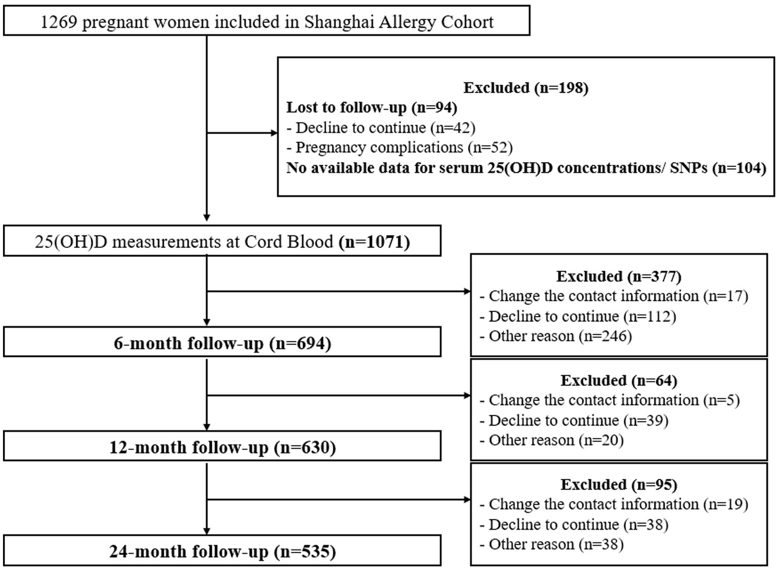
Figure 1. Flow chart of the study protocol. 25(OH)D, 25-hydroxyvitamin D; SNPs, single-nucleotide polymorphisms.
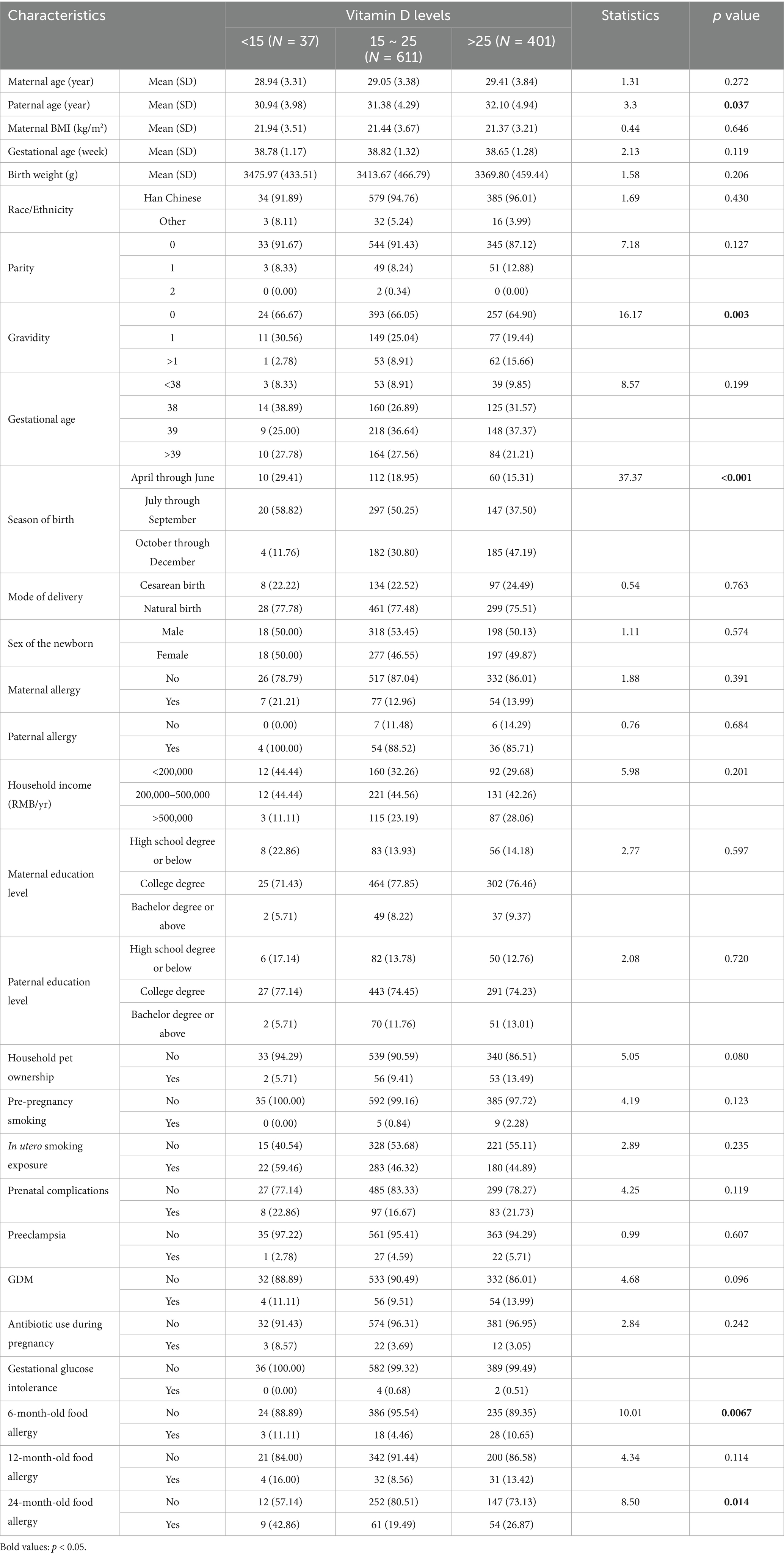
Table 1. Baseline characteristics of the cohort population and descriptive analysis of groups stratified by vitamin D levels.
3.2 Associations between vitamin D levels and food allergy
In univariate analysis, both low (<15 ng/mL) and high (>25 ng/mL) vitamin D levels were associated with increased risk of food allergy at 6 months (OR = 2.40, 95% CI: 1.18–4.90, p = 0.016; OR = 2.56, 95% CI: 1.38–4.72, p = 0.003) and 12 months (OR = 2.08, 95% CI: 1.35–3.19, p = 0.001; OR = 1.62, 95% CI: 1.12–2.36, p = 0.011) (Supplementary Table S1). Restricted cubic spline curves model confirmed a significant U-shaped dose–response relationship (Figure 2). Risk increased by 18–22% per 5 ng/mL decrease below 20 ng/mL and 10–18% per 5 ng/mL increase above 25 ng/mL at 6–12 months (all p < 0.05). After adjusting for covariates (season of birth, sex, maternal allergy, pet ownership, smoking, antibiotic use, birth weight, APGAR score, maternal BMI, and age), these associations remained significant for 6-month (adjusted OR = 2.55, p = 0.015 for <15 ng/mL; adjusted OR = 2.38, p = 0.008 for >25 ng/mL) and 12-month allergies (adjusted OR = 1.98, p = 0.035 for <15 ng/mL) (Table 2). By 24 months, only low vitamin D retained marginal significance in univariate analysis (OR = 1.55, p = 0.027), but not after adjustment (adjusted OR = 1.14, p = 0.779) (Table 2). For cumulative 0–24 month allergy risk, high vitamin D (>25 ng/mL) showed a robust association (adjusted OR = 1.78, 95% CI: 1.19–2.67, p = 0.005), while low vitamin D trended toward significance (adjusted OR = 1.50, p = 0.044) (Table 2).
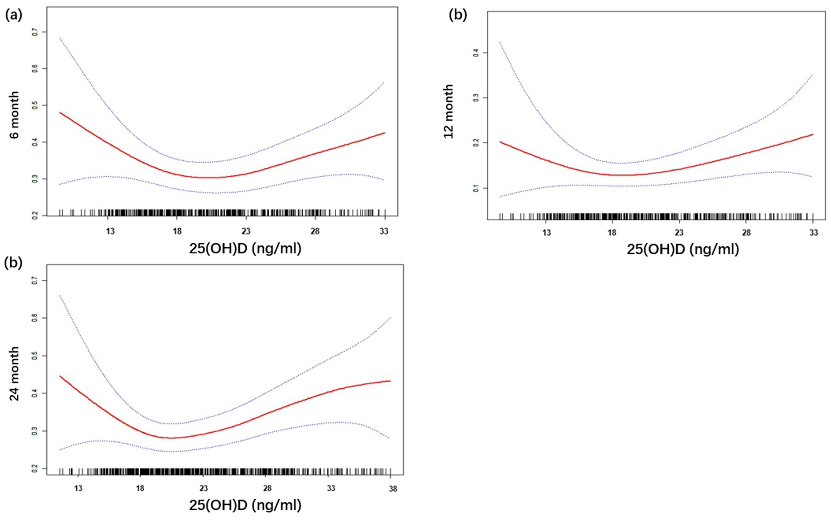
Figure 2. Relationship between cord blood vitamin D and food allergy status at: (a) 6-month; (b) 12-month; (c) 24-month. 25(OH)D, 25-hydroxyvitamin D.

Table 2. Associations between vitamin D levels and food allergy: categorical and continuous vitamin D model.
3.3 Genetic polymorphisms and food allergy susceptibility
Among five genetic loci analyzed, rs569108 (MS4A2 gene) demonstrated a significant association with food allergy under the dominant model (GG vs. AA+AG: adjusted OR = 1.68, 95% CI: 1.13–2.49, p = 0.011). No significant associations were observed for rs2243250 (IL-4), rs1801275 (IL-4R), rs1295686 (IL13), or rs20541 (IL13) in either dominant or recessive models after covariate adjustment. However, rs2243250 (IL-4) exhibited a trend toward reduced allergy risk in the dominant model (TT vs. TC+CC: adjusted OR = 0.59, 95% CI: 0.34–1.03, p = 0.062) (Table 3).
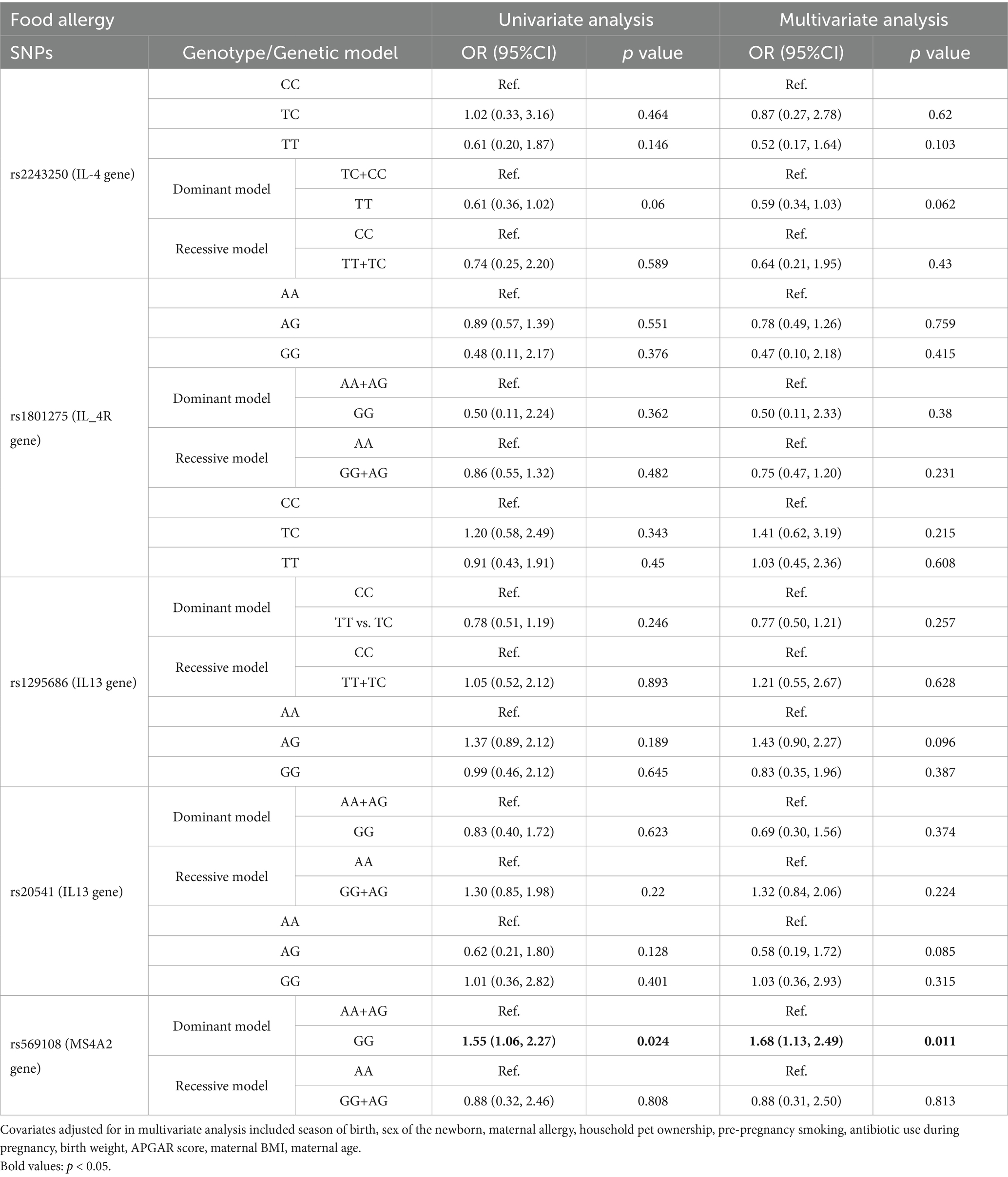
Table 3. Analysis of 0–24 month food allergy susceptibility based on the genotype and locus genetic model.
3.4 Gene-vitamin D interaction effects
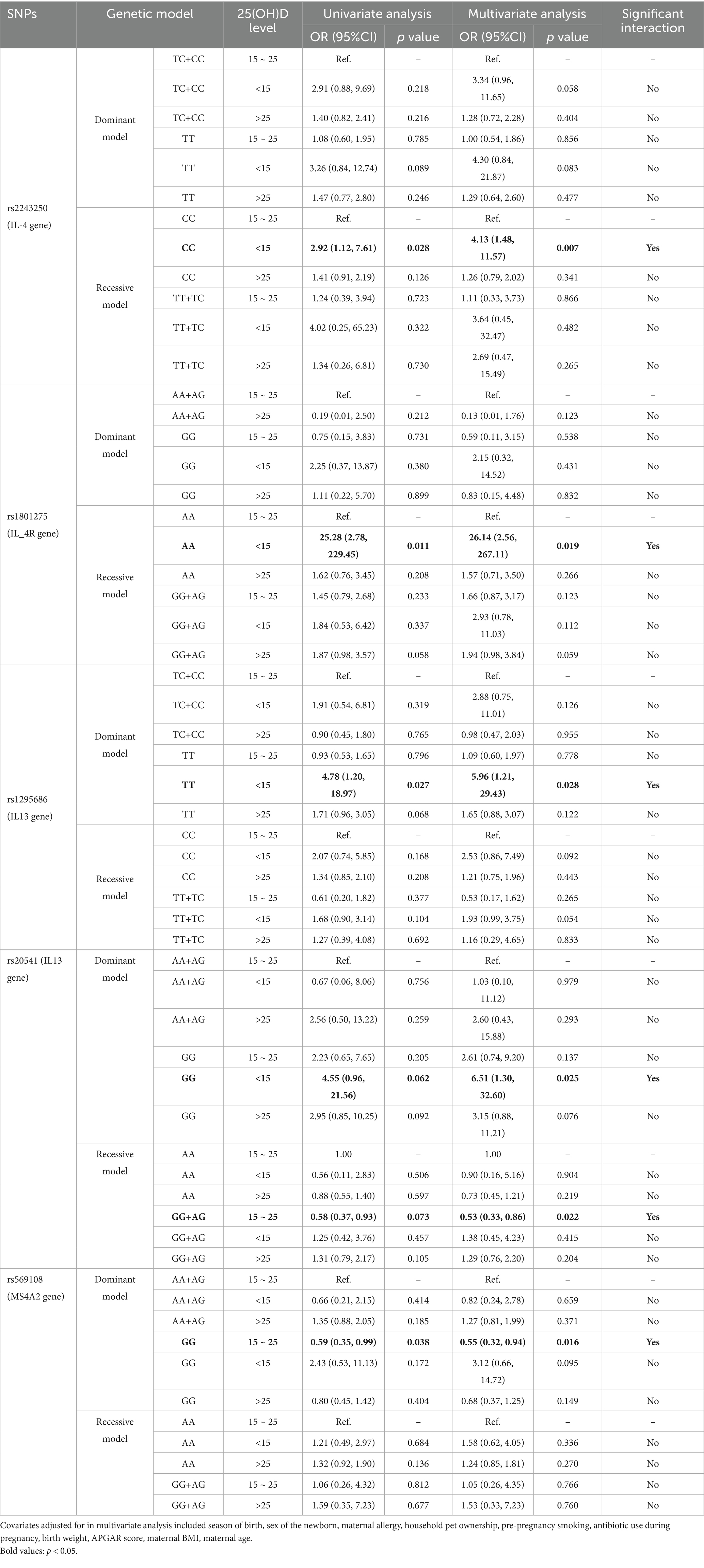
Stratified analyses revealed interactions between vitamin D levels and specific polymorphisms. For rs1801275 (IL-4R), infants with the AA genotype and low vitamin D (<15 ng/mL) had a significantly elevated allergy risk (adjusted OR = 26.14, 95% CI: 2.56–267.11, p = 0.019). Similarly, rs20541 (IL13) GG genotype infants with low vitamin D showed significantly elevated risk (adjusted OR = 6.51, 95% CI: 1.30–32.60, p = 0.025). Additionally, rs2243250 (IL-4) CC homozygotes with vitamin D deficiency exhibited elevated allergy risk (adjusted OR = 4.13, 95% CI: 1.48–11.57, p = 0.007). Notably, in the reference vitamin D group (15–25 ng/mL), the rs569108 (MS4A2) GG genotype conferred protection against allergy (adjusted OR = 0.55, 95% CI: 0.32–0.94, p = 0.016), but this effect was attenuated at high vitamin D levels (>25 ng/mL; adjusted OR = 0.68, 95% CI: 0.37–1.25, p = 0.149) (Table 4). The results of stratified analyses by genotype for all SNPs, evaluating both categorical and continuous vitamin D are comprehensively detailed in Supplementary Table S3. These interactions demonstrate that genetic susceptibility in Th2 pathway genes (IL4R, IL-4, IL13) dramatically amplifies food allergy risk under vitamin D deficiency, while MS4A2 variants modulate vitamin D’s protective effects.
4 Discussion
This prospective birth cohort study provides novel insights into the complex relationship between early-life vitamin D exposure, genetic susceptibility, and childhood allergy development. As visualized in Figure 2, the U-shaped relationship between cord blood 25(OH)D and food allergy risk was statistically robust (p < 0.001 for non-linearity) during infancy, echoing prior reports of non-linear vitamin D effects on respiratory sensitization (32). However, our study extends this paradigm in three key dimensions: First, our U-shaped risk curve reflects dual genetic modulation: (i) risk amplification via Th2-pathway variants (IL4R/IL-4/IL13) under low vitamin D; (ii) protection attenuation in FcεRIβ (MS4A2) carriers at high vitamin D. Second, we establish this U-shaped relationship for clinically confirmed food allergy—a distinct phenotype from respiratory outcomes. Third, we reveal temporal dynamics: vitamin D’s protective effects attenuate by 24 months, suggesting critical developmental windows for intervention. While prior studies predominantly emphasize vitamin D deficiency as a risk amplifier for allergic disorders (5), our observation of heightened FA susceptibility at both low [25(OH)D <15 ng/mL] and elevated [25(OH)D >25 ng/mL] vitamin D levels challenges the linear “deficiency-risk” paradigm. This aligns with emerging evidence suggesting that excessive vitamin D may paradoxically impair immune tolerance by over-suppressing Th1/Th2 balance (16), though contrasting with reports advocating higher postnatal vitamin D supplementation for allergy prevention (20). Our observation of a U-shaped relationship between cord blood 25(OH)D and clinical food allergy contrasts with null findings for food sensitization in the Danish COPSAC2000 cohort (12), where vitamin D deficiency (<50 nmol/L [20 ng/mL]) only associated with respiratory outcomes. This discrepancy may reflect key methodological differences: (i) COPSAC assessed IgE sensitization rather than clinically confirmed food allergy; (ii) their cohort had substantially lower mean cord 25(OH)D (12.9 ng/mL) with minimal high-exposure representation (<1.5% > 30 ng/mL), potentially limiting detection of non-linear effects; and (iii) population-specific genetic/epigenetic factors may modulate vitamin D’s immunoregulatory effects. Importantly, our gene–environment interaction analyses suggest vitamin D’s impact is modified by genetic susceptibility, a dimension absent in prior null reports.
The gene–environment interactions we identified reveal a triad of Th2-sensitizing genotypes—IL4R rs1801275 AA, IL-4 rs2243250 CC, and IL13 rs20541 GG—that confer 4- to 26-fold elevated food allergy risk under vitamin D deficiency. This synergistic effect aligns with vitamin D’s role in suppressing IL-4/STAT6 signaling and Th2 differentiation via VDR-mediated chromatin remodeling (6). Our observation that deficiency overrides this suppression in specific genotypes suggests certain variants may create a “genetic vulnerability window” during fetal immune programming. This contrasts with studies where vitamin D supplementation mitigated allergy risk even in high-risk genotypes (6), highlighting the need for genotype-stratified intervention trials. Our results suggest that vitamin D sufficiency may be particularly critical for children with genetic variants predisposing to enhanced Th2 responses, potentially through restoration of immune balance between regulatory T cells and Th2 effectors. Unlike the uniformly risk-amplifying effects of Th2-pathway variants (IL4R/IL-4/IL13) under vitamin D deficiency, MS4A2’s role is protective and vitamin D-dependent.
The dual role of MS4A2 (rs569108) in vitamin D-dependent allergy modulation refines our understanding of genetic regulation in IgE-mediated responses. MS4A2 encodes the β-chain of the high-affinity IgE receptor (FcεRIβ) (23), a key amplifier of mast cell signaling. Contrary to initial hypotheses, we observed that the GG genotype conferred significant protection against allergy at reference vitamin D levels (15–25 ng/mL; adjusted OR = 0.55, p = 0.016), suggesting FcεRIβ may participate in vitamin D-mediated tolerance induction. However, this protective effect was attenuated at high vitamin D levels (>25 ng/mL; OR = 0.68, p = 0.149), indicating a therapeutic ceiling for supplementation in GG carriers. Notably, the absence of risk amplification under vitamin D deficiency (OR = 3.12, p = 0.095) diverges from Th2 gene interactions but aligns with vitamin D’s dose-dependent suppression of FcεRIβ expression (20). This complex gene–environment interplay underscores that genetic effects on IgE pathways are contextually modified by vitamin D status. Thus, while our U-shaped association aligns with broader evidence of vitamin D’s dual immunomodulatory roles, the discovery of genotype-dependent risk thresholds and age-specific susceptibility windows provides unprecedented mechanistic and translational insights for allergy prevention in genetically predisposed infants.
The transient nature of vitamin D’s protective effects—strongest at 6–12 months but diminishing by 24 months—echoes murine models where neonatal dendritic cell programming by vitamin D dictates long-term immune trajectories (9), yet diverges from birth cohort studies reporting persistent vitamin D-FA associations beyond infancy (12). The temporal pattern of vitamin D’s protective effects aligns with emerging concepts of developmental immunotoxicity windows. Experimental models demonstrate that vitamin D regulates neonatal dendritic cell function and gut barrier integrity during critical periods of microbial colonization (33). Our observation that vitamin D insufficiency during this period correlates with persistent food allergy risk underscores the importance of prenatal and perinatal vitamin D optimization, particularly given the rapid placental transfer of 25(OH)D during late gestation.
Notably, our finding of increased allergy risk at cord blood 25(OH)D levels >25 ng/mL warrants careful consideration. This aligns with findings from the Boston Birth Cohort, which indicated that persistently low vitamin D status at birth and in early childhood increased the risk of food sensitization, particularly among children with specific genetic susceptibilities (34). While the Institute of Medicine defines 20 ng/mL as sufficient for bone health (23), our data suggest potential immune system hypersensitivity to higher concentrations (35). The immunomodulatory effects of vitamin D are further illustrated by its impact on regulatory T cells (Tregs), which play a crucial role in maintaining immune tolerance (35). A study investigating the Urban Environment and Childhood Asthma cohort found that higher cord blood 25(OH)D concentrations were inversely related to the number of Tregs, suggesting that elevated vitamin D levels might reduce the capacity for immune regulation, thereby increasing allergy risk (36). This is supported by another study that found a negative correlation between cord blood 25(OH)D levels and Treg numbers, indicating that high vitamin D levels might impair the development of these critical immune cells (11). Such alterations in immune cell populations could contribute to the increased risk of food allergies observed at elevated vitamin D levels. Moreover, the role of vitamin D in allergy development is complicated by genetic factors. Polymorphisms affecting vitamin D-binding protein (DBP) can modify the relationship between serum vitamin D levels and food allergy risk. A study found that genetic variations leading to lower DBP levels, which increase the bioavailability of vitamin D, could attenuate the association between low serum vitamin D levels and food allergy, suggesting that genetic factors may influence how vitamin D impacts immune responses (37). Our observed U-shaped risk curve—particularly the vulnerability at high vitamin D levels—likely stems from genotype-dependent saturation of vitamin D receptor (VDR) signaling pathways, where excessive ligand promotes pathological immune remodeling in predisposed infants. This underscores that optimal vitamin D ranges for allergy prevention may be genotype-specific, with deficiency posing greater threat to Th2-sensitized infants. This genetic interplay highlights the complexity of vitamin D’s role in allergy development and suggests that individual genetic makeup can significantly alter the effects of vitamin D on immune function. However, the biological plausibility of this U-shaped association requires further investigation through mechanistic studies.
Several limitations should be acknowledged. First, while we adjusted for key covariates (e.g., maternal allergy history, smoking, and antibiotic use), residual confounding by unmeasured environmental factors may influence observed associations, especially maternal dietary patterns during pregnancy (38)—such as intake of allergenic foods (e.g., peanuts, eggs) or micronutrients (e.g., omega-3 fatty acids)—were not captured. Future studies should integrate dietary assessments to disentangle nutritional influences on fetal immune development. Second, the single-center design and ethnic homogeneity (Chinese Han population) may limit generalizability. Third, while cord blood 25(OH)D serves as a well-validated indicator of late-gestation fetal vitamin D exposure (25), we lacked maternal prenatal or infant postnatal vitamin D measurements. This precludes analysis of how maternal status independently influences allergy risk or whether postnatal supplementation modifies associations observed at birth. Future studies integrating longitudinal maternal–infant vitamin D trajectories would strengthen causal inference. Fourth, while our U-shaped association aligns with emerging evidence of vitamin D’s dose-dependent immune effects, it diverges from cohorts with distinct genetic backgrounds or environmental exposures. Generalizability to populations with severe vitamin D insufficiency (e.g., high-latitude cohorts) requires verification. Future multi-ethnic cohorts with repeated vitamin D measurements and detailed immune profiling could address these limitations.
Data availability statement
The data analyzed in this study was obtained from the Shanghai Allergy Cohort, the following restrictions apply: data sensitivity and institutional limitations. Requests to access these datasets should be directed to the corresponding author.
Ethics statement
The studies involving humans were approved by the Ethics Committees of both Xinhua Hospital affiliated to Shanghai Jiao Tong University School of Medicine and the International Maternal and Children Care Hospital (approval number: XHEC-C-2012-003). The studies were conducted in accordance with the local legislation and institutional requirements. Written informed consent for participation in this study was provided by the participants’ legal guardians/next of kin.
Author contributions
XW: Data curation, Formal analysis, Methodology, Project administration, Software, Visualization, Writing – original draft, Writing – review & editing. YC: Data curation, Investigation, Methodology, Supervision, Validation, Writing – review & editing, Project administration, Visualization. JP: Data curation, Investigation, Project administration, Supervision, Writing – review & editing, Validation, Visualization. BW: Data curation, Formal analysis, Investigation, Methodology, Project administration, Software, Validation, Writing – review & editing. YT: Conceptualization, Methodology, Project administration, Resources, Supervision, Writing – review & editing. JZ: Conceptualization, Methodology, Project administration, Resources, Supervision, Writing – review & editing. XY: Conceptualization, Data curation, Funding acquisition, Methodology, Project administration, Resources, Validation, Writing – review & editing.
Funding
The author(s) declare that financial support was received for the research and/or publication of this article. This research was funded by the National Key Research and Development Program of China (no. 2022YFC2705203), the Major Science and Technology Projects of Fujian Province (no. 2021YZ034011), the High-level Medical Introduction Teams in Fujian Province (no. RT2022-022), Clinical Research Center of ADHD affiliated with Pediatrics College, Shanghai Jiao Tong University School of Medicine (no. ELYZX202207).
Acknowledgments
We are extremely grateful to all the families who took part in this study, and the teachers and nurses for their help in recruiting them.
Conflict of interest
The authors declare that the research was conducted in the absence of any commercial or financial relationships that could be construed as a potential conflict of interest.
Generative AI statement
The authors declare that no Gen AI was used in the creation of this manuscript.
Any alternative text (alt text) provided alongside figures in this article has been generated by Frontiers with the support of artificial intelligence and reasonable efforts have been made to ensure accuracy, including review by the authors wherever possible. If you identify any issues, please contact us.
Publisher’s note
All claims expressed in this article are solely those of the authors and do not necessarily represent those of their affiliated organizations, or those of the publisher, the editors and the reviewers. Any product that may be evaluated in this article, or claim that may be made by its manufacturer, is not guaranteed or endorsed by the publisher.
Supplementary material
The Supplementary material for this article can be found online at: https://www.frontiersin.org/articles/10.3389/fnut.2025.1652487/full#supplementary-material
Abbreviations
25(OH)D, 25-hydroxyvitamin D; SNPs, single-nucleotide polymorphisms; SD, standard deviation; HWE, Hardy–Weinberg equilibrium; ORs, odds ratios; CIs, confidence intervals; VD, Vitamin D.
References
1. Zhang, P, Xu, Q, and Zhu, R. Vitamin D and allergic diseases. Front Immunol. (2024) 15:1420883. doi: 10.3389/fimmu.2024.1420883
2. Lipińska-Opałka, A, Tomaszewska, A, Kubiak, JZ, and Kalicki, B. Vitamin D and immunological patterns of allergic diseases in children. Nutrients. (2021) 13:177. doi: 10.3390/nu13010177
3. Psaroulaki, E, Katsaras, GN, Samartzi, P, Chatziravdeli, V, Psaroulaki, D, Oikonomou, E, et al. Association of food allergy in children with vitamin D insufficiency: a systematic review and meta-analysis. Eur J Pediatr. (2023) 182:1533–54. doi: 10.1007/s00431-023-04843-2
4. Briceno Noriega, D, and Savelkoul, HFJ. Vitamin D and allergy susceptibility during gestation and early life. Nutrients. (2021) 13:1015. doi: 10.3390/nu13031015
5. Ly, NP, Litonjua, A, Gold, DR, and Celedón, JC. Gut microbiota, probiotics, and vitamin D: interrelated exposures influencing allergy, asthma, and obesity? J Allergy Clin Immunol. (2011) 127:1087–94; quiz 95-6. doi: 10.1016/j.jaci.2011.02.015
6. Ananthakrishnan, AN, Khalili, H, Higuchi, LM, Bao, Y, Korzenik, JR, Giovannucci, EL, et al. Higher predicted vitamin D status is associated with reduced risk of Crohn's disease. Gastroenterology. (2012) 142:482–9. doi: 10.1053/j.gastro.2011.11.040
7. Battistini, C, Ballan, R, Herkenhoff, ME, Saad, SMI, and Sun, J. Vitamin D modulates intestinal microbiota in inflammatory bowel diseases. Int J Mol Sci. (2020) 22:362. doi: 10.3390/ijms22010362
8. Prescott, SL. Early-life environmental determinants of allergic diseases and the wider pandemic of inflammatory noncommunicable diseases. J Allergy Clin Immunol. (2013) 131:23–30. doi: 10.1016/j.jaci.2012.11.019
9. Adams, JS, and Hewison, M. Unexpected actions of vitamin D: new perspectives on the regulation of innate and adaptive immunity. Nat Clin Pract Endocrinol Metab. (2008) 4:80–90. doi: 10.1038/ncpendmet0716
10. Chun, RF, Liu, PT, Modlin, RL, Adams, JS, and Hewison, M. Impact of vitamin D on immune function: lessons learned from genome-wide analysis. Front Physiol. (2014) 5:151. doi: 10.3389/fphys.2014.00151
11. Weisse, K, Winkler, S, Hirche, F, Herberth, G, Hinz, D, Bauer, M, et al. Maternal and newborn vitamin D status and its impact on food allergy development in the German LINA cohort study. Allergy. (2013) 68:220–8. doi: 10.1111/all.12081
12. Chawes, BL, Bønnelykke, K, Jensen, PF, Schoos, AM, Heickendorff, L, and Bisgaard, H. Cord blood 25(OH)-vitamin D deficiency and childhood asthma, allergy and eczema: the COPSAC2000 birth cohort study. PLoS One. (2014) 9:e99856. doi: 10.1371/journal.pone.0099856
13. Wegienka, G, Havstad, S, Zoratti, EM, Kim, H, Ownby, DR, and Johnson, CC. Association between vitamin D levels and allergy-related outcomes vary by race and other factors. J Allergy Clin Immunol. (2015) 136:1309–14.e1–4. doi: 10.1016/j.jaci.2015.04.017
14. McGowan, EC, Bloomberg, GR, Gergen, PJ, Visness, CM, Jaffee, KF, Sandel, M, et al. Influence of early-life exposures on food sensitization and food allergy in an inner-city birth cohort. J Allergy Clin Immunol. (2015) 135:171–8. doi: 10.1016/j.jaci.2014.06.033
15. Chiu, CY, Huang, SY, Peng, YC, Tsai, MH, Hua, MC, Yao, TC, et al. Maternal vitamin D levels are inversely related to allergic sensitization and atopic diseases in early childhood. Pediatr Allergy Immunol. (2015) 26:337–43. doi: 10.1111/pai.12384
16. Paternoster, L, Standl, M, Waage, J, Baurecht, H, Hotze, M, Strachan, DP, et al. Multi-ancestry genome-wide association study of 21, 000 cases and 95, 000 controls identifies new risk loci for atopic dermatitis. Nat Genet. (2015) 47:1449–56. doi: 10.1038/ng.3424
17. Torgerson, DG, Ampleford, EJ, Chiu, GY, Gauderman, WJ, Gignoux, CR, Graves, PE, et al. Meta-analysis of genome-wide association studies of asthma in ethnically diverse north American populations. Nat Genet. (2011) 43:887–92. doi: 10.1038/ng.888
18. Van Eerdewegh, P, Little, RD, Dupuis, J, Del Mastro, RG, Falls, K, Simon, J, et al. Association of the ADAM33 gene with asthma and bronchial hyperresponsiveness. Nature. (2002) 418:426–30. doi: 10.1038/nature00878
19. Zhang, Y, Leaves, NI, Anderson, GG, Ponting, CP, Broxholme, J, Holt, R, et al. Positional cloning of a quantitative trait locus on chromosome 13q14 that influences immunoglobulin E levels and asthma. Nat Genet. (2003) 34:181–6. doi: 10.1038/ng1166
20. Liu, X, Wang, G, Hong, X, Wang, D, Tsai, HJ, Zhang, S, et al. Gene-vitamin D interactions on food sensitization: a prospective birth cohort study. Allergy. (2011) 66:1442–8. doi: 10.1111/j.1398-9995.2011.02681.x
21. Yu, X, Wang, W, Wei, Z, Ouyang, F, Huang, L, Wang, X, et al. Vitamin D status and related factors in newborns in Shanghai. China Nutr. (2014) 6:5600–10. doi: 10.3390/nu6125600
22. Li, L, Zhou, H, Yang, X, Zhao, L, and Yu, X. Relationships between 25-hydroxyvitamin D and nocturnal enuresis in five- to seven-year-old children. PLoS One. (2014) 9:e99316. doi: 10.1371/journal.pone.0099316
23. Ross, AC, Manson, JE, Abrams, SA, Aloia, JF, Brannon, PM, Clinton, SK, et al. The 2011 report on dietary reference intakes for calcium and vitamin D from the Institute of Medicine: what clinicians need to know. J Clin Endocrinol Metab. (2011) 96:53–8. doi: 10.1210/jc.2010-2704
24. Holick, MF, Binkley, NC, Bischoff-Ferrari, HA, Gordon, CM, Hanley, DA, Heaney, RP, et al. Evaluation, treatment, and prevention of vitamin D deficiency: an Endocrine Society clinical practice guideline. J Clin Endocrinol Metab. (2011) 96:1911–30. doi: 10.1210/jc.2011-0385
25. Wang, X, Jiao, X, Tian, Y, Zhang, J, Zhang, Y, Li, J, et al. Associations between maternal vitamin D status during three trimesters and cord blood 25(OH)D concentrations in newborns: a prospective Shanghai birth cohort study. Eur J Nutr. (2021) 60:3473–83. doi: 10.1007/s00394-021-02528-w
26. Liu, X, Beaty, TH, Deindl, P, Huang, SK, Lau, S, Sommerfeld, C, et al. Associations between specific serum IgE response and 6 variants within the genes IL4, IL13, and IL4RA in German children: the German multicenter atopy study. J Allergy Clin Immunol. (2004) 113:489–95. doi: 10.1016/j.jaci.2003.12.037
27. Hizawa, N, Yamaguchi, E, Jinushi, E, and Kawakami, Y. A common FCER1B gene promoter polymorphism influences total serum IgE levels in a Japanese population. Am J Respir Crit Care Med. (2000) 161:906–9. doi: 10.1164/ajrccm.161.3.9903128
28. Woszczek, G, Borowiec, M, Ptasinska, A, Kosinski, S, Pawliczak, R, and Kowalski, ML. Beta 2-ADR haplotypes/polymorphisms associate with bronchodilator response and total IgE in grass allergy. Allergy. (2005) 60:1412–7. doi: 10.1111/j.1398-9995.2005.00869.x
29. Hua, L, Zuo, XB, Bao, YX, Liu, QH, Li, JY, Lv, J, et al. Four-locus gene interaction between IL13, IL4, FCER1B, and ADRB2 for asthma in Chinese Han children. Pediatr Pulmonol. (2016) 51:364–71. doi: 10.1002/ppul.23322
30. Hua, L, Liu, Q, Li, J, Zuo, X, Chen, Q, Li, J, et al. Gene-gene and gene-environment interactions on cord blood total IgE in Chinese Han children. Allergy Asthma Clin Immunol. (2021) 17:69. doi: 10.1186/s13223-021-00571-0
31. Jiang, F, and Yan, A. IL-4 rs2243250 polymorphism associated with susceptibility to allergic rhinitis: a meta-analysis. Biosci Rep. (2021) 41:10522. doi: 10.1042/BSR20210522
32. Rothers, J, Wright, AL, Stern, DA, Halonen, M, and Camargo, CA Jr. Cord blood 25-hydroxyvitamin D levels are associated with aeroallergen sensitization in children from Tucson, Arizona. J Allergy Clin Immunol. (2011) 128:1093–9.e1–5. doi: 10.1016/j.jaci.2011.07.015
33. Neeland, MR, Koplin, JJ, Dang, TD, Dharmage, SC, Tang, ML, Prescott, SL, et al. Early life innate immune signatures of persistent food allergy. J Allergy Clin Immunol. (2018) 142:857–64.e3. doi: 10.1016/j.jaci.2017.10.024
34. Liu, X, Arguelles, L, Zhou, Y, Wang, G, Chen, Q, Tsai, HJ, et al. Longitudinal trajectory of vitamin D status from birth to early childhood in the development of food sensitization. Pediatr Res. (2013) 74:321–6. doi: 10.1038/pr.2013.110
35. Martineau, AR, Jolliffe, DA, Hooper, RL, Greenberg, L, Aloia, JF, Bergman, P, et al. Vitamin D supplementation to prevent acute respiratory tract infections: systematic review and meta-analysis of individual participant data. BMJ. (2017) 356:i6583. doi: 10.1136/bmj.i6583
36. Chi, A, Wildfire, J, McLoughlin, R, Wood, RA, Bloomberg, GR, Kattan, M, et al. Umbilical cord plasma 25-hydroxyvitamin D concentration and immune function at birth: the urban environment and childhood asthma study. Clin Exp Allergy. (2011) 41:842–50. doi: 10.1111/j.1365-2222.2011.03712.x
37. Koplin, JJ, Suaini, NH, Vuillermin, P, Ellis, JA, Panjari, M, Ponsonby, AL, et al. Polymorphisms affecting vitamin D-binding protein modify the relationship between serum vitamin D (25[OH]D3) and food allergy. J Allergy Clin Immunol. (2016) 137:500–6.e4. doi: 10.1016/j.jaci.2015.05.051
Keywords: vitamin D, genetic polymorphisms, food allergy, birth cohort, gene–environment interaction
Citation: Wang X, Cai Y, Pei J, Wang B, Tian Y, Zhang J and Yu X (2025) Association of cord blood vitamin D and genetic polymorphisms with childhood food allergy in Shanghai, China: a prospective cohort. Front. Nutr. 12:1652487. doi: 10.3389/fnut.2025.1652487
Edited by:
Rosa Casas Rodriguez, August Pi i Sunyer Biomedical Research Institute (IDIBAPS), SpainReviewed by:
Fatme Al Anouti, Zayed University, United Arab EmiratesLiang Chen, Copenhagen Prospective Studies on Asthma in Childhood (COPSAC), Denmark
Copyright © 2025 Wang, Cai, Pei, Wang, Tian, Zhang and Yu. This is an open-access article distributed under the terms of the Creative Commons Attribution License (CC BY). The use, distribution or reproduction in other forums is permitted, provided the original author(s) and the copyright owner(s) are credited and that the original publication in this journal is cited, in accordance with accepted academic practice. No use, distribution or reproduction is permitted which does not comply with these terms.
*Correspondence: Xiaodan Yu, eGRfeXUyMDAzQDEyNi5jb20=
 Xirui Wang
 Yingying Cai2
 Bin Wang
 Xiaodan Yu