- 1Institute of Nanotectology, Consiglio Nazionale delle Ricerche, Rome, Italy
- 2NeuroImaging Lab, IRCCS Santa Lucia Foundation, Rome, Italy
- 3Department of Engineering and Architecture, University of Trieste, Trieste, Italy
- 4Diamond Light Source, Harwell Science and Innovation Campus, Didcot, United Kingdom
- 5Advanced Photon Source, Argonne National Laboratory, Lemont, IL, United States
- 6Department of Physics, University of Milano-Bicocca, Milano, Italy
- 7Elettra-Sincrotrone Trieste S.C.p.A, Basovizza, Trieste, Italy
- 8MARBILab, Museo Storico della Fisica e Centro Studi e Ricerche Enrico Fermi, Rome, Italy
- 9Department of Medical Physics and Biomedical Engineering, University College London, London, United Kingdom
- 10A. I. Virtanen Institute for Molecular Sciences, University of Eastern Finland, Kuopio, Finland
The choice of fixative is critical in X-ray phase-contrast tomography (XPCT) because it affects tissue preservation, contrast enhancement and compatibility with other imaging techniques. A careful selection and optimization of fixatives can lead to significant improvements in the quality and accuracy of imaging results, which is especially important when studying complex biological systems such as those involved in neurodegeneration, where it is crucial to maintain the fine details of the Grey Matter (GM) and White Matter (WM) structures. Dehydration with ethanol and xylene is commonly used as it effectively removes water while minimising structural alterations. Using perfusion in ethanol and dehydration in xylene as a secondary fixative can increase the contrast, thereby improving the visibility of myelinated fibers without using a contrast agent. In this paper we discuss an optimised fixation method to significantly enhance the contrast and boost the signal to noise ratio (SNR) in XPCT images of WM in the central nervous system (CNS).
1 Introduction
White matter (WM) and grey matter (GM) are two components of the central nervous system (CNS) with distinct structures and functions. The key difference between WM and GM lies in their composition and function. GM is primarily composed of neuronal cell bodies, dendrites, and unmyelinated axons and it is involved in information processing and regulation of outgoing signals. On the other hand, WM consists mainly of myelinated axons, which form the connections between different areas of the brain and spinal cord, allowing for the transmission of nerve signals [1]. The WM acts as the “wiring” of the brain, enabling various parts of the nervous system to work together effectively. Their integrity is essential for normal neurological function. In particular, damage to the WM can have significant implications for motor skills, sensory perception, and overall brain function [2,3]. The study of WM structure and function in the context of injuries and neurodegenerative diseases is crucial for understanding the underlying mechanisms and developing potential therapeutic strategies. Previous studies have shown that spinal cord injury can lead to widespread chronic changes in cerebral WM, affecting axonal integrity and cortical reorganisation [4,5]. Additionally, traumatic spinal cord injury causes damage to WM [6], leading to progressive degeneration and neuroplastic changes [4]. The degeneration and demyelination of WM are hallmark features of traumatic, ischemic, and neurodegenerative conditions, leading to neurological deficits [7]. Furthermore, WM injury is well documented in various neurodegenerative disorders, such as Alzheimer’s disease [8].
White matter neurodegeneration can be assessed using neuroimaging techniques based on magnetic resonance imaging (MRI), particularly diffusion MRI techniques (such as Diffusion Tensor Imaging, DTI), which have been used to investigate white matter degeneration and disruption of white matter tracts in brain and spinal cord [9–13].
Diffusion MRI is particularly important because it provides detailed insights into tissue microstructure and structural connectivity, which add value to other structural techniques [14].
In particular, Diffusion Tensor Imaging (DTI) offers valuable information into the microstructural characteristics of WM and its relationship to neurological conditions. The GM to WM signal ratio (GWR) has been also proposed as a novel biomarker of neurodegeneration in Alzheimer’s disease [15, 16].
MRI is a completely non-invasive technique in the absence of contrast agents like gadolinium (Gd), providing valuable evidence to assess WM damage and neurodegeneration in vivo. In addition, unlike X-ray imaging, MRI does not involve exposure to ionizing radiation, making it safer for repeated use in human subjects, especially in vivo studies. This characteristic allows for longitudinal studies where the same subjects can be monitored over time without the associated risks of radiation exposure. However, it also has limitations that lie in the quantification of WM changes limited in conventional MRI (T1 and T2 weighted) by the low spatial resolution (order of mm), a variety of biophysical models based on diffusion MRI, and the lack of standardisation in data acquisition and analysis. In addition, in vivo MRI lacks biological interpretation, only provided by histology. Addressing these challenges is crucial for leveraging the full potential of MRI in assessing WM integrity and its relationship with neurological cognitive function. There are several studies on the morphology of WM based on advanced high resolution X-ray imaging techniques such as X-ray phase contrast Tomography (XPCT) using synchrotron radiation. Compared to traditional methods such as myelin staining on histological samples, XPCT offers several advantages such as simple and fast sample preparation and high-contrast resolution of brain structures, including WM fibres. In addition, XPCT represents a significant advancement over conventional computed tomography (CT) in imaging soft tissues, particularly in the brain. Unlike traditional CT, which relies on X-ray attenuation to create images, XPCT utilizes phase shifts of X-rays as they pass through biological tissues. This method enhances the contrast resolution dramatically, allowing for the visualization of soft tissue structures that are typically difficult to discern with standard CT techniques. Notably, XPCT can achieve this improved resolution at radiation doses approximately 400 times lower than those required for conventional CT scans [18]. This capability also facilitates detailed imaging of neuronal and vascular networks within the brain without the need for contrast agents, thus preserving tissue integrity [19]. Moreover, XPCT allows to image a whole-brain of small animal models in 3D, which is extremely valuable for investigating WM injury and neurodegenerative diseases [17–20]. Indeed, XPCT has been successfully used to map WM in injury animal models [6] and to reveal early vascular alterations and neuronal loss in multiple sclerosis models [17–20].
Undoubtedly there is great potential in using XPCT for advancing our understanding of WM in the CNS. However, there are still some challenges to be overcome to harness the full capabilities of XPCT:
1. Depth discrimination: while XPCT provides the benefit of discriminating different depths within the sample, this can also pose a challenge, particularly in the context of studying the complex structures of WM[20].
2. Contrast enhancement: despite the advantages of XPCT, there are inherent limitations in its ability to enhance contrast for certain structures, such as white matter tracts [17]. XPCT may not provide adequate differentiation between WM tracts due to their subtle density differences compared to surrounding tissues. This limitation suggests that while XPCT can enhance overall image quality, it may not be sufficient for detailed visualization of specific neural pathways.
3. Spatial resolution and acquisition time: XPCT enables high-contrast resolution, but achieving it may require long acquisition times (hours to tens of hours) with conventional laboratory sources because it is limited by the x-ray flux of collimated/filtered laboratory sources and the need to acquire projections in several steps to recover phase information, representing a big challenge for the application of XPCT in vivo [18]. Several technical challenges still need to be overcome before obtaining XPCT images in vivo in the laboratory. Several groups worldwide are presently making an effort to translate such multi-contrast x-ray imaging for preclinical/clinical application. Good results are obtained by the group of F. Pfeiffer that obtained phase contrast radiography in vivo with 10 s of exposure time [34].
The aim of this manuscript is to investigate and optimise sample preparation methods to maximise image quality and information content, and how sample preparation methods affect the contrast-to-noise ratio in WM imaging.
2 Methods
Synchrotron XPCT consists of several experimental steps, all of them play a crucial role for the overall efficiency, image quality, accuracy, and data utility.
The main step for XPCT imaging is the sample preparation. While fresh samples can be imaged, a proper sample fixation is essential to prevent sample modifications and movements during scanning and, more importantly, to preserve it for subsequent investigations.
The data acquisition parameters such as exposure time, X-ray energy, and detector settings are also fundamental for collecting high quality images. The tailoring of the experimental parameters needs to consider the nature of the sample and the desired resolution. Finding the trade-off between spatial resolution and scanning time is essential for avoiding radiation damage.
Finally the last two steps to optimize image quality include:
a) data processing and reconstruction methods using advanced image processing techniques for noise reduction, artefact correction and relevant efficient algorithms such as iterative 3D image reconstruction [30].
b) post-processing procedures like segmentation and data analysis to obtain quantitative information about internal structures. Selecting suitable visualisation techniques like volume rendering and surface rendering assists in comprehending intricate 3D structures.
In the following section, we review different sample preparation techniques and discuss their effect in terms of image contrast. In addition, we present XPCT imaging of a sample perfused in ethanol and one perfused in ethanol and fixed with xylene. We discuss the results in terms of contrast enhancement for structural studies of WM in the CNS of a mouse model.
2.1 Sample preparation methods
XPCT offers high SNR without the need for staining agents. It provides micrometric spatial resolution and isotropic spatial resolution making it ideal for imaging the entire and intact rodent brain and spinal cord.
However, sample preparation for XPCT involves several steps such as sample stabilisation, dehydration, and mounting. A protocol for the preparation of large biological samples for high-resolution, hierarchical, synchrotron phase-contrast tomography with multimodal imaging compatibility has been described in a Nature Protocols article [21]. The protocol is applicable to a range of biological samples, including complete organisms, and aims to enhance contrast and prevent sample motion during scanning. Additionally, a study focused on establishing sample-preparation protocols for XPCT of rodent spinal cords has tested different nervous-tissue fixation procedures for this purpose [22, 23]. These resources provide valuable insights into the specific steps and techniques involved in preparing samples for XPCT. These methods are important for achieving effective and targeted neuroimaging via XPCT, particularly for optimal nervous-tissue structural preservation. The specific fixation method chosen should be tailored to the sample type and the preservation goals of the imaging study.
In particular, to optimise sample preparation for enhancing the signal in XPCT WM imaging in CNS, several key strategies can be employed such as ethanol dehydration which enhances the contrast between myelinated axons and surrounding tissue by modulating the refractive indices [35]. Ethanol fixation is a well-accepted technique in neuroscientific communities and has been used for virtual XPCT histology of various organs, including the brain. Ethanol fixation preserves critical structures, such as cell membranes and organelles, more effectively than formalin, which can lead to alterations in cell morphology. In particular, a graded ethanol series (e.g., 80%, 95%, 100%) can be used to ensure dehydration without causing excessive shrinkage or cracking of the tissue. This protocol also helps to maintain cellular integrity and morphology better than a constant ethanol concentration, even if lower than the maximum achieved in the graded series [35].
2.2 Tissue optimization strategies for multimodal approach
Fixation is critical for preserving tissue morphology and enhancing imaging contrast. Tissue optimization strategies for multimodal approaches such as XPCT and DTI are crucial for enhancing imaging quality and data accuracy. These strategies involve meticulous preparation techniques that preserve tissue integrity and improve imaging outcomes.
Various fixatives, such as paraformaldehyde (PFA), can be used, and the concentration should be optimised based on the specific imaging technique and tissue type. Proper fixation helps maintain cellular architecture and improves the SNR in imaging modalities like DTI [4, 5].
Using a reduced fixative concentration (e.g., 2% paraformaldehyde) can improve the SNR efficiency of fixed tissue. This approach helps in maintaining the tissue’s microstructure and diffusivity, which are pivotal for high-quality MRI diffusion imaging, for example [11, 36].
In addition, sufficient rehydration time (e.g., more than 20 days) can help in reducing the impact of fixative-induced changes on the tissue’s properties. This step is essential for achieving optimal SNR and image quality in MRI [4] and XPCT [23].
By combining these strategies, researchers can optimise sample preparation to enhance the signal in XPCT WM imaging and combine the results with other imaging techniques such as histology or DTI, providing high-quality data for the study of CNS microstructure and the investigation of neurological diseases. We report some preliminary results relative to the comparison between two different sample preparation protocols that underline the fibers in the WM and the vessels.
2.3 Xylene and ethanol sample preparation methods for brain and spinal cord
Two adult male JAXC57BL/6J mice (5–6 weeks old, weight 21–28g, Charles River) are used in the study. The animals were housed in a room (22°C ± 1°C, 50%–60% humidity) with 12 h light/dark cycle and free access to food and water. All animal procedures were approved by the Animal Care and Use Committee of the Provincial Government of Southern Finland and performed according to the guidelines set by the European Community Council Directives 86/609/EEC.
The mice are anaesthetized intraperitoneally with a ketamine (80 mg/kg)/xylazine (10 mg/kg) mixture and perfused transcardially with 0.9% saline solution containing heparin (50 U/ml) for 3 min at 2 ml/min. After that, the mice were perfused with 4% paraformaldehyde (PFA) in 0.1M phosphate buffer saline (PBS) (pH 7.4) for 15 min at 2 mL/min followed by absolute ethanol for 15 min at 2 mL/min. After the perfusion, the brain and spinal cord of each mouse were extracted from the skull and the spine respectively and placed in the incubation solution that consists for the sample “A” absolute ethanol (for further information see the [23] and for sample “B” 4 days in absolute ethanol and then stored in xylene [24, 25].
Before the XPCT measurement the samples were embedded in agar to prevent sample movement during scanning [26].
We perfuse the samples with PFA and ethanol in order to visualise the vascular network and the fibres without contrast agents, because they show a marked increase in the stiffness of the tissue, an effect that effectively avoids the problem of vessel and fiber collapse by increasing the SNR [23].
In this case, the specimens are then placed in absolute (100%) ethanol to increase the speed of the fixation of the external tissue. While for the case of the graded ethanol fixation the samples are perfused with physiological solution, so need a slow process to fix the all tissue without artefacts.
2.4 Brain and spinal cord XPCT measurements and results
XPCT offers high SNR without the need for staining agents, making it ideal for imaging WM fibres in whole and intact rodent brain and spinal cord. However, the method of specimen preparation and fixation is critical to improve the contrast-to-noise ratio of some details in XPCT images. In this framework we compare two different fixation methods for brain and spinal cord to study the effect on image quality of some details such as WM microarchitecture and vascularisation.
The XPCT acquisition of the spinal cord was performed at different XPCT beamlines (Syrmep @Elettra, ID17 @ ESRF, PO5 @PetraIII). Here we report the result obtained at the I13-1 beamline at Diamond Light Source [27]. The sample was illuminated with the X-ray beam from the undulator filtered with 0.95 mm of graphite and 0.5 mm of aluminium foils corresponding to an average X-ray energy of 22 keV.
The detector used for the XPCT acquisition is a PCO4000 CMOS camera (Complementary metal–oxide–semiconductor) coupled to a ×8 objective corresponding to an effective pixel size of 1.125 um, placed 23 cm downstream the sample. The tomography acquisition consisted of 2400 projections acquired over 180°, with an exposure time of 0.02 s for each projection.
The whole brain XPCT experiments were carried out at ID17 beamline at ESRF. The monochromatic incident X-ray energy was 30 keV. The sample was set at a distance of 2.3 m from the camera with a pixel size of 3.01 µm. As the sample was larger than the horizontal field of view of the camera, projections were acquired using the half-acquisition method [28]. The tomography was produced by means of 2000 projections covering a total angle range of 360° and with an acquisition time of 0.3 s per point. The phase retrieval algorithm was applied to the projections of the tomographic scans using the single distance method proposed by Paganin [29] and all projections were processed and reconstructed using SYRMEP Tomo Project (STP) [30]. We thus obtained a set of high-spatial resolution tomographic images, where the different grey-levels show the phase shift induced by the electron density of the different tissues inside the sample. The reconstructed images were analysed by means of ImageJ [37] (ImageJ bundled with 64-bit Java 8), using available and specifically developed plugins such as steerable 3D and skeletonize, together with Matlab (R2022a (MATLAB 9.12)) routine.
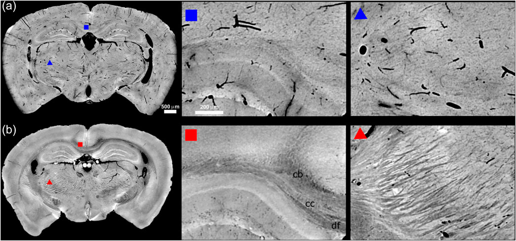
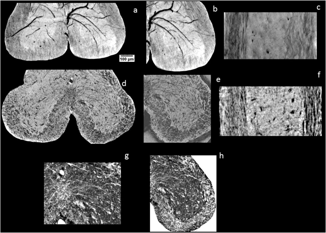
In order to discuss the various features detectable in the tomographic slices with several sample preparations, we report in Figures 1, 2 the XPCT reconstructions of the mouse brain and of the spinal cord (of the same sample) treated with ethanol and with xylene respectively (Figures 1A, 2A-C, 1B, 2D-H).

Figure 1. Panel (A, B) show a minimum intensity projection section across 45 μm of mouse midbrain following ethanol and xylene preparation protocols, respectively. The insets indicated by red and blue squares/triangles show a zoomed-in view of the region around the hippocampus including the corpus callosum (cc) and the fibres forming the thalamus nuclei. In panel (A) are well visible the vessels in black, while in panel (B) are well visible in light gray the fibers.

Figure 2. A minimum intensity projection section across 50 μm of spinal cord following ethanol (A–C) and xylene preparation protocols (D–H) respectively. In (A, B) the ventral horn is shown in the axial view. It is observed that in the ethanol preparation, the typical “H” shape of GM is well preserved in contrast, and a different greyscale associated to the phase shift, between WM and GM is noted. While in the sample preparation with xylene there is an enhanced contrast of the border between the GM and the WM. However, the contrast in the GM is too low due probably to the high dehydration that helps to enhance the contrast of the fibers reducing the contrast of the cells in the GM that contain water and lipids. In (C, F) the sagittal view of the spinal cord. (B, E) zoom of the left ventral horn where the vessels and the fibres are segmented respectively. (G, H) segmentation of the ventral white commissure and the nerve fibers in the ventral horn respectively.
We report for each sample the minimum intensity projections across a reconstructed coronal section for the brain in Figure 1 and axial and sagittal cross section for the spinal cord in Figure 2 [31]. A general inspection reveals as Xylene based sample preparation significantly enhances the contrast of the WM by effectively removing water and lipids from the tissue, which leads to a clearer delineation of the protein matrix against the surrounding air. This preparation technique generates a novel contrast without the need for additional staining or embedding procedures. As a result, the contrast between the WM and surrounding tissues is markedly improved, allowing for better visualisation of axon fibres such as in the brain region around the hippocampus including the corpus callosum (cc) and the fibres forming the thalamus nuclei. Specifically, according to the mouse brain Atlas the dorsal fornix (df) and the cingulum bundle (cb) can be identified.
In addition, the anterior or ventral white commissure, a collection of nerve fibres that cross the midline of the spinal cord and transmit information from or to the contralateral side of the brain, is visible [1, 31].
These crossing fibres make the anterior white commissure an important link in communication between the brain and the contralateral side of the body for both sensory and motor pathways.
On the other hand, the ethanol perfusion significantly enhances tissue rigidity, preventing the collapse of vessel walls [23]. This results in high-contrast images that clearly depict the morphology of vascular structures, making ethanol perfusion particularly effective for visualising brain and spinal cord vascularization (see Figures 1A, 2A). The vascular networks that supply blood to the spinal cord become distinctly visible without the need for a contrast agent.
We evaluate the efficiency of different sample preparation methods by calculating the gain, defined as the ratio between the contrast-to-background ratio (CBR) in xylene- and ethanol-perfused samples (for CBR, see. e.g., [38], Supplementary Materials and Supplementary Figure S1) CBR xylene/CBR ethanol. The same component and the same region were considered in both sample preparation for the quantification (see Supplementary Figure S1). We observe a gain of 18,1 in the brain, on the other hand in the spinal cord we estimate a gain of 0.5 probably due to the small cross section of the spinal cord and the important effect of the shrinkage due to the sample preparation. However, there is a contrast increase of the fibers as a consequence of the sample preparation with xylen.
3 Discussion
The perfusion and sample preparation is crucial for imaging techniques, particularly when studying neurodegenerative diseases. The choice of solvents, such as xylene and ethanol, plays a significant role in preserving the structural integrity of biological tissues and enhancing imaging quality. This is particularly important in neurodegenerative research, where subtle changes in tissue structure can provide insights into disease mechanisms. For instance, the use of aldehyde fixatives, followed by dehydration in ethanol, helps to preserve cellular architecture while minimizing artifacts that could obscure important details in imaging [24, 25, 32].
Ethanol is commonly used for dehydration, as it effectively removes water from tissues while maintaining their structural integrity. Xylene, on the other hand, is often employed in the clearing process, making tissues more transparent and suitable for embedding in paraffin. The sequential use of these solvents allows for optimal sample preparation, facilitating high-resolution imaging. For example, one study demonstrated that tissues prepared with 100% ethanol showed different structural characteristics compared to those processed with xylene, highlighting the impact of solvent choice on imaging outcomes [33].
On the other hand, it is important to note that xylene and ethanol perfusion substantially alters the actual dimensions and three-dimensional morphology of cells. Consequently, it may not be suitable for accurately detecting neurons, especially the motor neurons in the spinal cord ventral horn (refer to Figure 2B).
XPCT benefits from well-prepared samples, as it relies on differences in refractive index rather than absorption. This technique can reveal fine details of soft tissues that traditional X-ray methods might miss. By using appropriate sample preparation techniques, researchers can achieve high-resolution images that provide valuable information about the microanatomy of tissues affected by neurodegeneration [10, 22, 23].
In summary, meticulous sample preparation using solvents like xylene and ethanol coupled with a suitable fixative procedure, is vital for effective XPCT. It ensures the preservation of tissue structure, enhances imaging quality, and ultimately contributes to a better understanding of neurodegenerative diseases. Proper protocols can lead to significant advancements in the field by allowing researchers to visualize and analyze complex biological systems in detail.
Data availability statement
The raw data supporting the conclusions of this article will be made available by the authors, without undue reservation.
Ethics statement
The animal procedures were approved by the Animal Ethics Committee of the Province Government of Southern Finland and carried out according to the guidelines set by the European Community Council Directives 2010/63/EEC.
Author contributions
MF: Conceptualization, Data curation, Formal Analysis, Funding acquisition, Investigation, Supervision, Writing–review and editing, Writing–original draft. LM: Formal Analysis, Methodology, Writing–review and editing. FB: Data curation, Formal Analysis, Investigation, Writing–review and editing. DB: Formal Analysis, Methodology, Writing–review and editing. IB: Data curation, Writing–review and editing. AM: Formal Analysis, Methodology, Writing–review and editing. AB: Methodology, Writing–review and editing. EL: Data curation, Writing–review and editing. GT: Methodology, Writing–review and editing. FG: Funding acquisition, Investigation, Writing–review and editing. SC: Data curation, Methodology, Writing–original draft. AS: Conceptualization, Data curation, Methodology, Writing–review and editing, Writing–original draft.
Funding
The author(s) declare that financial support was received for the research, authorship, and/or publication of this article. The work was partially funded by: the European Union– Next Generation EU– NRRP M6C2– Investment 2.1 Enhancement and strengthening of biomedical research in the NHS under the grant PNRR-MAD-2022-12376889. The Marie Skłodowska-Curie Individual Fellowship action (Grant Agreement 101062305, BioDir-X). The EPSRC New Investigator Award EP/X020657/1.
Acknowledgments
The authors thank Diamond Light source for the beamtime MG32221-1 and they are grateful to the I13-1 beamline staff at the Diamond Light Source (UK) for assistance and useful discussion during and after x ray phase-contrast micro-tomography measurements. The presented work was realized using the infrastructure provided by the EuroBioImaging initiative: (https://www.eurobioimaging.eu/). Michela Fratini and Federico Giove thank the italian project PNRR-MAD-2022-12376889 from the Health Italian Ministry for support. Massimi Lorenzo thanks the EU Marie Sklodowska-Curie Postdoctoral Fellowship (Grant Agreement 101062305, BioDir-X) for founding. Silvia Cipiccia thanks the the EPSRC New Investigator Award EP/X020657/1 for founding. Alberto Bravin acknowledges the support of the National Plan for NRRP Complementary Investments (PNC, established with the decree-law 6 May 2021, n. 59, converted by law n. 101 of 2021) in the call for the funding of research initiatives for technologies and innovative trajectories in the health and care sectors (Directorial Decree n. 931 of 06-06-2022) - project n. PNC0000003 - AdvaNced Technologies for Human-centrEd Medicine (project acronym: ANTHEM). This work reflects only the authors' views and opinions, neither the Ministry for University and Research nor the European Commission can be considered responsible for them.
Conflict of interest
The authors declare that the research was conducted in the absence of any commercial or financial relationships that could be construed as a potential conflict of interest.
Publisher’s note
All claims expressed in this article are solely those of the authors and do not necessarily represent those of their affiliated organizations, or those of the publisher, the editors and the reviewers. Any product that may be evaluated in this article, or claim that may be made by its manufacturer, is not guaranteed or endorsed by the publisher.
Supplementary material
The Supplementary Material for this article can be found online at: https://www.frontiersin.org/articles/10.3389/fphy.2025.1479573/full#supplementary-material
References
2. Kandel ER, Schwartz JH, Jessell TM, Siegelbaum S, Hudspeth AJ, Mack S. Principles of neural science. McGraw-hill New York (2000).
3. Assaf Y, Cohen Y. Inferring microstructural information of white matter from diffusion MRI. Diffusion MRI. Elsevier. (2014).
4. Huynh V, Staempfli P, Luetolf R, Luechinger R, Curt A, Kollias S, et al. Investigation of cerebral white matter changes after spinal cord injury with a measure of fiber density. Front Neurol (2021) 12. doi:10.3389/fneur.2021.598336
5. Alizadeh A, Dyck SM, Karimi-Abdolrezaee S. Traumatic spinal cord injury: an overview of pathophysiology, models and acute injury mechanisms. Front Neurol (2019) 10:282. doi:10.3389/fneur.2019.00282
6. Maugeri L, Jankovski A, Malucelli E, Mangini F, Vandeweerd J-M, Gilloteaux J, et al. Lesion extension and neuronal loss after spinal cord injury using X-ray phase-contrast tomography in mice. J neurotrauma (2023) 40:939–51. doi:10.1089/neu.2021.0451
7. Festa LK, Grinspan JB, Jordan-Sciutto KL White matter injuryacross neurodegenerative disease. Trends Neurosciences(2024) 47:47–57. doi:10.1016/j.tins.2023.11.003
8. Shahsavani N, Kataria H, Karimi-Abdolrezaee S. Mechanisms and repair strategies for white matter degeneration in CNS injury and diseases. Biochim Biophys Acta (BBA)-Molecular Basis Dis (2021) 1867:166117. doi:10.1016/j.bbadis.2021.166117
9. Underwood CK, Kurniawan ND, Butler TJ, Cowin GJ, Wallace RH. Non-invasive diffusion tensor imaging detects white matter degeneration in the spinal cord of a mouse model of amyotrophic lateral sclerosis. Neuroimage (2011) 55:455–61. doi:10.1016/j.neuroimage.2010.12.044
10. Wen Q, Mustafi SM, Li J, Risacher SL, Tallman E, Brown SA, et al. White matter alterations in early-stage Alzheimer's disease: a tract-specific study. Alzheimer's and Demen Diagn Assess and Dis Monit (2019) 11:576–87. doi:10.1016/j.dadm.2019.06.003
11. Schwartz ED, Duda J, Shumsky JS, Cooper ET, Gee J. Spinal cord diffusion tensor imaging and fiber tracking can identify white matter tract disruption and glial scar orientation following lateral funiculotomy. J neurotrauma (2005) 22:1388–98. doi:10.1089/neu.2005.22.1388
12. Schwartz ED, Chin C-L, Shumsky JS, Jawad AF, Brown BK, Wehrli S, et al. Apparent diffusion coefficients in spinal cord transplants and surrounding white matter correlate with degree of axonal dieback after injury in rats. Am J neuroradiology (2005) 26:7–18.
13. Schwartz ED, Cooper ET, Chin C-L, Wehrli S, Tessler A, Hackney DB. Ex vivo evaluation of ADC values within spinal cord white matter tracts. Am J neuroradiology (2005) 26:390–7.
14. Johansen-Berg H, Behrens TEeds. Diffusion MRI: from quantitative measurement to in vivo neuroanatomy. Academic Press (2013).
15. Putcha D, Katsumi Y, Brickhouse M, Flaherty R, Salat DH, Touroutoglou A, et al. Gray to white matter signal ratio as a novel biomarker of neurodegeneration in Alzheimer’s disease. NeuroImage: Clin (2023) 37:103303. doi:10.1016/j.nicl.2022.103303
16. Pievani M, Agosta F, Pagani E, Canu E, Sala S, Absinta M, et al. Assessment of white matter tract damage in mild cognitive impairment and Alzheimer's disease. Hum Brain Mapp (2010) 31:1862–75. doi:10.1002/hbm.20978
17. Chourrout M, Rositi H, Ong E, Hubert V, Paccalet A, Foucault L, et al. Brain virtual histology with X-ray phase-contrast tomography Part I: whole-brain myelin mapping in white-matter injury models. Biomed Opt express (2022) 13:1620–39. doi:10.1364/boe.438832
18. Croton LCP, Morgan KS, Paganin DM, Kerr LT, Wallace MJ, Crossley KJ, et al. In situ phase contrast X-ray brain CT. Scientific Rep (2018) 8:11412. doi:10.1038/s41598-018-29841-5
19. Palermo F, Pieroni N, Maugeri L, Provinciali GB, Sanna A, Bukreeva I, et al. X-ray phase contrast tomography serves preclinical investigation of neurodegenerative diseases. Front Neurosci (2020) 14:584161. doi:10.3389/fnins.2020.584161
20. Cedola A, Bravin A, Bukreeva I, Fratini M, Pacureanu A, Mittone A, et al. X-ray phase contrast tomography reveals early vascular alterations and neuronal loss in a multiple sclerosis model. Scientific Rep (2017) 7:5890. doi:10.1038/s41598-017-06251-7
21. Brunet J, Walsh CL, Wagner WL, Bellier A, Werlein C, Marussi S, et al. Preparation of large biological samples for high-resolution, hierarchical, synchrotron phase-contrast tomography with multimodal imaging compatibility. Nat Protoc (2023) 18:1441–61. doi:10.1038/s41596-023-00804-z
22. Barbone GE, Bravin A, Mittone A, Kraiger MJ, DE Angelis MH, Bossi M, et al. Establishing sample-preparation protocols for X-ray phase-contrast CT of rodent spinal cords: aldehyde fixations and osmium impregnation. J Neurosci Methods (2020) 339:108744. doi:10.1016/j.jneumeth.2020.108744
23. Stefanutti E, Sierra A, Miocchi P, Massimi L, Brun F, Maugeri L, et al. Assessment of the effects of different sample perfusion procedures on phase-contrast tomographic images of mouse spinal cord. J Instrumentation (2018) 13:C03027. doi:10.1088/1748-0221/13/03/c03027
24. Li Y, Almassalha LM, Chandler JE, Zhou X, Stypula-Cyrus YE, Hujsak KA, et al. The effects of chemical fixation on the cellular nanostructure. Exp Cel Res (2017) 358:253–9. doi:10.1016/j.yexcr.2017.06.022
25. Spencer L, Bancroft J, Bancroft J, Gamble M. Tissue processing. In: Bancroft's theory and practice of histological techniques. 7nd ed. Netherlands, Amsterdam: Elsevier Health Sciences (2012). p. 105–23.
26. Massimi L, Bukreeva I, Santamaria G, Fratini M, Corbelli A, Brun F, et al. Exploring Alzheimer's disease mouse brain through X-ray phase contrast tomography: from the cell to the organ. NeuroImage (2019) 184:490–5. doi:10.1016/j.neuroimage.2018.09.044
27. Cipiccia S, Batey D, Shi X, Williams S, Wanelik K, Wilson A, et al. Multi-scale multi-dimensional imaging at I13-coherence branchline in diamond light source. AIP Conference Proceedings (2019) 2054.
28. Sztrókay A, Diemoz PC, Schlossbauer T, Brun E, Bamberg F, Mayr D, et al. High-resolution breast tomography at high energy: a feasibility study of phase contrast imaging on a whole breast. Phys Med and Biol (2012) 57:2931–42. doi:10.1088/0031-9155/57/10/2931
29. Paganin D, Gureyev TE, Pavlov KM, Lewis RA, Kitchen M. Phase retrieval using coherent imaging systems with linear transfer functions. Opt Commun (2004) 234:87–105. doi:10.1016/j.optcom.2004.02.015
30. Brun F, Massimi L, Fratini M, Dreossi D, Billé F, Accardo A, et al. SYRMEP Tomo Project: a graphical user interface for customizing CT reconstruction workflows. Adv Struct Chem Imaging (2017) 3:4–9. doi:10.1186/s40679-016-0036-8
31. Watson C, Paxinos G, Kayalioglu G, Heise C. Atlas of the mouse spinal cord. The spinal cord. Elsevier (2009).
32. Stirling JW. General tissue preparation methods. Diagnostic electron microscopy—a practical Guide to Interpretation and technique. Sussex: Wiley Publication (2013). p. 341–52.
33. Kelley AR, Colley M, Dyer S, Bach SBH, Zhu X, Perry G. Ethanol-fixed, paraffin-embedded tissue imaging: implications for Alzheimer’s disease research. J Am Soc Mass Spectrom (2020) 31:2416–20. doi:10.1021/jasms.0c00195
34. Bech M, Tapfer A, Velroyen A, Yaroshenko A, Pauwels B, Hostens J, et al. In-vivo dark-field and phase-contrast x-ray imaging. Sci Rep (2013) 3:3209. doi:10.1038/srep03209
35. Dewi AK, Anwar C, Komohara Y. Brain structure morphology after being fixated with ethanol on electron microscope. Int J Morphol (2020) 38(2):305–308.
36. Caporale A, Bonomo GB, Tani Raffaelli G, Tata AM, Avallone B, Wehrli FW, et al. Transient anomalous diffusion MRI in excised mouse spinal cord: Comparison among different diffusion metrics and validation with histology. Frontiers in neuroscience (2022) 15:797642.
37. Rueden CT, Eliceiri KW. ImageJ for the next generation of scientific image data. Microsc. Microanal (2019) 25:142–3.
Keywords: white matter (WM), central nervous system, xylene, X ray phase-contrast tomography, advanced imaging techniques
Citation: Fratini M, Massimi L, Brun F, Batey D, Bukreeva I, Mittone A, Bravin A, Longo E, Tromba G, Giove F, Cipiccia S and Sierra A (2025) Optimising sample preparation to enhance contrast-to-noise ratio in X-ray phase contrast tomography white matter (WM) imaging of the central nervous system. Front. Phys. 13:1479573. doi: 10.3389/fphy.2025.1479573
Received: 12 August 2024; Accepted: 14 February 2025;
Published: 26 June 2025.
Edited by:
Jian Lian, Shandong Management University, ChinaReviewed by:
Alessandra Stella Caporale, Università degli Studi G. d’Annunzio Chieti e Pescara, ItalyCopyright © 2025 Fratini, Massimi, Brun, Batey, Bukreeva, Mittone, Bravin, Longo, Tromba, Giove, Cipiccia and Sierra. This is an open-access article distributed under the terms of the Creative Commons Attribution License (CC BY). The use, distribution or reproduction in other forums is permitted, provided the original author(s) and the copyright owner(s) are credited and that the original publication in this journal is cited, in accordance with accepted academic practice. No use, distribution or reproduction is permitted which does not comply with these terms.
*Correspondence: Michela Fratini, bWljaGVsYS5mcmF0aW5pQGNuci5pdA==; Lorenzo Massimi, bC5tYXNzaW1pcGhkQGdtYWlsLmNvbQ==
†These authors have contributed equally to this work
 Michela Fratini
 Lorenzo Massimi
 Francesco Brun
 Darren Batey4
 Inna Bukreeva
 Alberto Mittone
 Alberto Bravin
 Elena Longo
 Giuliana Tromba
 Federico Giove
 Alejandra Sierra