 Peter Agbo1
 Rebecca J. Abergel
- 1Chemical Sciences Division, Lawrence Berkeley National Laboratory, Berkeley, CA, United States
- 2Department of Chemistry, University of California, Berkeley, Berkeley, CA, United States
- 3Department of Nuclear Engineering, University of California, Berkeley, Berkeley, CA, United States
This report details spectroscopic characterizations of rare-earth, core-shell nanoparticles decorated with the f-element chelator 3,4,3-LI(1,2-HOPO). Evidence of photon downconversion is corroborated through detailed power dependence measurements, which suggest two-photon decay paths are active in these materials, albeit only representing a minority contribution of the sum luminescence, with emission being dominated by normal, Stokes' shifted fluorescence. Specifically, ultraviolet ligand photosensitization of Nd3+ ions in a NaGdF4 host shell results in energy transfer to a Nd3+/Yb3+-doped NaGdF4 nanoparticle core. The population and subsequent decay of core, Yb3+ 2F5/2 states result in a spectral shift of 620 nm, manifested in a NIR emission displaying luminescence profiles diagnostic of Yb3+ and Nd3+ excited state decays. Emphasis is placed on the generality of this material architecture for realizing ligand-pumped, multi-photon downconversion, with the Nd3+/Yb3+ system presented here functioning as a working prototype for a design principle that may be readily extended to other lanthanide pairs.
Introduction
Broadening the spectral bandwidth of conventional photovoltaics remains one of the chief avenues for generating photocurrent at the detailed-balance limit described by Shockley and Queisser (1961). Realization of such advanced single-junction devices mandates the address of spectral mismatching between semiconductor absorption profiles and the terrestrial solar spectrum. Despite a sizable body of scholarship devoted to light upconversion (UC), comparatively little work has addressed the challenge of ultraviolet (UV) downconversion (DC) toward the low-energy visible and near-infrared (NIR) regimes, where the photocurrent response for bulk Si is highest. Currently, the practical implementation of downconverting lanthanide (Ln) materials in solar arrays has largely been limited by the low absorption intensities typical of f-f transitions (~ 10 M−1 cm−1), resulting in low external quantum efficiencies (Shavaleev et al., 2005; Werts, 2005; Zhang et al., 2007; Charbonnière et al., 2008; Moore et al., 2008, 2009; Van Der Ende et al., 2009; Bünzli and Eliseeva, 2010; Janssens et al., 2011; Li S. et al., 2012; Gauthier et al., 2013; Bünzli and Chauvin, 2014; Li S. W. et al., 2014; Binnemans, 2015; Irfanullah et al., 2015, 2016; Goetz et al., 2016; Song et al., 2016). Methods previously explored include the relaxation of Laporte selection rules through the embedding of Ln ions in low-symmetry crystal hosts, and the photosensitization of f-states through the use of either ligand-to-metal charge transfer transitions in transition metal ions, or inter-band (d-f ) charge transfer in Ln such as Ce3+ or Eu2+ (Sun et al., 2017). By contrast, the possibility of assisting spectral conversion yields in Ln nanoparticles (NPs) through the use of organic ligands remains a relatively novel method of enhancing f-block NP light absorption (Garfield et al., 2018). Recent work by Meijerink et al. described a dye-sensitized NP system showing successful Pr3+/Yb3+ energy transfer, but excluded explicit proof of two-photon production through power dependence or quantum yield determinations (Wang and Meijerink, 2018). As a complement to an earlier patent application disclosure (Agbo and Abergel, 2017), this report relates the concept of UV-NIR nanocrystals featuring the hydroxypyridinone-derived, metal chelator 3,4,3-LI(1,2-HOPO) (343) as a UV photosensitizer of NaGd1−x−yNdxYbyF4 | NaGd1−xNdxF4 core|shell NPs. Our previous work demonstrated the utility of sensitizing Ln excited states in nanocrystals through energy transfer from the 343 triplet state following UV ligand absorption. Aside from a demonstration of dramatic wavelength shifts from the UV to the NIR, the material described here achieves this spectral transformation through partial utilization of decay channels that permit the production of two NIR photons per UV photon absorbed, as illustrated by a detailed power dependence study.
Results and Discussion
Preparation of these core-shell structures proceeded through the stepwise synthesis of doubly-doped Nd3+/Yb3+ nanocrystal cores from Ln acetate precursors (Wang et al., 2014), followed by a second synthetic round using only Nd3+ dopant to yield the corresponding NaGd1−x−yNdxYbyF4 | NaGd1−xNdxF4 NPs. Successful synthesis was inferred from the results of transmission electron microscopy (TEM) and powder x-ray diffraction (XRD). Diffraction showed persistence of a single crystal phase consistent with the Bragg diffraction lines expected for the hexagonal-form, β-NaGdF4 (Figure 1). NP morphologies interrogated through TEM revealed monodisperse nanocrystals roughly 10 nm in diameter. High resolution imaging and electron diffraction permitted resolution of lattice plane spacings consistent with those observed in NaGdF4 hosts (Supplementary Figures 1–5). Modification of NP surfaces with 343 was found to promote aggregation among NPs (Supplementary Figures 1–4), consistent with earlier observations (Agbo and Abergel, 2016; Agbo et al., 2016). and was also demonstrated by UV-vis absorption (Figure 2A, inset). Absorption measurements of the unmodified particles are characterized solely by Rayleigh scatter. 343 surface chelation of these NPs results in the evolution of a broad π → π* (ε~17, 000M−1cm−1) transition between ca. 300 and 360 nm, with a maximum at 325 nm (Abergel et al., 2009).
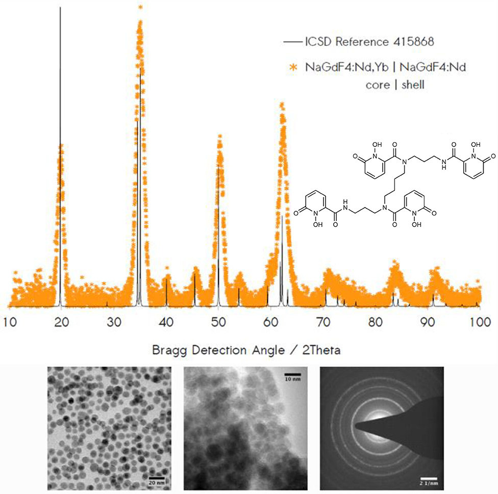
Figure 1. Top: Powder XRD data confirm colloidal synthesis of the hexagonal NaGdF4:Nd,Yb | NaGdF4:Nd. Inset: 343 surface chelator. Bottom: TEM reveals monodisperse nanocrystals of 10 nm in diameter in cases of unmodified (left) and 343-chelated (center) samples. Electron diffraction (right) provides evidence of dominant Bragg diffraction from lattice planes of hkl indices: (100), (110) + (101), (201), and (300) + (121).
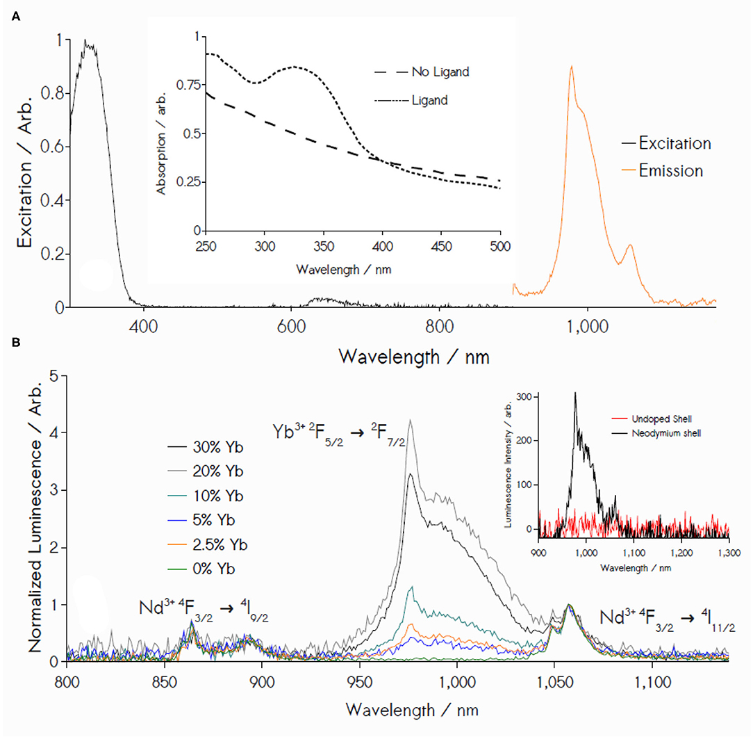
Figure 2. (A) Black: Excitation spectrum of 3% Nd3+/20% Yb3+-doped NPs monitoring 980 nm Yb3+ emission. Orange trace: NIR emission under 355 nm excitation. Inset: UV-vis absorption in ethanol. (B) Luminescence from Nd3+, Yb3+ core-shell NPs upon excitation at 355 nm, showing increasing Yb3+ emission (normalized relative to Nd3+ emission at 1059 nm) as Yb3+ content increases (0 to 20 mol %). Nd3+ content is held constant at 3 mol %. Inset: Removal of Nd3+ from the shell blocks energy transfer to terminal Yb3+ acceptor ions, arresting photoemission.
NP luminescence reveals a ligand-sensitized emission in the NIR that is a composite of Yb3+ (2F5/2 → 2F7/2) and Nd3+ (4F3/2 → 4IJ=9/2,11/2) transitions (Figure 2B). Specifically, excitation spectra of these materials indicate their irradiation between ca. 300 and 360 nm results in an IR emission representing a superposition of Yb3+ radiative decay and peaks diagnostic of Nd3+ transitions at 860 nm (4F3/2 → 4I9/2) and 1059 nm (4F3/2 → 4I11/2). Varying Yb3+ content results in an increased Yb3+ luminescence until a 20% dopant concentration is reached (Figure 2B) (Lakshminarayana et al., 2008a; Li L. et al., 2012; Wang et al., 2015), which was therefore used for all subsequent measurements in this study.
These results are consistent with a deliberate nanocrystal design where 343 functions as a terminal light absorber, transmitting energy exclusively to Nd3+ ions in the adjacent NP shell via Dexter transfer, followed by an energy migration step between shell Nd3+ ions and the nanocrystalline core. Partial energy localization in the core is presumed to result in the reversible energy transfer between Nd3+ 4F3/2 (~11, 460 cm−1) and Yb3+ 2F5/2 (10,400 cm−1), populating an equilibrium mixture of Yb3+ excited states under steady-state illumination; radiative deactivation of these states produces an NIR luminescence centered around 979 nm. Inspection of control data bolsters this narrative: NPs featuring a Nd3+/Yb3+-doped core and an undoped shell (NaGd1−x−yNdxYbyF4 | NaGdF4 -343) display no NIR emission (Figure 2B, inset). Yb3+-free controls (NaGd1−xNdxF4 | NaGd1−xNdxF4 -343) yield the intuitive result of an IR emission defined solely by Nd3+ transitions. UV illumination in the absence of the 343 ligand yields no detectable luminescence under identical conditions from either Nd3+ or Yb3+ lumiphores. Furthermore, ligand incorporation is shown to significantly increase luminescence yield upon excitation at 350 nm, with a resulting signal intensity at 979 nm approximately 1100-fold greater for the 343-decorated particles than observed for the unmodified nanostructures (Figure 3). It is noteworthy that none of the higher energy emissions that typically result from transitioning between Nd3+ stark levels are found in the visible range between 360 and 860 nm (Koningstein and Geusic, 1964; Zhang et al., 2007, 2015; Chen et al., 2012; Li X. et al., 2013; Wang et al., 2015), a surprising fact, given the high density of energetic states in Nd3+ electronic structure, and a standing precedent in the literature of Nd3+-doped, rare-earth hosts yielding emission spectra reflective of the ion's several f-f transitions (Li X. et al., 2013; Mimun et al., 2013; Wang et al., 2015). One, or a combination, of four likely possibilities may explain such phenomena: (1) the overall equilibrium between Yb3+ 2F5/2 and the Nd3+ states serving as acceptor states from the 343 triplet manifold favors production of Yb3+ excited states; (2) The resonant exchange between Yb3+ 2F5/2 and Nd3+ donor states largely favors Yb3+ 2F5/2 production; (3) Nd3+ states are rapidly quenched by phonon-coupled effects involving either ligand or host lattice vibrational states; (4) Rates of energy migration between Nd3+ ions significantly outpace the rates of radiative decay from individual Nd3+ stark levels. The scope of data acquired in this current study make assertions (1) and (4) difficult to confirm, as it demands a comprehensive knowledge of the rates and efficiency of all steps from the initial point of photon absorption by the ligand to Yb3+ ion emission. However, the nature of energy exchange between Nd3+ and Yb3+ is moderately exothermic, with an energy gap that is sufficiently large to possibly make phonon-assisted, energetic back-transfer from Yb3+ (2F5/2) → Nd3+ (4F3/2) kinetically prohibitive in NaGd(Y)F4 hosts (1060 cm−1 energy gap vs. ~ 400 cm−1 phonon energy), suggesting (2) as a partial explanation. Finally, (3) also seems possible. Despite numerous reports of Nd3+ visible-frequency luminescence in rare-earth fluoride particles featuring surface ligands bound via Ln coordination by oxygen donors (Li X. et al., 2013; Wang et al., 2015), such studies are generally conducted in nonpolar solvents. In this work, the ethanol solvent needed for stabilizing particles with 343 contain OH oscillators that are likely conduits for non-radiative decays that may result in quenched visible emission form Nd3+. Such a possibility is also consistent with previous findings that the 343-Nd3+ molecular complex dissolved in aqueous buffer only yields NIR Nd3+ luminescence (Sturzbecher-Hoehne et al., 2011).
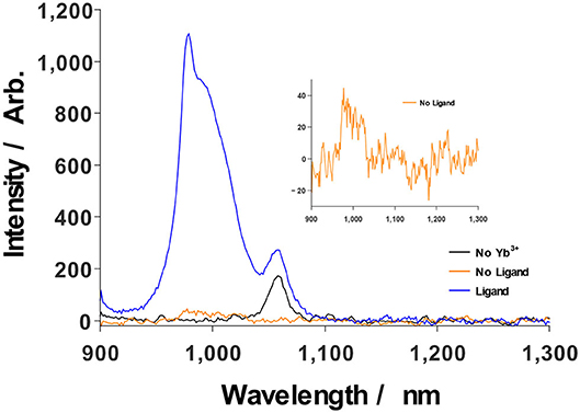
Figure 3. Ligand sensitization significantly enhances Yb3+ luminescence upon excitation at 350 nm (blue), relative to the case of NP illumination in the absence of photosensitization by 343 (orange, inset). Sensitized NPs without Yb3+ only show Nd3+ emission at 1057 nm (black).
Excited State Dynamics and Energy Transfer
Extraction of 343-Nd3+, Nd3+-Yb3+ energy transfer rates and Nd3+/Yb3+ excited state lifetimes were achieved through time-resolved luminescence measurements. Rates of energy transfer between the ligand and shell-confined Nd3+ were found through cryogenic (77 K) monitoring of the 343 triplet luminescence at 525 nm (Supplementary Figure 6). Mean decay times for the ligand triplet state in the presence of Nd3+ quenching were calculated according to Equation (1) (where I(t) is the time-dependent emission intensity, and I0 is its initial value), yielding values of 626 ± 48 μs. 343 triplet deactivation displayed tri-exponential behavior, occurring in decay phases with rates of 2197 ± 392 s−1, 382 ± 42 s−1, and 25.5 ± 19 s−1 at respective amplitudes of 0.60, 0.34, and 0.06, consistent with data previously reported for a 343-sensitized, Eu3+-doped system (Agbo and Abergel, 2016; Agbo et al., 2016). Extracting mean lifetimes by calculating the weighted sum of these decay rates (Equation 2) according to their respective contributions toward the measured decay spectrum yields a similar lifetime of 694 ± 127 μs, a value within error of the integral method. Ligand-Nd3+ energy transfer efficiencies were then determined according to Equation (3), where τDA is the measured 343 donor lifetime in the presence of Nd3+ acceptor ions and τD is the measured 343 donor lifetime in the absence of acceptor quenching. Values for τD were determined previously (1.61 ms) (Agbo and Abergel, 2016; Agbo et al., 2016). Accordingly, energy transfer between ligands on the NP surface and Nd3+ atoms in the nanocrystalline shell was found to proceed with an efficiency of 61 ± 3%.
Quenching of Nd3+ excited states by Yb3+ ions was resolved through monitoring of Nd3+ transitions at 860 nm (4F3/2 → 4I9/2) and 1057 nm (4F3/2 → 4I11/2), as the 4F3/2 state in Nd3+ lies at a similar energy to the 2F5/2 state of Yb3+ (Figure 4C). Transient luminescence of the 860 nm transition proceeds in two phases, with treatments showing rates of 2.13 x 10⁵ ± 6150 s−1 and 798 ± 67.7 s−1; 4F3/2 → 4I11/2 decay at 1057 nm displays quenching rates of 1.56 x 10⁵ ± 2760 s−1 and 4940 ± 140 s−1 (Figures 4A,B). Controls containing no core Yb3+ (NaGdF4:Nd|NaGdF4:Nd-343 core|shell NPs) yielded Nd3+ decay rates of 4.14 x 10⁵ s−1 at 860 nm and 1.43 x 10⁵ s−1 at 1057 nm. These data permit the calculation of Nd3+-Yb3+ energy transfer efficiencies (Equation 3) of 86.8 ± 2.0 and 82.4 ± 1.4%, using the respective 860 nm and 1057 nm transitions as spectroscopic handles (Supplementary Figure 7).
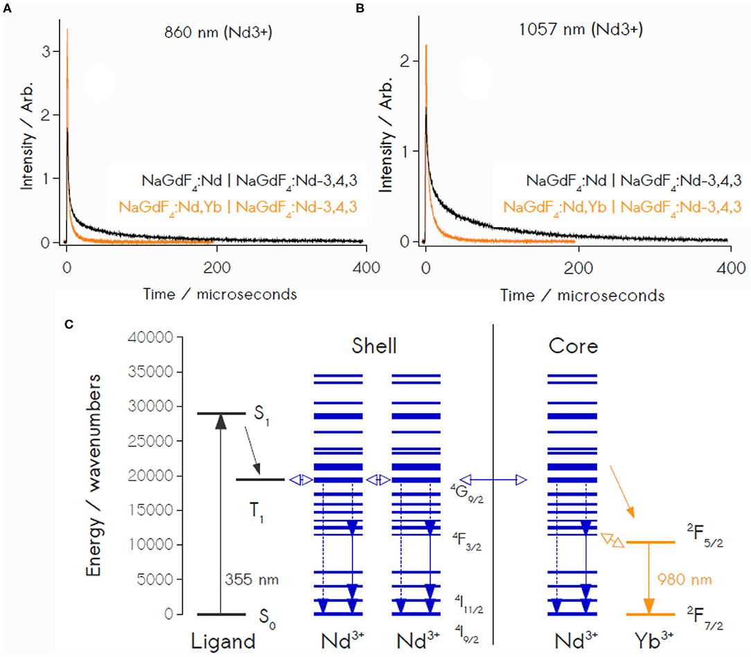
Figure 4. Time resolved Nd3+ luminescence at both 860 nm (A) and 1057 nm (B) reveals quenching in the presence of Yb3+ acceptors, confirming significant energy transfer between Nd3+ and Yb3+ ions. (C) Schematic Jablonski depiction of energy transfer processes in the sensitized Nd3+, Yb3+ core-shell particles. Downconversion involves direct transitioning from the 4G9/2 donor level in Nd3+ to the 2F5/2 level in Yb3+.
Downconversion Order via Power Dependence
Luminescence measurements as a function of incident light power were conducted to determine the order of photon production by the coated nanocrystals featuring Nd3+ and Yb3+ doping levels of 3 and 20%, respectively (Figure 5) (Suyver et al., 2005; Zou et al., 2012; Lin et al., 2015). Luminescence measured at light intensities over the range of 6.2–2.1 mW cm−2 (3.0 μW to 1.0 μW as measured at the fiber optic) displays linear correlations between the source power logarithms and integrated emission intensity in the NIR regime, with an average slope of 0.86 ± 0.04 (Supplementary Figures 8–9). Notably, this behavior occurs under diffuse illumination conditions relevant to the terrestrial solar spectrum (~100 mW cm−2 total), a stark contrast to the high excitation powers generally required for Ln luminescence. Furthermore, the excitation powers used here are far below the regimes for power saturation that are generally observed in these and related materials (Suyver et al., 2005; Chen G, et al., 2009; Lin et al., 2015; Loiko et al., 2016; Chen et al., 2017), demonstrating that our reported power dependencies are not mere artifacts of excited state saturation. The observed sub-unity slopes are diagnostic of a multi-photon production process involving direct transitions from the 4G9/2 donor level in Nd3+ to the 2F5/2 level in Yb3+. Generally, an idealized two-photon DC process yields a theoretical slope of n = 0.5, in contrast to the slope of 1 found in the power dependence for a typical, 1:1 Stokes-shifted emission and the n = 2 slope for two-photon UC (Suyver et al., 2005; Chen G, et al., 2009; Zou et al., 2012; Lin et al., 2015; Loiko et al., 2016; Chen et al., 2017). Here, the intermediate slope observed in the NIR luminescence suggests a photoemission that is a function of both 1:1 Stokes-shifted luminescence and 1:2 photon DC pathways. Furthermore, while our slopes values are far from the ideal two-photon slope, the small measurement error strongly suggests the measured effect is a statistically significant one, and that the multi-photon effect is therefore genuine (if minor); this is bolstered by control power dependencies conducted with Nd3+-only, 343 NPs, which yield a slope value of 1, for Nd3+ luminescence detected at 1057 nm (Supplementary Figure 10). The relative contributions of these various decay channels to the overall photoluminescence are difficult to precisely quantify but are estimated with the system described in Equation (4), where terms ak are weights representing the respective contributions of the kth unique decay channel (i.e., 1:1 photon emission, 2-photon DC), mk are the theoretical slopes for a photon DC process of order 1/mk and m is the total slope derived from power dependence, a value in which the contributions of all active luminescent decay channels are implicit. For our particular case, where only two unique processes (one-photon, m = 1 luminescence and m = 0.5, two-photon conversion) are present, solving for the coefficients for m = 0.86 yields a1 = 0.72 and a2 = 0.28, suggesting that, while two-photon mechanisms are operative (28%), luminescence in the NIR remains dominated by single-photon processes (72%) (Strek et al., 2001). These results are notable, as a handful of previous reports had pointed to claims of ligand-sensitized, Stokes-shifted luminescence in nanocrystals, however these studies had not produced any explicit evidence of multi-photon DC (Charbonnière et al., 2008; Cross et al., 2010; Chen et al., 2013; Li S. et al., 2013, 2014; Irfanullah et al., 2015; Goetz et al., 2016).
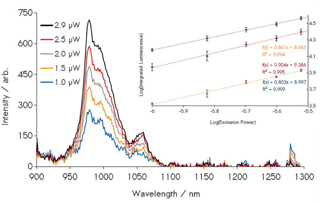
Figure 5. Power dependence study of 3/20% doped Nd3+/Yb3+ NPs. Emission spectra are integrated in the region of Yb3+ luminescence (940–1040 nm) and plotted against the excitation power (inset). The power dependence of these material exhibits a sub-linear slope, with a total of 10 independent measurements yielding an average slope of 0.86 ± 0.04 among three replicates, confirming the presence of two-photon emissions.
Quantum Efficiency of Sensitized Emission
343's efficacy as a photosensitizer was further evaluated by determining the internal quantum yields observed for Yb3+ infrared emission. Direct Nd3+ excitation was achieved through laser excitation at 456 nm ((4I9/2 → 4G9/2, SI). Direct excitation pathways display the low external quantum yields that are a natural consequence of the low f-f absorptivities of Ln transitions, with direct generation of Nd3+ excited states yielding an average efficiency of 0.15, 0.14, and 0.34% (0.21 ± 0.11%) for Yb3+ luminescence integrated over the range 940–1040 nm. Using the relation Φ343−Yb = ΦNd−Ybηsens, where ηsens is the ligand-Nd3+ sensitization efficiency, allows for determination of an approximate quantum yield of 0.13% for Yb3+ emission originating from 355 nm ligand excitation. In addition to our measured quantum yields, we compare these results to a treatment for calculating DC quantum yields commonly employed throughout photon DC literature (Vergeer et al., 2005; Lakshminarayana et al., 2008a,b; Ye et al., 2008, 2011; Chen X, et al., 2009; Van Der Ende et al., 2009; Li K.-Y. et al., 2014; Zhu et al., 2014). As defined in Equation (5), QYNd−Yb is the quantum efficiency of DC from a Nd3+ donor to an Yb3+ acceptor, whereas ηNd is the Nd3+ donor quantum efficiency and is generally assumed to be 100% (no non-radiative losses); (Vergeer et al., 2005; Lakshminarayana et al., 2008a,b; Ye et al., 2008, 2011; Chen X, et al., 2009; Van Der Ende et al., 2009; Li K.-Y. et al., 2014; Zhu et al., 2014). ETE is the Nd3+-Yb3+ energy transfer efficiency (0.87). Here, such approach yields a maximum two-photon quantum yield of 182% for these nanocrystals in the case of luminescence driven by photoexcitation of the Nd3+ absorption band at 456 nm. Determination of the ligand-sensitized Yb3+ quantum yields are found by factoring in the additional ligand-Nd3+ energy transfer step according to Equation (6). Applying our experimentally determined value of 61% for the ligand-Nd3+ sensitization efficiency yields a maximum quantum yield of 114% for a two-photon Yb3+ emission in these crystals, where the ligand is the terminal light absorber, rather than Nd3+. It must be stressed that such values of two-photon emission that are calculated using Equations (5, 6) only represent theoretical upper limits on the two-photon quantum yields, as they assume the absence of non-radiative decay paths (Vergeer et al., 2005; Lakshminarayana et al., 2008a,b; Ye et al., 2008, 2011; Chen X, et al., 2009; Van Der Ende et al., 2009; Li K.-Y. et al., 2014; Zhu et al., 2014). Here, we have provided the actual quantum yields of these materials as a comparison, with the substantial gap between the calculated and experimental values reflecting the considerable degree of non-radiative losses that exist in these nanocrystals. This observation should prompt a conservative interpretation of any two-photon quantum efficiencies determined through calculated methods assuming an absence of non-radiative decays. Such approaches run in stark contrast to physical measurements of two-photon production, where an accounting of non-radiative excited-state deactivation is necessarily an implicit feature of acquired experimental data.
Conclusion
This work demonstrates the value in considering not only the respective energies of Ln donor/acceptor levels in the design of multi-photon emitters, but also the role that core-shell structures can play. Specifically, the use of layered crystal domains permits the critical segregation of ligand absorber and metal donors/acceptors (Nd3+/Yb3+), enabling an efficient, stepwise ligand-donor-acceptor energy transfer that mitigates the possibility of quantum efficiency losses arising from the direct sensitization of Yb3+ by 343 (Sturzbecher-Hoehne et al., 2011). Though availability of a resonant 4F3/2 donor level in Nd3+ facilitates energy transfer to the Yb3+ excited state, the high density of Nd3+ f-states falling between energies of the 343 triplet (21,500 cm−1) and the Nd3+4 F3/2 donor (11,460 cm−1) allows for significant losses through multi-phonon relaxation during 343 → Nd3+ sensitization, a physical basis for the low contribution of two-photon emission to the overall Yb3+ luminescence (while the measured energy transfer efficiency for this step is 0.61, this is only a remark on the total energy siphoned from 343 phosphors by Nd3+ acceptor levels; it provides no information on non-radiative losses incurred by excited Nd3+ ions during internal conversion to yield the 4F3/2 intermediate). These results build on our initial studies of ligand sensitization of Eu3+ in NaGd(Y)F4 crystals (Agbo et al., 2016), extending the general principle of sensitized Ln NP luminescence to the particular case of two-photon generation in the NIR. The present findings detail a material capable of shifting luminescence over a range of 600 nm, from UV to IR, with a NIR emission profile that is a partial function of two-photon processes. These results bear significance to the challenge of addressing energetic mismatches between terrestrial solar illumination and the spectral response of commercial photovoltaics.
Data Availability Statement
All datasets generated for this study are included in the article/ Supplementary Material.
Author Contributions
PA and RA designed the research. PA performed experimental work. All authors performed data analysis and contributed to the writing of the manuscript.
Funding
This work was supported by the Director, Office of Science, Office of Basic Energy Sciences, and the Division of Chemical Sciences, Geosciences, and Biosciences of the U.S. Department of Energy at the Lawrence Berkeley National Laboratory under Contract No. DE-AC02-05CH11231.
Conflict of Interest
RA and PA are listed as inventors on a patent application filed by the Lawrence Berkeley National Laboratory and describing inventions related to the research results presented here.
The remaining author declares that the research was conducted in the absence of any commercial or financial relationships that could be construed as a potential conflict of interest.
Acknowledgments
We thank Dr. S. Fischer and J. Swabeck for help acquiring quantum yield data on a custom system in the laboratory of Prof. P. Alivisatos at UC Berkeley.
Supplementary Material
The Supplementary Material for this article can be found online at: https://www.frontiersin.org/articles/10.3389/fchem.2020.579942/full#supplementary-material
Supporting Information: Experimental procedures, quantum yield data, time-resolved luminescence, energy transfer calculations, and Eu3+-Cm3+ excitation spectra are included in the Supporting Information. This material is available free of charge via the internet at http://pubs.acs.org.
References
Abergel, R. J., D'Aléo, A., Ng Pak Leung, C., Shuh, D. K., and Raymond, K. N. (2009). Using the antenna effect as a spectroscopic tool: photophysics and solution thermodynamics of the model luminescent hydroxypyridonate complex [EuIII (3, 4, 3-LI (1, 2-HOPO))]−. Inorg. Chem. 48, 10868–10870. doi: 10.1021/ic9013703
Agbo, P., and Abergel, R. J. (2016). Ligand-sensitized lanthanide nanocrystals: merging solid-state photophysics and molecular solution chemistry. Inorg. Chem. 55, 9973–9980. doi: 10.1021/acs.inorgchem.6b00879
Agbo, P., and Abergel, R. J. (2017). Ligand-Sensitized Lanthanide Nanocrystals as Ultraviolet Downconverters. U.S. Patent.
Agbo, P., Xu, T., Sturzbecher-Hoehne, M., and Abergel, R. J. (2016). Enhanced ultraviolet photon capture in ligand-sensitized nanocrystals. ACS Photon. 3, 547–552. doi: 10.1021/acsphotonics.6b00118
Binnemans, K. (2015). Interpretation of europium (III) spectra. Coord. Chem. Rev. 295, 1–45. doi: 10.1016/j.ccr.2015.02.015
Bünzli, J.-C. G., and Chauvin, A.-S. (2014). “Lanthanides in solar energy conversion,” in Handbook on the Physics and Chemistry of Rare Earths. Vol. 44, eds J.-C. G. Bünzli and V. K. Pecharsky (Amsterdam: Elsevier Science B.V.), 169–281. doi: 10.1016/B978-0-444-62711-7.00261-9
Bünzli, J.-C. G., and Eliseeva, S. V. (2010). “Basics of lanthanide photophysics,” in Lanthanide Luminescence, eds P. Hänninen and H. Härmä (Berlin; Heidelberg: Springer), 1–45. doi: 10.1007/4243_2010_3
Charbonnière, L. J., Rehspringer, J.-L., Ziessel, R., and Zimmermann, Y. (2008). Highly luminescent water-soluble lanthanide nanoparticles through surface coating sensitization. New. J. Chem. 32, 1055–1059. doi: 10.1039/b719700d
Chen, G., Liang, H., Liu, H., Somesfalean, G., and Zhang, Z. (2009). Near vacuum ultraviolet luminescence of Gd3+ and Er3+ ions generated by super saturation upconversion processes. Opt. Express. 17, 16366–16371. doi: 10.1364/OE.17.016366
Chen, G., Ohulchanskyy, T. Y., Liu, S., Law, W.-C., Wu, F., Swihart, M. T., et al. (2012). Core/shell NaGdF4: Nd3+/NaGdF4 nanocrystals with efficient near-infrared to near-infrared downconversion photoluminescence for bioimaging applications. ACS nano. 6, 2969–2977. doi: 10.1021/nn2042362
Chen, J., Meng, Q., May, P. S., Berry, M. T., and Lin, C. (2013). Sensitization of Eu3+ luminescence in Eu: YPO4 nanocrystals. J. Mat. Chem. 117, 5953–5962. doi: 10.1021/jp3109072
Chen, X., Huang, X., and Zhang, Q. (2009). Concentration-dependent near-infrared quantum cutting in NaYF4: Pr3+, Yb3+ phosphor. J. Appl. Phys. 106:063518. doi: 10.1063/1.3224906
Chen, X., Zhu, Y., Zhou, D., Xu, W., Zhu, J., Pan, G., et al. (2017). Size-dependent downconversion near-infrared emission of NaYF4: Yb3+, Er3+ nanoparticles. J. Mat. Chem. 5, 2451–2458. doi: 10.1039/C7TC00267J
Cross, A. M., May, P. S., Veggel, F. C. v, and Berry, M. T. (2010). Dipicolinate sensitization of europium luminescence in dispersible 5% Eu: LaF3 nanoparticles. J. Mat. Chem. 114, 14740–14747. doi: 10.1021/jp103366j
Garfield, D. J., Borys, N. J., Hamed, S. M., Torquato, N. A., Tajon, C. A., Tian, B., et al. (2018). Enrichment of molecular antenna triplets amplifies upconverting nanoparticle emission. Nat. Photon. 12, 402–407. doi: 10.1038/s41566-018-0156-x
Gauthier, N., Raccurt, O., Imbert, D., and Mazzanti, M. (2013). Efficient sensitization of Ln3+-doped NaYF4 nanocrystals with organic ligands. J. Nanoparticle Res. 15:1723. doi: 10.1007/s11051-013-1723-1
Goetz, J., Nonat, A., Diallo, A., Sy, M., Sera, I., Lecointre, A., et al. (2016). Ultrabright lanthanide nanoparticles. ChemPlusChem. 81, 526–534. doi: 10.1002/cplu.201600007
Irfanullah, M., Bhardwaj, N., and Chowdhury, A. (2016). Sensitized luminescence from water-soluble LaF3: Eu nanocrystals via partially-capped 1, 10-phenanthroline: time-gated emission and multiple lifetimes. Dalton Trans. 45, 12483–12495. doi: 10.1039/C6DT01917J
Irfanullah, M., Sharma, D. K., Chulliyil, R., and Chowdhury, A. (2015). Europium-doped LaF3 nanocrystals with organic 9-oxidophenalenone capping ligands that display visible light excitable steady-state blue and time-delayed red emission. Dalton Trans. 44, 3082–3091. doi: 10.1039/C4DT03249G
Janssens, S., Williams, G., and Clarke, D. (2011). Systematic study of sensitized LaF3: Eu3+ nanoparticles. J. Appl. Phys. 109, 023506. doi: 10.1063/1.3531994
Koningstein, J., and Geusic, u. J. (1964). Energy levels and crystal-field calculations of neodymium in yttrium aluminum garnet. Phys. Rev. 136, A711. doi: 10.1103/PhysRev.136.A711
Lakshminarayana, G., Yang, H., Ye, S., Liu, Y., and Qiu, J. (2008a). Cooperative downconversion luminescence in Pr3+/Yb3+: SiO2-Al2O3-BaF2-GdF3 glasses. J. Mater. Res. 23, 3090–3095. doi: 10.1557/JMR.2008.0372
Lakshminarayana, G., Yang, H., Ye, S., Liu, Y., and Qiu, J. (2008b). Co-operative downconversion luminescence in Tm3+/Yb3+: SiO2-Al2O3-LiF-GdF3 glasses. J. Phys. D. 41:175111. doi: 10.1088/0022-3727/41/17/175111
Li, K.-Y., Liu, L.-Y., Wang, R.-Z., Xiao, S.-G., Zhou, H., and Yan, H. (2014). Broadband sensitization of downconversion phosphor YPO4 by optimizing TiO2 substitution in host lattice co-doped with Pr3+-Yb3+ ion-couple. J. Appl. Phys. 115:123103. doi: 10.1063/1.4869659
Li, L., Xiantao, W., Yonghu, C., Changxin, G., and Min, Y. (2012). Energy transfer in Tb3+, Yb3+ codoped Lu2O3 near-infrared downconversion nanophosphors. J. Rare Earths. 30, 197–201. doi: 10.1016/S1002-0721(12)60022-2
Li, S., Hou, Z., Cheng, Z., Lian, H., Li, C., and Lin, J. (2013). Enhanced near-infrared quantum cutting luminescence in 1, 2, 4, 5-benzenetetracarboxylic acid/NaYF4: Tb3+, Yb3+ hybrid nanoparticles. RSC Adv. 3, 5491–5497. doi: 10.1039/c3ra23439h
Li, S., Li, X., Jiang, Y., Hou, Z., Cheng, Z., Ma, P., et al. (2014). Highly luminescent lanthanide fluoride nanoparticles functionalized by aromatic carboxylate acids. RSC Adv. 4, 55100–55107. doi: 10.1039/C4RA09266J
Li, S., Zhang, X., Hou, Z., Cheng, Z., Ma, P., and Lin, J. (2012). Enhanced emission of ultra-small-sized LaF3: RE3+(RE= Eu, Tb) nanoparticles through 1, 2, 4, 5-benzenetetracarboxylic acid sensitization. Nanoscale. 4, 5619–5626. doi: 10.1039/c2nr31206a
Li, S. W., Ren, H. J., and Ju, S. G. (2014). Sensitized Luminescence of LaF3: Eu3+ Nanoparticles through Pyromellitic Acid. J. Nanosci. Nanotech. 14, 3677–3682. doi: 10.1166/jnn.2014.7968
Li, X., Wang, R., Zhang, F., Zhou, L., Shen, D., Yao, C., et al. (2013). Nd3+ sensitized up/down converting dual-mode nanomaterials for efficient in-vitro and in-vivo bioimaging excited at 800 nm. Sci. Rep. 3:3536. doi: 10.1038/srep03536
Lin, J., Fujita, Y., and Neogi, A. (2015). Saturation of two photon emission in ZnO nanoparticles with second order nonlinearity. RSC Adv. 5, 10921–10926. doi: 10.1039/C4RA08380F
Loiko, P., Khaidukov, N., Méndez-Ramos, J., Vilejshikova, E., Skoptsov, N., and Yumashev, K. (2016). Up-and down-conversion emissions from Er3+ doped K2YF5 and K2YbF5 crystals. J. Luminesc. 170, 1–7. doi: 10.1016/j.jlumin.2015.10.016
Mimun, L. C., Ajithkumar, G., Pokhrel, M., Yust, B. G., Elliott, Z. G., Pedraza, F., et al. (2013). Bimodal imaging using neodymium doped gadolinium fluoride nanocrystals with near-infrared to near-infrared downconversion luminescence and magnetic resonance properties. J. Mater. Chem. B. 1, 5702–5710. doi: 10.1039/c3tb20905a
Moore, E. G., Samuel, A. P., and Raymond, K. N. (2009). From antenna to assay: lessons learned in lanthanide luminescence. Acc. Chem. Res. 42, 542–552. doi: 10.1021/ar800211j
Moore, E. G., Szigethy, G., Xu, J., and Pålsson, L.-O. (2008). 3, 2-HOPO Complexes of Near Infra-Red (NIR) Emitting Lanthanides: sensitization of HoIII and PrIII in Aqueous Solution. Angew. Chem. 47:9500. doi: 10.1002/anie.200802337
Shavaleev, N. M., Accorsi, G., Virgili, D., Bell, Z. R., Lazarides, T., Calogero, G., et al. (2005). Syntheses and crystal structures of dinuclear complexes containing d-block and f-block luminophores. Sensitization of NIR luminescence from Yb (III), Nd (III), and Er (III) centers by energy transfer from Re (I)– and Pt (II)– bipyrimidine metal centers. Inorg. Chem. 44, 61–72. doi: 10.1021/ic048875s
Shockley, W., and Queisser, H. J. (1961). Detailed balance limit of efficiency of p-n junction solar cells. J. Appl. Phys. 32, 510–519. doi: 10.1063/1.1736034
Song, Y., Shao, B., Feng, Y., L¨u, W., Huo, J., Zhao, S., et al. (2016). Emission enhancement and color tuning for GdVO4: Ln3+ (Ln= Dy, Eu) by surface modification at single wavelength excitation. Inorg. Chem. 56, 282–291. doi: 10.1021/acs.inorgchem.6b02125
Strek, W., Bednarkiewicz, A., and Dere,ń, P. (2001). Power dependence of luminescence of Tb3+-doped KYb (WO4)2crystal. J. Luminesc. 92, 229–235. doi: 10.1016/S0022-2313(00)00263-5
Sturzbecher-Hoehne, M., Ng Pak Leung, C., D'Aléo, A., Kullgren, B., Prigent, A.-L., Shuh, D. K., et al. (2011). 3,4,3-LI(1,2-HOPO): In vitro formation of highly stable lanthanide complexes translates into efficacious in vivo europium decorporation. Dalton Trans. 40, 8340–8346. doi: 10.1039/c1dt10840a
Sun, T., Chen, X., Jin, L., Li, H.-W., Chen, B., Fan, B., et al. (2017). Broadband Ce (III)-sensitized quantum cutting in core-shell nanoparticles: mechanistic investigation and photovoltaic application. J. Phys. Chem. Lett. 8, 5099–5104. doi: 10.1021/acs.jpclett.7b02245
Suyver, J., Aebischer, A., García-Revilla, S., Gerner, P., and Güdel, H. (2005). Anomalous power dependence of sensitized upconversion luminescence. Phys. Rev. B. 71:125123. doi: 10.1103/PhysRevB.71.125123
Van Der Ende, B. M., Aarts, L., and Meijerink, A. (2009). Lanthanide ions as spectral converters for solar cells. Phys. Chem. Chem. Phys. 11, 11081–11095. doi: 10.1039/b913877c
Vergeer, P., Vlugt, T., Kox, M., Den Hertog, M., Van der Eerden, J., and Meijerink, A. (2005). Quantum cutting by cooperative energy transfer in Ybx Y1−x P O4: Tb3+. Phys. Rev. B. 71:014119. doi: 10.1103/PhysRevB.71.014119
Wang, F., Deng, R., and Liu, X. (2014). Preparation of core-shell NaGdF4 nanoparticles doped with luminescent lanthanide ions to be used as upconversion-based probes. Nat. Prot. 9:1634. doi: 10.1038/nprot.2014.111
Wang, K., Qincheng, W., Zhang, Y., Qiao, R., Li, S., and Li, Z. (2015). Synthesis of Nd3+/Yb3+ sensitized upconversion core-shell nanocrystals with optimized hosts and doping concentrations. RSC Adv. 5, 62899–62904. doi: 10.1039/C5RA09873D
Wang, Z., and Meijerink, A. (2018). Dye-sensitized downconversion. J. Phys. Chem. Lett. 9, 1522–1526. doi: 10.1021/acs.jpclett.8b00516
Werts, M. H. (2005). Making sense of lanthanide luminescence. Sci. Prog. 88, 101–131. doi: 10.3184/003685005783238435
Ye, S., Katayama, Y., and Tanabe, S. (2011). Down conversion luminescence of Tb3+-Yb3+ codoped SrF2 precipitated glass ceramics. J. Non-Cryst. Solids. 357, 2268–2271. doi: 10.1016/j.jnoncrysol.2010.11.083
Ye, S., Zhu, B., Luo, J., Chen, J., Lakshminarayana, G., and Qiu, J. (2008). Enhanced cooperative quantum cutting in Tm3+-Yb3+ codoped glass ceramics containing LaF3 nanocrystals. Opt. Express. 16, 8989–8994. doi: 10.1364/OE.16.008989
Zhang, J., Shade, C. M., Chengelis, D. A., and Petoud, S. (2007). A strategy to protect and sensitize near-infrared luminescent Nd3+ and Yb3+: organic tropolonate ligands for the sensitization of Ln3+-doped NaYF4 nanocrystals. J. Am. Chem. Soc. 129, 14834–14835. doi: 10.1021/ja074564f
Zhang, X., Zhao, Z., Zhang, X., Cordes, D. B., Weeks, B., Qiu, B., et al. (2015). Magnetic and optical properties of NaGdF4: Nd3+, Yb3+, Tm3+ nanocrystals with upconversion/downconversion luminescence from visible to the near-infrared second window. Nano Res. 8, 636–648. doi: 10.1007/s12274-014-0548-2
Zhu, W., Chen, D., Lei, L., Xu, J., and Wang, Y. (2014). An active-core/active-shell structure with enhanced quantum-cutting luminescence in Pr-Yb co-doped monodisperse nanoparticles. Nanoscale. 6, 10500–10504. doi: 10.1039/C4NR02785J
Keywords: sensitization, lanthanide, two-photon, downconversion, energy transfer
Citation: Agbo P, Kanady JS and Abergel RJ (2020) Infrared Photon Pair-Production in Ligand-Sensitized Lanthanide Nanocrystals. Front. Chem. 8:579942. doi: 10.3389/fchem.2020.579942
Received: 03 July 2020; Accepted: 08 October 2020;
Published: 04 November 2020.
Edited by:
Qianqian Su, Shanghai University, ChinaReviewed by:
Guanying Chen, Harbin Institute of Technology, ChinaLimin Jin, Harbin Institute of Technology, Shenzhen, China
Copyright © 2020 Agbo, Kanady and Abergel. This is an open-access article distributed under the terms of the Creative Commons Attribution License (CC BY). The use, distribution or reproduction in other forums is permitted, provided the original author(s) and the copyright owner(s) are credited and that the original publication in this journal is cited, in accordance with accepted academic practice. No use, distribution or reproduction is permitted which does not comply with these terms.
*Correspondence: Rebecca J. Abergel, YWJlcmdlbEBiZXJrZWxleS5lZHU=