 Xin Wang
 Haiyan Zhao1
 Angzhen Li
 Pengfei Wang
- 1Key Laboratory of In-fiber Integrated Optics of Ministry of Education, College of Science, Harbin Engineering University, Harbin, China
- 2Optoelectronics Research Centre, University of Southampton, Southampton, United Kingdom
- 3Key Laboratory of Optoelectronic Devices and Systems of Ministry of Education and Guangdong Province, College of Optoelectronic Engineering, Shenzhen University, Shenzhen, China
In this letter, an Nd3+-doped compound fluorosilicate glass was successfully fabricated with the method of melt-quenching. Under the excitation of a 808 nm laser, enhanced near-infrared photoluminescence emission at the range from 1,065 to 1,140 nm was observed in the glass sample. To characterize its stability and resistance to environmental effects, transmission spectra at the range of NIR-MIR were measured under different environments, including humidity and temperature. In addition, the obtained fluorosilicate glass was also developed as a microsphere resonator by using CO2 laser heating method. When the diameter of the microsphere was controlled at 61.5 μm, coupling with a tapered fiber, single-multimode lasing in the wavelength range λ1,056–1,071 nm was achieved with a low lasing threshold of 1.5 mW. Compared to silica and phosphate glasses, this fluorosilicate glasses have lower phonon energy, which can reduce the probability of non-radiative transitions and improve the photoluminescence efficiency. Therefore, using it as the raw material, the developed microsphere resonator offers a high transition temperature and with a low lasing threshold, which are promising it for high performance sensing and detection applications.
Introduction
Optical microcavity refers to a laser resonator with a volume in the order of the light wavelength. In recent decades, microsphere resonators have attracted considerable attention due to their wide range of applications such as low threshold laser (Ilchenko et al., 1998), non-linear optics (Wang et al., 2012), cavity quantum electrodynamics (von Klitzing et al., 2001), and high sensitivity sensing (Sanchez-Martin et al., 2006). In 1961, Garrett et al. firstly demonstrated that microsphere resonators can be used as laser resonators, and observed Whispering Gallery Modes (WGMs) in a CaF2: Sm3+ crystal at the hydrogen temperature. Since then, various optical microcavities have been reported in different host materials, including drops, glasses, and crystals (Cai et al., 2000; Humar et al., 2009; Yang et al., 2017a). Glass microspheres with diameters from few microns to hundreds of microns could intrinsically be a resonator, because of their high quality factor and small modes per unit volume. The first laser emission from rare-earth doped glass microsphere resonators was reported by Miura et al. (1997). From then on, numerous investigations about microcavity resonators have focused on rare-earth doped or codoped glass microspheres (Wang et al., 2004; Mescia et al., 2010; Rasoloniaina et al., 2012; Li et al., 2015, 2019; Fang et al., 2017; Yang et al., 2017b).
Glasses with different compositions usually exhibit different optical properties, including refractive index, optical transmittance, coefficient of thermal expansion, and absorption coefficient. According to the glass composition, microspheres can be divided into silicate, tellurate, borate, fluoride, and chalcogenide glasses. Li et al. achieved 2.0 μm lasing from a Tm3+ doped silica glass microsphere pumped by a 808 nm laser (Li et al., 2018). The threshold power was as low as 1.2 mW and the quality factor as high as 105. Silica has numerous advantages, such as great chemical stability, simple preparation and excellent processability, but its high phonon energy increases the probability of non-radiative transitions, resulting in a very low photoluminescence efficiency. Compared to silica, compound glasses exhibit interesting properties, such as high transmittance and low phonon energy. Xiang et al. achieved 1,556 nm lasing in a Er3+ doped tellurite glass microsphere under the excitation of a 975 nm laser (Xiang et al., 2003). The laser threshold of glass microsphere was measured to be smaller than 2 mW. Fluoride glasses were widely applied in upconversion and MIR emission due to their low phonon energy. In 2000, von Klitzing demonstrated a green upconversion laser emission in an Er3+-doped fluoride glass microsphere (von Klitzing et al., 2000). Under diode laser pumping at λ ~801 nm, lasing at λ ~540 nm was obtained, providing a threshold smaller than 30 μW. Up to now, chalcogenide glasses have been the material of choice for MIR-FIR lasers, as they exhibit the highest transmittance at long wavelengths (over 4,000 nm). In 1996, Heo and Shin observed fluorescence at λ ~4.38 μm from a Dy3+ doped Ge–As chalcogenides glass under the excitation of a λ ~808 nm laser (Heo and Shin, 1996). Nevertheless, the compound glass exhibited poor chemical and mechanical properties. Research on novel materials with both low phonon energy and great stability is currently a hotspot.
In this letter, we fabricated a fluorosilicate glass and glass microsphere doped with Nd3+. Compared to silica and compound glasses, this fluorosilicate glass has both low phonon energy (Wang et al., 2018) and great stability, which is a potential material for micro-nano photonics.
Experiment
Nd3+-doped glass samples with the composition of (in mol%) 16ZnF2-68SiO2-16KF-xNd2O3 (x = 0.1, 0.2, 0.3, 0.4, and 0.5) were fabricated by the traditional method of melt-quenching. All the raw materials were acquired from Aladdin Company and their purity was 99% (ZnF2), 99.99% (SiO2), 99.5% (KF), and 99.99% (Nd2O3). Fifty gram of raw powders were placed in an agate mortar with smooth wall and were then stirred for 20 min to mix them uniformly in a platinum rhodium crucible. The crucible was placed into a high temperature electric furnace heated by silicon molybdenum rods at 1,520°C for 30 min. The melt was poured onto a 1.5 cm thick copper plate and pressed with another copper plate for a rapid cooling, in order to prevent crystallization, and then annealed at 430°C for 4 h to remove residual stresses. After cooling to room-temperature, the Nd3+ doped fluorosilicate glass was cut into samples. To confirm these samples were amorphous with no nanocrystals, X-ray diffraction (XRD) measurements were recorded by an X-ray diffractometer (ADVANCE D8, BRUKER, GERMANY). The absorption and photoluminescence emission spectra were measured by a spectrophotometer (LAMBDA, PERKINELMER, USA) and a fluorescence spectrometer (ZOLIX, CHINA), respectively. The details about the fabrication of the fluorosilicate glass fiber and microsphere were reported in our previous work (Wang et al., 2018).
Results and Discussion
Figure 1A shows the absorption spectra of xNd3+ doped fluorosilicate glass samples with composition of 16ZnF2-68SiO2-16KF–xNd2O3 (x = 0.1, 0.2, 0.3, 0.4, and 0.5 mol%). The absorption peaks located at 521, 579, 683, 734, 800, and 868 nm were attributed to the transitions from the ground state level to the 4G7/2, 4G5/2/2G7/2, 4F9/2, 4F7/2/4S3/2, 4F5/2/2H9/2, and 4F3/2 levels, respectively. As the strongest absorption peaks are located at λ ~800 nm, a diode laser at λ ~808 nm was chosen as excitation source, and intense near infrared photoluminescence emission was observed at λ ~1,059 nm, as shown in Figure 1B. When the Nd3+content increased from 0.2 to 0.8 mol%, the intensity of the infrared emission initially increased and then decreased. This can be explained as follows: when the dopant content increases, firstly more Nd3+ ions take part in the process of photoluminescence, and both the pump absorption and photoluminescence efficiency increases. As the content of Nd3+ continues to increase, the cross relaxation between Nd3+ ions becomes more and more significant and radiation-free transitions are more likely to occur due to the proximity between Nd3+ ions. This fluorosilicate glass had a phase-separated structure, and dopants were predominantly located in the F-rich area (Wang et al., 2018), that is why the concentration quenching value was as low as 0.8 mol%.
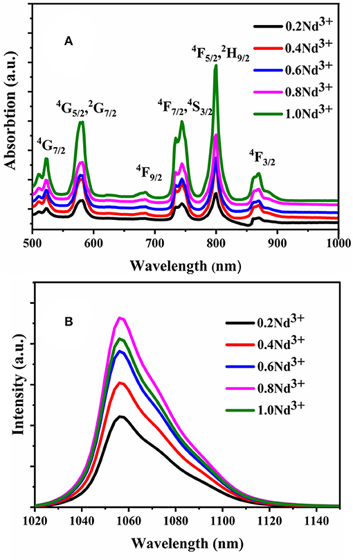
Figure 1. (A) Absorption spectra of x mol% Nd3+ doped fluorosilicate glass sample and (B) photoluminescence emission spectra of x mol% Nd3+ doped fluorosilicate glass sample under the excitation of a λ ~808 nm laser. (x = 0.2, 0.4, 0.6, 0.8, and 1.0).
The photoluminescence emission mechanism is shown in Figure 2. Under the excitation of a λ ~808 nm laser, the Nd3+ ions absorb photons and the electrons located at the ground states (4F9/2) transfer to the 4F5/2 level via ground state absorption (GSA) process, before going down to the 4F3/2 level with no photon emission. The electrons located at the 4F3/2 level can relax to the 4I13/2 level, 4I11/2 level or to the ground state directly. The transition from 4F3/2 to 4I11/2 results in the photoluminescence emission at λ ~1,056 nm.
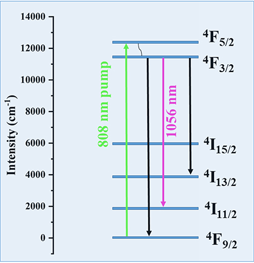
Figure 2. Schematic energy level and related transitions of the near-infrared photoluminescence emission processes.
Compared to silica, most of the compound glasses have relatively low transition and crystallization temperatures, resulting in a tendency to crystallize easily. Figures 3A,B show that both glass and glass microsphere are transparent. To further investigate the degree of crystallinity of the samples, XRD patterns of the bulk glass and glass microsphere samples (Figure 3C). Both patterns exhibit broad bands with no sharp peaks, indicating that both samples are amorphous, and microspheres can be fabricated from this fluorosilicate glass by fiber heating.
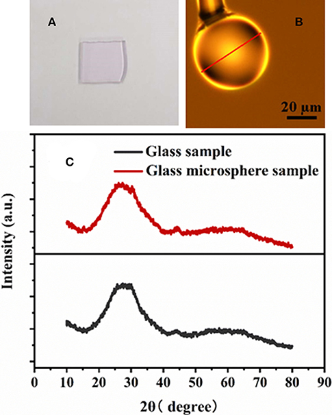
Figure 3. (A) Photograph of the bulk glass samples with composition 16ZnF2-68SiO2-16KF-0.4Nd2O3 (in mol%). (B) Microscope image of the glass microsphere. (C) XRD of the bulk glass and glass microsphere samples.
The diagram of the experimental set-up used to characterize the Nd3+ doped fluorosilicate glass microsphere laser under the excitation at λ ~808 nm is shown in Figure 4. The tapered fiber used for coupling the pump light into the glass microsphere was fabricated by heating a standard silica single mode optical fiber under tension in a conventional fiber tapering rig. This tapered silica single mode fiber has advantages, such as efficient transmission at both the pump laser and the gain laser wavelengths. When heated by the hydrogen-oxygen flame, the fiber section softened and was tapered under the action of axial stress. The glass microsphere was attached to a fiber clamp and positioned in close proximity to the tapered fiber to achieve critical coupling. To monitor the relative location of the taper with respect to the glass microsphere, a CCD camera attached to a 20x microscope eyepieces was used. The pump laser was launched into the single mode tapered fiber and coupled into the Nd3+ doped glass microsphere through the evanescent field. An optical spectrum analyzer (OSA) was used to detect the gain laser output from the Nd3+ doped fluorosilicate glass microsphere.
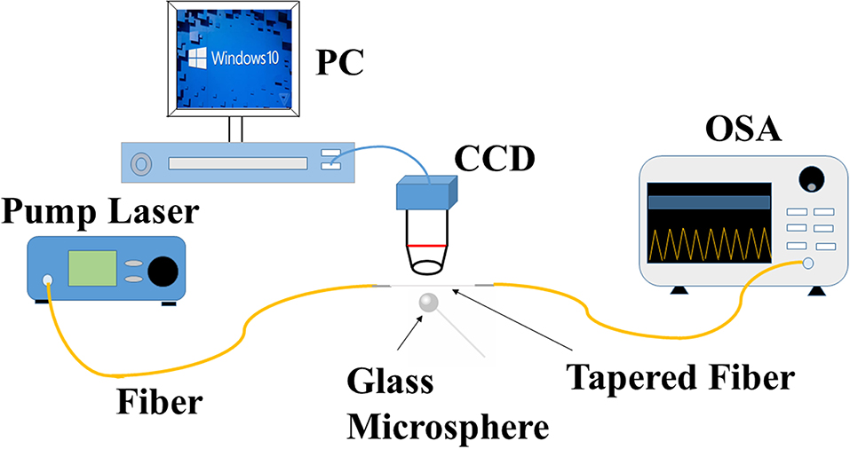
Figure 4. Diagram of the experimental set-up used to characterize the Nd3+ doped fluorosilicate glass microsphere laser.
Under the excitation at λ ~808 nm, single mode laser emission from the glass microsphere was observed by the OSA. The relationship between the intensity of the single mode laser output power and the pump power is shown in Figure 5A. The threshold of lasing is 1.5 mW, and with increasing pump power, the output-pump power exhibits a linear relationship.
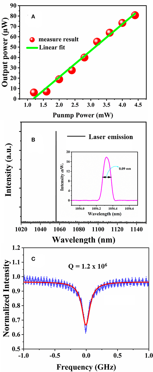
Figure 5. (A) Relation between laser output power and pump power in the glass microsphere; (B) single mode laser emission from 0.8 mol% Nd3± doped fluorosilicate glass microsphere. Inset: zoom-in of the laser emission peak; (C) normalized transmission in the microsphere using 1,550 nm tunable laser. The red line represents the Lorentz fitting.
Figure 5B shows the typical single mode laser emission spectra from the 0.8 mol% Nd3± doped glass microsphere samples. The wavelength of laser emission peak is at λ ~1,056.3 nm, the inset shows a zoom-in of the laser emission peak. When the pump power was set to 2.0 mW, the output power reached 18 nW and the linewidth was 0.09 nm. The coupling efficiency between the tapered fiber and the glass microsphere is only about 0.5% (Wang et al., 2018), thus can explain the low efficiency output of ~1.8 × 10⁻³. The Q factor was measured by scanning the frequency of the tuneable laser with the center wavelength of 1,550 nm, and the repetition frequency of the signal generator and the scan frequency of one period was set as 50 Hz and 15 GHz, respectively. The transmission curve was shown in Figure 5C and the corresponding Q factor of this Nd3± doped fluorosilicate glass microsphere is calculated to be 1.2 × 10⁶.”
In addition, the stability of this fluorosilicate compounds glass was also studied by measuring its transmittance under different environmental conditions, as shown in Figure 6. The untreated sample shows a transmittance very similar to that of the sample left for 2 h at 50°C and 90% humidity, especially at the absorption band of OH− (3,000 nm). This seems to indicate that this compound glass material can work in a high temperature and high humidity environment.
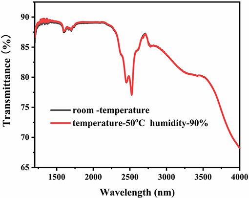
Figure 6. Transmittance spectra of the Nd3± doped fluorosilicate glass at room temperature (black) and after treatment at T ~50°C, ~90% humidity for 2 h (red).
Conclusion
Nd3+ doped compound fluorosilicate glass and glass microsphere samples were fabricated. Under the excitation of a λ ~808 nm laser, enhanced near-infrared photoluminescence was observed. To determine the microstructure of the glass sample, XRD was performed, and the broad bands indicated that both the glass and glass microsphere samples are amorphous with no significant presence of nanocrystals. Furthermore, a single mode near-infrared laser emission was observed from the Nd3+ doped fluorosilicate glass microsphere under the excitation at λ ~808 nm through a tapered standard single mode fiber. The excellent stability and resistance to environmental changes was confirmed by the transmittance spectra recorded in the mid-infrared region, which indicated that this glass and glass microsphere are potential optical gain materials, capable to operate at high temperature and humidity.
Data Availability Statement
The datasets generated for this study are available on request to the corresponding author.
Author Contributions
XW and PW designed the experiment and wrote the draft. HZ, AL, and KT did the measurements. GB discussed the result and help for editing the manuscript. All the authors have reviewed the manuscript.
Funding
National Natural Science Foundation of China (NSFC) (61575050); National Key R&D Program (2016YF-E0126500); The Fundamental Research Funds of the Central Universities (HEUCFG201841); Key Program for Natural Science Foundation of Heilongjiang Province of China (ZD2016012); Open Fund of the State Key Laboratory on Integrated Optoelectronics (IOSKL2016KF03); 111 Project (B13015); Recruitment Program for Young Professionals (The Young Thousand Talents Plan). Ph.D. Student Research and Innovation Fund of the Fundamental Research Funds for the Central Universities (3072019GIP2518).
Conflict of Interest
The authors declare that the research was conducted in the absence of any commercial or financial relationships that could be construed as a potential conflict of interest.
References
Cai, M., Painter, O., Vahala, K. J., and Sercel, P. C. (2000). Fiber-coupled microsphere laser. Opt. Lett. 25, 1430–1432. doi: 10.1364/OL.25.001430
Fang, Z. J., Chormaic, S. N., Wang, S. Y., Wang, X., Yu, J. B., Jiang, Y. X., et al. (2017). Bismuth-doped glass microsphere lasers. Photonics Res. 5, 740–744. doi: 10.1364/PRJ.5.000740
Heo, J., and Shin, Y. B. (1996). Absorption and mid-infrared emission spectroscopy of Dy3+ in Ge-As (or Ga) -S glasses. J. Non Cryst. Solids 196, 162–167. doi: 10.1016/0022-3093(95)00579-X
Humar, M., Ravnik, M., Pajk, S., and Musevic, I. (2009). Electrically tunable liquid crystal optical microreson-ators. Nat. Photonics 3, 595–600. doi: 10.1038/nphoton.2009.170
Ilchenko, V. S., Volikov, P. S., Velichansky, V. L., Treussart, F., Lefe‘vre-Seguin, V., Raimond, J.-M., et al. (1998). Strain-tunable high-Q optical microsphere resonator. Opt. Commun. 145, 86–90. doi: 10.1016/S0030-4018(97)00439-2
Li, A. Z., Li, W. H., Zhang, M., Zhang, Y. D., Wang, S. B., Yang, A. P., et al. (2019). Tm3+-Ho3+ codoped tellurite glass microsphere laser in the 1.47 μm wavelength region. Opt. Lett. 44, 511–513. doi: 10.1364/OL.44.000511
Li, A. Z., Zhang, J. Q., Zhang, M., Li, W. H., Wang, S. B., Lewis, E., et al. (2018). Effect of Tm3+ concentration on the emission wavelength shift in Tm3+-doped silica microsphere lasers. Opt. Lett. 43, 4325–4328. doi: 10.1364/OL.43.004325
Li, C. R., Dai, S. X., Zhang, Q. Y., Shen, X., Wang, X. S., Zhang, P. Q., et al. (2015). Low threshold fiber taper coupled rare earth ion-doped chalcogenide microsphere laser. Chin. Phys. B 24, 044208/1–044208/4. doi: 10.1088/1674-1056/24/4/044208
Mescia, L., Prudenzano, F., De Sario, M., Palmisano, T., Ferrari, M., and Righini, G. C. (2010). Design of rare-earth-doped microspheres. IEEE Photonics Technol. Lett. 22, 422–424. doi: 10.1109/LPT.2009.2039932
Miura, K., Tanaka, K., and Hirao, K. (1997). CW laser oscillation on both the 4F3/2-4I11/2 and 4F3/2-4I13/2 transitions of Nd3+ ions using a fluoride glass micros-phere. J. Non Cryst. Solids 213, 276–280. doi: 10.1016/S0022-3093(96)00671-0
Rasoloniaina, A., Trebaol, S., Huet, V., Le Cren, E., Conti, G. N., Serier-Brault, H., et al. (2012). High-gain wavelength-selective amplification and cavity ring down spectroscopy in a fluoride glass erbium-doped microsphere. Opt. Lett. 37, 4735–4737. doi: 10.1364/OL.37.004735
Sanchez-Martin, R. M., Cuttle, M., Mittoo, S., and Bradley, M. (2006). Microsphere-based real-time calcium sensing. Angew. Chem. Int. Ed. 45, 5472–5474. doi: 10.1002/anie.200601242
von Klitzing, W., Jahier, E., Long, R., Lissillour, F., Lefevre-Seguin, V., Hare, J., et al. (2000). Very low threshold green lasing in microspheres by up-conversion of IR photons. J. Opt. B Quantum Semiclass. Opt. 2, 204–206. doi: 10.1088/1464-4266/2/2/324
von Klitzing, W., Long, R., Ilchenko, V. S., Hare, J., and Lefevre-Seguin, V. (2001). Frequency tuning of the whispering-gallery modes of silica microspheres for cavity quantum electrodynamics and spectroscopy. Opt Lett. 26, 166–168. doi: 10.1364/OL.26.000166
Wang, J. Y., Ji, G. R., Jin, P., Zhao, L. J., and Zhang, C. Z. (2004). Upconversion emission of a Er3+-doped glass microsp-here under 633 nm excitation. Microelectr. J. 35, 627–627. doi: 10.1016/j.mejo.2004.04.005
Wang, P., Murugan, G. S., Lee, T., Ding, M., Brambilla, G., Semenova, Y., et al. (2012). High-Q bismuth-silicate nonlinear glass microsphere resonators. IEEE Photonics J. 4, 1013–1020. doi: 10.1109/JPHOT.2012.2202385
Wang, X., Yu, Y., Wang, S., Ward, J. M., Chormaic, S. N., and Wang, P. (2018). Single mode green lasing and multicolor luminescent emission from an Er3+-Yb3+ co-doped compound fluorosilicate glass microsphere resonator. OSA Continuum 1, 261–273. doi: 10.1364/OSAC.1.000261
Xiang, P., Feng, S., Jiang, S. B., Gonokami, M., and Peyghambarian, N. (2003). Fiber-taper-coupled L-band Er3+-doped tellurite glass microsphere laser. Proc. Soc. Photo Opt. 4990, 22–29. doi: 10.1117/1.1869995
Yang, Z. S., Wu, Y. H., Yang, K., Xu, P. P., Zhang, W., Dai, S. X., et al. (2017a). Fabrication and characterization of Tm3+-Ho3+ co-doped tellurite glass microsphere lasers operating at ~2.1 μm. Opt Mater. 72, 524–528. doi: 10.1016/j.optmat.2017.06.057
Keywords: fluorosilicate glass, glass microsphere, near-infrared, photoluminescence, laser
Citation: Wang X, Zhao H, Li A, Tian K, Brambilla G and Wang P (2019) Near-Infrared Luminescence and Single-Mode Laser Emission From Nd3+ Doped Compound Glass and Glass Microsphere. Front. Mater. 6:237. doi: 10.3389/fmats.2019.00237
Received: 02 July 2019; Accepted: 11 September 2019;
Published: 25 September 2019.
Edited by:
Guoping Dong, South China University of Technology, ChinaReviewed by:
Zhixu Jia, Jilin University, ChinaJayasankar Chalicheemalapalli Kulala, Sri Venkateswara University, India
Danping Chen, Shanghai Institute of Optics and Fine Mechanics (CAS), China
Copyright © 2019 Wang, Zhao, Li, Tian, Brambilla and Wang. This is an open-access article distributed under the terms of the Creative Commons Attribution License (CC BY). The use, distribution or reproduction in other forums is permitted, provided the original author(s) and the copyright owner(s) are credited and that the original publication in this journal is cited, in accordance with accepted academic practice. No use, distribution or reproduction is permitted which does not comply with these terms.
*Correspondence: Pengfei Wang, cGVuZ2ZlaS53YW5nQGRpdC5pZQ==