- Department of Obstetrics, Obstetrics and Gynaecology Center, The First Hospital of Jilin University, Changchun, China
Early-onset Preeclampsia (EOPE) is a severe pregnancy complication that poses significant risks to both maternal and fetal health, often leading to fetal growth restriction and maternal morbidity. Despite extensive research, the etiology of EOPE remains unclear, though emerging evidence suggests that vitamin D (VD) may play an important role in placental development and function. Recent studies associate VD deficiency with adverse pregnancy outcomes, including EOPE, through mechanisms such as impaired trophoblast invasion and immune dysregulation at the maternal-fetal interface. This review aimed to synthesize current literature on the role of VD in the pathogenesis of EOPE. We reviewed in vitro, in vivo, and clinical studies to evaluate the impact of VD on immune modulation, angiogenesis, oxidative stress, and trophoblast migration and invasion in the placenta. This comprehensive review aims to provide insights into how VD deficiency exacerbates placental dysfunction, contributing to the development of EOPE. These insights support the rationale for VD supplementation as a potential preventive strategy and highlight the need for further clinical investigation.
1 Introduction
Early-onset preeclampsia (EOPE) is a severe pregnancy complication defined by hypertension and proteinuria before 34 weeks of gestation, affecting approximately 2–8% of pregnancies worldwide (1). It is associated with adverse maternal and fetal outcomes including fetal growth restriction (FGR), preterm birth and increased maternal morbidity. While the exact etiology of EOPE is multifactorial, abnormal placental development is recognized as a central feature (2).
Among the potential upstream contributors, vitamin D (VD) deficiency has emerged as a candidate risk factor. VD is known to influence key biological processes such as trophoblast invasion, immune tolerance, angiogenesis, and oxidative balance—all of which are commonly disrupted in EOPE (3–5). However, the mechanistic role of VD in EOPE remains less thoroughly explored compared to other maternal and placental factors, and the potential for VD-targeted interventions has yet to be fully elucidated.
This review aims to comprehensively examine the current evidence linking VD deficiency to EOPE, with a focus on mechanistic insights. We synthesize findings from in vitro, in vivo, and clinical studies to evaluate how VD regulates placental function through its effects on trophoblast biology, vascular integrity, immune balance, and oxidative stress. We also discuss the emerging potential of VD supplementation as a modifiable risk factor in EOPE, particularly in high-risk pregnancies. By integrating molecular mechanisms with clinical relevance, this review seeks to bridge existing knowledge gaps and inform future research directions.
2 Pathophysiology of early-onset preeclampsia
Preeclampsia (PE) is a complex, multifactorial disorder of pregnancy characterized by new-onset hypertension and proteinuria, typically after 20 weeks of gestation (6). Among its subtypes, EOPE, which occurs before 34 weeks of gestation, is distinguished by greater severity, higher rates of maternal and fetal morbidity, and a closer association with placental dysfunction compared to late-onset preeclampsia (LOPE) (7).
The pathophysiology of EOPE involves a combination of abnormal placentation, dysregulated maternal immune adaptation, oxidative stress, and impaired vascular remodeling (8–10). Abnormal placentation refers to defective development or function of the placenta, often resulting in inadequate nutrient and oxygen delivery to the fetus (11). In a healthy pregnancy, cytotrophoblasts (specialized placental cells) differentiate into extravillous trophoblasts (EVTs). Trophoblast invasion is the process by which these EVTs migrate into the maternal uterine lining, allowing the placenta to anchor securely and interact with maternal tissues (12). A key aspect of this interaction is spiral artery remodeling, during which EVTs contribute to the transformation of maternal uterine spiral arteries from narrow, high-resistance vessels into wider, lower-resistance channels (13). This process is thought to facilitate adequate maternal blood flow to the placenta and, consequently, to the developing fetus. In EOPE, evidence suggests that trophoblast invasion and spiral artery remodeling may be insufficient, which could contribute to persistently high-resistance blood flow, placental hypoperfusion, hypoxia, and increased oxidative stress (14).
Immune dysregulation is an important aspect in the pathogenesis of EOPE (15). This may involve, but is not limited to, altered maternal immune responses to fetal antigens; other contributing factors such as embryo or systemic damage may also play a role (16). Multiple studies have demonstrated that changes in the maternal immune system can contribute to abnormal placentation and the development of EOPE (17). At the molecular level, EOPE is associated with an imbalance between pro-angiogenic and anti-angiogenic factors, such as reduced placental expression of vascular endothelial growth factor (VEGF) and placental growth factor (PlGF), alongside increased levels of soluble fms-like tyrosine kinase-1 (sFlt-1) and endoglin (18, 19). These changes disrupt angiogenesis, further impairing placental vascularization (20). Additionally, abnormal release of inflammatory cytokines [e.g., tumor necrosis factor-α (TNF-α), IL-6], heightened activation of the maternal immune system, and insufficient generation of regulatory T cells (Tregs) contribute to a pro-inflammatory environment at the maternal-fetal interface, exacerbating placental dysfunction (21, 22).
EOPE is also characterized by heightened oxidative stress due to the accumulation of reactive oxygen species (ROS) and insufficient antioxidant defenses in the placenta (9, 23). This exacerbates endothelial dysfunction, maternal hypertension, and further restricts fetal growth (24). The combined effects of impaired trophoblast invasion, defective vascular remodeling, angiogenic imbalance, and oxidative injury underlie the unique clinical and pathological features of EOPE (25).
Importantly, while LOPE is often linked to maternal metabolic and cardiovascular risk factors, EOPE is more directly associated with placental pathology and abnormal early pregnancy adaptation (8). This review therefore focuses on placental-associated mechanisms of EOPE, particularly those processes that are potentially modulated by vitamin D—including trophoblast function, angiogenesis, immune regulation, and oxidative stress.
3 VD and its association with EOPE
VD exists in two primary forms in humans: vitamin D2 (ergocalciferol) and vitamin D3 (cholecalciferol), with the latter synthesized endogenously through ultraviolet exposure (26). Vitamin D3 is the dominant form involved in human physiology and is the focus of this review. As a fat-soluble vitamin, VD is naturally found in dietary sources such as cod liver oil, fatty fish, mushrooms and egg yolks. Although VD has been traditionally associated with calcium and phosphorus metabolism, it has also been implicated in broader physiological functions including immune regulation, vascular health, and placental development (27–29).
VD metabolism was once believed to occur primarily in the kidneys (30). However, recent studies have revealed that VD is actively metabolized in multiple tissues, including the female reproductive system (31). Both 25-hydroxyvitamin D3 (25 (OH)D3) and its receptor, VD receptor (VDR), are expressed in various organs, including the uterus, ovaries, fallopian tubes, mammary glands, and placenta (15). The expression of α-hydroxylase enzymes in the decidua and placenta during pregnancy further underscores the crucial role of VD at the maternal-fetal interface (15). VD may assist in maintaining healthy placental development and function by regulating calcium transport and immune modulation within the placenta (32).
During healthy pregnancy, maternal serum 25 (OH)D₃ levels typically rise from early to mid-gestation, supporting fetal skeletal development and placental growth (33, 34). However, individuals with EOPE often exhibit significantly lower serum VD levels compared to normotensive pregnancies, with reported deficits of approximately 10–20% (35). Although optimal VD status remains debated, levels below 20 ng/mL are generally considered deficient (36). According to an earlier classification proposed in 2015, serum 25 (OH)D3 levels in healthy pregnant individuals are generally reported to range from 20 to 30 ng/mL (37). Several studies suggest that serum VD levels below this threshold in early pregnancy may be associated with an increased risk of EOPE, likely due to impaired placental adaptation during the first and second trimesters (37, 38).
Numerous meta-analyses, case–control studies, and randomized controlled trials have consistently shown that low maternal vitamin D status is associated with an increased risk of preeclampsia and EOPE, and that vitamin D supplementation may have a protective effect, particularly in high-risk pregnancies. The key clinical evidence is summarized in Table 1.
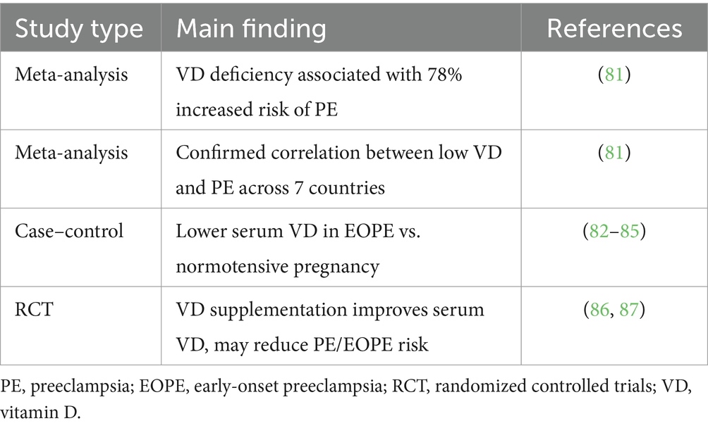
Table 1. Summary of clinical and epidemiological studies linking vitamin D status and risk of PE/EOPE.
4 Mechanisms involving VD in EOPE
EOPE remains poorly understood. Beyond its traditional role in regulating calcium and phosphorus metabolism, VD influences early placental development and function through multiple biological pathways, including gene expression, immune modulation, angiogenesis, and antioxidant activity (39). Low serum VD levels are associated with abnormal placental implantation and disrupted uterine spiral artery remodeling, leading to impaired angiogenesis and insufficient placental blood supply (40). These pathological processes may exacerbate placental hypoxia and oxidative stress, thereby contributing to the early onset of EOPE (41).
A growing body of research suggests that VD deficiency may promote the onset and progression of EOPE through both direct and indirect mechanisms (39). In early pregnancy, VD is involved in placental immune regulation and trophoblast cell invasion, both of which are essential for ensuring adequate placental blood flow (42). Therefore, further investigation into the role of VD in immune modulation, angiogenesis, oxidative stress, and trophoblast invasion may clarify the pathogenesis of EOPE and provide a theoretical basis for considering VD as a potential preventive strategy. The following sections will explore these key mechanisms in detail, highlighting the specific effects and influences of VD in EOPE.
4.1 Role of VD in maternal-fetal immune tolerance
Dysregulation of immune adaptation at the maternal-fetal interface has been widely reported in EOPE. Studies suggest that VD may be involved in the regulation of maternal immune tolerance by promoting Treg function and modulating T helper cell differentiation (43). VD deficiency has therefore been proposed as a potential contributor to placental immune imbalance observed in EOPE (Figure 1).
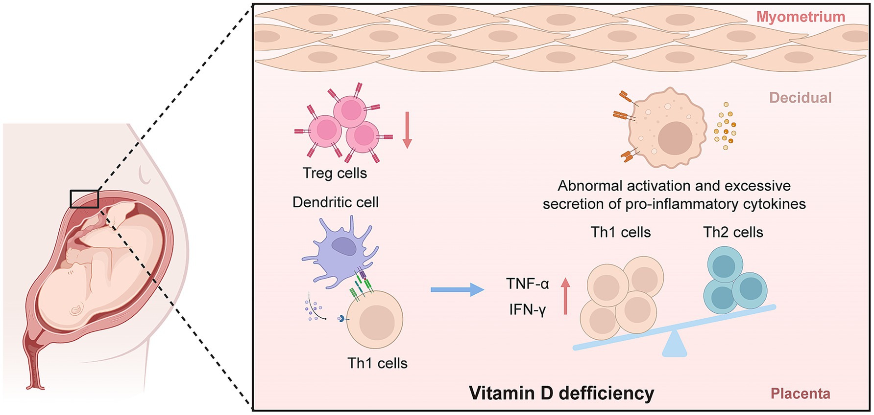
Figure 1. The immunoregulatory role of vitamin D deficiency in EOPE. Created with BioRender and designed by the authors. EOPE, early-onset preeclampsia; IFN-γ, interferon gamma; Th, T-helper cells; TNF-α, tumor necrosis factor alpha; Treg, regulatory T cells.
Experimental and clinical evidence indicates that the active form of VD, 1,25(OH)₂D₃, enhances the expansion and suppressive function of FoxP3 + regulatory Tregs, which are essential for maintaining immune homeostasis at the maternal-fetal interface (15, 44). For example, in patients with EOPE, both peripheral and decidual Treg counts are significantly decreased compared to normotensive pregnant controls, and these alterations have been correlated with lower maternal 25(OH)D₃ concentrations (45, 46). In vitro studies using human immune cells have further demonstrated that VD/VDR signaling directly upregulates FoxP3 expression, supporting Treg differentiation and activity (44).
In addition to Treg modulation, VD also influences the Th1/Th2 balance, a key immunological axis in pregnancy. VD has been shown to suppress the production of pro-inflammatory Th1 cytokines, including TNF-α and interferon-γ, while promoting anti-inflammatory Th2 cytokines such as interleukin-4 (IL-4), interleukin-5 (IL-5), and interleukin-10 (IL-10) (47–50). This effect has been observed in both in vitro human T cell studies and clinical cohorts, where VD deficiency is associated with elevated Th1/Th2 ratios and increased placental inflammation in EOPE (46, 50).
Furthermore, studies have demonstrated that VD regulates the activity of dendritic cells (DCs), which play a central role in antigen presentation at the maternal-fetal interface (51, 52). VD inhibits the maturation of DCs and reduces their capacity to activate T cells, thereby limiting local inflammatory responses in the placenta (52, 53). Insufficient VD enhances DC-mediated T cell activation and promotes a pro-inflammatory environment, which has been implicated in abnormal placental development and increased EOPE risk (53, 54).
VD also modulates placental macrophage polarization. VD promotes the M2 anti-inflammatory phenotype while inhibiting the M1 pro-inflammatory phenotype, leading to reduced secretion of TNF-α and interleukin-6 (IL-6) in the placenta (55, 56). Both animal models and human studies have linked VD deficiency to increased M1 macrophage infiltration and heightened local inflammation in EOPE placentas (21, 57).
Collectively, these findings from in vitro, animal, and clinical studies indicate that adequate VD status supports maternal-fetal immune tolerance by enhancing Treg function, regulating the Th1/Th2 axis, suppressing excessive dendritic cell and macrophage activation, and mitigating placental inflammation. Contrarily, VD deficiency, disrupts these immunological processes, contributing to the immune pathophysiology of EOPE.
4.2 Role of VD in reducing impaired uterine spiral artery remodeling
Disruption of placental angiogenesis and inadequate remodeling of the uterine spiral arteries are frequently described features in EOPE (41, 58, 59). Current evidence indicates that VD can influence angiogenic pathways in the placenta, including the regulation of VEGF expression and the renin-angiotensin-aldosterone system (RAAS) (60, 61). The relationship between VD status and placental vascular development remains an area of active research (Figure 2).
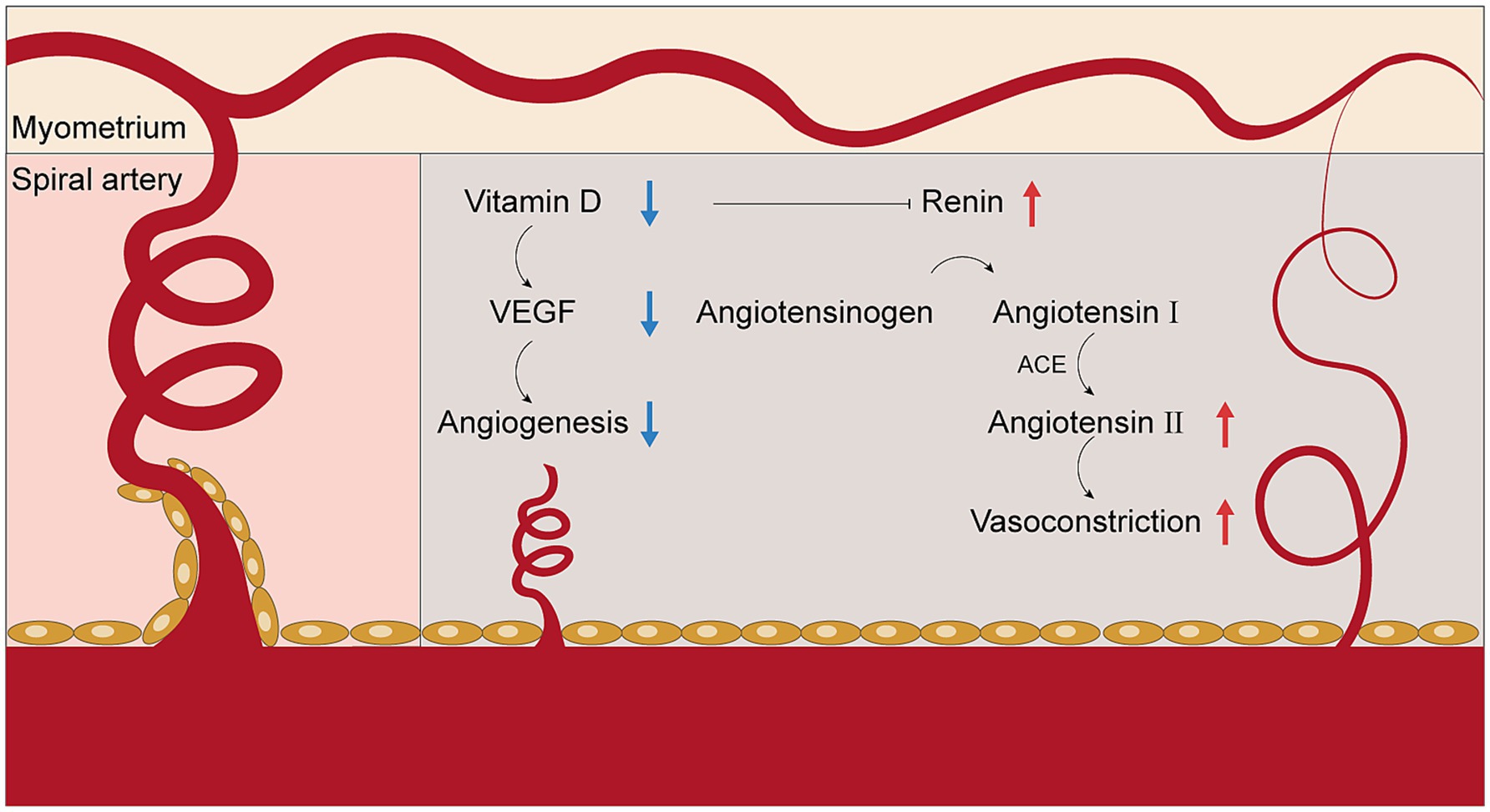
Figure 2. The role of vitamin D deficiency in regulating angiogenesis in the placenta. ACE, angiotensin-converting enzyme; VEGF, vascular endothelial growth factor.
Clinical research has corroborated these findings. Analyses of EOPE placental tissue and maternal serum reveal lower levels of VEGF and PlGF, along with elevated concentrations of the anti-angiogenic factor sFlt-1 in women with low VD status (58, 62, 63). These molecular changes correlate with reduced spiral artery remodeling and increased placental vascular resistance, as observed in Doppler ultrasound and histopathology studies.
In addition to directly regulating angiogenic factors, VD is known to modulate the RAAS pathway within the placenta. Experimental animal studies demonstrate that VD suppresses the transcription of the renin gene, leading to lower angiotensin II production and decreased vasoconstriction (61, 64). Clinical data indicate that VD deficiency is associated with increased RAAS activity, contributing to hypertension and further compromising placental perfusion in EOPE (64, 65).
Placental VD receptor (VDR) expression is also reduced in EOPE, which may decrease the placenta’s responsiveness to circulating VD and further limit angiogenic signaling (66–68). Notably, studies report that lower maternal and placental VD/VDR levels are associated with higher risk of FGR secondary to impaired placental blood flow (66, 69).
Although considerable progress has been made in delineating the relationship between vitamin D and placental vascular development, the precise molecular mechanisms—particularly the interplay between VD/VDR signaling, angiogenic factor expression, and RAAS regulation in EOPE—require further investigation in experimental models and large-scale clinical studies.
4.3 Role of VD in oxidative stress
Elevated oxidative stress has been implicated in the pathophysiology of EOPE, particularly in relation to placental dysfunction and endothelial injury (23, 70). Experimental and clinical studies have examined the antioxidant properties of VD, including its regulation of key antioxidant enzymes and its effects on oxidative stress pathways (Figure 3) (70–72). The role of VD in modulating placental oxidative stress is being increasingly explored.
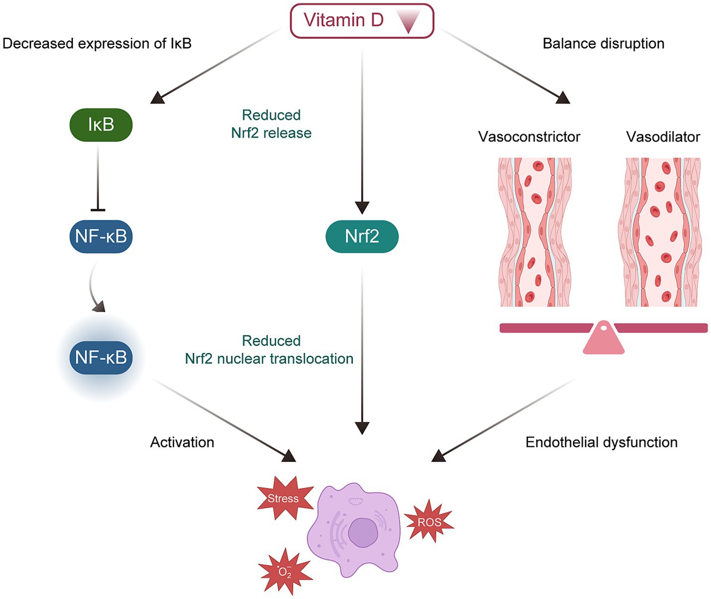
Figure 3. Potential molecular mechanisms of vitamin D deficiency in the development of EOPE. EOPE, early-onset preeclampsia.
In vitro studies have shown that 1,25(OH)₂D₃ can upregulate antioxidant enzymes such as superoxide dismutase (SOD) and glutathione peroxidase in placental cells, thereby reducing levels of ROS and lipid peroxidation (70, 73). Consistent with this, women with EOPE and VD deficiency display increased placental malondialdehyde (a marker of oxidative stress) and reduced SOD activity compared to healthy pregnancies (70, 74).
Mechanistically, vitamin D has been reported to inhibit activation of the nuclear factor kappa-light-chain-enhancer of activated B cells pathway in trophoblasts, thereby reducing the expression of pro-inflammatory and pro-oxidant genes and mitigating oxidative injury (70, 72). Furthermore, animal models of preeclampsia have demonstrated that VD supplementation increases nuclear factor erythroid 2-related factor 2 transcriptional activity in the placenta and lowers oxidative stress biomarkers (75).
Although these findings support an antioxidant role for vitamin D in the placenta, the precise molecular mechanisms, especially involving VDR, NF-κB, and downstream effectors such as Nrf2, require further clarification.
4.4 Role of VD in EVT migration and invasion
Limited trophoblast invasion and suboptimal remodeling of the maternal uterine arteries have been associated with EOPE in both experimental and clinical observations (76, 77). Research has suggested that VD, via the VDR expressed in trophoblasts, may be involved in the regulation of EVT migration and invasion (78, 79). The possible impact of VD deficiency on these cellular processes is the subject of ongoing investigation.
In vitro experiments with human trophoblast cell lines have demonstrated that 1,25(OH)₂D₃ upregulates the expression of matrix metalloproteinases (MMP2 and MMP9), which are essential for extracellular matrix degradation and successful EVT invasion (79). Placental samples from EOPE pregnancies show decreased VDR and MMP9 expression, which are associated with reduced EVT invasive capacity (79, 80). Additionally, vitamin D signaling modulates other molecules involved in cell migration, such as E-cadherin and integrins, which play roles in cell adhesion and motility (59, 78). Importantly, 1,25(OH)₂D₃ stimulates the secretion of human chorionic gonadotropin (hCG) via the cAMP/PKA pathway, which is a well-known regulator of trophoblast motility and invasion (78). Animal studies further indicate that vitamin D deficiency impairs trophoblast invasion and spiral artery remodeling, resulting in phenotypes similar to EOPE (59).
Overall, these findings suggest that vitamin D may facilitate EVT migration and invasion by regulating MMPs, adhesion molecules, and hCG-related signaling pathways, but more research is needed to clarify its exact molecular targets in the context of EOPE (Figure 4).
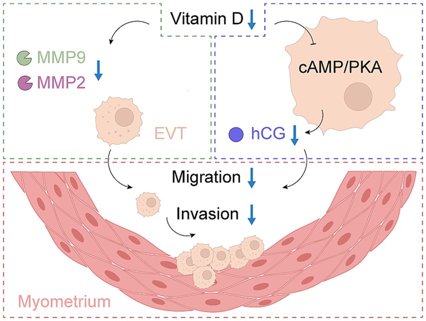
Figure 4. The role of vitamin D in EVT invasion and migration in EOPE. EOPE, early-onset preeclampsia; EVT, extravillous trophoblast; hCG, human chorionic gonadotropin.
5 Conclusion
VD has been proposed to play a role in the pathogenesis of EOPE, as its deficiency has been associated with impaired placental development, increased oxidative stress, and immune dysregulation at the maternal-fetal interface (Figure 5). Findings from individual studies suggest that VD may influence processes such as angiogenesis and vascular remodeling, which are considered important for supporting healthy pregnancy outcomes. Low VD levels during pregnancy have been associated with an increased risk of EOPE and FGR, and VD supplementation has been proposed as a potential area for therapeutic exploration. Understanding the molecular mechanisms through which VD influences EOPE offers a promising approach to clinical management and prevention. In clinical practice, monitoring and managing VD levels has been suggested as a potentially beneficial approach, especially in high-risk pregnancies.
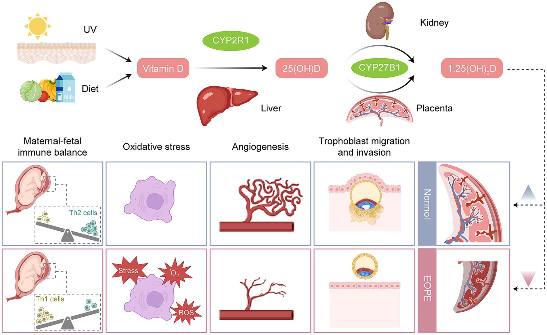
Figure 5. The metabolism of vitamin D and the functional differences in the regulation of placentas between EOPE and normal pregnancy. Created with BioRender and designed by the authors. EOPE, early-onset preeclampsia; ROS, reactive oxygen species; Th cells, T-helper cells; UV, ultraviolet.
Author contributions
SZ: Conceptualization, Writing – review & editing, Writing – original draft. SD: Writing – review & editing. HS: Investigation, Writing – original draft. PX: Writing – original draft. CS: Funding acquisition, Writing – review & editing.
Funding
The author(s) declare that financial support was received for the research and/or publication of this article. This research was funded by Wu Jieping Medical Foundation (grant number 320.6750.2021–06-32), Finance Department of Jilin Province, China (grant number JLSCZD2019-053), Natural Science Funds in Science and Technology Department of Jilin Province, China (grant number 20210101295JC), and Open Project of Key Laboratory of Organ Regeneration and Transplantation, Ministry of Education (grant number 2020JC07).
Acknowledgments
We would like to thank Editage (www.editage.cn) for English language editing.
Conflict of interest
The authors declare that the research was conducted in the absence of any commercial or financial relationships that could be construed as a potential conflict of interest.
Generative AI statement
The authors declare that no Gen AI was used in the creation of this manuscript.
Publisher’s note
All claims expressed in this article are solely those of the authors and do not necessarily represent those of their affiliated organizations, or those of the publisher, the editors and the reviewers. Any product that may be evaluated in this article, or claim that may be made by its manufacturer, is not guaranteed or endorsed by the publisher.
Abbreviations
EOPE, Early-onset preeclampsia; LOPE, Late-onset preeclampsia; FGR, Fetal growth restriction; VD, Vitamin D; VDR, Vitamin D receptor; 25(OH)D3, 25-hydroxyvitamin D3; 1,25(OH)2D3, 1,25-dihydroxyvitamin D3; PE, Preeclampsia; Tregs, Regulatory T cells; Th, T helper; DC, Dendritic cell; RAAS, Renin-angiotensin-aldosterone system; VEGF, Vascular endothelial growth factor; NF-κB, Nuclear factor κB; EVT, Extravillous trophoblast; MMP, Matrix metalloproteinase; hCG, Human chorionic gonadotropins.
References
1. Voto, LS, and Zeitune, MG. Preeclampsia In: RA Moreira de Sá and EB Fonseca, editors. Perinatology: Evidence-based best practices in perinatal medicine. Cham: Springer International Publishing (2022). 707–46.
2. Xu, Y, Qin, X, Zeng, W, Wu, F, Wei, X, Li, Q, et al. Dock1 deficiency drives placental trophoblast cell dysfunction by influencing inflammation and oxidative stress, hallmarks of preeclampsia. Hypertens Res. (2024) 47:3. doi: 10.1038/s41440-024-01920-3
3. Nunes, PR, Romao-Veiga, M, Ribeiro, VR, de Oliveira, LRC, Zupelli, TG, Abbade, JF, et al. Vitamin D decreases cell death and inflammation in human umbilical vein endothelial cells and placental explants from pregnant women with preeclampsia cultured with Tnf-Α. Immunol Investig. (2022) 51:1630–46. doi: 10.1080/08820139.2021.2017452
4. Vanherwegen, A-S, Gysemans, C, and Mathieu, C. Vitamin D endocrinology on the cross-road between immunity and metabolism. Mol Cell Endocrinol. (2017) 453:52–67. doi: 10.1016/j.mce.2017.04.018
5. Heyden, EL, and Wimalawansa, SJ. Vitamin D: effects on human reproduction, pregnancy, and fetal well-being. J Steroid Biochem Mol Biol. (2018) 180:41–50. doi: 10.1016/j.jsbmb.2017.12.011
6. Ives, CW, Sinkey, R, Rajapreyar, I, Tita, ATN, and Oparil, S. Preeclampsia—pathophysiology and clinical presentations: Jacc state-of-the-art review. J Am Coll Cardiol. (2020) 76:1690–702. doi: 10.1016/j.jacc.2020.08.014
7. Gohar, S, and Syed, W. Comparison of adverse fetomaternal outcome in early and late onset pre eclampsia. J Postgraduate Med Inst. (2021) 35:15–8. doi: 10.54079/jpmi.35.1.2732
8. Aneman, I, Pienaar, D, Suvakov, S, Simic, TP, Garovic, VD, and McClements, L. Mechanisms of key innate immune cells in early- and late-onset preeclampsia. Front Immunol. (2020) 11:864. doi: 10.3389/fimmu.2020.01864
9. Aouache, R, Biquard, L, Vaiman, D, and Miralles, F. Oxidative stress in preeclampsia and placental diseases. Int J Mol Sci. (2018) 19:1496. doi: 10.3390/ijms19051496
10. Bakrania, BA, Spradley, FT, Drummond, HA, LaMarca, B, Ryan, MJ, and Granger, JP. Preeclampsia: linking placental ischemia with maternal endothelial and vascular dysfunction. Compr Physiol. (2020) 11:1315–49. doi: 10.1002/cphy.c200008
11. Arechavaleta-Velasco, F, Koi, H, Strauss, JF, and Parry, S. Viral infection of the trophoblast: time to take a serious look at its role in abnormal implantation and placentation? J Reprod Immunol. (2002) 55:113–21. doi: 10.1016/S0165-0378(01)00143-7
12. Pijnenborg, R, Dixon, G, Robertson, WB, and Brosens, I. Trophoblastic invasion of human decidua from 8 to 18 weeks of pregnancy. Placenta. (1980) 1:3–19. doi: 10.1016/S0143-4004(80)80012-9
13. Brosens, JJ, Pijnenborg, R, and Brosens, IA. The Myometrial junctional zone spiral arteries in Normal and abnormal pregnancies: a review of the literature. Am J Obstet Gynecol. (2002) 187:1416–23. doi: 10.1067/mob.2002.127305
14. Myatt, L. Role of placenta in preeclampsia. Endocrine. (2002) 19:103–12. doi: 10.1385/ENDO:19:1:103
15. Cyprian, F, Lefkou, E, Varoudi, K, and Girardi, G. Immunomodulatory effects of vitamin D in pregnancy and beyond. Front Immunol. (2019) 10:739. doi: 10.3389/fimmu.2019.02739
16. Ma, Y, Ye, S, Liu, Y, Zhao, X, Wang, Y, and Wang, Y. Interferon regulatory factor 1 mediated inhibition of Treg cell differentiation induces maternal-fetal immune imbalance in preeclampsia. Int Immunopharmacol. (2024) 141:112988. doi: 10.1016/j.intimp.2024.112988
17. Alijotas-Reig, J, Llurba, E, and Gris, JM. Potentiating maternal immune tolerance in pregnancy: a new challenging role for regulatory T cells. Placenta. (2014) 35:241–8. doi: 10.1016/j.placenta.2014.02.004
18. Bhattacharjee, J, Mohammad, S, Goudreau, AD, and Adamo, KB. Physical activity differentially regulates Vegf, Plgf, and their receptors in the human placenta. Physiol Rep. (2021) 9:e14710. doi: 10.14814/phy2.14710
19. Patel, B, Bakrania, B, and Granger, J. Abstract P3047: soluble Guanylyl cyclase stimulators and activators inhibit hypoxia-induced placental Sflt-1 levels. Hypertension. (2019) 74:3047. doi: 10.1161/hyp.74.suppl_1.P3047
20. Reynolds, LP, and Redmer, DA. Angiogenesis in the Placenta1. Biol Reprod. (2001) 64:1033–40. doi: 10.1095/biolreprod64.4.1033
21. Shaw, J, Tang, Z, Schneider, H, Saljé, K, Hansson, SR, and Guller, S. Inflammatory processes are specifically enhanced in endothelial cells by placental-derived Tnf-Α: implications in preeclampsia (Pe). Placenta. (2016) 43:1–8. doi: 10.1016/j.placenta.2016.04.015
22. Eghbal-Fard, S, Yousefi, M, Heydarlou, H, Ahmadi, M, Taghavi, S, Movasaghpour, A, et al. The imbalance of Th17/Treg Axis involved in the pathogenesis of preeclampsia. J Cell Physiol. (2019) 234:5106–16. doi: 10.1002/jcp.27315
23. Marín, R, Chiarello, DI, Abad, C, Rojas, D, Toledo, F, and Sobrevia, L. Oxidative stress and mitochondrial dysfunction in early-onset and late-onset preeclampsia. Biochim Biophys Acta. (2020) 1866:165961. doi: 10.1016/j.bbadis.2020.165961
24. Bezemer, RE, Schoots, MH, Timmer, A, Scherjon, SA, Erwich, JJHM, van Goor, H, et al. Altered levels of decidual immune cell subsets in fetal growth restriction, stillbirth, and placental pathology. Front Immunol. (2020) 11:898. doi: 10.3389/fimmu.2020.01898
25. Chen, JZJ, Wong, MH, Brennecke, SP, and Keogh, RJ. The effects of human chorionic gonadotrophin, progesterone and oestradiol on trophoblast function. Mol Cell Endocrinol. (2011) 342:73–80. doi: 10.1016/j.mce.2011.05.034
26. Houghton, LA, and Vieth, R. The case against ergocalciferol (vitamin D2) as a vitamin supplement1,2. Am J Clin Nutr. (2006) 84:694–7. doi: 10.1093/ajcn/84.4.694
27. Roseland, JM, Phillips, KM, Patterson, KY, Pehrsson, PR, and Taylor, CL. Chapter 60—vitamin D in foods: an evolution of knowledge. In: D Feldman, editor. Vitamin D. 4th ed. San Diego, CA, USA: Academic Press (2018). 41–77.
28. Cardwell, G, Bornman, JF, James, AP, and Black, LJ. A review of mushrooms as a potential source of dietary vitamin D. Nutrients. (2018) 10:1498. doi: 10.3390/nu10101498
29. Ismailova, A, and White, JH. Vitamin D, infections and immunity. Rev Endocr Metabolic Disord. (2022) 23:265–77. doi: 10.1007/s11154-021-09679-5
30. Tebben, PJ, and Kumar, R. Chapter 26—vitamin D and the kidney. In: D Feldman, editor. Vitamin D. Fourth ed. San Diego, CA, USA: Academic Press (2018). 437–59.
31. Shahrokhi, SZ, Ghaffari, F, and Kazerouni, F. Role of vitamin D in female reproduction. Clin Chim Acta. (2016) 455:33–8. doi: 10.1016/j.cca.2015.12.040
32. Tamblyn, JA, Hewison, M, Wagner, CL, Bulmer, JN, and Kilby, MD. Immunological role of vitamin D at the maternal–fetal interface. J Endocrinol. (2015) 224:R107–21. doi: 10.1530/joe-14-0642
33. Kovacs, CS. Maternal mineral and bone metabolism during pregnancy, lactation, and post-weaning recovery. Physiol Rev. (2016) 96:449–547. doi: 10.1152/physrev.00027.2015
34. Wagner, CL, and Hollis, BW. The implications of vitamin D status during pregnancy on mother and her developing child. Front Endocrinol. (2018) 9:500. doi: 10.3389/fendo.2018.00500
35. Christoph, P, Challande, P, Raio, L, and Surbek, D. High prevalence of severe vitamin D deficiency during the first trimester in pregnant women in Switzerland and its potential contributions to adverse outcomes in the pregnancy. Swiss Med Wkly. (2020) 150:w20238. doi: 10.4414/smw.2020.20238
36. Ryan, BA, and Kovacs, CS. Chapter 33—the role of vitamin D physiology in regulating calcium and bone metabolism in mother and child: pregnancy, lactation, postweaning, fetus, and neonate In: M Hewison, R Bouillon, E Giovannucci, D Goltzman, M Meyer, and J Welsh, editors. Feldman and pike' s vitamin D (fifth edition) San Diego, CA, USA: Academic Press (2024). 693–759.
37. El-Hajj Fuleihan, G, Bouillon, R, Clarke, B, Chakhtoura, M, Cooper, C, McClung, M, et al. Serum 25-Hydroxyvitamin D levels: variability, knowledge gaps, and the concept of a desirable range. J Bone Miner Res. (2015) 30:1119–33. doi: 10.1002/jbmr.2536
38. Nunes, PR, Romao-Veiga, M, Ribeiro, VR, Oliveira, LRC, Carvalho Depra, I, Oliveira, LG, et al. Inflammasomes in placental explants of women with preeclampsia cultured with monosodium urate may be modulated by vitamin D. Hypertens Pregnancy. (2022) 41:139–48. doi: 10.1080/10641955.2022.2063330
39. Poniedziałek-Czajkowska, E, and Mierzyński, R. Could vitamin D be effective in prevention of preeclampsia? Nutrients. (2021) 13:3854. doi: 10.3390/nu13113854
40. Liu, NQ, and Hewison, M. Vitamin D, the placenta and pregnancy. Arch Biochem Biophys. (2012) 523:37–47. doi: 10.1016/j.abb.2011.11.018
41. Sheridan, MA, Yang, Y, Jain, A, Lyons, AS, Yang, P, Brahmasani, SR, et al. Early onset preeclampsia in a model for human placental trophoblast. Proc Natl Acad Sci. (2019) 116:4336–45. doi: 10.1073/pnas.1816150116
42. Karras, SN, Wagner, CL, and Castracane, VD. Understanding vitamin D metabolism in pregnancy: from physiology to pathophysiology and clinical outcomes. Metabolism. (2018) 86:112–23. doi: 10.1016/j.metabol.2017.10.001
43. Schröder-Heurich, B, Springer, CJP, and von Versen-Höynck, F. Vitamin D effects on the immune system from Periconception through pregnancy. Nutrients. (2020) 12:1432. doi: 10.3390/nu12051432
44. Chambers, ES, and Hawrylowicz, CM. The impact of vitamin D on regulatory T cells. Curr Allergy Asthma Rep. (2011) 11:29–36. doi: 10.1007/s11882-010-0161-8
45. Silalahi, ER, Wibowo, N, Prasmusinto, D, Djuwita, R, Rengganis, I, and Mose, JC. Decidual dendritic cells 10 and Cd4+Cd25+Foxp3 regulatory T cell in preeclampsia and their correlation with nutritional factors in Pathomechanism of immune rejection in pregnancy. J Reprod Immunol. (2022) 154:103746. doi: 10.1016/j.jri.2022.103746
46. Muyayalo, KP, Huang, X-B, Qian, Z, Li, Z-H, Mor, G, and Liao, A-H. Low circulating levels of vitamin D may contribute to the occurrence of preeclampsia through deregulation of Treg /Th17 cell ratio. Am J Reprod Immunol. (2019) 82:e13168. doi: 10.1111/aji.13168
47. Hewison, M. Vitamin D and immune function: an overview. Proc Nutr Soc. (2012) 71:50–61. doi: 10.1017/S0029665111001650
48. Romanowska-Próchnicka, K, Felis-Giemza, A, Olesińska, M, Wojdasiewicz, P, Paradowska-Gorycka, A, and Szukiewicz, D. The role of Tnf-Α and anti-Tnf-Α agents during preconception, pregnancy, and breastfeeding. Int J Mol Sci. (2021) 22:2922. doi: 10.3390/ijms22062922
49. Mitchell, RE, Hassan, M, Burton, BR, Britton, G, Hill, EV, Verhagen, J, et al. Il-4 enhances Il-10 production in Th1 cells: implications for Th1 and Th2 regulation. Sci Rep. (2017) 7:11315. doi: 10.1038/s41598-017-11803-y
50. Skapenko, A, Niedobitek, GU, Kalden, JR, Lipsky, PE, and Schulze-Koops, H. The Th2 cytokines Il-4 and Il-10 are internal controllers of human Th1-biased immunity in vivo. Arthritis Res Ther. (2003) 5:88. doi: 10.1186/ar889
51. Hewison, M. Vitamin D, immunity and human disease. Clin Rev Bone Miner Metab. (2010) 8:32–9. doi: 10.1007/s12018-009-9062-6
52. Bartels, LE, Hvas, CL, Agnholt, J, Dahlerup, JF, and Agger, R. Human dendritic cell antigen presentation and chemotaxis are inhibited by intrinsic 25-Hydroxy vitamin D activation. Int Immunopharmacol. (2010) 10:922–8. doi: 10.1016/j.intimp.2010.05.003
53. Bishop, L, Ismailova, A, Dimeloe, S, Hewison, M, and White, JH. Vitamin D and immune regulation: antibacterial, antiviral, anti-inflammatory. JBMR Plus. (2020) 5:405. doi: 10.1002/jbm4.10405
54. Piccinni, M-P, Robertson, SA, and Saito, S. Editorial: adaptive immunity in pregnancy. Front Immunol. (2021) 12:242. doi: 10.3389/fimmu.2021.770242
55. Barrera, D, Díaz, L, Noyola-Martínez, N, and Halhali, A. Vitamin D and inflammatory cytokines in healthy and Preeclamptic pregnancies. Nutrients. (2015) 7:6465–90. doi: 10.3390/nu7085293
56. Zhu, X, Zhu, Y, Li, C, Yu, J, Ren, D, Qiu, S, et al. 1,25-Dihydroxyvitamin D regulates macrophage polarization and ameliorates experimental inflammatory bowel disease by suppressing Mir-125b. Int Immunopharmacol. (2019) 67:106–18. doi: 10.1016/j.intimp.2018.12.015
57. Liu, X, Jiang, M, Ren, L, Zhang, A, Zhao, M, Zhang, H, et al. Decidual macrophage M1 polarization contributes to adverse pregnancy induced by toxoplasma Gondii Pru strain infection. Microb Pathog. (2018) 124:183–90. doi: 10.1016/j.micpath.2018.08.043
58. Abascal-Saiz, A, Duque-Alcorta, M, Fioravantti, V, Antolín, E, Fuente-Luelmo, E, Haro, M, et al. The relationship between Angiogenic factors and energy metabolism in preeclampsia. Nutrients. (2022) 14:2172. doi: 10.3390/nu14102172
59. James, JL, Boss, AL, Sun, C, Allerkamp, HH, and Clark, AR. From stem cells to spiral arteries: a journey through early placental development. Placenta. (2022) 125:68–77. doi: 10.1016/j.placenta.2021.11.004
60. Jamali, N, Sorenson, CM, and Sheibani, N. Vitamin D and regulation of vascular cell function. Am J Phys Heart Circ Phys. (2018) 314:H753–65. doi: 10.1152/ajpheart.00319.2017
61. Qiao, G, Kong, J, Uskokovic, M, and Li, YC. Analogs of 1α,25-Dihydroxyvitamin D3 as novel inhibitors of renin biosynthesis. J Steroid Biochem Mol Biol. (2005) 96:59–66. doi: 10.1016/j.jsbmb.2005.02.008
62. Schulz, EV, Cruze, L, Wei, W, Gehris, J, and Wagner, CL. Maternal vitamin D sufficiency and reduced placental gene expression in Angiogenic biomarkers related to comorbidities of pregnancy. J Steroid Biochem Mol Biol. (2017) 173:273–9. doi: 10.1016/j.jsbmb.2017.02.003
63. Raia-Barjat, T, Sarkis, C, Rancon, F, Thibaudin, L, Gris, J-C, Alfaidy, N, et al. Vitamin D deficiency during late pregnancy mediates placenta-associated complications. Sci Rep. (2021) 11:20708. doi: 10.1038/s41598-021-00250-5
64. Kota, SK, Kota, SK, Jammula, S, Meher, LK, Panda, S, Tripathy, PR, et al. Renin–angiotensin system activity in vitamin D deficient, obese individuals with hypertension: an urban Indian study. Indian J Endocrinol Metab. (2011) 15:S395–401. doi: 10.4103/2230-8210.86985
65. Gathiram, P, and Moodley, J. The role of the renin-angiotensin-aldosterone system in preeclampsia: a review. Curr Hypertens Rep. (2020) 22:89. doi: 10.1007/s11906-020-01098-2
66. Murthi, P, Yong, HEJ, Ngyuen, TPH, Ellery, S, Singh, H, Rahman, R, et al. Role of the placental vitamin D receptor in modulating feto-placental growth in fetal growth restriction and preeclampsia-affected pregnancies. Front Physiol. (2016) 7:43. doi: 10.3389/fphys.2016.00043
67. Knabl, J, Vattai, A, Ye, Y, Jueckstock, J, Hutter, S, Kainer, F, et al. Role of placental Vdr expression and function in common late pregnancy disorders. Int J Mol Sci. (2017) 18:340. doi: 10.3390/ijms18112340
68. Hutabarat, M, Wibowo, N, Obermayer-Pietsch, B, and Huppertz, B. Impact of vitamin D and vitamin D receptor on the trophoblast survival capacity in preeclampsia. PLoS One. (2018) 13:e0206725. doi: 10.1371/journal.pone.0206725
69. Ortega, MA, Fraile-Martínez, O, García-Montero, C, Sáez, MA, Álvarez-Mon, MA, Torres-Carranza, D, et al. The pivotal role of the placenta in Normal and pathological pregnancies: a focus on preeclampsia, fetal growth restriction, and maternal chronic venous disease. Cells. (2022) 11:568. doi: 10.3390/cells11030568
70. Sosa-Díaz, E, Hernández-Cruz, EY, and Pedraza-Chaverri, J. The role of vitamin D on redox regulation and cellular senescence. Free Radic Biol Med. (2022) 193:253–73. doi: 10.1016/j.freeradbiomed.2022.10.003
71. Wimalawansa, SJ. Vitamin D deficiency: effects on oxidative stress, epigenetics, gene regulation, and aging. Biology. (2019) 8:30. doi: 10.3390/biology8020030
72. Miao, D, and Goltzman, D. Chapter eleven—mechanisms of action of vitamin D in delaying aging and preventing disease by inhibiting oxidative stress. In: G Litwack, editor. Vitamins and hormones, vol. 121. San Diego, CA, USA: Academic Press (2023). 293–318.
73. Mokhtari, Z, Hekmatdoost, A, and Nourian, M. Antioxidant efficacy of vitamin D. J Parathyroid Dis. (2017) 5:11–6. Available at: https://jparathyroid.com/Article/JPD_20160924145139
74. Chen, H, Zhang, H, Xie, H, Zheng, J, Lin, M, Chen, J, et al. Maternal, umbilical arterial metabolic levels and placental Nrf2/Cbr1 expression in pregnancies with and without 25-Hydroxyvitamin D deficiency. Gynecol Endocrinol. (2021) 37:807–13. doi: 10.1080/09513590.2021.1942451
75. Nakai, K, Fujii, H, Kono, K, Goto, S, Kitazawa, R, Kitazawa, S, et al. Vitamin D activates the Nrf2-Keap1 antioxidant pathway and ameliorates nephropathy in diabetic rats. Am J Hypertens. (2013) 27:586–95. doi: 10.1093/ajh/hpt160
76. Velicky, P, Meinhardt, G, Plessl, K, Vondra, S, Weiss, T, Haslinger, P, et al. Genome amplification and cellular senescence are hallmarks of human placenta development. PLoS Genet. (2018) 14:e1007698. doi: 10.1371/journal.pgen.1007698
77. Kim, RH, Ryu, BJ, Lee, KM, Han, JW, and Lee, SK. Vitamin D facilitates trophoblast invasion through induction of epithelial-mesenchymal transition. Am J Reprod Immunol. (2018) 79:e12796. doi: 10.1111/aji.12796
78. Ganguly, A, Tamblyn, JA, Finn-Sell, S, Chan, S-Y, Westwood, M, Gupta, J, et al. Vitamin D, the placenta and early pregnancy: effects on trophoblast function. J Endocrinol. (2018) 236:R93–R103. doi: 10.1530/joe-17-0491
79. Chan, SY, Susarla, R, Canovas, D, Vasilopoulou, E, Ohizua, O, McCabe, CJ, et al. Vitamin D promotes human extravillous trophoblast invasion in vitro. Placenta. (2015) 36:403–9. doi: 10.1016/j.placenta.2014.12.021
80. Velicky, P, Windsperger, K, Petroczi, K, Pils, S, Reiter, B, Weiss, T, et al. Pregnancy-associated diamine oxidase originates from Extravillous trophoblasts and is decreased in early-onset preeclampsia. Sci Rep. (2018) 8:6342. doi: 10.1038/s41598-018-24652-0
81. Van der Pligt, P, Willcox, J, Szymlek-Gay, EA, Murray, E, Worsley, A, and Daly, RM. Associations of maternal vitamin D deficiency with pregnancy and neonatal complications in developing countries: a systematic review. Nutrients. (2018) 10:640. doi: 10.3390/nu10050640
82. Baker, AM, Haeri, S, Camargo, CA Jr, Espinola, JA, and Stuebe, AM. A nested case-control study of Midgestation vitamin D deficiency and risk of severe preeclampsia. J Clin Endocrinol Metabol. (2010) 95:5105–9. doi: 10.1210/jc.2010-0996
83. Bodnar, LM, Catov, JM, Simhan, HN, Holick, MF, Powers, RW, and Roberts, JM. Maternal vitamin D deficiency increases the risk of preeclampsia. J Clin Endocrinol Metabol. (2007) 92:3517–22. doi: 10.1210/jc.2007-0718
84. Halhali, A, Díaz, L, Avila, E, Ariza, AC, Garabédian, M, and Larrea, F. Decreased fractional urinary calcium excretion and serum 1,25-dihydroxyvitamin D and Igf-I levels in preeclampsia. J Steroid Biochem Mol Biol. (2007) 103:803–6. doi: 10.1016/j.jsbmb.2006.12.055
85. Halhali, A, Villa, AR, Madrazo, E, Soria Ma, C, Mercado, E, Dı́az, L, et al. Longitudinal changes in maternal serum 1,25-dihydroxyvitamin D and insulin like growth factor I levels in pregnant women who developed preeclampsia: comparison with normotensive pregnant women. J Steroid Biochem Mol Biol. (2004) 89:553–6. doi: 10.1016/j.jsbmb.2004.03.069
86. Aghajafari, F, Nagulesapillai, T, Ronksley, PE, Tough, SC, O’Beirne, M, and Rabi, DM. Association between maternal serum 25-hydroxyvitamin D level and pregnancy and neonatal outcomes: systematic review and meta-analysis of observational studies. BMJ. (2013) 346:f1169. doi: 10.1136/bmj.f1169
Keywords: vitamin D, early-onset preeclampsia, placental angiogenesis, oxidative stress, immune modulation
Citation: Zheng S, Dong S, Shen H, Xu P and Shu C (2025) Role of vitamin D in the pathogenesis of early-onset preeclampsia: a narrative review. Front. Nutr. 12:1598691. doi: 10.3389/fnut.2025.1598691
Edited by:
Dorota Formanowicz, Poznan University of Medical Sciences, PolandReviewed by:
MyeongJin Yi, National Institute of Environmental Health Sciences (NIH), United StatesGulsym Serikbaivna Manasova, Odessa National Medical University, Ukraine
Estabraq A. Mahmoud, University of Baghdad, Iraq
Copyright © 2025 Zheng, Dong, Shen, Xu and Shu. This is an open-access article distributed under the terms of the Creative Commons Attribution License (CC BY). The use, distribution or reproduction in other forums is permitted, provided the original author(s) and the copyright owner(s) are credited and that the original publication in this journal is cited, in accordance with accepted academic practice. No use, distribution or reproduction is permitted which does not comply with these terms.
*Correspondence: Chang Shu, c2h1X2NoYW5nQGpsdS5lZHUuY24=
 Shu Zheng
 Chang Shu