- 1Department of Biochemistry & Microbiology, Rutgers University, New Brunswick, NJ, United States
- 2Department of Biochemistry & Molecular Biology, The University of British Columbia, Vancouver, BC, Canada
- 3Humans and the Microbiome Program, Canadian Institute for Advanced Research, Toronto, ON, Canada
- 4Manitoba Interdisciplinary Lactation Centre (MILC), Children’s Hospital Research of Manitoba and Department of Pediatrics and Child Health, University of Manitoba, Winnipeg, MB, Canada
- 5Infectious Disease and Microbiome Program, Broad Institute of MIT and Harvard, Cambridge, MA, United States
- 6Fungal Kingdom: Threats & Opportunities Program, Canadian Institute for Advanced Research, Toronto, ON, Canada
- 7Department of Molecular Genetics, University of Toronto, Toronto, ON, Canada
- 8Department of Microbiology, School of Medicine, University of Puerto Rico, San Juan, Puerto Rico
- 9Department of Nutrition and Food Sciences, Complutense University of Madrid, Madrid, Spain
- 10Pluridisciplinar Institute, Complutense University of Madrid, Madrid, Spain
Background: The human milk microbiota is one of the biologically active components of human milk, and factors affecting it and the effect size are not well understood. Assessments of human milk microbiota have mainly been done in small cohorts and/or in single geographical locations, and most have been restricted to the bacteriome. Here we assessed the bacterial, archaeal and fungal composition of human milk and the potential inter-kingdom interactions in milk collected from women living in a wide spectrum of countries, environments, and socio-economical settings.
Materials and methods: About 518 human milk samples were collected in 16 countries. After DNA extraction, bacterial and fungal metataxonomic analyses were performed via amplification and sequencing of the 16S rDNA and the ITS2 genes, respectively. In parallel, the presence of methanogenic archaea was determined by qPCR.
Results: Bacterial analysis revealed significant Country variations in human milk microbiota diversity and taxa distribution. Core genera such as Staphylococcus, Streptococcus, and Bifidobacterium were universally prevalent, and their abundance varied geographically. Methanogenic sequences were found in the amplicon sequences, mostly of Methanobrevibacter (11.8% of samples), while qPCR only detected 0.7% (2 out of 268) methanogens. Fungi—mostly Candida—were detected in 7% of samples, with wide country variations. Co-abundance network analysis revealed mostly positive bacterial correlations and negative inter-kingdom interactions.
Conclusion: This study shows substantial global variation in the human milk microbiome with bacterial-fungal interactions, highlighting the importance of global-scale studies to understand the human microbiome and its role in maternal and infant health.
1 Introduction
Human milk is the natural diet in early life, characterized by its unique and adaptable diversity of nutrients and bioactive components (1–3). In addition to proteins, carbohydrates and fats essential for infant nourishment, human milk contains hormones (4–8), immune factors (9–11), microbes (12–19) and microbial nourishment components such as human milk oligosaccharides (HMOs), which are indigestible for infants (20–22). Human milk represents one of nature’s most complex biological systems, and the mechanisms by which it influences infant development are currently being characterized (23–26).
Most research on human milk microbiomes has concentrated on bacteria and examined the impact of various factors including genetics, maternal age, diet, maternal BMI, mode of delivery, feeding practices, gestational age, and temporal changes (15–17, 27–30). Relatively few studies have explored non-bacterial microbes in human milk, such as archaea (31), fungi (32, 33), viruses (34–36), or assessed multi-kingdom microbial associations and their role in shaping microbial communities (37–40).
Limitations of studies so far include a focus primarily on bacterial communities, restricted sample sizes, and limited geographical diversity. Furthermore, variability in methods for sample collection, storage, and processing complicates determining the actual impact of any specific factor (41). Large-scale studies encompassing diverse geographic locations and socio-economic backgrounds, employing standardized methodologies, are necessary to characterize the variability of the human milk microbiome accurately. Such studies will help define distinct microbial community networks or “lactotypes” influenced by maternal, infant, and environmental factors (40, 42–45). This study aimed to contribute to a better knowledge of the bacterial, archaeal, and fungal composition of human milk and the potential inter-kingdom interactions in milk samples collected from women living in a wide spectrum of countries, including different environments and socio-economical settings.
2 Materials and methods
2.1 Subjects and sampling
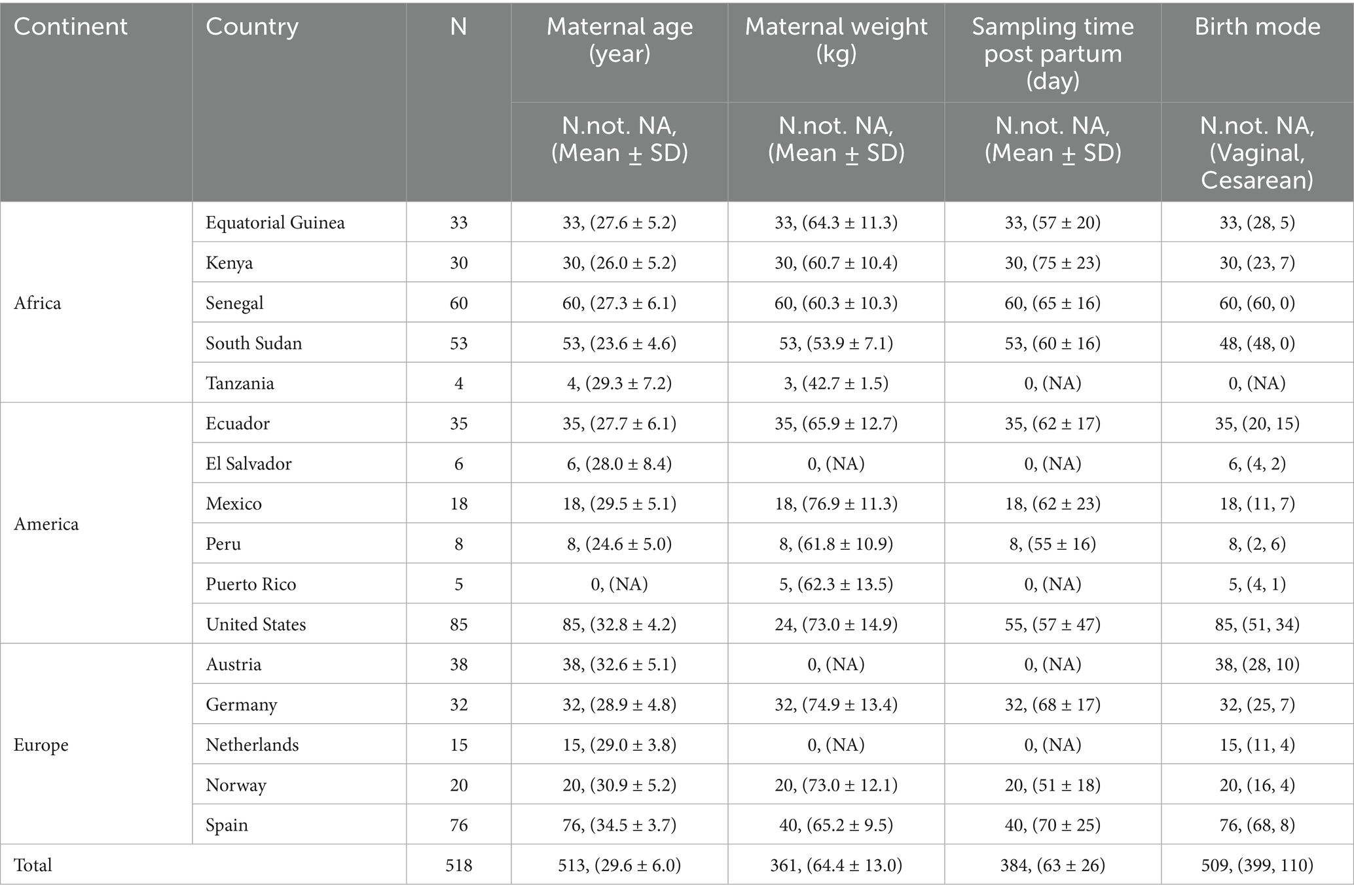
Milk samples from 518 healthy mothers (one individual sample per mother) were obtained in 16 different countries, including cohorts from Equatorial Guinea (n = 33), Kenya (n = 30), Senegal (n = 60), South Sudan (n = 53), Tanzania (n = 4), Ecuador (n = 35), El Salvador (n = 6), Mexico (n = 18), Peru (n = 8), Puerto Rico (n = 5), mainland United States (n = 85), Austria (n = 38), Germany (n = 32), The Netherlands (n = 15), Norway (n = 20), and Spain (n = 76). The subject’s age, body weight, sampling time, and birth mode of the baby were listed in Table 1.
The study procedures related to the samples obtained from Equatorial Guinea, Kenya, Senegal, South Sudan, Ecuador, El Salvador, Mexico, Peru, Austria, Germany, The Netherlands, Norway, and Spain were approved by the overarching Institutional Review Board of the European Commission in the frame of the EU project “Variations in biochemical and microbiological milk composition among highly diverse human populations and their impact on infant gut ecosystem” (call FP-7-PEOPLE-2013-IEF) (protocol #624773, approved on 14 February 2014) and at each study location, and consent was obtained from each participating woman. Human milk samples from Puerto Rico were approved by the University of Puerto Rico, Medical Sciences Campus (IRB ProB2310120, approved on 24 March 2021). The United States samples were approved by the Rutgers University-New Brunswick Health Sciences Institutional Review Board (Protocol #Pro2018002781, approved on 17 September 2021; Protocol #Pro2020002169, approved on 9 June 2021), and consent was obtained from each participating woman. The samples from Tanzania are from a previous study approved by the IRB reference number 164-12-21052012 (46).
Collection of human milk was performed by the mothers as described by McGuire et al. (47); briefly, the aureola skin was wiped with antiseptic wipes containing chlorhexidine digluconate (bactiseptic wipes, Vesismin Health, Barcelona, Spain) using gloved hands, and milk was manually expressed into disposable sterile containers. Samples were shipped on dry ice to the Complutense University of Madrid (Spain), where they were stored at −80°C until an aliquot was sent on dry ice to Rutgers University (USA) for analyses. Human milk samples from Puerto Rico, the U.S. mainland, and Tanzania were collected either by hand expression or using the mother’s own sterile pump. Samples were frozen at the participants’ homes and transported on ice to the laboratory, where they were stored at −80°C until analysis.
2.2 DNA isolation, amplification and sequencing
The samples (1 mL) were centrifuged (15,000×g for 10 min at 4°C), and the fat layer was removed using a sterile swab. This step was repeated twice more to remove all fat. Then, the pellets together with a 200 μL fraction of the supernatants, were used for total DNA extraction employing the Dneasy PowerSoil Pro Kit (QIAGEN, Hilden, Germany), following the manufacturer’s instructions.
2.3 16S rRNA gene sequencing
Primers 515 F (5’-GTGYCAGCMGCCGCGGTAA-3′) and 806R (5′-GGACTACNVGGGTWTCTAAT-3′) were used to amplify the V4 hypervariable region of the bacterial 16S rRNA gene following the protocols for the Earth Microbiome Project.1 The concentration of the pooled, purified and barcoded DNA amplicons was determined using the Qubit dsDNA HS assay kit (Thermo Fisher, Waltham, MA, USA). Amplicons were sequenced at Genewiz, LLC. (South Plainfield, NJ, USA) using the Illumina MiSeq platform (Illumina, CA, USA) with the Illumina MiSeq 2 × 150 bp paired-end protocol (Illumina Inc., San Diego, CA, USA) using the Illumina MiSeq platform.
2.4 ITS sequencing
For fungi, the Internal Transcribed Spacer 2 (ITS2) region was amplified from DNA obtained from extracted milk samples with a single ITS3 forward primer (5′-AATGATACGGCGACCACCGAG ATCTACACTATGGTAATTGTGCATCGATGAAGAACGCAGC-3′) and a barcoded ITS4 reverse primer (5′-CAAGCAGAAGACGGCATA CGAGATTCCCTTGTCTCCAGTCAGTCAGCCTCCTCCGCTTAT TGATATGC-3′) (38). Primers included Illumina adapters and the reverse primer included a unique 12 base Golay barcode (XX) for pooled demultiplexing (5′-CAAGCAGAAGACGGCATACGAG ATXXXXXXXXXXXXCGGCTGCGTTCTTCATCGATGC-3′). To determine PCR cycle counts appropriate for the fraction of fungal material, qPCR was performed on 518 extracted human milk samples. For samples with a Ct value less than 30, 25 cycles of PCR was performed. For samples with a Ct value between 30–31, 33 cycles of PCR was performed; the higher cycle number was needed to ensure there was enough fungal material for sequencing. Samples with a Ct value above 33 were considered to contain no fungal material (matching a no-template control). Samples were purified by SPRI, pooled, and sequenced on the Illumina MiSeq platform using the Illumina MiSeq Reagent v3 600-cycle (2 × 300 bp).
2.5 qPCR detection of methanogen
For the detection of Methanogen from the samples, we performed qPCR to detect the copy number of Methanobacteriales with specific primers (Fwd: 5’-AGGAATTGGCGGGGGAGCAC-3′, Rev.: 5′-TGGGTCTCGCTCGTTG-3′) targeting the 16S rRNA gene fragment between positions 915 and 1,100 (48, 49). First, we use a universal primer (Fwd: 5′-ACTCCTACGGGAGGCAGCAG-3′, Rev.: 5′-ATTACCGCGGCTGCTGG-3′) targeting the 16S rRNA gene between position 314 and 540 (50) to detect the number of total bacteria and archaea. Part of the E. coli 16S gene fragment (NR_112558) was synthesized and diluted to 107, 106, 105, 104, 103, 102, and 10 copies as standards. The PCR program for total bacteria is initial at 95°C for 5 min, followed by 45 cycles of 10 s at 95°C, 10 s at 60°C, and 10 s at 72°C. Only the samples that had total bacterial copy numbers > 105 were used to detect Methanogens. The standards for Methanogen were synthesized from part of the Methanobacterium espanolae 16S rRNA gene (NR_104983.1) and diluted to range from 107 to 10 copies. The PCR program for Methanogen detection started with an initial step at 95°C for 10 min, followed by 40 cycles of 10 s at 95°C, 30 s at 57°C, and 20 s at 72°C. Both PCR was performed using a Quantstudio 3 system (Thermo Fisher, Waltham, MA, USA) with a Quantinova SYBR green PCR kit (Qiagen, Hilden, Germany) and 1uL of extracted DNA was used as the template. To avoid extrapolating beyond the standard curve, we set the detection limit at 10 copies for both PCR experiments.
2.6 Data analyses
Raw reads were demultiplexed and quality-filtered using the QIIME2 pipeline (v2022.2) (51). The quality-filtered reads were denoised and concatenated with DADA2 (52). Taxonomy was assigned to Amplicon sequence variants (ASVs) against the SILVA database v138.1 (53), using in QIIME 2 (q2-feature-classifier). The phylogenetic tree was generated by the FastTree algorithm (54). Negative control samples were also sequenced, and ASVs identified as contaminated by decontam (55) were removed from further analysis. The reads count was rarefied to 2,857 reads per sample (21 samples were excluded, Supplementary Table 1). The ASVs determined as contamination are listed in Supplementary Table 2.
Faith’s Phylogenetic diversity, observed ASV, and Shannon Index were calculated as alpha diversity metrics. The difference in alpha diversity between countries or continents was tested using the Kruskal-Wallis group test and with FDR p-value adjustment.
Jaccard, Bray Curtis, unweighted and weighted Unifrac distances were calculated to obtain pairwise beta-diversity, and dimensionality reduction on the distances was performed using Principal Coordinates Analysis (PCoA) method (ape v5.8). All alpha and beta diversity metrics are calculated on ASV level abundance. Permutational multivariate analysis of variance (PERMANOVA) was used to test the significance of different groups with 999 permutations (vegan v2.6-8).
Differentiated taxa were detected with ANCOMBC (2.6.0) (56) with Holm–Bonferroni correction, with a default prevalence cutoff of 10%. The shared taxa between countries were shown as an upset plot by UpSetR (1.4.0). The co-abundance network was performed on the genus level with taxa with abundance > 0.01 and prevalence > 10% using the SparCC method (57) (SpiecEasi v1.1.3), and the correlation cutoff was set to ± 0.3.
3 Results
3.1 Human milk bacteriome
The 16S rRNA gene (V4 region) sequencing analysis of the 497 milk samples included in this work (Supplementary Table 3) yielded 10,767,189 high-quality filtered sequences in total, ranging from 18 to 76,462 reads per sample [mean = 20,786 reads per sample; median (IQR) = 18,069 (9,542–28,173) sequences per sample]. The samples were rarefied to 2,857 sequences per sample (Supplementary Figure 1).
First, we applied a linear model to analyze the effect of country as well as some potential cofounders like maternal age, maternal body weight, birth mode, and the postpartum days of milk collection on the alpha diversity. Only the country showed a significant effect on the milk alpha diversity, and the effect size is bigger than the other variables (Supplementary Table 4). Next, we only focused on the difference between countries. As assessed by using the Faith PD diversity index, the milk alpha diversity oscillated between Equatorial Guinea and Mexico (Figure 1A); the same range between Equatorial Guinea (lowest) and Mexico (highest) in Observed ASVs and Shannon index (Supplementary Figure 2). Overall, there was a significant difference in alpha diversity between different countries. Among all the pairwise comparisons, Ecuador and Mexico had significantly higher Faith PD diversity than Germany, Norway, Spain, the United States, and Equatorial Guinea, Equatorial Guinea had significantly lower alpha diversity than Peru, Senegal, South Sudan, Austria, Mexico, and Ecuador. Most other pairwise comparisons were not significant after adjustment for multiple comparisons. Similar trends are shown in Observed ASVs, but not in Pielou evenness or Shannon index (Supplementary Table 5). When aggregated into the Continent level, America showed significantly higher alpha diversity in Faith PD and richness (observed ASVs), but metrics accounting for evenness (Pielou and Shannon index) showed higher evenness in Africa (Supplementary Figure 3).
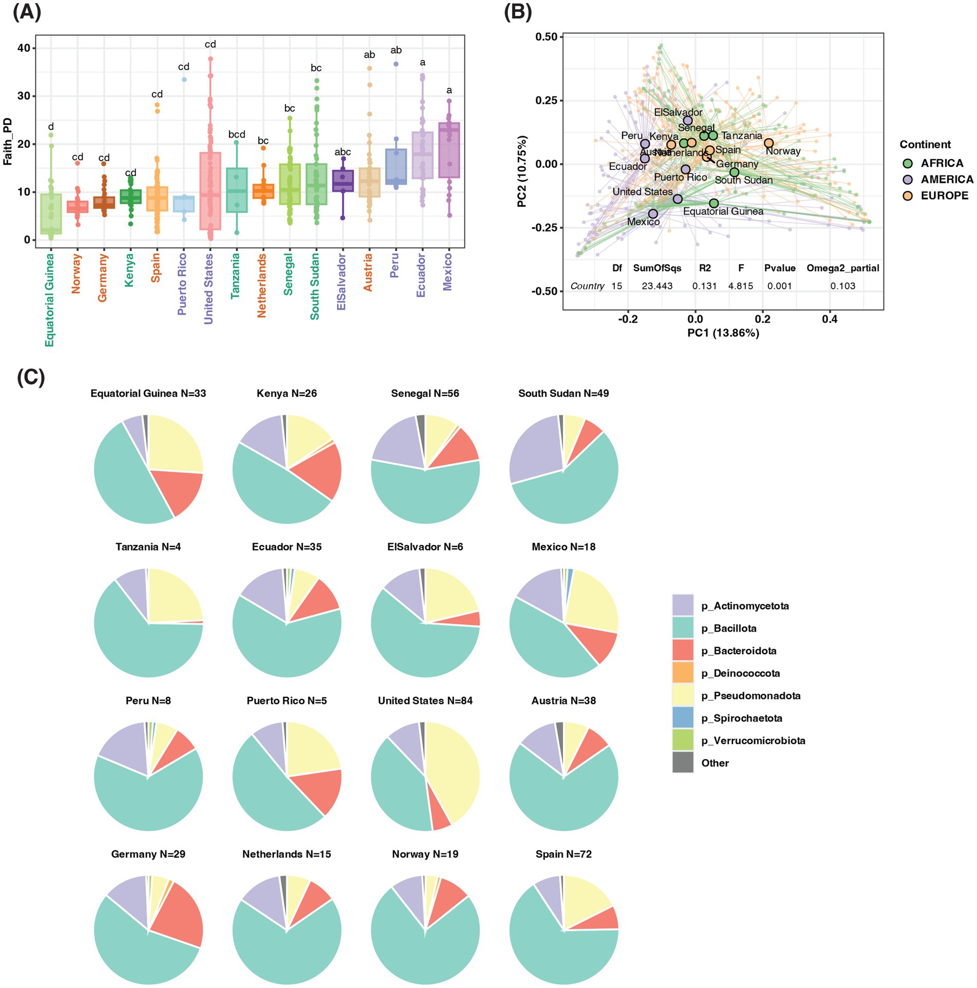
Figure 1. The bacterial difference between countries. (A) Alpha diversity in Faith PD, country ordered by median of Faith PD from low to high, Different letters show significant differences (Kruskal-Wallis test with FDR adjustment, p < 0.05). (B) PCoA plot based on Bray Curtis distance, the center of each country is in large dots, and individual samples are in small dots. PERMANOVA test of country effect is listed below. (C) The top abundant bacterial Phyla by country.
For beta diversity, we also included all potential cofounders in the first PERMANOVA analysis and found that only country showed a significant effect on beta diversity (Supplementary Table 6). Therefore, in the following analysis, we only included the country or continent. We detected a significant effect by country with the omega square effect size is 0.103 (meaning about 10.3% of the variation is from the country effect; p-value = 0.001). Mexico, Equatorial Guinea, and Norway showed the most segregated centroids in PCoA analysis based on Bray Curtis distance (Figure 1B). Some European and African countries were close to each other in the PCoA plot, including Spain, Germany, Austria, Kenya, Senegal, and Tanzania, indicating similar bacterial structures. The countries from different continents also differ in PC1, the countries from America all had negative PC1 values while most of the rest countries had positive PC1 values. Jaccard and Unifrac distance showed a similar effect, but the effect size of weighted Unifrac (0.120) is bigger than Jaccard (0.068) or unweighted Unifrac (0.064), suggesting the difference between countries was due to abundant taxa (Supplementary Figure 4). The effect of the continent on beta diversity was also significant but had a smaller effect size (omega square range from 0.022 to 0.043), and based on Bray Curtis and Jaccard, the distance of the center of samples from Europe and Africa was closer than the distance to America (Supplementary Figure 5).
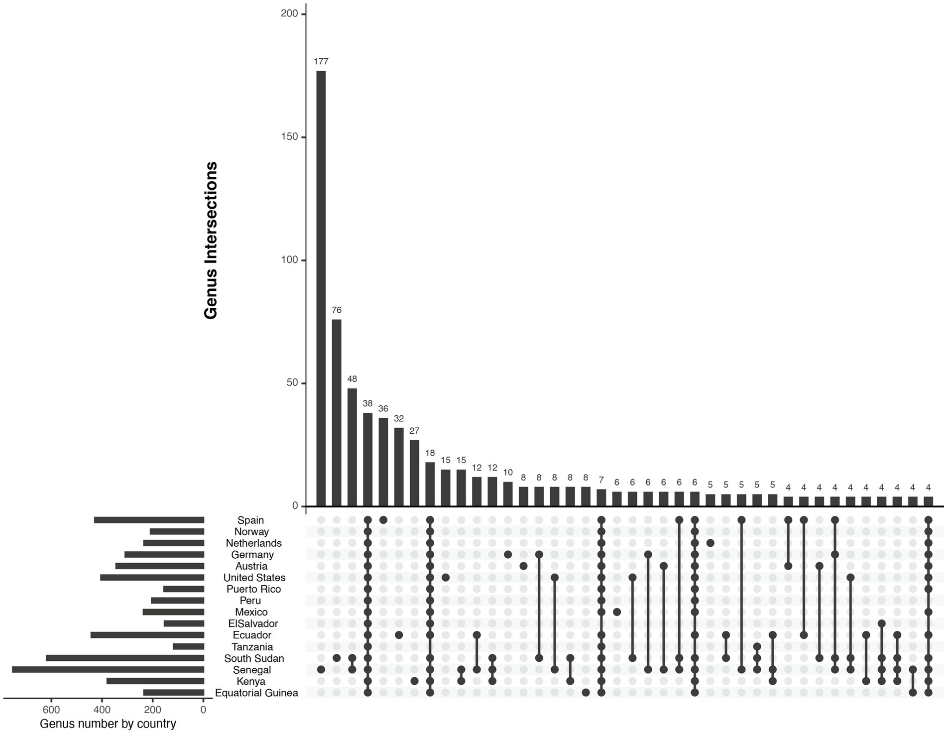
In total 14,581 clean ASVs were generated from 16S sequencing, belonging to 41 phyla, 1,333 genera and 2,774 species. Most of them corresponded to seven major phyla: Bacillota, Pseudomonadota, Actinomycetota, Bacteroidota, Spirochaetota, Verrucomicrobiota, and Deinococcota, (Figure 1C). Among the 1,333 genera detected in this work, only 38 of them were found in samples from all the countries (Figure 2), while 18 were shared by all countries except for those from Tanzania and 7 were shared by all countries except for those from El Salvador. The genera with the top three highest frequency of detection were Staphylococcus (99.2% of samples; present in samples from all countries), Streptococcus (97.4% of samples; present in all countries), and Bifidobacterium (93.2% of samples; present in all countries). Overall, the top three most abundant genera were the same as the top three prevalent genera, Streptococcus (mean abundance 23%), Staphylococcus (mean abundance 16.8%), and Bifidobacterium (mean abundance 5.9%). Other highly abundant (> 1%) and universal genera include Pseudomonas, Corynebacterium, Acinetobacter, Rothia, Lactobacillus, Prevotella, Bacteroides, and unclassified genera from Muribaculaceae, Enterobacteriaceae, and Lachnospiraceae (Supplementary Table 7).
Samples from mainland USA are enriched in Pseudomonas, and depleted in Streptococcus, and Lactobacillus. Mexico and Ecuador are enriched in Bifidobacterium, Bacteroides, and Prevotella. Many countries from the European cohort are depleted in Bacillus and Alistipes (except Germany), enriched in Staphylococcus. Many countries from the African cohort are enriched in Alistipes and Bacillus and depleted in Bacteroides and Gemella. Kenya enriched in Acinetobacter, Alistipes, Lactobacillus, and Muribaculaceae (Figure 3 and Supplementary Figure 6).
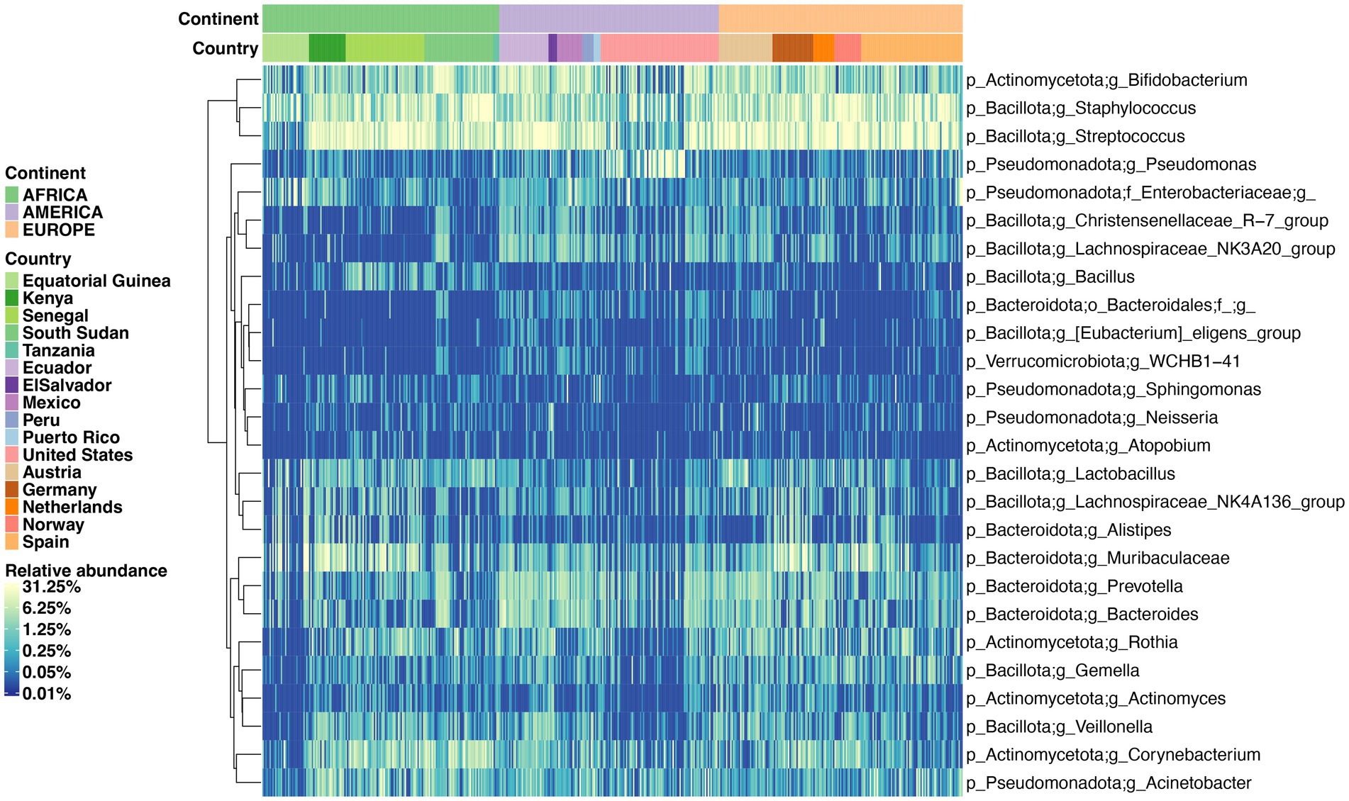
Figure 3. Supervised heatmap of genera relative abundance significantly different between countries. The differentiated genera were detected by ANCOM global comparison with Holm–Bonferroni correction with adjusted p-value < 0.05.
Co-abundance network analysis of human milk bacteria only showed positive associations, particularly Bacteroides, Prevotella, and Bifidobacterium formed positive connections between each other. Other positive connections are between Staphylococcus and Corynebacterium; Streptococcus and Rothia; Pseudomonas and Acinetobacter (Figure 4A). That different bacterial genera occupied central positions, indicates country-specific structures and ecological roles within the microbial communities. For example, in Austria, Blautia and Rothia served as central nodes with strong associations to multiple genera. In South Sudan, Lactobacillus, Corynebacterium, and Kocuria were the most highly connected, while in Ecuador, Streptococcus, Veillonella, Staphylococcus, Blautia, Treponema, Bifidobacterium, and Prevotella showed the highest connectivity. These variations likely reflect underlying differences in genus abundance. Indeed, genera such as Bifidobacterium, Staphylococcus, Streptococcus, Lactobacillus, Corynebacterium, Rothia, Bacteroides, Prevotella, and Pseudomonas were identified as differentially abundant and consistently appeared as key hubs in their respective networks. Notably, the global network combining all countries’ data was the most robust and densely interconnected, highlighting overarching co-abundance patterns across diverse populations.
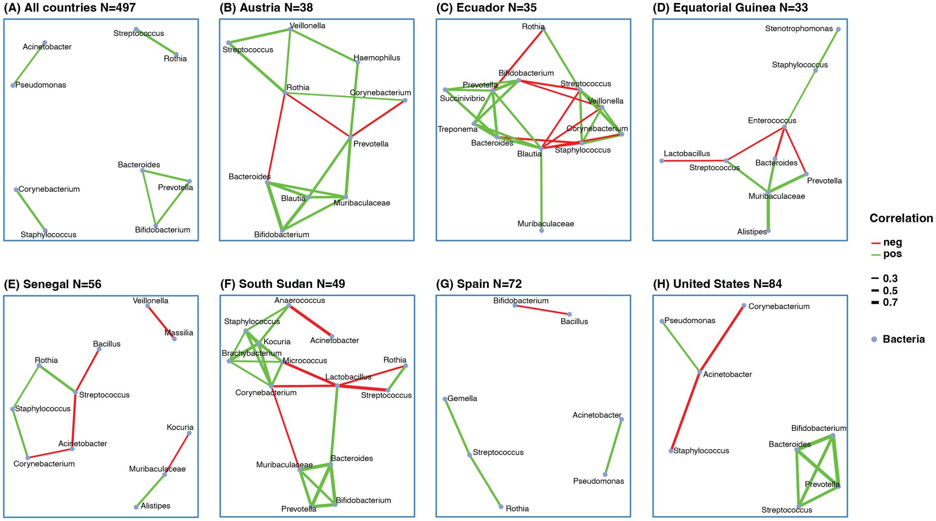
Figure 4. Co-abundance network of human milk bacteria. (A) Co-abundance network with samples from all countries. Co-abundance network from individual country, (B) Austria, (C) Ecuador, (D) Equatorial Guinea, (E) Senegal, (F) South Sudan, (G) Spain, (H) United States. The correlation was calculated based on sparCC with abundance of genera > 0.01 and prevalence > 10%. Only correlations > 0.3 or <−0.3 and pseudo-p-value < 0.05 were selected. The red line indicated negative correlation, the green line indicated positive correlation, line width indicated correlation value.
Despite these differences, we also observed some recurring correlation patterns across countries, similar to those in the global network. For instance, connections among Bacteroides, Prevotella, and Bifidobacterium were seen in Austria, Ecuador, South Sudan, and the United States. However, specific patterns varied: in Austria, Blautia and Muribaculaceae were also connected (Figure 4B); in Ecuador, Blautia, Succinivibrio, Treponema, and Bacteroides formed key links (Figure 4C); in South Sudan, Muribaculaceae showed strong associations (Figure 4F); and in the United States, Streptococcus emerged as a connected node (Figure 4H). Networks in Austria, Ecuador, Equatorial Guinea, and South Sudan exhibited greater complexity and connectivity compared to others.
3.2 Detection of total bacteria methanogenic archaea
Based on 16S sequencing, we were able to detect three genera from Methanobacteriales (Methanobrevibacter (0–2.6%), Methanosphaera (0–16.7%), and Methanobacterium (0–33.3%) Supplementary Table 8).
We also applied a standard curve qPCR method to detect Methanobacteriales. Based on our 16S rRNA sequencing results, which showed that the methanogen relative abundance ranged from 0.03 to 1% in samples where methanogen reads were detected. Given these proportions, if a sample contained fewer than 10⁵ total bacterial cells, the number of methanogen cells could be as low as 30 copies -close to the detection limit-. Therefore, we performed methanogen qPCR only on samples with a total bacterial cell count exceeding 10⁵. When examining a subset of the samples (268 samples from 7 countries) with qPCR, we found that only 21 (8%) had total bacteria/archaea 16S gene copy number >105 per 1 μL DNA, evidencing the low bacterial density in these human milk samples from healthy women. Furthermore, this threshold of detection of 105 gene copies found in our study likely overestimates the number of bacterial cells in the samples, since bacteria can have multiple 16S gene copies. Among the 21 samples, only two, one from South Sudan and the other from Equatorial Guinea had detectable methanogenic archaea (Supplementary Table 9).
3.3 Human milk mycobiome
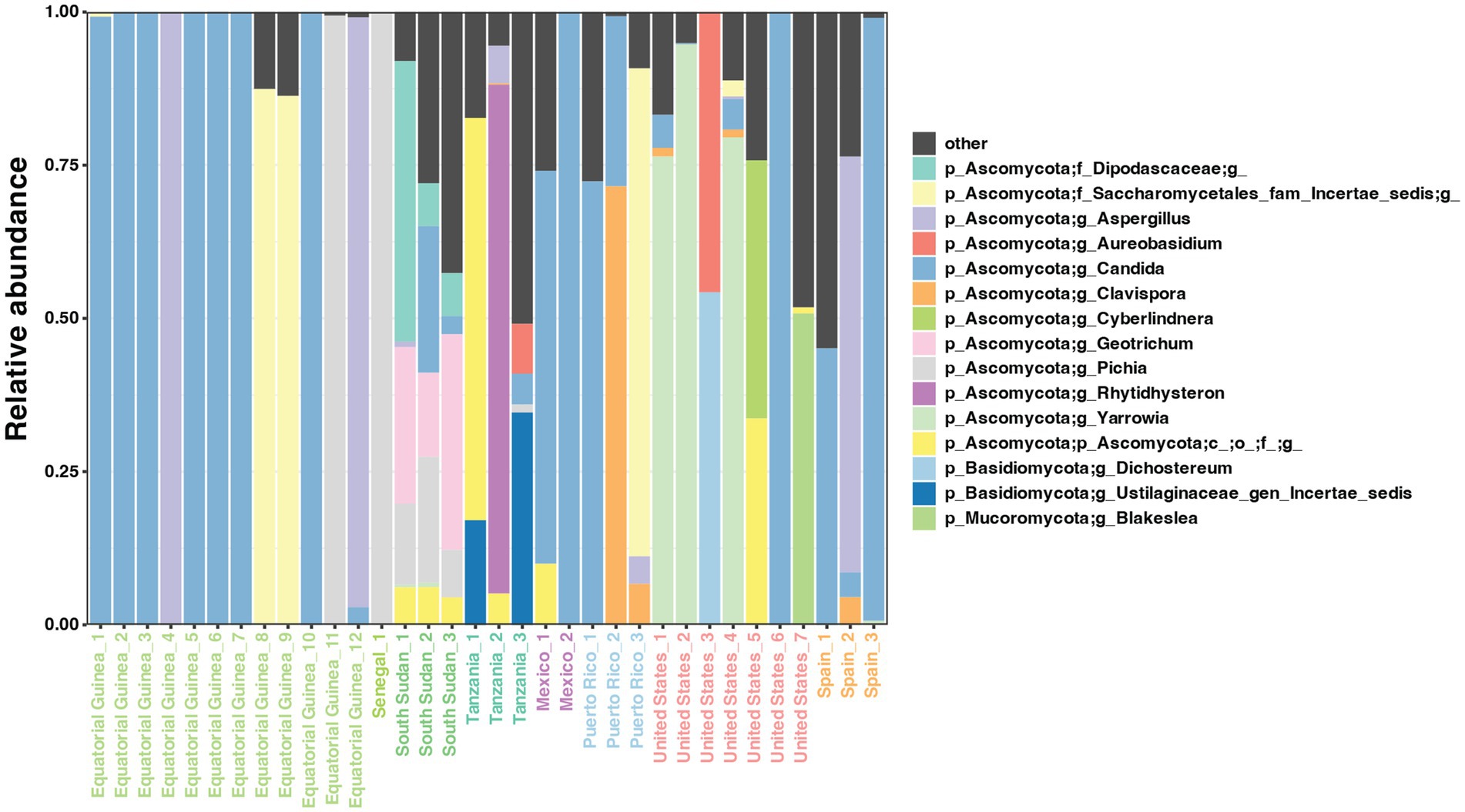
The presence of fungi was evaluated in the whole collection of 518 samples (Supplementary Table 10). Only 6.6% of samples (n = 34) were positive based on ITS2 amplification and they belonged to the following cohorts: Equatorial Guinea (n = 12, 36.4%), Senegal (n = 1, 1.7%), South Sudan (n = 3, 5.7%), Tanzania (n = 3, 75%), Mexico (n = 2, 11.1%), mainland US (n = 7, 8.2%), Puerto Rico (n = 3, 60%), and Spain (n = 3, 3.9%). Among the fungi-positive samples, three fungal phyla (Ascomycota, Basidiomycota, and Mucoromycota) and 12 major genera were detected, dominated by Candida (Figure 4). Some of the fungal genera, such as Candida, and Malassezia, are frequent inhabitants of the human skin while others, such as Dichostereum or Blakeslea, are typically associated other to air, soil, and decaying plant matter, suggesting environmental or laboratory contamination of milk samples (Figure 5, Supplementary Table 11).
3.4 Interkingdom co-abundance networks
The interkingdom co-abundance network showed that Prevotella, Corynebacterium and Clavispora were the centers of the network (Figure 6A). Besides Clavispora, there were three fungal genera involved in the network, including Candida, Pichia, and Yarrowia. Most bacterial genera involved were the common genera found in the bacteria-only networks, including Bacteroides, Prevotella, Bifidobacterium, Staphylococcus, Corynebacterium, Streptococcus, Pseudomonas, Lactobacillus, and Rothia. Compared with the bacteria-only network with all samples (Figure 4A) or the same sample size as the interkingdom network (Figure 6B), the bacteria correlations remained similar, and the fungal genera acted as “bridges” connecting the isolated parts of the bacteria-only network.
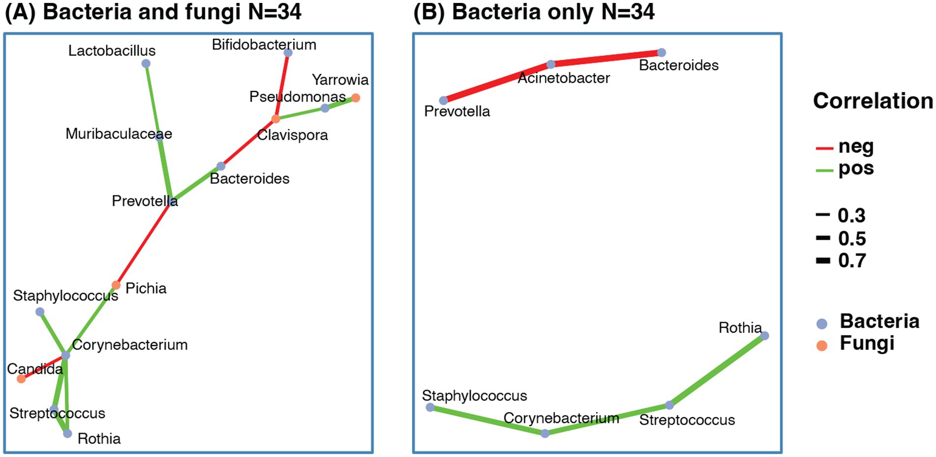
Figure 6. Co-abundance network of human milk microbes. (A) Co-abundance network with both bacteria and fungi. (B) Co-abundance network with only bacteria using the same sample as the interkingdom network. The correlation was calculated based on sparCC with abundance of genera > 0.01, and prevalence > 10%. Only correlations > 0.3 or <−0.3 and pseudo-p-value < 0.05 were selected. The red line indicated negative correlation, the green line indicated positive correlation, line width indicated correlation value.
4 Discussion
Human milk poses some challenges for microbiome studies since the procedures used for milk and milk DNA processing and analysis, its low biomass in healthy women (as observed in this work by qPCR), and the risk of skin and environmental contamination during sampling may also be a cause of artifacts or biases in these studies (40, 58). In this work, the application of the same methodology, from DNA extraction to data analysis, to a large collection of milk samples from 16 countries confirmed the existence of large inter-individual and inter-cohort variations in the composition of the human milk bacteriome and mycobiome, as described previously (22, 30, 38, 39, 59). The lack of clear separation between milk samples from women in different geographical regions may be due to the high intraindividual variability among milk samples from the same continent or country (40). Our findings align with previous studies focused on the bacteriome of human milk samples worldwide, particularly with those obtained within the framework of the INSPIRE study (30, 40). Staphylococcus and Streptococcus were the dominant genera by prevalence and abundance, detected in 99.2 and 97.4% of our samples, closely matching the 98.7 and 97.7% reported in INSPIRE (30), and consistent with other studies (22).
Similarly, the INSPIRE study reported a high abundance of Streptococcus in Peruvian and Pseudomonas in U.S. samples (30), consistent with our findings. Both studies also observed a higher relative abundance of Pseudomonadota (formerly Proteobacteria) in African versus European cohorts. However, as in INSPIRE, additional core taxa varied across cohorts. In our study, other frequent genera included Bifidobacterium, Pseudomonas, Corynebacterium, Acinetobacter, Rothia, Lactobacillus, Prevotella, and Bacteroides, with no clear geographical differences. Unlike INSPIRE, we did not analyze infant fecal samples, limiting our ability to assess the role of milk microbiota in seeding the infant gut. INSPIRE’s comparison of milk and infant feces provided evidence of this relationship (30). Future studies are needed to further explore vertical transmission through breastfeeding.
In this study, potential intrakingdom and interkingdom relationships were investigated through the establishment of co-abundance networks of bacteria and bacteria-fungi, respectively. In relation to intrakingdom associations, there was a positive correlation between the presence of Staphylococcus and Corynebacterium. The same observation has been performed repeatedly when studying other human niches and, particularly, the skin (60–62). It is noteworthy that mammary glands are highly specialized organs, likely evolving from ancestral cutaneous apocrine-like glands (63). The populations of Staphylococcus and Corynebacterium appear to respond synchronously, both in health and disease, to shared host or environmental factors within the mammary ecosystem (64). This synchrony is also observed in various skin regions, including the nasal sinuses and ocular surfaces and glands (62, 65–68). Additionally, skin secretions and milk share common components such as exfoliated epithelial cells and nutrients, including urea, amino acids, peptides, glycoproteins, glycerol, phospholipids, and others. Coagulase-negative staphylococci utilize amino acids mainly provided by their own proteolytic activities and, in turn, corynebacteria need these same amino acids and are cross-fed by their skin partners (69). The proteolytic properties of resident staphylococci also have a protective role for corynebacteria since they are able to inactivate antibacterial proteins and peptides (69, 70). In addition, lipophilic corynebacteria lack fatty acid synthase and, consequently, are fatty acid auxotrophs (71). Interestingly, it seems that Staphylococcus epidermidis and Corynebacterium spp. use different glycans as molecular decoys for binding to human skin and sweat (72). More specifically, sialic acid and fucose, which are also key components of human milk oligosaccharides, are binding epitopes for staphylococci while N-glycans did not provide binding epitopes for Corynebacterium, consistent with a lack of competition between them for these substrates.
A positive correlation was also observed among Bacteroides, Prevotella, and Bifidobacterium. DNA from these three genera has already been detected in human milk (15, 30, 73–75), and they are common inhabitants of the human gut (76). Their abundance is higher in children than in adults (77) and is reduced in caesarean-delivered infants in comparison with vaginally-delivered infants (78). These three genera have been proposed as biomarkers of diet and lifestyle (79). They may form metabolic networks in the infant gut, playing synergistic and complementary roles. For example, certain Bifidobacterium species are well adapted to the human milk and infant gut environment due to their ability to metabolize human milk oligosaccharides (HMOs) (80). In the process, they produce lactic and acetic acids, which promote the growth of short-chain fatty acid (SCFA) producers like Prevotella and Bacteroides. These species further enhance glucose metabolism and generate SCFAs and vitamins that support health (81). During lactation, Prevotella and Bacteroides also metabolize milk-derived amino acids (82), and later become key to breaking down complex polysaccharides once solid foods are introduced. Thus, human milk may seed the infant gut with bacteria that support gut health both early and later in life.
Supporting this idea, a fecal microbiota transplant (FMT) study in patients with autism spectrum disorder (ASD) found that 2 years post-treatment, the gut microbiome was dominated by Bacteroides, Prevotella, and Bifidobacterium, alongside a reduction in ASD-associated taxa (82), Similarly, these genera are often depleted in individuals colonized by Clostridioides difficile (83), further highlighting their potential health benefits.
In this study, Bifidobacterium was negatively associated with Pseudomonas, consistent with previous results showing the antagonistic activity of bifidobacteria against some species of Pseudomonas and, particularly, against Pseudomonas aeruginosa (84–86). In the same direction, pretreatment of human corneal epithelial cells with a strain of Bifidobacterium longum subsp. infantis protected them from infection by a P. aeruginosa strain (87). Evidence suggests that the protection against P. aeruginosa cellular infection is through modulation of the expression of IL-8 and beta-defensin-2 (88). Indeed, this protection has been harnessed to slow the decay of the microbiological quality caused by P. aeruginosa by B. longum subsp. infantis biofilms in the inner surface of cheese packages (89). Therefore, it seems that bifidobacteria displays, to some extent, mechanisms of competitive exclusion against pseudomonas.
In a study including 80 mothers from four countries [Finland, Spain, South Africa, and China; (28)], in another of 65 mothers from Spain (32), detection of the fungi ranged from 35 to 86%. In a Canadian cohort with 271 mothers, it was lower, 21% of the human milk samples (38), but still higher than the 7% in the current study (ranging from 2 to 36%), for reasons that are not clear. This could be due to methodological issues, or to higher density of bacteria that inhibit fungi. Interactions between bacteria and fungi are poorly known although they may be relevant for health (90, 91). Two major negative correlations were detected in our study, one between Bifidobacterium and Clavispora, and the second between Corynebacterium and Candida. These results are consistent with those in a recent study reporting negative correlation between Bifidobacterium and C. albicans in the milk of mothers who delivered vaginally (39). A body of evidence suggests that in the gut, bifidobacteria provide resistance against colonization by yeasts, particularly by Candida albicans (92–96). More recently, another study in children and adolescents reported negative correlation between Bifidobacterium and Candida, with higher Candida low Bifidobacterium associated with depression (97). Other studies have not found fungal-bacterial interactions in the feces of healthy subjects (98). As for Corynebacterium and Candida, it is long known their negative association and the protective effect of the former against infections (99–101). Interestingly, high oral C. albicans and low Corynebacterium appears to be a signature of oral carcinoma and head and neck cancer (102).
The qPCR analysis detected the presence of detectable methanogenic archaea (Methanobacteriales) in two African samples, one from South Sudan and one from Equatorial Guinea. Consistent with the qPCR results, the methanogenic archaea detection rate was higher in African and American countries based on 16S sequencing. However, the detection rate of 16S sequencing is much higher than that of qPCR, possibly because the DNA concentration in the milk sample is low, and the 1 μL of input DNA used for qPCR may not be sufficient to detect the methanogenic archaea. Archaea are among the neglected microbes in human microbiome studies because of technical challenges in detection (103), although their presence has been previously reported in human colostrum and milk (31, 104), and in the gut of babies (105–107). Archaeal DNA has been detected in human milk, albeit at low frequency and abundance. Togo et al. (31) identified Methanobrevibacter smithii in approximately 25% of colostrum and milk samples using species-specific qPCR, whereas Methanobrevibacter oralis was not detected. Another study reported the presence of DNA from Methanoculleus, Methanosarcina, and Methanobrevibacter in Mexican mother-infant dyads, suggesting that colostrum may serve as a source of neonatal archaea (103). Similarly, Grine et al. (105) proposed maternal transmission of M. smithii. Methanogenic archaea—including Methanobrevibacter spp., Methanosphaera stadtmanae, and members of the Methanomassiliicoccales—are recognized as consistent but low-abundance constituents of the infant gut microbiome (103, 106, 108). However, detection remains limited by methodological biases favoring bacterial over archaeal targets, such as inefficiencies in DNA extraction, primer design, and reference databases. Notably, a recent gut microbiome survey in Africa—where our archaeal-positive samples were most prevalent -did not assess archaeal DNA (109). To advance characterization of archaeal diversity and function, we recommend: (a) the use of archaeal-specific or dual-target primers and nested PCR; (b) shotgun metagenomics for unbiased genomic and functional profiling; (c) optimized DNA extraction protocols tailored to the resilient archaeal cell wall; (d) expansion of archaeal reference databases; (e) integration of multi-omics approaches to detect archaeal metabolites such as methane; and (f) improved cultivation methods using anaerobic conditions and archaeal-specific substrates.
This study has several limitations. There was a big variation in sample size between countries (from 4 to 85), which weakens the statistical power. Also, sample collection was not absolutely standardized, which may introduce a systematic bias between cohorts. Despite the limitations, this work highlights differences in the human milk bacterial and fungal microbiome across geographies, emphasizing the need for global studies that provide a better understanding of the human microbiome and the interkingdom relationships that may explain microbiota structures, with implications for maternal and infant health.
Data availability statement
The datasets presented in this study can be found in online repositories. The names of the repository/repositories and accession number(s) can be found at: https://www.ncbi.nlm.nih.gov/, PRJNA1216262.
Ethics statement
The studies involving humans were approved by Overarching Institutional Review Board of the European Commission, Rutgers University New Brunswick Health Sciences Institutional Review Board, University of Leipzig Ethik-Kommission Review Board, Institutional Review Board of the University of Puerto Rico. The studies were conducted in accordance with the local legislation and institutional requirements. The participants provided their written informed consent to participate in this study.
Author contributions
HS: Writing – review & editing, Methodology, Investigation, Writing – original draft, Formal analysis, Visualization. BF: Methodology, Formal analysis, Writing – review & editing. MA: Methodology, Writing – review & editing, Formal analysis. CC: Formal analysis, Methodology, Writing – review & editing. LEC: Methodology, Formal analysis, Writing – review & editing. BB: Writing – review & editing, Investigation, Methodology, Formal analysis. JL: Formal analysis, Writing – review & editing, Methodology, Investigation. TS: Formal analysis, Visualization, Writing – review & editing, Methodology. EA: Formal analysis, Methodology, Writing – review & editing. FG-V: Writing – review & editing, Formal analysis, Resources, Methodology. MW: Resources, Writing – review & editing, Methodology, Investigation. MS: Writing – review & editing, Investigation, Formal analysis. CA: Formal analysis, Resources, Data curation, Writing – review & editing, Validation. JR: Supervision, Writing – review & editing, Conceptualization, Funding acquisition, Writing – original draft, Resources. MD-B: Funding acquisition, Resources, Conceptualization, Supervision, Writing – review & editing, Writing – original draft.
Funding
The author(s) declare that financial support was received for the research and/or publication of this article. The authors acknowledge financial support from CIFAR Manulife Fund, EMCH Fund, grant 624773 (FP-7-PEOPLE-2013-IEF, European Commission), and project AGL2013-4190-P (Ministry of Economy and Competitiveness, Spain).
Conflict of interest
LEC is co-founder and shareholder in Bright Angel Therapeutics, a platform company for the development of novel antifungal therapeutics. LEC is a Science Advisor for Kapoose Creek, a company that harnesses the therapeutic potential of fungi. LEC is a Canada Research Chair (Tier 1) in Microbial Genomics & Infectious Disease and co-Director of the CIFAR Fungal Kingdom: Threats & Opportunities program.
The remaining authors declare that the research was conducted in the absence of any commercial or financial relationships that could be construed as a potential conflict of interest.
Generative AI statement
The authors declare that no Gen AI was used in the creation of this manuscript.
Publisher’s note
All claims expressed in this article are solely those of the authors and do not necessarily represent those of their affiliated organizations, or those of the publisher, the editors and the reviewers. Any product that may be evaluated in this article, or claim that may be made by its manufacturer, is not guaranteed or endorsed by the publisher.
Supplementary material
The Supplementary material for this article can be found online at: https://www.frontiersin.org/articles/10.3389/fnut.2025.1610346/full#supplementary-material
SUPPLEMENTARY FIGURE 1 | Bacterial 16S rarefaction curve (A) and goods coverage of samples (B). Rarefy depth set to 2857.
SUPPLEMENTARY FIGURE 2 | Alpha diversity between countries. (A) Observed ASVs, (B) Pielou evenness, (C) Shannon index. Countries ordered by median of diversity from low to high, different letters show significant differences (Kruskal-Wallis test with FDR adjustment, p < 0.05).
SUPPLEMENTARY FIGURE 3 | Alpha diversity by continent. (A) Faith PD, (B) observed ASVs, (C) Pielou evenness, (D) Shannon Index. Difference between continents tested by Kruskal-Wallis and with FDR adjustment. *p < 0.05, **p < 0.01, ***p < 0.001, ****p < 0.0001.
SUPPLEMENTARY FIGURE 4 | PCoA plots based on beta diversity in different countries. (A) Jaccard distance, (B) unweighted Unifrac distance, (C) weighted Unifrac distance. The center of each country is in large dots, and individual samples are in small dots. PERMANOVA test of country effect is listed below.
SUPPLEMENTARY FIGURE 5 | PCoA plots based on beta diversity in different continents. (A) Bray Curtis distance, (B) Jaccard distance, (C) unweighted Unifrac distance, (D) weighted Unifrac distance. The center of each country is in large dots, and individual samples are in small dots. PERMANOVA test of country effect is listed below.
SUPPLEMENTARY FIGURE 6 | Difference of relative abundance between countries of selected genera. Boxplot of the log-transformed relative abundance of differentiated genera selected by ANCOM. In each panel, the countries are ordered by the median abundance of the genus. Different letters show significant differences (Kruskal-Wallis test, adjusted with FDR, p < 0.05).
Footnotes
References
1. Meek, JY, and Noble, LSection on Breastfeeding. Policy statement: breastfeeding and the use of human Milk. Pediatrics. (2022) 150:e2022057988. doi: 10.1542/peds.2022-057988
2. Le Doare, K, Holder, B, Bassett, A, and Pannaraj, PS. Mother's Milk: a purposeful contribution to the development of the infant microbiota and immunity. Front Immunol. (2018) 9:361. doi: 10.3389/fimmu.2018.00361
3. Perrella, S, Gridneva, Z, Lai, CT, Stinson, L, George, A, Bilston-John, S, et al. Human milk composition promotes optimal infant growth, development and health. Semin Perinatol. (2021) 45:151380. doi: 10.1016/j.semperi.2020.151380
4. Honorio-França, AC, Pernet Hara, CC, Ormonde, JVS, Triches Nunes, G, and Luzía França, E. Human colostrum melatonin exhibits a day-night variation and modulates the activity of colostral phagocytes. J Appl Biomed. (2013) 11:153–62. doi: 10.2478/v10136-012-0039-2
5. Aparicio, M, Browne, PD, Hechler, C, Beijers, R, Rodríguez, JM, de Weerth, C, et al. Human milk cortisol and immune factors over the first three postnatal months: relations to maternal psychosocial distress. PLoS One. (2020) 15:e0233554. doi: 10.1371/journal.pone.0233554
6. Hinde, K, Skibiel, AL, Foster, AB, del Rosso, L, Mendoza, SP, and Capitanio, JP. Cortisol in mother's milk across lactation reflects maternal life history and predicts infant temperament. Behav Ecol. (2015) 26:269–81. doi: 10.1093/beheco/aru186
7. Hahn-Holbrook, J, le, TB, Chung, A, Davis, EP, and Glynn, LM. Cortisol in human milk predicts child BMI. Obesity (Silver Spring). (2016) 24:2471–4. doi: 10.1002/oby.21682
8. Lemas, DJ, Young, BE, Baker, PR II, Tomczik, AC, Soderborg, TK, Hernandez, TL, et al. Alterations in human milk leptin and insulin are associated with early changes in the infant intestinal microbiome. Am J Clin Nutr. (2016) 103:1291–300. doi: 10.3945/ajcn.115.126375
9. Mastromarino, P, Capobianco, D, Campagna, G, Laforgia, N, Drimaco, P, Dileone, A, et al. Correlation between lactoferrin and beneficial microbiota in breast milk and infant's feces. Biometals. (2014) 27:1077–86. doi: 10.1007/s10534-014-9762-3
10. Rogier, EW, Frantz, AL, Bruno, MEC, Wedlund, L, Cohen, DA, Stromberg, AJ, et al. Secretory antibodies in breast milk promote long-term intestinal homeostasis by regulating the gut microbiota and host gene expression. Proc Natl Acad Sci USA. (2014) 111:3074–9. doi: 10.1073/pnas.1315792111
11. Morais, TC, Honorio-França, AC, Silva, RR, Fujimori, M, Fagundes, DLG, and França, EL. Temporal fluctuations of cytokine concentrations in human milk. Biol Rhythm Res. (2015) 46:811–21. doi: 10.1080/09291016.2015.1056434
12. Martín, R, Langa, S, Reviriego, C, Jimínez, E, Marín, ML, Xaus, J, et al. Human milk is a source of lactic acid bacteria for the infant gut. J Pediatr. (2003) 143:754–8. doi: 10.1016/j.jpeds.2003.09.028
13. Martín, R, Langa, S, Reviriego, C, Jiménez, E, Marín, ML, Olivares, M, et al. The commensal microflora of human milk: new perspectives for food bacteriotherapy and probiotics. Trends Food Sci Technol. (2004) 15:121–7. doi: 10.1016/j.tifs.2003.09.010
14. Heikkilä, MP, and Saris, PE. Inhibition of Staphylococcus aureus by the commensal bacteria of human milk. J Appl Microbiol. (2003) 95:471–8. doi: 10.1046/j.1365-2672.2003.02002.x
15. Hunt, KM, Foster, JA, Forney, LJ, Schütte, UME, Beck, DL, Abdo, Z, et al. Characterization of the diversity and temporal stability of bacterial communities in human milk. PLoS One. (2011) 6:e21313. doi: 10.1371/journal.pone.0021313
16. Williams, JE, Carrothers, JM, Lackey, KA, Beatty, NF, York, MA, Brooker, SL, et al. Human Milk microbial community structure is relatively stable and related to variations in macronutrient and micronutrient intakes in healthy lactating women. J Nutr. (2017) 147:1739–48. doi: 10.3945/jn.117.248864
17. Fernández, L, Pannaraj, PS, Rautava, S, and Rodríguez, JM. The microbiota of the human mammary ecosystem. Front Cell Infect Microbiol. (2020) 10:586667. doi: 10.3389/fcimb.2020.586667
18. Smilowitz, JT, Allen, LH, Dallas, DC, McManaman, J, Raiten, DJ, Rozga, M, et al. Ecologies, synergies, and biological systems shaping human milk composition-a report from "breastmilk ecology: genesis of infant nutrition (BEGIN)" working group 2. Am J Clin Nutr. (2023) 117:S28–s42. doi: 10.1016/j.ajcnut.2022.11.027
19. Henrick, BM, Rodriguez, L, Lakshmikanth, T, Pou, C, Henckel, E, Arzoomand, A, et al. Bifidobacteria-mediated immune system imprinting early in life. Cell. (2021) 184:3884–3898.e11. doi: 10.1016/j.cell.2021.05.030
20. Sela, DA, and Mills, DA. Nursing our microbiota: molecular linkages between bifidobacteria and milk oligosaccharides. Trends Microbiol. (2010) 18:298–307. doi: 10.1016/j.tim.2010.03.008
21. Zivkovic, AM, German, JB, Lebrilla, CB, and Mills, DA. Human milk glycobiome and its impact on the infant gastrointestinal microbiota. Proc Natl Acad Sci USA. (2011) 108:4653–8. doi: 10.1073/pnas.1000083107
22. Moossavi, S, Atakora, F, Miliku, K, Sepehri, S, Robertson, B, Duan, QL, et al. Integrated analysis of human Milk microbiota with oligosaccharides and fatty acids in the CHILD cohort. Front Nutr. (2019) 6:58. doi: 10.3389/fnut.2019.00058
23. Christian, P, Smith, ER, Lee, SE, Vargas, AJ, Bremer, AA, and Raiten, DJ. The need to study human milk as a biological system. Am J Clin Nutr. (2021) 113:1063–72. doi: 10.1093/ajcn/nqab075
24. Shenhav, L, and Azad, MB. Using community ecology theory and computational microbiome methods to study human Milk as a biological system. mSystems. (2022) 7:e0113221. doi: 10.1128/msystems.01132-21
25. Donovan, SM, Aghaeepour, N, Andres, A, Azad, MB, Becker, M, Carlson, SE, et al. Evidence for human milk as a biological system and recommendations for study design-a report from "breastmilk ecology: genesis of infant nutrition (BEGIN)" working group 4. Am J Clin Nutr. (2023) 117:S61–s86. doi: 10.1016/j.ajcnut.2022.12.021
26. Shenhav, L, Fehr, K, Reyna, ME, Petersen, C, Dai, DLY, Dai, R, et al. Microbial colonization programs are structured by breastfeeding and guide healthy respiratory development. Cell. (2024) 187:5431–5452.e20. doi: 10.1016/j.cell.2024.07.022
27. Selma-Royo, M, Calvo-Lerma, J, Bäuerl, C, Esteban-Torres, M, Cabrera-Rubio, R, and Collado, MC. Human milk microbiota: what did we learn in the last 20 years? Microbiome Res Rep. (2022) 1:19. doi: 10.20517/mrr.2022.05
28. Boix-Amorós, A, Puente-Sánchez, F, du Toit, E, Linderborg, KM, Zhang, Y, and Yang, B. Mycobiome profiles in breast Milk from healthy women depend on mode of delivery, geographic location, and interaction with Bacteria. Appl Environ Microbiol. (2019) 85:e02994-18. doi: 10.1128/AEM.02994-18
29. Fehr, K, Moossavi, S, Sbihi, H, Boutin, RCT, Bode, L, Robertson, B, et al. Breastmilk feeding practices are associated with the co-occurrence of Bacteria in mothers' Milk and the infant gut: the CHILD cohort study. Cell Host Microbe. (2020) 28:285–297.e4. doi: 10.1016/j.chom.2020.06.009
30. Lackey, KA, Williams, JE, Meehan, CL, Zachek, JA, Benda, ED, Price, WJ, et al. What's Normal? Microbiomes in human Milk and infant feces are related to each other but vary geographically: the INSPIRE study. Front Nutr. (2019) 6:45. doi: 10.3389/fnut.2019.00045
31. Togo, AH, Grine, G, Khelaifia, S, des Robert, C, Brevaut, V, Caputo, A, et al. Culture of methanogenic Archaea from human colostrum and Milk. Sci Rep. (2019) 9:18653. doi: 10.1038/s41598-019-54759-x
32. Boix-Amorós, A, Martinez-Costa, C, Querol, A, Collado, MC, and Mira, A. Multiple approaches detect the presence of Fungi in human breastmilk samples from healthy mothers. Sci Rep. (2017) 7:13016. doi: 10.1038/s41598-017-13270-x
33. Heisel, T, Nyaribo, L, Sadowsky, MJ, and Gale, CA. Breastmilk and NICU surfaces are potential sources of fungi for infant mycobiomes. Fungal Genet Biol. (2019) 128:29–35. doi: 10.1016/j.fgb.2019.03.008
34. Duranti, S, Lugli, GA, Mancabelli, L, Armanini, F, Turroni, F, James, K, et al. Maternal inheritance of bifidobacterial communities and bifidophages in infants through vertical transmission. Microbiome. (2017) 5:66. doi: 10.1186/s40168-017-0282-6
35. Pannaraj, PS, Ly, M, Cerini, C, Saavedra, M, Aldrovandi, GM, Saboory, AA, et al. Shared and distinct features of human milk and infant stool viromes. Front Microbiol. (2018) 9:1162. doi: 10.3389/fmicb.2018.01162
36. Mohandas, S, and Pannaraj, PS. Beyond the bacterial microbiome: virome of human milk and effects on the developing infant. Nestle Nutr Inst Workshop Ser. (2020) 94:86–93. doi: 10.1159/000504997
37. Ward, TL, Hosid, S, Ioshikhes, I, and Altosaar, I. Human milk metagenome: a functional capacity analysis. BMC Microbiol. (2013) 13:116. doi: 10.1186/1471-2180-13-116
38. Moossavi, S, Fehr, K, Derakhshani, H, Sbihi, H, Robertson, B, Bode, L, et al. Human milk fungi: environmental determinants and inter-kingdom associations with milk bacteria in the CHILD cohort study. BMC Microbiol. (2020) 20:146. doi: 10.1186/s12866-020-01829-0
39. Heisel, T, Johnson, AJ, Gonia, S, Dillon, A, Skalla, E, Haapala, J, et al. Bacterial, fungal, and interkingdom microbiome features of exclusively breastfeeding dyads are associated with infant age, antibiotic exposure, and birth mode. Front Microbiol. (2022) 13:1050574. doi: 10.3389/fmicb.2022.1050574
40. Ruiz, L, Alba, C, García-Carral, C, Jiménez, EA, Lackey, KA, McGuire, MK, et al. Comparison of two approaches for the metataxonomic analysis of the human milk microbiome. Front Cell Infect Microbiol. (2021) 11:622550. doi: 10.3389/fcimb.2021.622550
41. LeMay-Nedjelski, L, Copeland, J, Wang, PW, Butcher, J, Unger, S, Stintzi, A, et al. Methods and strategies to examine the human breastmilk microbiome. Methods Mol Biol. (2018) 1849:63–86. doi: 10.1007/978-1-4939-8728-3_5
42. Hoashi, M, Meche, L, Mahal, LK, Bakacs, E, Nardella, D, Naftolin, F, et al. Human milk bacterial and glycosylation patterns differ by delivery mode. Reprod Sci. (2016) 23:902–7. doi: 10.1177/1933719115623645
43. Pace, RM, Williams, JE, Robertson, B, Lackey, KA, Meehan, CL, Price, WJ, et al. Variation in human milk composition is related to differences in milk and infant fecal microbial communities. Microorganisms. (2021) 9:1153. doi: 10.3390/microorganisms9061153
44. Castro, I, García-Carral, C, Furst, A, Khwajazada, S, García, J, Arroyo, R, et al. Interactions between human milk oligosaccharides, microbiota and immune factors in milk of women with and without mastitis. Sci Rep. (2022) 12:1367. doi: 10.1038/s41598-022-05250-7
45. Fang, ZY, Stickley, SA, Ambalavanan, A, Zhang, Y, Zacharias, AM, Fehr, K, et al. Networks of human milk microbiota are associated with host genomics, childhood asthma, and allergic sensitization. Cell Host Microbe. (2024) 32:1838–1852.e5. doi: 10.1016/j.chom.2024.08.014
46. Schnorr, SL, Candela, M, Rampelli, S, Centanni, M, Consolandi, C, Basaglia, G, et al. Gut microbiome of the Hadza hunter-gatherers. Nat Commun. (2014) 5:3654. doi: 10.1038/ncomms4654
47. McGuire, MK, Meehan, CL, McGuire, MA, Williams, JE, Foster, J, Sellen, DW, et al. What's normal? Oligosaccharide concentrations and profiles in milk produced by healthy women vary geographically. Am J Clin Nutr. (2017) 105:1086–100. doi: 10.3945/ajcn.116.139980
48. Embley, TM, Finlay, BJ, Thomas, RH, and Dyal, PL. The use of rRNA sequences and fluorescent probes to investigate the phylogenetic positions of the anaerobic ciliate Metopus palaeformis and its archaeobacterial endosymbiont. J Gen Microbiol. (1992) 138:1479–87. doi: 10.1099/00221287-138-7-1479
49. Jeyanathan, J, Kirs, M, Ronimus, RS, Hoskin, SO, and Janssen, PH. Methanogen community structure in the rumens of farmed sheep, cattle and red deer fed different diets. FEMS Microbiol Ecol. (2011) 76:311–26. doi: 10.1111/j.1574-6941.2011.01056.x
50. Barman, M, Unold, D, Shifley, K, Amir, E, Hung, K, Bos, N, et al. Enteric salmonellosis disrupts the microbial ecology of the murine gastrointestinal tract. Infect Immun. (2008) 76:907–15. doi: 10.1128/IAI.01432-07
51. Bolyen, E, Rideout, JR, Dillon, MR, Bokulich, NA, Abnet, CC, al-Ghalith, GA, et al. Reproducible, interactive, scalable and extensible microbiome data science using QIIME 2. Nat Biotechnol. (2019) 37:852–7. doi: 10.1038/s41587-019-0209-9
52. Callahan, BJ, McMurdie, PJ, Rosen, MJ, Han, AW, Johnson, AJA, and Holmes, SP. DADA2: high-resolution sample inference from Illumina amplicon data. Nat Methods. (2016) 13:581–3. doi: 10.1038/nmeth.3869
53. Yilmaz, P, Parfrey, LW, Yarza, P, Gerken, J, Pruesse, E, Quast, C, et al. The SILVA and "all-species living tree project (LTP)" taxonomic frameworks. Nucleic Acids Res. (2014) 42:D643–8. doi: 10.1093/nar/gkt1209
54. Price, MN, Dehal, PS, and Arkin, AP. FastTree 2 – approximately maximum-likelihood trees for large alignments. PLoS One. (2010) 5:e9490. doi: 10.1371/journal.pone.0009490
55. Davis, NM, Proctor, DM, Holmes, SP, Relman, DA, and Callahan, BJ. Simple statistical identification and removal of contaminant sequences in marker-gene and metagenomics data. Microbiome. (2018) 6:226. doi: 10.1186/s40168-018-0605-2
56. Lin, H, and Peddada, SD. Analysis of compositions of microbiomes with bias correction. Nat Commun. (2020) 11:3514. doi: 10.1038/s41467-020-17041-7
57. Friedman, J, and Alm, EJ. Inferring correlation networks from genomic survey data. PLoS Comput Biol. (2012) 8:e1002687. doi: 10.1371/journal.pcbi.1002687
58. Moossavi, S, Sepehri, S, Robertson, B, Bode, L, Goruk, S, Field, CJ, et al. Composition and variation of the human Milk microbiota are influenced by maternal and early-life factors. Cell Host Microbe. (2019) 25:324–335.e4. doi: 10.1016/j.chom.2019.01.011
59. Kumar, H, du Toit, E, Kulkarni, A, Aakko, J, Linderborg, KM, Zhang, Y, et al. Distinct patterns in human Milk microbiota and fatty acid profiles across specific geographic locations. Front Microbiol. (2016) 7:1619. doi: 10.3389/fmicb.2016.01619
60. Callewaert, C, Kerckhof, FM, Granitsiotis, MS, van Gele, M, van de Wiele, T, and Boon, N. Characterization of Staphylococcus and Corynebacterium clusters in the human axillary region. PLoS One. (2013) 8:e70538. doi: 10.1371/journal.pone.0070538
61. Kwaszewska, A, Sobiś-Glinkowska, M, and Szewczyk, EM. Cohabitation—relationships of corynebacteria and staphylococci on human skin. Folia Microbiol (Praha). (2014) 59:495–502. doi: 10.1007/s12223-014-0326-2
62. Kim, JH, Son, SM, Park, H, Kim, BK, Choi, IS, Kim, H, et al. Taxonomic profiling of skin microbiome and correlation with clinical skin parameters in healthy Koreans. Sci Rep. (2021) 11:16269. doi: 10.1038/s41598-021-95734-9
63. Oftedal, OT. The evolution of milk secretion and its ancient origins. Animal. (2012) 6:355–68. doi: 10.1017/S1751731111001935
64. Sam Ma, Z, Guan, Q, Ye, C, Zhang, C, Foster, JA, and Forney, LJ. Network analysis suggests a potentially 'evil' alliance of opportunistic pathogens inhibited by a cooperative network in human milk bacterial communities. Sci Rep. (2015) 5:8275. doi: 10.1038/srep08275
65. Lux, CA, Wagner Mackenzie, B, Johnston, J, Zoing, M, Biswas, K, Taylor, MW, et al. Antibiotic treatment for chronic rhinosinusitis: prescription patterns and associations with patient outcome and the sinus microbiota. Front Microbiol. (2020) 11:595555. doi: 10.3389/fmicb.2020.595555
66. Ma, L, Zhang, H, Jia, Q, Bai, T, Yang, S, Wang, M, et al. Facial physiological characteristics and skin microbiomes changes are associated with body mass index (BMI). Clin Cosmet Investig Dermatol. (2024) 17:513–28. doi: 10.2147/CCID.S447412
67. Naqvi, M, Fineide, F, Utheim, TP, and Charnock, C. Culture- and non-culture-based approaches reveal unique features of the ocular microbiome in dry eye patients. Ocul Surf. (2024) 32:123–9. doi: 10.1016/j.jtos.2024.02.002
68. Tian, X, Sun, H, Huang, Y, Sui, W, Zhang, D, Sun, Y, et al. Microbiological isolates and associated complications of dacryocystitis and canaliculitis in a prominent tertiary ophthalmic teaching hospital in northern China. BMC Ophthalmol. (2024) 24:56. doi: 10.1186/s12886-024-03323-x
69. Bojar, RA, and Holland, KT. Review: the human cutaneous microflora and factors controlling colonisation. World J Microbiol Biotechnol. (2002) 18:889–903. doi: 10.1023/A:1021271028979
70. Chiller, K, Selkin, BA, and Murakawa, GJ. Skin microflora and bacterial infections of the skin. J Investig Dermatol Symp Proc. (2001) 6:170–4. doi: 10.1046/j.0022-202x.2001.00043.x
71. Tauch, A, Trost, E, Tilker, A, Ludewig, U, Schneiker, S, Goesmann, A, et al. The lifestyle of Corynebacterium urealyticum derived from its complete genome sequence established by pyrosequencing. J Biotechnol. (2008) 136:11–21. doi: 10.1016/j.jbiotec.2008.02.009
72. Lin, CH, Peterson, RA, Gueniche, A, de Beaumais, SA, Hourblin, V, Breton, L, et al. Differential involvement of glycans in the binding of Staphylococcus epidermidis and Corynebacterium spp. to human sweat. Microbiol Res. (2019) 220:53–60. doi: 10.1016/j.micres.2018.12.007
73. Martín, R, Jiménez, E, Heilig, H, Fernández, L, Marín, ML, Zoetendal, EG, et al. Isolation of bifidobacteria from breast milk and assessment of the bifidobacterial population by PCR-denaturing gradient gel electrophoresis and quantitative real-time PCR. Appl Environ Microbiol. (2009) 75:965–9. doi: 10.1128/AEM.02063-08
74. Cabrera-Rubio, R, Collado, MC, Laitinen, K, Salminen, S, Isolauri, E, and Mira, A. The human milk microbiome changes over lactation and is shaped by maternal weight and mode of delivery. Am J Clin Nutr. (2012) 96:544–51. doi: 10.3945/ajcn.112.037382
75. Jost, T, Lacroix, C, Braegger, CP, Rochat, F, and Chassard, C. Vertical mother-neonate transfer of maternal gut bacteria via breastfeeding. Environ Microbiol. (2014) 16:2891–904. doi: 10.1111/1462-2920.12238
76. Rahayu, ES, Utami, T, Mariyatun, M, Hasan, PN, Kamil, RZ, Setyawan, RH, et al. Gut microbiota profile in healthy Indonesians. World J Gastroenterol. (2019) 25:1478–91. doi: 10.3748/wjg.v25.i12.1478
77. Zhong, H, Penders, J, Shi, Z, Ren, H, Cai, K, Fang, C, et al. Impact of early events and lifestyle on the gut microbiota and metabolic phenotypes in young school-age children. Microbiome. (2019) 7:2. doi: 10.1186/s40168-018-0608-z
78. Yap, GC, Chee, KK, Hong, PY, Lay, C, Satria, CD, Sumadiono,, et al. Evaluation of stool microbiota signatures in two cohorts of Asian (Singapore and Indonesia) newborns at risk of atopy. BMC Microbiol. (2011) 11:193. doi: 10.1186/1471-2180-11-193
79. Gorvitovskaia, A, Holmes, SP, and Huse, SM. Interpreting Prevotella and Bacteroides as biomarkers of diet and lifestyle. Microbiome. (2016) 4:15. doi: 10.1186/s40168-016-0160-7
80. Cheema, AS, Trevenen, ML, Turlach, BA, Furst, AJ, Roman, AS, Bode, L, et al. Exclusively breastfed infant microbiota develops over time and is associated with human Milk oligosaccharide intakes. Int J Mol Sci. (2022) 23:2804. doi: 10.3390/ijms23052804
81. Kovatcheva-Datchary, P, Nilsson, A, Akrami, R, Lee, YS, de Vadder, F, Arora, T, et al. Dietary fiber-induced improvement in glucose metabolism is associated with increased abundance of Prevotella. Cell Metab. (2015) 22:971–82. doi: 10.1016/j.cmet.2015.10.001
82. Morton, JT, Jin, DM, Mills, RH, Shao, Y, Rahman, G, McDonald, D, et al. Multi-level analysis of the gut-brain axis shows autism spectrum disorder-associated molecular and microbial profiles. Nat Neurosci. (2023) 26:1208–17. doi: 10.1038/s41593-023-01361-0
83. Martinez, E, Taminiau, B, Rodriguez, C, and Daube, G. Gut microbiota composition associated with Clostridioides difficile colonization and infection. Pathogens. (2022) 11:781. doi: 10.3390/pathogens11070781
84. Delcaru, C, Alexandru, I, Podgoreanu, P, Cristea, VC, Bleotu, C, Chifiriuc, MC, et al. Antagonistic activities of some Bifidobacterium sp. strains isolated from resident infant gastrointestinal microbiota on gram-negative enteric pathogens. Anaerobe. (2016) 39:39–44. doi: 10.1016/j.anaerobe.2016.02.010
85. Fredua-Agyeman, M, and Gaisford, S. Assessing inhibitory activity of probiotic culture supernatants against Pseudomonas aeruginosa: a comparative methodology between agar diffusion, broth culture and microcalorimetry. World J Microbiol Biotechnol. (2019) 35:49. doi: 10.1007/s11274-019-2621-1
86. Ghiaei, A, Ghasemi, SM, and Shokri, D. Investigating the antagonistic effect of indigenous probiotics on Carbapenem-resistant Pseudomonas aeruginosa strains. Biomed Res Int. (2023) 2023:6645657. doi: 10.1155/2023/6645657
87. Paterniti, I, Scuderi, SA, Cambria, L, Nostro, A, Esposito, E, and Marino, A. Protective effect of probiotics against Pseudomonas aeruginosa infection of human corneal epithelial cells. Int J Mol Sci. (2024) 25:1770. doi: 10.3390/ijms25031770
88. Huang, FC, Lu, YT, and Liao, YH. Beneficial effect of probiotics on Pseudomonas aeruginosa-infected intestinal epithelial cells through inflammatory IL-8 and antimicrobial peptide human beta-defensin-2 modulation. Innate Immun. (2020) 26:592–600. doi: 10.1177/1753425920959410
89. Speranza, B, Liso, A, Russo, V, and Corbo, MR. Evaluation of the potential of biofilm formation of Bifidobacterium longum subsp. infantis and Lactobacillus reuteri as competitive biocontrol agents against pathogenic and food spoilage Bacteria. Microorganisms. (2020) 8:177. doi: 10.3390/microorganisms8020177
90. Ward, TL, Dominguez-Bello, MG, Heisel, T, al-Ghalith, G, Knights, D, and Gale, CA. Development of the human mycobiome over the first month of life and across body sites. mSystems. (2018) 3:e00140-17. doi: 10.1128/mSystems.00140-17
91. Mercer, EM, Ramay, HR, Moossavi, S, Laforest-Lapointe, I, Reyna, ME, Becker, AB, et al. Divergent maturational patterns of the infant bacterial and fungal gut microbiome in the first year of life are associated with inter-kingdom community dynamics and infant nutrition. Microbiome. (2024) 12:22. doi: 10.1186/s40168-023-01735-3
92. Ricci, L, Mackie, J, Donachie, GE, Chapuis, A, Mezerová, K, Lenardon, MD, et al. Human gut bifidobacteria inhibit the growth of the opportunistic fungal pathogen Candida albicans. FEMS Microbiol Ecol. (2022) 98:fiac095. doi: 10.1093/femsec/fiac095
93. Wagner, RD, Warner, T, Pierson, C, Roberts, L, Farmer, J, and Dohnalek, M. Biotherapeutic effects of Bifidobacterium spp. on orogastric and systemic candidiasis in immunodeficient mice. Rev Iberoam Micol. (1998) 15:265–70.
94. Romeo, MG, Romeo, DM, Trovato, L, Oliveri, S, Palermo, F, Cota, F, et al. Role of probiotics in the prevention of the enteric colonization by Candida in preterm newborns: incidence of late-onset sepsis and neurological outcome. J Perinatol. (2011) 31:63–9. doi: 10.1038/jp.2010.57
95. Roy, A, Chaudhuri, J, Sarkar, D, Ghosh, P, and Chakraborty, S. Role of enteric supplementation of probiotics on late-onset sepsis by Candida species in preterm low birth weight neonates: a randomized, double blind, placebo-controlled trial. N Am J Med Sci. (2014) 6:50–7. doi: 10.4103/1947-2714.125870
96. Mirhakkak, MH, Schäuble, S, Klassert, TE, Brunke, S, Brandt, P, Loos, D, et al. Metabolic modeling predicts specific gut bacteria as key determinants for Candida albicans colonization levels. ISME J. (2021) 15:1257–70. doi: 10.1038/s41396-020-00848-z
97. Hao, SR, Zhang, Z, Zhou, YY, Zhang, X, Sun, WJ, Yang, Z, et al. Altered gut bacterial-fungal interkingdom networks in children and adolescents with depression. J Affect Disord. (2023) 332:64–71. doi: 10.1016/j.jad.2023.03.086
98. Maas, E, Penders, J, and Venema, K. Fungal-bacterial interactions in the human gut of healthy individuals. J Fungi (Basel). (2023) 9:139. doi: 10.3390/jof9020139
99. Sher, NA, Chaparas, SD, Greenberg, LE, and Bernard, S. Effects of BCG, Corynebacterium parvum, and methanol-extraction residue in the reduction of mortality from Staphylococcus aureus and Candida albicans infections in immunosuppressed mice. Infect Immun. (1975) 12:1325–30. doi: 10.1128/iai.12.6.1325-1330.1975
100. Sawyer, RT, Moon, RJ, and Beneke, ES. Trapping and killing of Candida albicans by Corynebacterium parvum-activated livers. Infect Immun. (1981) 32:945–50. doi: 10.1128/iai.32.2.945-950.1981
101. Gladysheva, IV, Chertkov, KL, Cherkasov, SV, Khlopko, YA, Kataev, VY, and Valyshev, AV. Probiotic potential, safety properties, and antifungal activities of Corynebacterium amycolatum ICIS 9 and Corynebacterium amycolatum ICIS 53 strains. Probiotics Antimicrob Proteins. (2023) 15:588–600. doi: 10.1007/s12602-021-09876-3
102. Constantin, M, Chifiriuc, MC, Mihaescu, G, Vrancianu, CO, Dobre, EG, Cristian, RE, et al. Implications of oral dysbiosis and HPV infection in head and neck cancer: from molecular and cellular mechanisms to early diagnosis and therapy. Front Oncol. (2023) 13:1273516. doi: 10.3389/fonc.2023.1273516
103. Chibani, CM, Mahnert, A, Borrel, G, Almeida, A, Werner, A, Brugère, JF, et al. A catalogue of 1,167 genomes from the human gut archaeome. Nat Microbiol. (2022) 7:48–61. doi: 10.1038/s41564-021-01020-9
104. Jiménez, E, de Andrés, J, Manrique, M, Pareja-Tobes, P, Tobes, R, Martínez-Blanch, JF, et al. Metagenomic analysis of milk of healthy and mastitis-suffering women. J Hum Lact. (2015) 31:406–15. doi: 10.1177/0890334415585078
105. Grine, G, Boualam, MA, and Drancourt, M. Methanobrevibacter smithii, a methanogen consistently colonising the newborn stomach. Eur J Clin Microbiol Infect Dis. (2017) 36:2449–55. doi: 10.1007/s10096-017-3084-7
106. Wampach, L, Heintz-Buschart, A, Hogan, A, Muller, EEL, Narayanasamy, S, Laczny, CC, et al. Colonization and succession within the human gut microbiome by archaea, bacteria, and microeukaryotes during the first year of life. Front Microbiol. (2017) 8:738. doi: 10.3389/fmicb.2017.00738
107. Salas-López, M, Vélez-Ixta, JM, Rojas-Guerrero, DL, Piña-Escobedo, A, Hernández-Hernández, JM, Rangel-Calvillo, MN, et al. Human milk archaea associated with neonatal gut colonization and its co-occurrence with bacteria. Microorganisms. (2025) 13:85. doi: 10.3390/microorganisms13010085
108. Borrel, G, Brugère, JF, Gribaldo, S, Schmitz, RA, and Moissl-Eichinger, C. The host-associated archaeome. Nat Rev Microbiol. (2020) 18:622–36. doi: 10.1038/s41579-020-0407-y
Keywords: human milk, microbiome, bacteriome, mycobiome, global
Citation: Sun H, Finlay B, Azad MB, Cuomo CA, Cowen LE, Berdy B, Livny J, Shea T, Aquino EE, Godoy-Vitorino F, Woortman MA, Shumaker M, Alba C, Rodríguez JM and Domínguez-Bello MG (2025) The human milk bacteriome and mycobiome and their inter-kingdom interactions viewed across geography. Front. Nutr. 12:1610346. doi: 10.3389/fnut.2025.1610346
Edited by:
Francisco José Pérez-Cano, University of Barcelona, SpainReviewed by:
Ioannis A. Giantsis, Aristotle University of Thessaloniki, GreeceErin C. Davis, University of Rochester, United States
Copyright © 2025 Sun, Finlay, Azad, Cuomo, Cowen, Berdy, Livny, Shea, Aquino, Godoy-Vitorino, Woortman, Shumaker, Alba, Rodríguez and Domínguez-Bello. This is an open-access article distributed under the terms of the Creative Commons Attribution License (CC BY). The use, distribution or reproduction in other forums is permitted, provided the original author(s) and the copyright owner(s) are credited and that the original publication in this journal is cited, in accordance with accepted academic practice. No use, distribution or reproduction is permitted which does not comply with these terms.
*Correspondence: María G. Domínguez-Bello, bWQxMzYwQHNlYnMucnV0Z2Vycy5lZHU=; Juan M. Rodríguez, am1yb2RyaWdAdmV0LnVjbS5lcw==
 Haipeng Sun
 Brett Finlay2,3
 Meghan B. Azad
 Christina A. Cuomo
 Leah E. Cowen
 Brittany Berdy
 Filipa Godoy-Vitorino
 Claudio Alba
 Juan M. Rodríguez