- 1State Key Laboratory of Discovery and Utilization of Functional Components in Traditional Chinese Medicine, Guizhou Medical University, Guiyang, China
- 2Natural Products Research Center of Guizhou Province, Guiyang, China
- 3China National Research Institute of Food & Fermentation Industries Co., Ltd., Beijing, China
- 4College of Food and Biotechnology Engineering, Xuzhou University of Technology, Xuzhou, China
Background: Non-alcoholic fatty liver disease (NAFLD) is a chronic liver disease characterized by the excessive accumulation of lipids as a pathological feature. Previous studies have demonstrated that Rosa roxburghii Tratt. fruit vinegar (RFV) played an important role in intervening in obesity and related complications by regulating the intestinal microbiota in high-fat diet mice.
Methods: This study investigated the mechanisms by which RFV improves NAFLD from multiple perspectives. Potential targets were predicted by network pharmacology and molecular docking analyses. Intestinal microbial communities were detected and analyzed using 16S rRNA gene sequencing technology. Liver metabolites were detected and analyzed using ultra high performance liquid chromatography quadrupole-exactive high field-X mass spectrometer (UHPLC-Q-Exactive HF-X) and Progenesis QI software. Hepatic protein expression levels were detected and quantified using Western blotting analysis and gray-value analysis, respectively.
Results: The results indicated that, RFV could improve the diversity of intestinal microbiota in NAFLD mice, reduce the ratio of Firmicutes to Bacteroidetes (F/B), and reverse the relative abundance of differential bacteria genera related to lipid accumulation and energy metabolism. The intestinal microbiota was correlated with the levels of lipid metabolism and oxidative stress in the serum and liver of mice with NAFLD. The primary bacteria genera involved were Allobaculum, Faecalibaculum, Dubosiella, Blautia, and unclassified_f_Lachnospiraceae. A total of 441 liver metabolites were identified in NAFLD mice and participating in 21 metabolic pathways. Glycerophospholipid metabolism may be an important pathway regulating NAFLD by RFV. Phosphatidylcholines (PC) and lysophosphatidylcholinergic (LPC) metabolites were significantly regulated by RFV and had significant correlation with differential microbiota. RFV may improve NAFLD by regulating lipid synthesis in the adenosine 5’-monophosphate (AMP)-activated protein kinase (AMPK) pathway. Western blotting analysis showed that, RFV could activate the AMPK phosphorylation, and reduce the expression of fatty acid synthase (FASN) and sterol regulatory element-binding protein 1 (SREBP-1c), resulting in the inhibition of fatty acids de novo synthesis and lipid accumulation.
Conclusion: As a functional food, RFV has been proven to be effective in improving NAFLD. The underlying mechanisms involve the modulation of the intestinal microbiota and metabolites balance, and regulation on lipid disorders through AMPK signaling pathway.
1 Introduction
Rosa roxburghii Tratt., also known as Cili, is a member of the genus Rosa and family Rosaceae. It has high edible and medicinal values as a source of homologous medicine and food in China (1, 2). It is rich in vitamin C (3), polyphenols (4), triterpenoids (5), superoxide dismutase (6), polysaccharide, etc. (7), and has the effects of anti-oxidation (8), hypolipidemia (9), hypoglycemia (10), anti-aging, etc. (11). However, the sour and astringent taste of Rosa roxburghii Tratt. products in the market limit their promotion and development. Numerous studies have demonstrated that probiotic fermentation can effectively remove or reduce anti-nutritional factors, such as tannins and phytic acid in raw materials, producing foods with higher nutritional value (12–14). Rosa roxburghii Tratt. fruit vinegar (RFV) was fermented using a mixture of Lactobacillus plantarum, Acetobacter pasteurianus, and Saccharomyces cerevisiae. The sour and astringent taste of Rosa roxburghii Tratt. was significantly improved by this fermentation method. Vinegar is a fermented product produced through alcohol fermentation followed by acetic acid fermentation. Studies have demonstrated that vinegar can improve hypertension, hyperlipidemia, obesity, and diabetes and regulate intestinal microbiota (15–17). Mohamad et al. (18) reported that the gavage of obese mice using coconut water vinegar augmented the abundance of Akkermansia and Bacteroides. Similarly, Hosoda et al. (19) found that ginkgo vinegar inhibited high fat diet-induced weight gain in mice and reduced the size of adipocytes. Obesity is associated with many chronic diseases such as atherosclerosis, cardiovascular disease, inflammatory bowel disease, hyperlipidemia, and non-alcoholic fatty liver disease (NAFLD). Previous studies have demonstrated that RFV plays an important role in intervening in obesity and related complications by regulating the intestinal microbiota in high-fat diet mice (20). Consequently, we hypothesize that RFV, a type of fruit-fermented vinegar, may ameliorate obesity-induced NAFLD.
NAFLD is a metabolic disorder caused by a high-fat diet. In recent years, the prevalence of NAFLD has continued to rise, affecting nearly one billion individuals globally. Approximately 40% of patients with NAFLD progress to severe non-alcoholic steatohepatitis (NASH) (21). The pathogenesis of NAFLD is complex. The widely accepted “multiple hits” hypothesis posits that various factors collectively contribute to the onset of the disease, including lipid accumulation, oxidative stress, endoplasmic reticulum stress, and lipotoxicity, all of which are implicated in NAFLD development (22). These factors can influence the fat content of hepatocytes and the inflammatory environment of the liver, leading to chronic liver inflammation (23). Due to the complex composition of the RFV, the specific mechanism by which it improved NAFLD was not yet clear.
Recently, numerous studies have demonstrated that an imbalance in intestinal microbiota may lead to metabolic disorders such as obesity, diabetes, metabolic syndrome, and cardiovascular disease. Studies have demonstrated that the “gut–liver” axis pathway plays a crucial role in regulating liver disease through the intestinal microbiota and its metabolites (24). The occurrence and development of NAFLD are often accompanied by a steady-state imbalance in the intestinal microbiota (25). The liver is the primary source of endogenous metabolites, precursors of signaling molecules, and enzymes involved in detoxification. Therefore, the liver metabolome comprises dynamic and complex small-molecule metabolites (26). Through metabolomics analysis, Udan Lipid-reducing prescription was found to reverse lipid metabolism disorders caused by a high-fat diet and improve hepatocyte steatosis by downregulating arachidonic acid, phosphatidylethanolamine (PE), and triglyceride (TG) and upregulating acylcarnitine (27). AMPK is an important metabolic sensor that can regulate the energy homeostasis of cells (28), and its activity is suppressed in energy surplus. Studies have demonstrated that AMPK inhibition may stimulate anabolic pathways (lipid synthesis) and attenuate the catabolic pathway (β-oxidation). Furthermore, it aggravates the degeneration of adipocytes, liver injury, and liver fibrosis and subsequently accelerates the transformation of NASH to cirrhosis and hepatocellular carcinoma (29, 30). Consequently, AMPK activity could be used as a target to regulate NAFLD.
In this study, we systematically explored the regulatory mechanism of RFV in NAFLD mice. UHPLC-Q-Exactive HF-X, network pharmacological methods, and molecular docking were used to predict the targets. Illumina MiSeq high-throughput sequencing, non-targeted liquid chromatography–mass spectrometry (LC–MS) metabolomics, and Western blotting analysis were used to analyze fecal intestinal microbiota, liver metabolites, and liver protein expression, respectively.
2 Materials and methods
2.1 Materials and reagents
Rosa roxburghii Tratt. fruits were obtained from Guizhou Saisi Rosa roxburghii Tratt Health Industry Co., Ltd. (Qiannan, Guizhou, China). A DNA extraction kit was purchased from Omega Bio-tek Co., Ltd. (Omega Bio-tek, Norcross, GA, USA). The library building kit was procured from Bioo Scientific Co., Ltd. (Bioo Scientific, Norcross, GA, USA). A sequencing kit was obtained from Illumina Co., Ltd. (Illumina, Norcross, GA, USA). The bicinchoninic acid (BCA) protein concentration determination kit was received from Beyotime Biological Reagent Company (Guangxi, China). Polyvinylidene fluoride membrane (PDVF) was sourced from Millipore Co., Ltd. (Millipore, Bedford, MA). AMPK monoclonal antibody, AMPK phosphorylated monoclonal antibody P-AMPK monoclonal antibody, SREBP-1c monoclonal antibody, FASN monoclonal antibody, and β-actin monoclonal antibody were supplied by Abcam Co., Ltd. (Abcam, Cambridge, USA). Horseradish peroxidase (HRP)-labeled goat anti-rabbit/mouse IgG secondary antibody was provided by Servicebio Co., Ltd. (Wuhan, China).
2.2 Preparation of RFV
RFV was prepared as previously described (31). In brief, the fresh Rosa roxburghii Tratt. fruits were cleaned and beaten thoroughly sequentially, and water was added at a ratio of 1:3 (g:mL). The Rosa roxburghii Tratt. juice (RRJ) was obtained. The mixed fermentation starter (Lactobacillus plantarum, Acetobacter pasteurianus, and Saccharomyces cerevisiae at a ratio of 1:1:1) was activated with warm water and added at a mass ratio of 0.02%. Finally, at a constant temperature of 25–27°C, the fermentation broth was supplemented with 10% (m/m) sucrose for aerobic fermentation for up to 20 days. The fermentation broth was stirred every 3 days. The fermentation was completed with a solid content of 7–8%, and a pH value of approximately 4. RFV was obtained by filtration and used for subsequent experiments.
2.3 Network pharmacology analysis based on active compounds in RFV
UPLC–MS was used to identify the compounds of RFV. The specific method has been previously described (32). According to the previous experimental results, 70 compounds were identified in RFV through UPLC–MS analysis, including 27 phenolic compounds, 14 terpenoids, 5 organic acids, 4 sugars, 3 lactones, 3 amino acids, 2 lipids, 1 vitamin, and 11 other compounds (32). Based on the active compounds in RFV, network pharmacology methods were employed to predict their targets. In brief, the network pharmacology methods primarily included the following steps: first, potential target genes for the compounds in RFV and NAFLD were obtained through the Swiss Target Prediction1 and GeneCards databases (see text Footnote 1), respectively. The intersecting targets were screened using the Venny 2.1 online tool.2 The intersecting targets were then imported into the STRING database to construct a protein–protein interaction (PPI) network.3 Cytoscape software was used for visualization. The CytoNCA plugin was used to select and visualize core targets. Subsequently, gene ontology (GO) and Kyoto encyclopedia of genes and genomes (KEGG) analyses were performed using the DAVID database.4 The results were visualized using the Wei Sheng Xin Cloud Platform.5 Finally, the compounds, targets, and pathways were integrated to construct a network relationship diagram, revealing the mechanism by which RFV improves NAFLD.
2.4 Molecular docking
The spatial data files (SDF format) of the active compounds were retrieved from the PubChem database.6 Three-dimensional structures of the core target proteins were obtained from the protein data bank (PDB, https://www.rcsb.org/). Active compounds and target proteins were prepared through dehydration, hydrogenation, and structural refinement using PyMOL. PyMOL software was utilized to visualize and analyze the protein–ligand interactions. Binding energy values were used to evaluate molecular docking results.
2.5 Animal experiment
Experimental grouping: Animal studies were performed in accordance with the Chinese regulations on the management of laboratory animals (State Council Decree No. 676, 2017) and the animal research: reporting of in vivo experiments (ARRIVE) guidelines. All protocols were approved by the Animal Ethics Committee of Guizhou Medical University (SYXK (Gui) 2023–0002). Forty C57BL/6 J male mice (8-week-old, 20–22 g) were obtained from Beijing Xiao Shu You Tai Biotechnology Co., Ltd. (Beijing, China). These mice were housed in a standard animal facility maintained at 23–25°C with 55–60% humidity. They underwent an adaptive feeding period for 1 week. Subsequently, 40 mice were randomly divided into five groups (n = 8): a normal diet (Control) group, a high-fat diet (NAFLD) group, an RRJ group, a low-dose RFV (RFV-L) group, and a high-dose RFV (RFV-H) group.
One week later, a NAFLD model was established based on the method described in this reference (33). Except for the Control group, the mice in the other groups were fed a high-fat diet for 12 weeks. During the modeling period, mice in the RRJ, low, and high-dose RFV groups were intragastrically administered with RRJ and RFV, respectively. The Control and NAFLD group received saline. All treatments were administered at 0.01 mL/10 g body weight, during which the body weight of mice was recorded every 5 days. Based on the previous studies (34) and results of our pre-experiments, RRJ was diluted 2-fold (50.0%, v/v), and RFV was diluted 8-fold and 4-fold to prepare as low-dose RFV (12.5%, v/v) and high-dose RFV (25.0%, v/v), respectively.
2.6 16S rRNA gene sequencing of intestinal microbiota
Total DNA was extracted using the E.Z.N.A.® Soil DNA Kit (Omega Bio-tek, Norcross, GA, USA). The concentration and purity were determined using NanoDrop 2000 (Thermo Fisher Scientific, San Jose, CA, USA). DNA integrity was assessed using 1% agarose gel electrophoresis at a voltage of 5 V/cm for 20 min (Biowest Co., Ltd., Spain). The Illumina MiSeq platform (Microeco Tech Co., Ltd., Shenzhen, China) was used to amplify and sequence the highly variable region of the 16S rRNA gene V3–V4 using the following primers: 338F: ACTCCTACGGGAGGCAGCAG and 806R: GGACTACHVGGGTWTCTAAT.
2.7 Liver metabolites detection by non-targeted LC-MS
Chromatographic conditions: The chromatographic column was ACQUITY UPLC® HSS T3 (100 × 2.1 mm, 1.8 μm; Waters, Milford, USA). The parameters were set as follows: mobile phase A consisted of 95% water and 5% acetonitrile (containing 0.1% formic acid), and mobile phase B consisted of 47.5% acetonitrile, 47.5% isopropyl alcohol, and 5% water (containing 0.1% formic acid). The injection volume was 3 μL, and the column temperature was 40°C.
Mass spectral conditions: Electrospray was used to ionize the sample. The spectra were obtained using electrospray ionization with both positive and negative ionization modes, using a Q-Exactive Focus (Thermo Scientific, Waltham, MA, USA). The parameters were set as follows: The negative ion spray voltage and positive ion spray voltage were set to 3.50 kV, the sheath gas was 50 arb, the auxiliary gas was 13 arb, the scanning range was 70–1,050, the capillary temperature was 325°C, the resolution was 3,500, and collision voltage was set to 40 eV.
2.8 Western blotting analysis
The expression levels of AMPK, P-AMPK, SREBP-1c, and FASN in the livers of each group were detected by Western blotting. Proteins were extracted from the protein lysate and denatured at 100°C for 10 min. Protein samples were subjected to sodium dodecyl sulfate-polyacrylamide gel electrophoresis (SDS-PAGE), and the proteins were transferred to polyvinylidene fluoride (PVDF) membranes, which were then blocked with a 5% skimmed milk solution. Subsequently, the membranes were incubated overnight at 4°C with rabbit anti-AMPK, rabbit anti-P-AMPK, rabbit anti-SREBP-1c, rabbit anti-FASN, and rabbit anti-β-actin antibodies at a dilution of 1:1000. Finally, the membranes were incubated with the corresponding anti-mouse secondary antibody (1:3000) at room temperature for 1 h. The signal was detected using a chemiluminescent imaging system (XRS; Bio-Rad, Fort Hershey, USA). Protein expression was determined by grayscale analysis of the target protein bands using ImageJ software. The relative expression levels of SREBP-1c and FASN were expressed as the gray ratio of SREBP-1c, FASN, and β-actin, and the relative expression level of P-AMPK was expressed as the gray ratio of P-AMPK and AMPK.
2.9 Statistical analysis
GraphPad Prism (version 5) and Origin (version 2022) software were used for mapping analysis. The t-test was used for comparisons between two groups. If p < 0.05, the difference between the two groups was considered statistically significant.
3 Results
3.1 Prediction of potential targets based on active compounds in RFV
The compounds in RFV were identified as previously described. A total of 70 compounds were identified in RFV through UPLC-MS analysis, including 27 phenolic compounds, 14 terpenoids, 5 organic acids, 4 sugars, 3 lactones, 3 amino acids, 2 lipids, 1 vitamin, and 11 other compounds. Based on these findings, network pharmacology was used to predict the targets and pathways by which RFV regulates NAFLD. A total of 1,816 potential targets of RFV were identified using the ITCM database (with a probability > 0). Simultaneously, 1,399 targets associated with NAFLD were identified using the GeneCards database. Among 1,399 targets, 377 with a relevance score > 0.88 (twice the median relevance score) were selected for further analysis. A Venn diagram indicated that 44 common targets were identified across these datasets (Figure 1A). Using the CytoNCA plugin, a PPI network was constructed for the top 20 target genes based on their degree (Figure 1B). Protein nodes, such as PPARG, TNF, IL6, AKT1, ALB, and FASN, exhibited higher connectivity, indicating their central roles within the network.
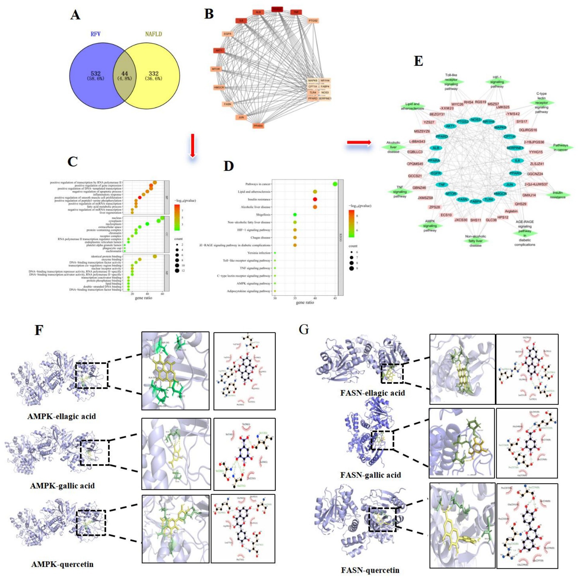
Figure 1. Results of network pharmacology and molecular docking. Venn diagram of potential targets of RFV on NAFLD (A); PPI network diagram of key targets (B); GO function enrichment analysis of potential targets (C); KEGG pathway enrichment of potential targets (D); Compound-key target-pathway visualization network (E); The molecular docking diagram of AMPK and ellagic acid, gallic acid, quercetin. AMPK-ellagic acid (−9.1 kcal/mol), gallic acid (−5.9 kcal/mol), and quercetin (−8.7 kcal/mol) (F); The molecular docking diagram of FASN and ellagic acid, gallic acid, quercetin. FASN-ellagic acid (−8.2 kcal/mol), gallic acid (−6.6 kcal/mol), and quercetin (−8.4 kcal/mol) (G).
3.2 Prediction of potential pathways based on targets
The 20 intersecting targets were analyzed for GO functional annotation and KEGG pathway using the DAVID (version 6.8.0) database. The results were visualized on the Wei Sheng Xin Cloud Platform. GO results indicated that biological process (BP) included inflammatory responses, fatty acid metabolism, and liver regeneration (Figure 1C). The proteins encoded by the intersecting genes were primarily located in the nucleus, cytoplasm, and nucleoplasm. Molecular function (MF) primarily involves the binding of proteins, enzymes, and lipids. The results of KEGG pathway analysis are illustrated in Figure 1D. The following 11 relevant pathways were selected: lipid and atherosclerosis pathway, AGE-RAGE signaling pathway, AMPK pathway, C-type lectin receptor pathway, insulin resistance, HIF − 1 signaling pathway, toll-like receptor signaling pathway, TNF signaling pathway, alcoholic liver disease, NAFLD, and pathways in cancer. A compound-gene-pathway network diagram was created to visually illustrate the intricate relationships among compounds, targets, and pathways (Figure 1E). The lines in the diagram reflect the relationships among compounds, targets, and pathways. The network diagram further revealed that the active compounds of RFV might improve NAFLD through multiple targets and pathways.
3.3 Molecular docking
Molecular docking results revealed that the key active compounds (quercetin, ellagic acid, and gallic acid) were molecularly docked with core targets associated with the AMPK pathway (AMPK and FASN). Molecular docking results indicated that all three compounds could dock with AMPK and FASN proteins (Figures 1F,G). They all exhibited binding energies of <−5 kcal/mol, indicating a strong binding affinity. The AMPK pathway may be an important target in NAFLD regulation. This was experimentally validated below.
3.4 Effect of RFV on intestinal microbiota in NAFLD mice
The samples were analyzed using Illumina MiSeq high-throughput sequencing to investigate the effect of RFV on the intestinal microbiota composition (n = 6). The Venn diagram illustrates the total and overlapping numbers of operational taxonomic units (OTUs) in each group (Figure 2A). Control, NAFLD, RRJ, RFV-L, and RFV-H groups had 755, 465, 428, 404, and 490 OTUs, respectively. All five groups shared 152 OTUs, indicating the presence of at least 152 symbiotic bacteria common to each group. Furthermore, the data revealed that the Control group had 453 unique OTUs, the NAFLD group had 39 unique OTUs, and RFV-L and RFV-H groups had 39 and 53 unique OTUs, respectively. Alpha diversity estimation analyses revealed that the Chao1 indices (391) in the NAFLD group decreased significantly (p < 0.001) compared to the Control group, whereas the Simpson indices (0.08) did not indicate any significant change. After administration of high-dose RFV, Chao1 indices (465) and Simpson indices (0.30) increased significantly (p < 0.05, p < 0.001) (Figures 2C,D).
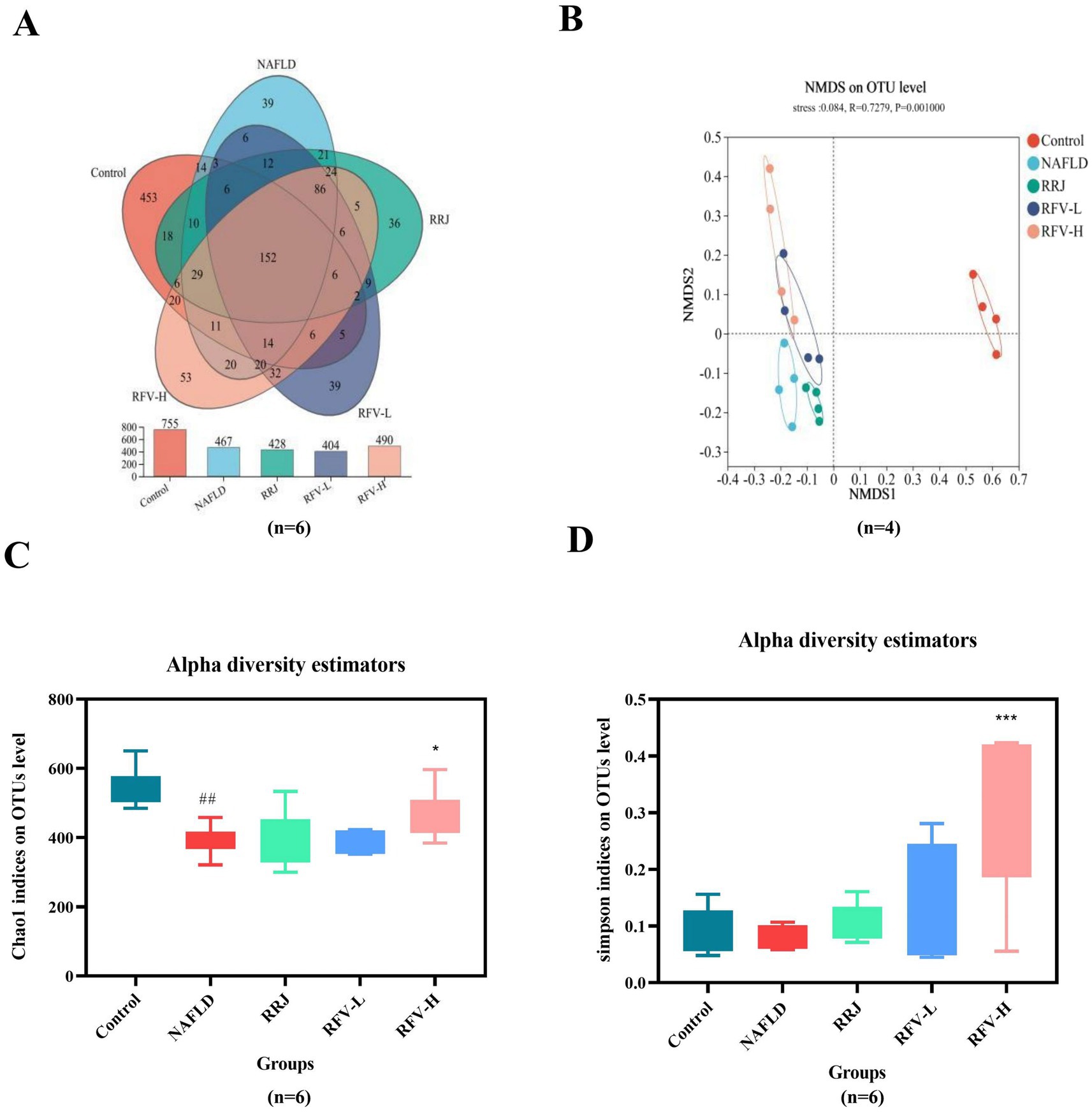
Figure 2. Effect of RFV on intestinal microbiota in NAFLD mice. Venn diagram of OTUs (A); NMDS analysis of β diversity (B); Chao1 indices of Alpha diversity (C); Simpson indices of Alpha diversity (D). The Control, NAFLD, RRJ, RFV-L, and RFV-H were normol diet group, high-fat diet group, Rosa roxburghii juice, low dosage Rosa roxburghii fruit vinegar group, and high dosage Rosa roxburghii fruit group, respectively. #p<0.05, compared with Control group, *p<0.05, compared with NAFLD group (n=6).
The microbial composition significantly changed after HFD and RFV intervention. There was a clear separation between the Control and NAFLD groups. Each administration group was significantly different from the NAFLD group (Figure 2B). The above results indicate that an HFD can alter the structure of the intestinal microbiota in mice, and RFV can improve the situation to a certain extent; therefore, the microbiota structure is closer to that of the Control group. The composition of the intestinal microbiota was analyzed at the phylum and genus levels (Figures 3A,B) and (Supplementary Figures 1A,B). At the phylum level, Firmicutes, Bacteroidetes, and Actinobacteria were dominant in each group. The relative abundances of Firmicutes in Control, NAFLD, RRJ, RFV-L, and RFV-H groups were 55.71, 0.57, 0.32, 1.44, and 4.86%, respectively. The relative abundances of Bacteroidetes were 55.71, 0.57, 0.32, 1.44, and 4.86%, respectively. The NAFLD group exhibited a significant increase in Firmicutes and a significant decrease in Bacteroidetes compared to the Control group (p < 0.001) (Figure 3C). As a result, the ratio of Firmicutes to Bacteroidetes (F/B) was highest in the NAFLD group. After the administration of high-dose RFV, the relative abundance of Firmicutes decreased significantly (p < 0.01), Bacteroidetes increased, and F/B decreased significantly (p < 0.01) (Figure 3D), indicating that RFV could improve the imbalance in microbial abundance caused by NAFLD, and the community composition was reversed to be similar to that of the Control group. Compared to the Control group, the relative abundance of Allobacterium, Faecalibaculum, Coriobacteriaceae_UCG-002, Dubosiella, Blautia, and unclassified_f_Lachnospiraceae increased in the NAFLD group (p < 0.05). Compared to the NAFLD group, RFV-H significantly reduced the relative abundance of Allobaculum, Faecalibaculum, Dubosiella, Blautia, Coriobacteriaceae_UCG-002, and unclassified_f_Lachnospiraceae (Figures 3E-J). Overall, RFV reversed intestinal microbiota disruption caused by NAFLD.
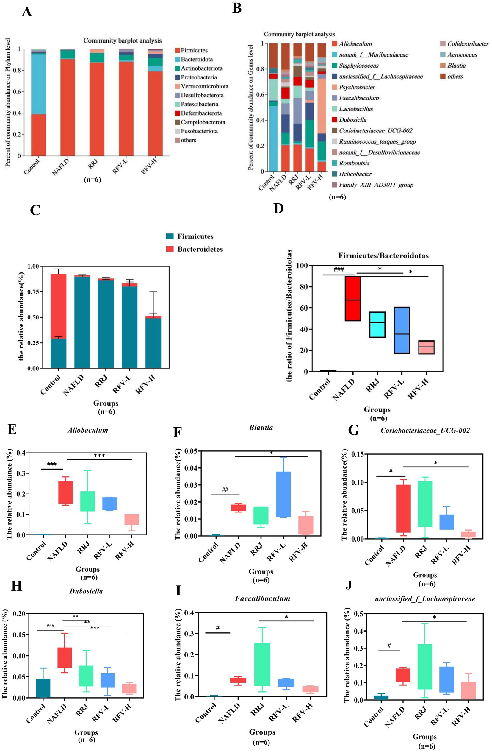
Figure 3. Species composition and bacteria with significant differences in relative abundanceat at the phylum and genus level. Histogram of relative abundance at phylum level (A); Histogram of relative abundance at genus level (B). Relative abundance of Firmicutes and Bacteroidetes (C); Ratio of F to B (F/B) (D); The relative abundance at genus level of Allobaculum (E); Blautia (F); Coriobacteriaceae_UCG-002 (G); Dubosiella (H); Faecalibaculum (I); Unclassified_f_Lachnospiraceae (J). #p<0.05, compared with Control group, *p<0.05, compared with NAFLD group.
3.5 Effect of RFV on liver metabolites in NAFLD mice
Partial least squares discriminant analysis (PLS-DA) demonstrated that liver metabolites were significantly different due to HFD and RFV intervention. The induction of NAFLD significantly changed liver metabolites. After the administration of RRJ and RFV, the metabolite composition was different from that of the NAFLD group and closer to the Control group (Figure 4A). A replacement test was performed using the PLS-DA model (R2Y = 0.64, Q2 = 0.256, p < 0.05), indicating that the model was stable and reliable (Figure 4B).
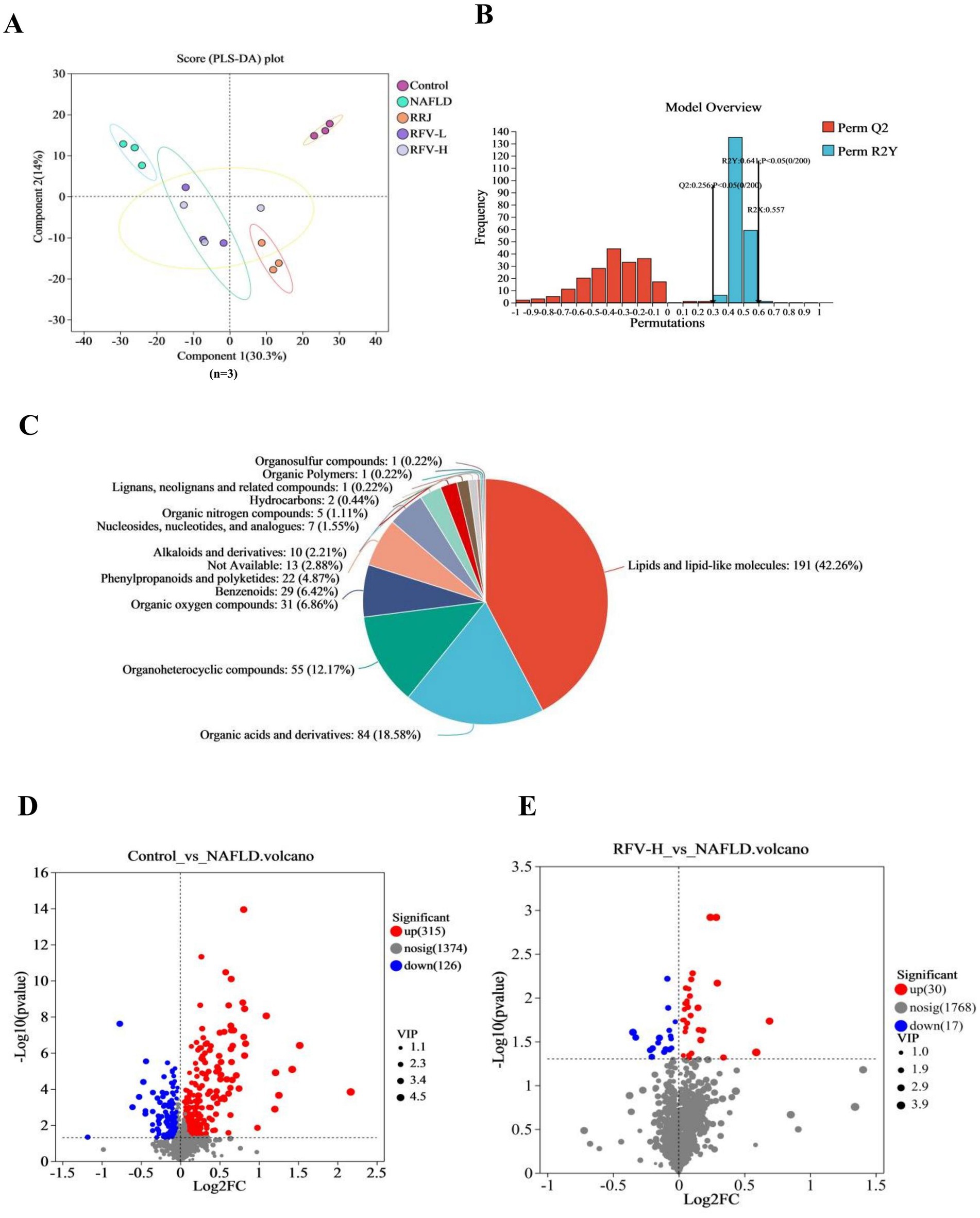
Figure 4. Effect of RFV on liver metabolites in NAFLD mice. PLS-DA score chart (A); PLS-DA displacement test map (B); Pie chart of differential metabolite classification (C); The trend of metabolites change in Control vs. NAFLD (D); The trend of metabolites change in RFV-H vs. NAFLD (E) (n=6).
Enrichment analysis of differential metabolites was performed using metaboAnalyst enrichment analysis. A total of 490 different metabolites were screened among the five groups (p < 0.05). These metabolites included 191 lipids and lipid-like molecules, 84 organic acids and their derivatives, 55 organic heterocyclic compounds, 31 benzenoid compounds, 29 organic oxygen compounds, 22 phenylpropanoids and polyketones, 10 nucleosides, nucleotides, and analogs, 7 alkaloids and their derivatives, 5 organic nitrogen compounds, 5 organic sulfur compounds, 2 hydrocarbon derivatives, 1 hydrocarbon compound, 1 lignin, new signaling, and related compounds, 1 organic polymer, and 13 other metabolites (Figure 4C).
There were 441 and 47 differential metabolites in the Control versus NAFLD group and NAFLD versus RFV-H group (p < 0.05, VIP value > 1, Fold change = 1), respectively. In both positive and negative ion modes, the volcano plot revealed that 315 metabolites were upregulated and 26 metabolites were downregulated in the Control versus NAFLD groups, whereas 30 metabolites were upregulated and 17 metabolites were downregulated in the NAFLD versus RFV-H groups (Figures 4D,E).
3.6 Metabolic pathway analysis
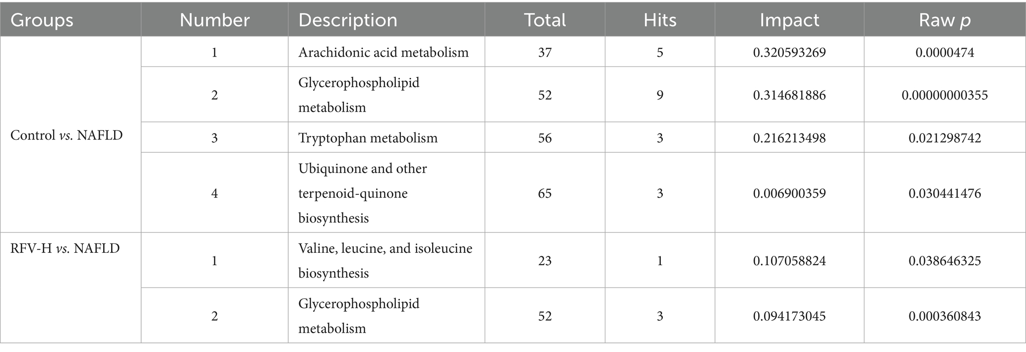
Metabolic sets were created for metabolites of the two comparison groups. KEGG topology analysis was used to examine the pathways associated with the metabolites (Figures 5A,B) and subsequently screened for key pathways based on p-values and impact values (Table 1). Except for metabolic pathways with an impact of 0, 21 metabolic pathways were involved between the Control and NAFLD groups. Arachidonic acid metabolism, glycerophospholipid metabolism, tryptophan metabolism, and the biosynthesis pathways of ubiquinone and other terpenoid quinones were the most significant. The impact values were 0.320, 0.314, 0.216, and 0.007, respectively, with p-values < 0.05.
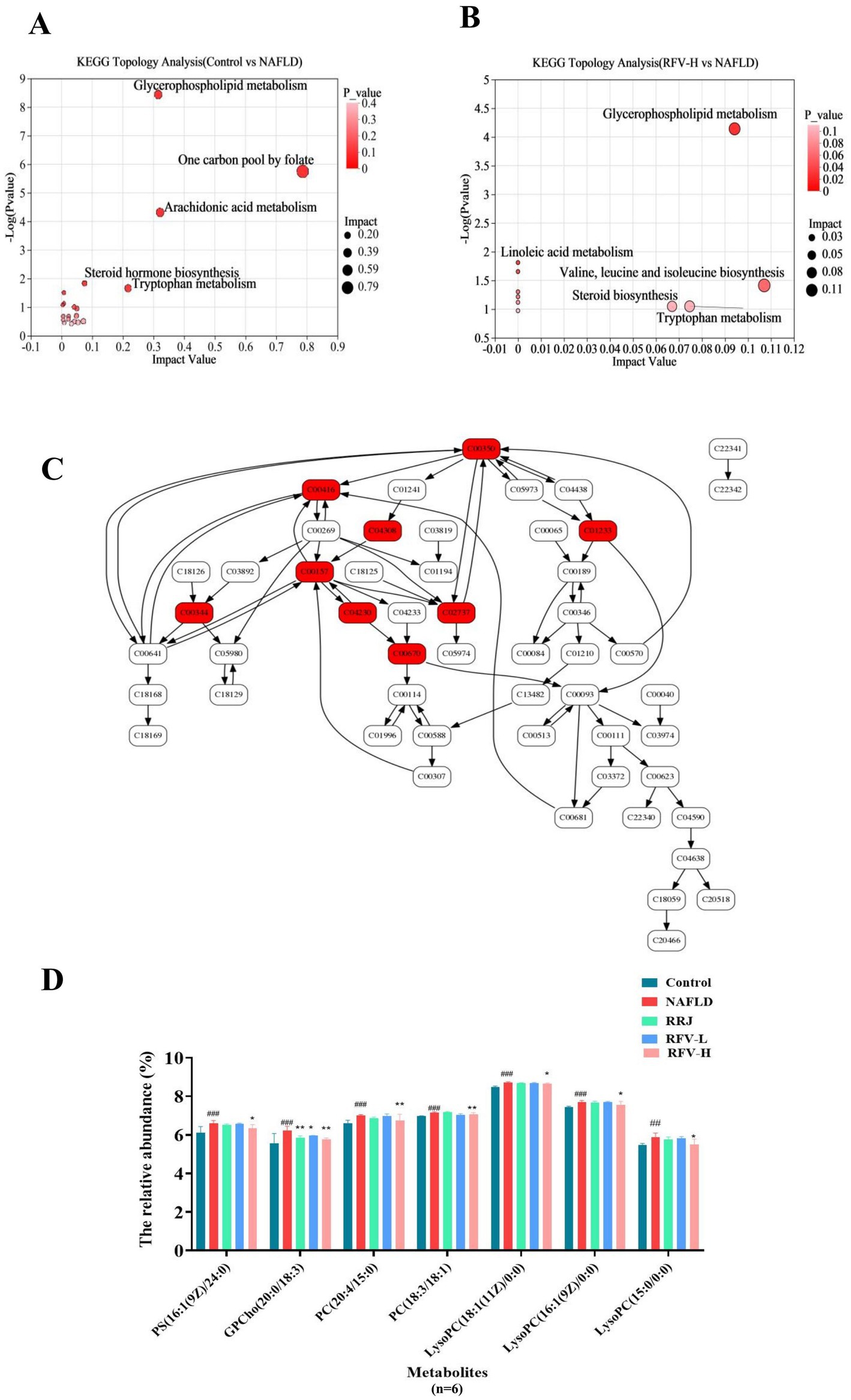
Figure 5. Analysis of enriched pathways of differential metabolites. Topology analysis diagram (A,B); Glycerophospholipid metabolism pathway diagram (C); Histogramgraph of the relative abundance of metabolites in each group (D). #p<0.05, compared with Control group, *p<0.05, compared with NAFLD group. C00350: phosphatidylethanolamine (PE), C00416: phosphatidic acid (PA; 18:0/18:2 (9Z and 12Z)), C04308: PE-NMe2 (14:0/14:0), C01233: Sn-glycoro-3-Phosphatethanolamine, C0015: phosphatidylcholine (PC), C00344: glycerophosphate (PGs), C04230: lysophosphatidylcholine (LysoPC), C02737: Phosphatidylserine (PS; 16:1(9Z)/(24:0)), C00670: Glycerophosphocholine and Glycerylphosphorylcholine.
In the RFV-H group, RFV-H significantly regulated NAFLD through multiple pathways, including valine, leucine, and isoleucine biosynthesis, as well as glycerophospholipid metabolism. The impact values were 0.107 and 0.094, with p < 0.05. The glycerophospholipid metabolic pathway was common to NAFLD and Control, RFV-H groups, suggesting that it is a potential target for RFV intervention in NAFLD. Therefore, the related metabolites are discussed in detail.
The glycerophospholipid metabolic pathway is illustrated in Figure 5C, where the red portion represents significantly different metabolites between the Control and NAFLD groups. The relative abundance of these metabolites was significantly regulated by RFV (Figure 5D) (n = 6). The high-fat diet significantly upregulated the levels of PCs, PS, and LysoPCs (p < 0.001). Following the intervention across various treatment groups, these metabolite levels varied during recovery, with RFV-H exhibiting the most significant effect (p < 0.01).
3.7 Correlation analysis between intestinal microbiota and lipid metabolism and oxidative stress indexes in NAFLD mice
To investigate the correlation between intestinal microbiota and superoxide dismutase (SOD), triglyceride (TG), cholesterol (TC), malondialdehyde (MDA), high-density lipoprotein (HDL-C), and low-density lipoprotein (LDL-C) in NAFLD mice, species were selected for correlation network analysis based on Spearman’s correlation, |r| > 0.6, p < 0.05 (Figures 6A,B). The heatmap of the correlation analysis demonstrated that Firmicutes exhibited a significant positive correlation with MDA, TC, LDL-C, and TG and a negative correlation with SOD. Patescibacteria was negatively correlated with MDA (p < 0.05), TC, LDL-C, and TG (p < 0.05) and positively correlated with SOD (p < 0.05). Bacteroidetes were negatively correlated with MDA (p < 0.001), and Unclassifiedk_norank_d_Bacteria were positively correlated with SOD (p < 0.05). TC and LDL-C exhibited a positive correlation with Allobaculum, Faecalibaculum (p < 0.01, p < 0.05), and Blautia (p < 0.01). TC was positively correlated with Dubosiella (p < 0.01), whereas LDL-C was negatively correlated with norank_f_Muribaculaceae (p < 0.05). MDA levels were positively correlated with Collinsella, Romboutsia, Family_XIII_AD3011_group (p < 0.05), Faecalibaculum, unclassified_f_Lachnospiraceae, Allobaculum, Ruminococcus_torques_group (p < 0.01), and Blautia (p < 0.001), whereas they were negatively correlated with norank_f_Muribaculaceae (p < 0.01). SOD exhibited a negative correlation with Romboutsia, Faecalibaculum, Blautia (p < 0.01), unclassified_f_Lachnospiraceae (p < 0.05), and Allobaculum (p < 0.001) and a positive correlation with norank_f_ Muribaculaceae (p < 0.05). Consequently, a correlation was found between the intestinal microbiota, lipid metabolism, and oxidative stress levels in mice with NAFLD. The main bacterial genera were Allobaculum, Faecalibaculum, Dubosiella, Blautia, unclassified_f_Lachnospiraceae, and norank_f_ Muribaculaceae.
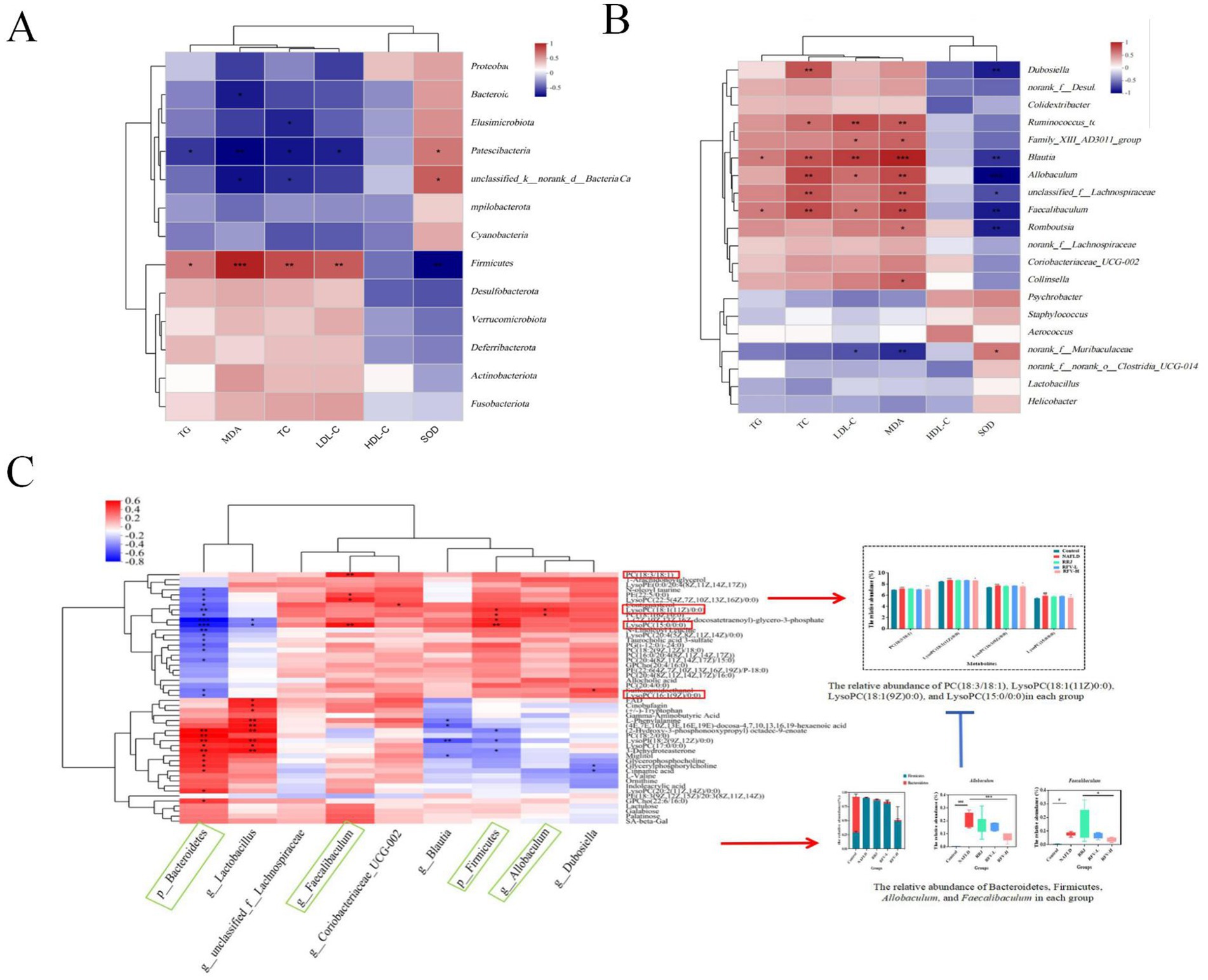
Figure 6. The correlation among intestinal microbiota, liver metabolites, and NAFLD-related indexes based on heatmap correlation analysis. The correlation between Bacteria at each taxonomic level and mouse lipid metabolism and oxidative stress indicators (A,B); The correlation between differential metabolites and differential microflora (C); p ≤ 0.05 is marked as *; p ≤ 0.01 is marked as **; p ≤ 0.001 is marked as ***. Blue box: dominant intestinal microbiota; Red box: significant metabolites.
3.8 Correlation analysis between differential metabolites and intestinal microbiota in NAFLD mice
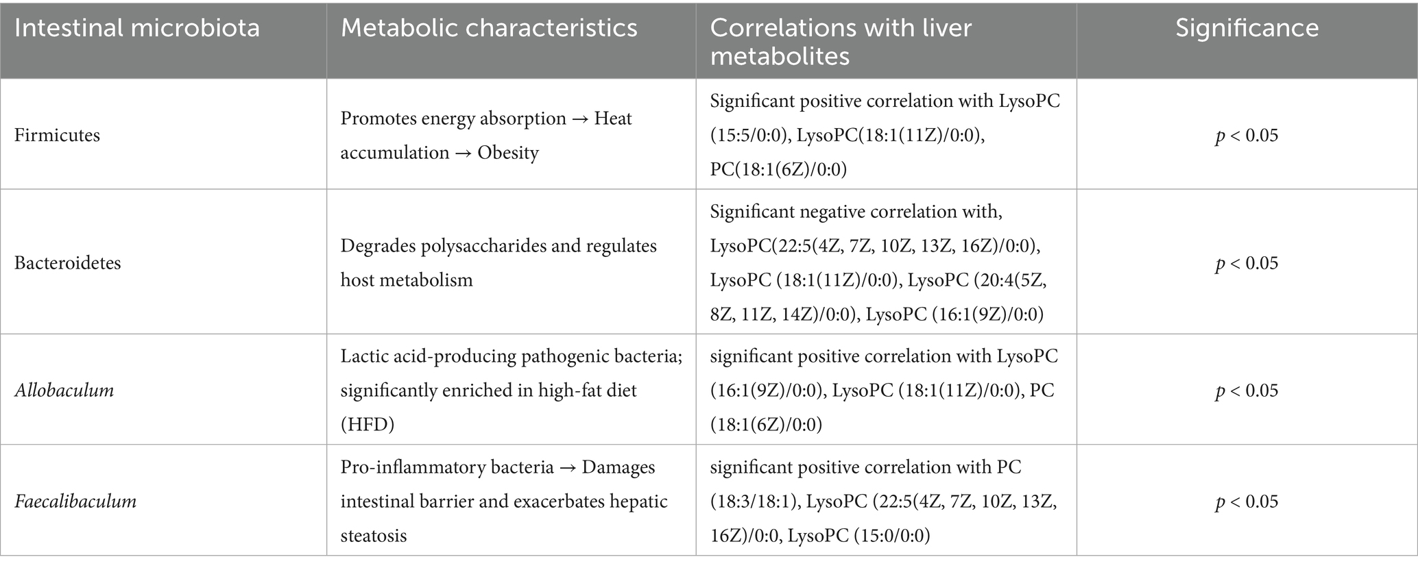
Spearman was used to analyze the correlation between the related bacteria and the differential metabolites affected by RFV (Figure 6C). Glycerophospholipids such as phosphatidylethanolamine: PE (22:5/0:0), phosphatidylcholine: PC (18:1(6Z)/0:0), PC (22:5/0:0), PC (18:2(9Z, 12Z)/18:0), PC (16:0/20:4(8Z, 11Z, 14Z, 17Z)), PC (20:4(8Z, 11Z, 14Z, 17Z)/15:0), lysophosphatidylcholine: LysoPC (22:5(4Z, 7Z, 10Z, 13Z, 16Z)/0:0), LysoPC (18:1(11Z)/0:0), LysoPC (20:4(5Z, 8Z, 11Z, 14Z)/0:0), and LysoPC (16:1(9Z)/0:0) had a significant negative correlation with Bacteroidetes (p < 0.05). Firmicutes had a significant positive correlation with LysoPC (15:5/0:0), LysoPC (18:1(11Z)/0:0), and PC (18:1(6Z)/0:0) (p < 0.05). Faecalibaculum had a significant positive correlation with PC (18:3/18:1), LysoPC (22:5(4Z, 7Z, 10Z, 13Z, 16Z)/0:0), and LysoPC (15:0/0:0) (p < 0.05). Blautia had a significant negative correlation with phenylalanine (p < 0.05), while Allobaculum had a significant positive correlation with LysoPC (18:1(11Z)/0:0) and PC (18:1(6Z)/0:0) (p < 0.05) (see Table 2).
3.9 Effect of RFV on the protein expression of AMPK signaling pathway in NAFLD mice
To further investigate the effect of RFV on the AMPK/FASN/SREBP-1c signaling pathway, Western blotting was performed to measure the expression levels of AMPK, FASN, and SREBP-1c in the liver.
Western blotting results demonstrated that compared to the Control group, the expression of P-AMPK in the NAFLD group (n = 3) was significantly inhibited (p < 0.05), whereas the expressions of SREBP-1c and FASN were significantly increased (p < 0.05) (Figure 7). After administration of RFV, AMPK expression was activated, and P-AMPK was significantly increased (p < 0.05, p < 0.01). Conversely, SREBP-1c and FASN expression levels were significantly reduced. In summary, RFV inhibited the expression of SREBP-1c and FASN by activating P-AMPK in the AMPK signaling pathway, thereby reducing lipid accumulation and improving NAFLD.
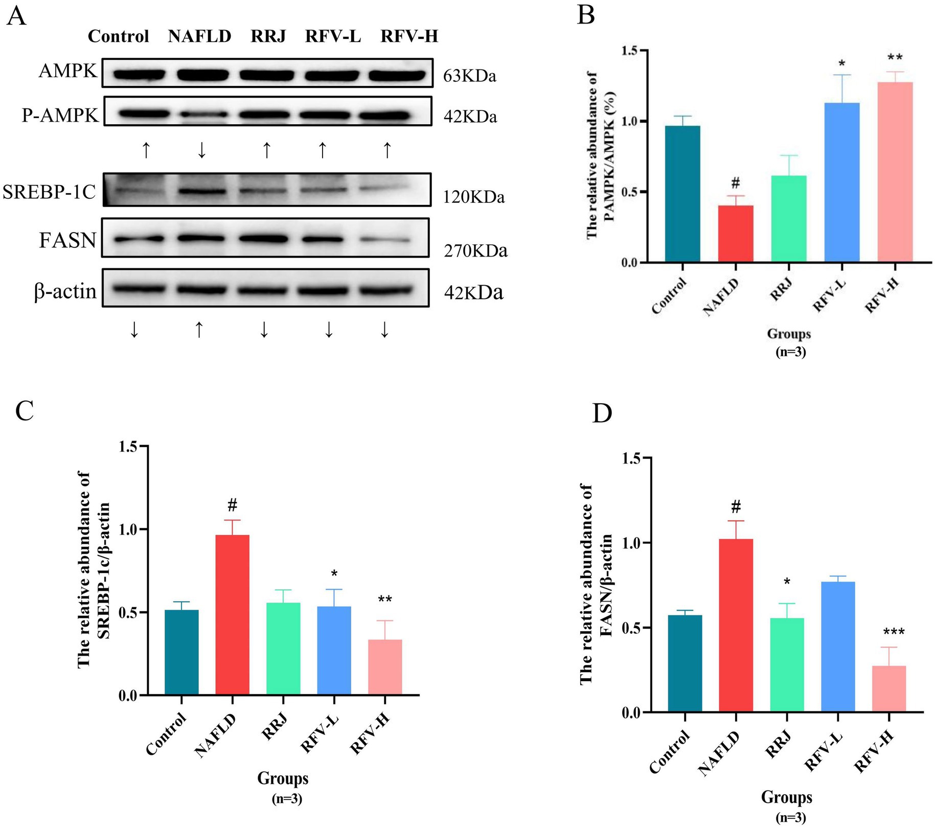
Figure 7. The protein expression and relative abundance of in liver. Protein bands diagram (A); the gray values of P-AMPK/AMPK (B); SREBBP-1c/β -actin (C); FASN/β -actin (D). #p<0.05, compared with Control group, *p<0.05, compared with NAFLD group (n=3).
4 Discussion
NAFLD is a liver metabolic syndrome caused by obesity. Previous studies have demonstrated that RFV improves obesity and its related complications. This effect was achieved by regulating the intestinal microbiota of mice fed a high-fat diet (20). Consequently, a NAFLD mouse model was established using a high-fat diet. The mechanism by which RFV ameliorates NAFLD was further investigated.
The chemical components of RFV are highly diverse. However, the mechanisms by which RFV regulates NAFLD remain unclear. To address this, UPLC-MS was used to characterize the major constituents of RFV, and 70 compounds were identified (32). Based on the compounds identified in RFV, this study used network pharmacology and molecular docking to predict the potential targets and pathways through which RFV might ameliorate NAFLD. These results indicate that RFV may exert its ameliorative effects on NAFLD through these key compounds, including ellagic acid, gallic acid, bayogenin, asiatic acid, α-linolenic acid, and quercetin. Among these, quercetin, ellagic acid, and gallic acid can suppress hepatic lipogenesis and improve hepatic steatosis (35–37). This was achieved by regulating the expression of proteins involved in the AMPK signaling pathway. Based on NAFLD pathogenesis, including lipid accumulation, inflammatory factors, and insulin resistance and its progression (such as obesity, diabetes, liver disease, and cancer) (38), the following 11 relevant pathways were selected: lipid and atherosclerosis pathway, AGE-RAGE signaling pathway, AMPK pathway, C-type lectin receptor pathway, insulin resistance, HIF − 1 signaling pathway, toll-like receptor signaling pathway, TNF signaling pathway, alcoholic liver disease, NAFLD, and pathways in cancer. They regulate the oxidative stress, inflammatory response, and lipid metabolic disorders (39, 40). Furthermore, the HIF-1, toll-like receptor, and TNF signaling pathways are inflammatory pathways that regulate the expression of inflammatory factors such as TNF-α, IL-6, and IL-1β (41, 42). In summary, RFV may improve NAFLD by regulating lipid synthesis via the AMPK pathway.
Numerous studies have demonstrated that the intestinal microbiota and its metabolites influence the occurrence and progression of NAFLD through various mechanisms. To clarify whether the regulation of RFV in NAFLD mice led to changes in intestinal microbiota, the influence of a high-fat diet and RFV on the intestinal microbiota in mice was observed (43). At the phylum level, Firmicutes and Bacteroidetes were the dominant genera, and Firmicutes can produce energy for the body to absorb heat and promote obesity (44). Bacteroidetes play an important role in host metabolism, including the absorption and degradation of polysaccharides (45). F/B is associated with the susceptibility of obesity disease states (45), and F/B can be used an index symbolized as the disorder of metabolic diseases (46). A study reported that the abundance of Firmicutes and the ratio of F/B increased in obese individuals (47). At the genus level, the dominant genera in the Control group were Lactobacillus and g_norank_f_Oscillospiraceae and in the NAFLD group were Faecalibaculum, Blautia, Allobaculum, Dubosiella, Blautia, and unclassified_f_Lachnospiraceae. Faecalibaculum has been reported to be positively associated with serum lipids and the development of NAFLD (48). Allobaculum is a conditioned pathogen that produces lactic acid, which was significantly higher in the HFD group (49). It has been reported that the flora of the Lachnospiraceae family is typically associated with weight gain in obese mice (50). In contrast, Blautia is associated with the development of glucose metabolism disorders and is significantly positively correlated with obesity, inflammation, intestinal permeability, and metabolic endotoxemia (51, 52).
Compared to the NAFLD group, RFV-H significantly increased the relative abundance of these bacteria, with Psychrobacter being the dominant species. Psychrobacter is a beneficial bacterium. It can co-regulate disturbances in intestinal microbiota with other bacteria in high-fat diet-induced type 2 diabetes (53). Numerous studies have revealed that the abundance of Akkermansia is negatively correlated with body weight and obesity in mice and humans (54). In our study, compared to the normal-fed mice (Control group), the relative abundance of Akkermansia increased in high-fat diet-fed mice (NAFLD group) and was higher than in the Control group after intervention with RFV-L and RFV-H, but still lower than that in the NAFLD group. In a study of the effects of Shenling Baizhu Powder on intestinal microbiota in NAFLD mice, the relative abundance of Akkermansia in NAFLD mice and various administration groups was consistent with the results of this study (55). Finally, the correlation analysis revealed that the above bacteria were significantly positively correlated with TC, LDL-C, and MDA and significantly negatively correlated with SOD. Therefore, we speculate that the effects of RFV on liver lipid, liver function impairment, and oxidative stress in NAFLD may be related to regulating the relative abundance of Bacteroidetes, Firmicutes, Allobaculum, Faecalibaculum, Dubosiella, Blautia, and unclassified_f_Lachnospiraceae.
The glycerophospholipid metabolic pathway related to lipid metabolism may be a potential target for RFV intervention in NAFLD. Diacylglycerol produced by TG metabolism is used as a raw material to continue the production of PC or PE in the glycerophospholipid metabolic pathway, and PC can further produce LysoPC (56). Lipid accumulation due to TG is one of the leading causes of NAFLD, which in turn affects the metabolism of PC and PE. Some studies have reported that the plasma levels of PC and PE were disturbed in patients with NAFLD, and the levels of PC and PE in the livers of mice with NAFLD induced by a high-fat diet were significantly increased (57). LysoPC, the main component of LDL-C, induces lipid peroxidation of the cell membrane and leads to inflammation. LysoPC plays an important role in cell membrane processes and mediates signal transduction (58). Moreover, previous studies have demonstrated that PC and LysoPC levels are associated with fat decay, inflammation, and oxidative stress in hepatocytes (59, 60). The results of this study indicate that a high-fat diet increased the content of some metabolites prefixed with LysoPC and PC in the liver. However, the contents of these metabolites decreased significantly and exhibited a trend toward normal levels after RFV-H administration. These results indicated that RFV could alleviate the occurrence and development of NAFLD by regulating the expression of metabolites related to lipid accumulation and energy metabolism in glycerophospholipid metabolism. The correlation analysis (Figure 7D) revealed a significant interaction between intestinal microbiota and liver metabolites. Specifically, the abundance of phosphatidylcholine (PC) and lysophospholipid (LPC) metabolites was significantly associated with intestinal microbiota dysbiosis, which was characterized by an increased relative abundance of Firmicutes and a decreased of Bacteroidetes. Notably, Firmicutes showed significant positive correlations with PC (22:5/0:0), LysoPC (18:1(11Z)/0:0), and PC (18:1(6Z)/0:0). Bacteroidetes showed significant negative correlations with LysoPC (22:5(4Z,7Z,10Z,13Z,16Z)/0:0), LysoPC (18:1(11Z)/0:0), LysoPC (20:4(5Z,8Z,11Z,14Z)/0:0), and LysoPC (16:1(9Z)/0:0). Our research findings indicate that RFV mitigates NAFLD through intestinal microbiota–metabolite axis. Importantly, alterations in the intestinal microbiota act as the driving force influencing hepatoprotective metabolites, thereby ultimately reducing steatosis and oxidative stress.
Through non-targeted metabolomics, it was revealed that RFV may mitigate lipid metabolism dysregulation in NAFLD mice by modulating the glycerophospholipid metabolism pathway (Figures 5A,B). Importantly, metabolites such as phosphatidylcholines (PC) and lysophosphatidylcholine (LPC) within lipid metabolism pathways were significantly influenced by RFV (Figure 5D). These metabolites or derivatives of gut microbial metabolites served as energy substrates for intestinal and hepatic tissues while concurrently enhancing intestinal secretory functions and lipid metabolism (61, 62). From an integrated metabolic perspective, lipid metabolism constitutes a principal mechanism through which RFV ameliorates NAFLD-associated dysregulation. Nevertheless, the molecular signaling pathways by which these metabolites induce lipid metabolic disorders remain to be elucidated.
In the glycerophospholipid metabolic pathway, differential metabolites primarily participate in fatty acid synthesis, oxidation, and degradation. They are directly regulated by PPARα. On the one hand, the activity of PPARα is regulated by adenosine 5′-monophosphate (AMP)-activated protein kinase (AMPK) (63). AMPK activation inhibits downstream targets fatty acid synthase (FASN) and sterol element regulatory protein (SREBP-1c), regulating hepatic lipid metabolism disorders (64, 65). On the other hand, based on network pharmacology and literature analysis, the active compounds in RFV may improve NAFLD. These compounds could activate the AMPK/SREBP-1c/FASN lipid synthesis pathway and reduce lipid accumulation (66). Therefore, we proposed that RFV ameliorated lipid metabolism dysregulation via the AMPK signaling pathway. Then, further analysis of the protein expression levels of FASN and SREBP-1C in lipid synthesis confirmed the results of this study. RFV was observed to activate AMPK phosphorylation and reduce the expression levels of FASN and SREBP-1c (Figure 7). The mechanism by which RFV improves lipid metabolism disorders in high-fat diet mice is by activating the AMPK pathway, regulating the expression of downstream target proteins, weakening fatty acid synthesis, and promoting fatty acid breakdown.
This study demonstrated RFV-mediated regulation of intestinal microbiota via 16S rRNA analysis and predicted multi-target effects against NAFLD through network pharmacology. As a functional food, RFV has been proven to be effective in improving NAFLD. Nonetheless, the investigation did not address the impact on the intestinal acid–base environment and the milieu of intestinal epithelial cells, thereby neglecting potential effects on essential functions such as nutrient absorption efficiency, gut barrier integrity, and mucosal immune responses. The experimental validation was confined to the AMPK signaling pathway and did not encompass the other prediction pathways, such as insulin resistance and PPARα. The real-time luminal pH mapping coupled with single-cell transcriptomic profiling of the intestinal epithelium can be integrated into future studies. This dual-axis approach will provide insights into how acid–base dynamics influence the gut–microbe–immune interface, particularly through pH-sensitive receptors such as GPR65 and electrolyte transporters such as NHE3. In addition, it is essential to detect the expression levels of intestinal epithelial tight junction proteins, such as Occludin, Claudins, and ZO-1, to further elucidate the mechanisms underlying the “gut–liver” axis. Insulin signaling and PPARα pathways will be validated to elucidate multi-target mechanisms of RFV.
In conclusion, as a functional food, RFV has been proven to be effective in improving NAFLD. The underlying mechanisms involve the modulation of the intestinal microbiota and metabolites balance and regulation on lipid disorders through AMPK signaling pathway. These findings not only furnish a theoretical foundation for the development and application of RFV but also propose a novel approach for the improvement of NAFLD by dietary therapy.
Data availability statement
The datasets presented in this study can be found in online repositories. The names of the repository/repositories and accession number(s) can be found in the article/Supplementary material.
Ethics statement
The animal study was approved by Guizhou Medical University (SYXK (Gui) 2023-0002). The study was conducted in accordance with the local legislation and institutional requirements.
Author contributions
YuW: Conceptualization, Formal analysis, Funding acquisition, Investigation, Methodology, Writing – original draft. RL: Conceptualization, Data curation, Funding acquisition, Methodology, Writing – original draft. LL: Data curation, Investigation, Writing – original draft. YoW: Data curation, Investigation, Writing – original draft. MG: Data curation, Investigation, Writing – original draft. JZ: Data curation, Investigation, Writing – original draft. JY: Data curation, Investigation, Writing – original draft. QL: Data curation, Investigation, Writing – original draft. LW: Data curation, Investigation, Writing – original draft. XY: Data curation, Investigation, Writing – original draft. XW: Funding acquisition, Resources, Writing – original draft, Writing – review & editing. CX: Funding acquisition, Resources, Writing – original draft, Writing – review & editing.
Funding
The author(s) declare that financial support was received for the research and/or publication of this article. This research was funded by the Science and Technology Projects of Guizhou Province (QKHCG[2024]YB095; QKHCG[2024]YB087; QKHZC[2023]YB472; QKHCG[2022]YB025), the Central Government-guided Local Science and Technology Development Fund (2025) (QKHZYD[2025]014), and the Guizhou Technical Innovation Center of Cili Industrial (QKHPTJSZX[2025]006).
Conflict of interest
XW was employed by China National Research Institute of Food & Fermentation Industries Co., Ltd.
The remaining authors declare that the research was conducted in the absence of any commercial or financial relationships that could be construed as a potential conflict of interest.
Generative AI statement
The author(s) declare that no Gen AI was used in the creation of this manuscript.
Publisher’s note
All claims expressed in this article are solely those of the authors and do not necessarily represent those of their affiliated organizations, or those of the publisher, the editors and the reviewers. Any product that may be evaluated in this article, or claim that may be made by its manufacturer, is not guaranteed or endorsed by the publisher.
Supplementary material
The Supplementary material for this article can be found online at: https://www.frontiersin.org/articles/10.3389/fnut.2025.1617931/full#supplementary-material
Footnotes
1. ^http://www.swisstargetprediction.ch/
2. ^https://bioinfogp.cnb.csic.es/tools/venny/
4. ^https://david.ncifcrf.gov/
References
1. Chen, G, and Kan, J. Characterization of a novel polysaccharide isolated from Rosa roxburghii Tratt fruit and assessment of its antioxidant in vitro and in vivo. Int J Biol Macromol. (2018) 107:166–74. doi: 10.1016/j.ijbiomac.2017.08.160
2. He, JY, Zhang, YH, Ma, N, Zhang, XL, Liu, MH, and Fu, WM. Comparative analysis of multiple ingredients in Rosa roxburghii and R. Sterilis fruits and their antioxidant activities. J Funct Foods. (2016) 27:29–41. doi: 10.1016/j.jff.2016.08.058
3. Xu, F, Zhu, Y, Lu, M, Zhao, D, Qin, L, and Ren, T. Exploring the mechanism of browning of Rosa roxburghii juice based on nontargeted metabolomics. J Food Sci. (2023) 88:1835–48. doi: 10.1111/1750-3841.16534
4. Chen, C, Tan, S, Ren, T, Wang, H, Dai, X, and Wang, H. Polyphenol from Rosaroxburghii Tratt fruit ameliorates the symptoms of diabetes by activating the P13K/AKT insulin pathway in db/db mice. Foods. (2022) 11:636. doi: 10.3390/foods11050636
5. Chen, F, Li, L, Peng, M, Yan, Y, Wang, L, Li, L, et al. Identification of triterpenoids and hepatoprotective property of Fructus Rosa roxburghii against alcohol-induced liver injury by regulating keap1- Nrf2 signaling. Phytomed Plus. (2021) 1:100102. doi: 10.1016/j.phyplu.2021.100102
6. Wang, L, Wang, L, Zhang, Z, Ma, M, Wang, R, Qian, M, et al. Genome-wide identification and comparative analysis of the superoxide dismutase gene family in pear and their functions during fruit ripening. Postharvest Biol Technol. (2018) 143:68–77. doi: 10.1016/j.postharvbio.2018.04.012
7. Wang, L, Wei, T, Zheng, L, Jiang, F, Ma, W, Lu, M, et al. Recent advances on main active ingredients, pharmacological activities of Rosa roxbughii and its development and utilization. Foods. (2023) 12:1051. doi: 10.3390/foods12051051
8. Wu, H, Li, M, Yang, X, Wei, Q, Sun, L, Zhao, J, et al. Extraction optimization, physicochemical properties and antioxidant and hypoglycemic activities of polysaccharides from roxburgh rose (Rosa roxburghii Tratt.) leaves. Int J Biol Macromol. (2020) 165:517–29. doi: 10.1016/j.ijbiomac.2020.09.198
9. Morikawa, T, Chaipech, S, Matsuda, H, Hamao, M, Umeda, Y, Sato, H, et al. Anti-hyperlipidemic constituents from the bark of Shorea roxburghii. J Nat Med. (2012) 66:516–24. doi: 10.1007/s11418-011-0619-6
10. Benattia, FK, Arrar, Z, Dergal, F, and Khabbal, Y. Synergistic effect, and therapeutic potential of aqueous prickly pear extract. In vivo neuroleptic, catatonic, and hypoglycemic activity. Curr Chem Biol. (2023) 17:216–25. doi: 10.2174/0122127968263950231127051454
11. Chen, Z, Zhu, Y, Lu, M, Yu, L, Tan, S, and Ren, T. Effects of Rosa roxburghii Tratt glycosides and quercetin on D-galactose-induced aging mice model. J Food Biochem. (2022) 46:e14425. doi: 10.1111/jfbc.14425
12. Canaviri-Paz, P, Oscarsson, E, Kjellström, A, Olsson, H, Jois, C, and Håkansson, Å. Effects on microbiota composition after consumption of quinoa beverage fermented by a novel xylose-metabolizing L. plantarum strain. Nutrients. (2021) 13:3318. doi: 10.3390/nu13103318
13. Chaturvedi, S, and Chakraborty, S. Optimization of extraction process for legume-based synbiotic beverages, followed by their characterization and impact on antinutrients. Int J Gastron Food Sci. (2022) 28:100506. doi: 10.1016/j.ijgfs.2022.100506
14. Shang, YF, Cao, H, Ma, YL, Zhang, C, Ma, F, Wang, CX, et al. Effect of lactic acid bacteria fermentation on tannins removal in Xuan Mugua fruits. Food Chem. (2019) 274:118–22. doi: 10.1016/j.foodchem.2018.08.120
15. Beh, BK, Mohamad, NE, Yeap, SK, Ky, H, Boo, SY, Chua, JYH, et al. Anti-obesity and anti-inflammatory effects of synthetic acetic acid vinegar and Nipa vinegar on high-fat-diet-induced obese mice. Sci Rep. (2017) 7:6664. doi: 10.1038/s41598-017-06235-7
16. Samad, A, Azlan, A, and Ismail, A. Therapeutic effects of vinegar: a review. Curr Opin Food Sci. (2016) 8:56–61. doi: 10.1016/j.cofs.2016.03.001
17. Xia, T, Zhang, B, Duan, W, Zhang, J, and Wang, M. Nutrients and bioactive components from vinegar: a fermented and functional food. J Funct Foods. (2020) 64:103681. doi: 10.1016/j.jff.2019.103681
18. Mohamad, NE, Yeap, SK, Ky, H, Ho, WY, Boo, SY, Chua, J, et al. Dietary coconut water vinegar for improvement of obesity-associated inflammation in high-fat-diet-treated mice. Food Nutr Res. (2017) 61:1368322. doi: 10.1080/16546628.2017.1368322
19. Hosoda, S, Kawazoe, Y, Shiba, T, Numazawa, S, and Manabe, A. Anti-obesity effect of ginkgo vinegar, a fermented product of ginkgo seed coat, in mice fed a high-fat diet and 3T3-L1 preadipocyte cells. Nutrients. (2020) 12:230. doi: 10.3390/nu12010230
20. Li, J, Zhang, J, Zhang, Y, Shi, Y, Feng, D, Zuo, Y, et al. Effect and correlation of Rosa roxburghii Tratt fruit vinegar on obesity, dyslipidemia and intestinal microbiota disorder in high-fat diet mice. Foods. (2022) 11:4108. doi: 10.3390/foods11244108
21. Xiao, P, Ye, Z, Li, X, Feng, Q, and Su, Y. Ginseng and its functional components in non-alcoholic fatty liver disease: therapeutic effects and multi-target pharmacological mechanisms. Front Pharmacol. (2025) 16:1540255. doi: 10.3389/fphar.2025.1540255
22. Guo, X, Yin, X, Liu, Z, and Wang, J. Non-alcoholic fatty liver disease (NAFLD) pathogenesis and natural products for prevention and treatment. Int J Mol Sci. (2022) 23:15489. doi: 10.3390/ijms232415489
23. Friedman, SL, Neuschwander-Tetri, BA, Rinella, M, and Sanyal, AJ. Mechanisms of NAFLD development and therapeutic strategies. Nat Med. (2018) 24:908–22. doi: 10.1038/s41591-018-0104-9
24. Kumavath, R, Pavithran, H, Paul, S, Anju, VT, Busi, S, and Dyavaiah, M. Effects of gut microbiome and obesity on the development, progression and prevention of cancer. Int J Oncol. (2024) 64:4. doi: 10.3892/ijo.2023.5592
25. Kolodziejczyk, AA, Zheng, D, Shibolet, O, and Elinav, E. The role of the microbiome in NAFLD and NASH. EMBO Mol Med. (2019) 11:e9302. doi: 10.15252/emmm.201809302
26. Schnackenberg, LK, Kaput, J, and Beger, RD. Metabolomics: a tool for personalizing medicine? Per Med. (2008) 5:495–504. doi: 10.2217/17410541.5.5.495
27. Lin, H. Animal experiment and serum metabolomics to reveal the improvement effect of Wudan lipid-reducing prescription on metabolic associated fatty liver disease in rats [Master’s Thesis]. Hengyang: University of South China (2022).
28. Coyral-Castel, S, Rame, C, Fatet, A, and Dupont, J. Characterization of AMP-activated protein kinase (AMPK) in goat ovary. Ljubljana: University of Ljubljana (2009).
29. Boudaba, N, Marion, A, Huet, C, Pierre, R, Viollet, B, and Foretz, M. AMPK re-activation suppresses hepatic steatosis but its downregulation does not promote fatty liver development. EBioMedicine. (2018) 28:194–209. doi: 10.1016/j.ebiom.2018.01.008
30. Cheung, PCF, Salt, IP, Davies, SP, Hardie, DG, and Carling, D. Characterization of AMP-activated protein kinase γ-subunit isoforms and their role in AMP binding. Biochem J. (2000) 346:659–69. doi: 10.1042/bj3460659
31. Wang, Y, Li, W, Yang, X, Li, L, Yang, J, Zhang, C, et al. Roxburgh rose enzymatic fruit vinegar and preparation process and application there of China patent Guizhou Saisi pear health industry co ltd. and key Laboratory of Natural Product Chemistry of Guizhou Academy of Sciences, [Beijing] (2022).
32. Lang, R, Wang, Y, Wen, Y, Li, L, Yan, Y, Yang, X, et al. Regulatory effects of Rosa roxburghii Tratt. Fruit vinegar on nonalcoholic fatty liver disease. Sci Technol Food Ind. (2025). doi: 10.13386/j.issn1002-0306.2024110039
33. Xia, QS, Gao, Y, Wu, WB, Wu, F, Dong, H, Xu, LJ, et al. Ban-xia-xie-xin-tang ameliorates hepatic steatosis by regulating Cidea and Cidec expression in HFD-fed mice. Phytomedicine. (2022) 105:154351. doi: 10.1016/j.phymed.2022.154351
34. Zhao, D, Cao, J, Jin, H, Shan, Y, Fang, J, and Liu, F. Beneficial impacts of fermented celery (Apium graveolens L.) juice on obesity prevention and gut microbiota modulation in high-fat diet fed mice. Food Funct. (2021) 12:9151–64. doi: 10.1039/D1FO00560J
35. Altamimi, JZ, Alshammari, GM, AlFaris, NA, Alagal, RI, Aljabryn, DH, Albekairi, NA, et al. Ellagic acid protects against non-alcoholic fatty liver disease in streptozotocin-diabetic rats by activating AMPK. Pharm Biol. (2022) 60:25–37. doi: 10.1080/13880209.2021.1990969
36. Katsaros, I, Sotiropoulou, M, Vailas, M, Papachristou, F, Papakyriakopoulou, P, Grigoriou, M, et al. The effect of quercetin on non-alcoholic fatty liver disease (NAFLD) and the role of Beclin1, P62, and LC3: an experimental study. Nutrients. (2024) 16:4282. doi: 10.3390/nu16244282
37. Lu, Y, Zhang, C, Song, Y, Chen, L, Chen, X, Zheng, G, et al. Gallic acid impairs fructose-driven de novo lipogenesis and ameliorates hepatic steatosis via AMPK-dependent suppression of SREBP-1/ACC/FASN cascade. Eur J Pharmacol. (2023) 940:175457. doi: 10.1016/j.ejphar.2022.175457
38. Tilg, H, and Moschen, AR. Evolution of inflammation in nonalcoholic fatty liver disease: the multiple parallel hits hypothesis. Hepatology. (2010) 52:1836–46. doi: 10.1002/hep.24001
39. Zhou, Y, Fu, Z, Dou, P, Yu, C, Dong, X, Hong, H, et al. Decoding nattokinase efficacy: from digestion and absorption to lipid pathway modulation in high-fat diet-induced atherosclerosis. Food Biosci. (2025) 66:106203. doi: 10.1016/j.fbio.2025.106203
40. Farrag, EAE, Hammad, MO, Safwat, SM, Hamed, S, and Hellal, D. Artemisinin attenuates type 2 diabetic cardiomyopathy in rats through modulation of AGE-RAGE/HMGB-1 signaling pathway. Sci Rep. (2023) 13:11043. doi: 10.1038/s41598-023-37678-w
41. Tan, S, Gu, J, Yang, J, Dang, X, Liu, K, Gong, Z, et al. L-theanine mitigates acute alcoholic intestinal injury by activating the HIF-1 signaling pathway to regulate the TLR4/NF-κB/HIF-1α axis in mice. Nutrients. (2025) 17:720. doi: 10.3390/nu17040720
42. Ruengthanoo, PM, Changthong, A, Sriraj, P, Prathumtet, J, Laikaew, N, and Aukkanimart, R. Anti-inflammatory effects of Andrographis paniculata (fah Talai Jone) via TNFα-JNK pathway and bioactive compound identification. Phytomed Plus. (2025) 5:100720. doi: 10.1016/j.phyplu.2024.100720
43. Safari, Z, and Gérard, P. The links between the gut microbiome and non-alcoholic fatty liver disease (NAFLD). Cell Mol Life Sci. (2019) 76:1541–58. doi: 10.1007/s00018-019-03011-w
44. Komaroff, AL. The microbiome and risk for obesity and diabetes. JAMA. (2017) 317:355–6. doi: 10.1001/jama.2016.20099
45. Kim, M, and Benayoun, BA. The microbiome: an emerging key player in aging and longevity. Transl Med Aging. (2020) 4:103–16. doi: 10.1016/j.tma.2020.07.004
46. Stojanov, S, Berlec, A, and Štrukelj, B. The influence of probiotics on the firmicutes/bacteroidetes ratio in the treatment of obesity and inflammatory bowel disease. Microorganisms. (2020) 8:1715. doi: 10.3390/microorganisms8111715
47. Xu, P, Li, M, Zhang, J, and Zhang, T. Correlation of intestinal microbiota with overweight and obesity in Kazakh school children. BMC Microbiol. (2012) 12:283. doi: 10.1186/1471-2180-12-283
48. Mu, H, Zhou, Q, Yang, R, Zeng, J, Li, X, Zhang, R, et al. Naringin attenuates high fat diet induced non-alcoholic fatty liver disease and gut bacterial dysbiosis in mice. Front Microbiol. (2020) 11:585066. doi: 10.3389/fmicb.2020.585066
49. Zheng, Z, Lyu, W, Ren, Y, Li, X, Zhao, S, Yang, H, et al. Allobaculum involves in the modulation of intestinal ANGPTLT4 expression in mice treated by high-fat diet. Front Nutr. (2021) 8:690138. doi: 10.3389/fnut.2021.690138
50. Kameyama, K, and Itoh, K. Intestinal colonization by a Lachnospiraceae bacterium contributes to the development of diabetes in obese mice. Microbes Environ. (2014) 29:427–30. doi: 10.1264/jsme2.ME14054
51. Duan, R, Guan, X, Huang, K, Zhang, Y, Li, S, Ja, X, et al. Flavonoids from whole-grain oat alleviated high-fat diet-induced hyperlipidemia via regulating bile acid metabolism and gut microbiota in mice. J Agric Food Chem. (2021) 69:7629–40. doi: 10.1021/acs.jafc.1c01813
52. Zhao, Q, Hou, D, Fu, Y, Xue, Y, Guan, X, and Shen, Q. Adzuki bean alleviates obesity and insulin resistance induced by a high-fat diet and modulates gut microbiota in mice. Nutrients. (2021) 13:3240. doi: 10.3390/nu13093240
53. Chen, J, Han, Y, Sun, E, Sun, J, Kang, X, Zhu, Z, et al. Bifidobacterium lactis MN-gup dairy products and its application in improving type 2 diabetes China patent Inner Mongolia Mengniu dairy group co ltd. [Beijing] (2023).
54. Everard, A, Belzer, C, Geurts, L, Ouwerkerk, JP, Druart, C, Bindels, LB, et al. Cross-talk between Akkermansia muciniphila and intestinal epithelium controls diet-induced obesity. Proc Natl Acad Sci USA. (2013) 110:9066–71. doi: 10.1073/pnas.1219451110
55. Zhang, Y, Tang, K, Deng, Y, Chen, R, Liang, S, Xie, H, et al. Effects of shenling baizhu powder herbal formula on intestinal microbiota in high-fat diet-induced NAFLD rats. Biomed Pharmacother. (2018) 102:1025–36. doi: 10.1016/j.biopha.2018.03.158
56. Zhou, Y, Dai, Z, Deng, K, Wang, Y, Ying, J, Chu, D, et al. Eight Zhes decoction ameliorates the lipid dysfunction of nonalcoholic fatty liver disease using integrated lipidomics, network pharmacology and pharmacokinetics. J Pharm Anal. (2023) 13:1058–69. doi: 10.1016/j.jpha.2023.05.012
57. Park, SH, Lee, JE, Lee, SM, Lee, J, Seo, CS, Hwang, GS, et al. An unbiased lipidomics approach identifies key lipid molecules as potential therapeutic targets of Dohongsamul-tang against non-alcoholic fatty liver diseases in a mouse model of obesity. J Ethnopharmacol. (2020) 260:112999. doi: 10.1016/j.jep.2020.112999
58. Han, MS, Park, SY, Shinzawa, K, Kim, S, Chung, KW, Lee, JH, et al. Lysophosphatidylcholine as a death effector in the lipoapoptosis of hepatocytes. J Lipid Res. (2008) 49:84–97. doi: 10.1194/jlr.M700184-JLR200
59. Han, X. Lipidomics for studying metabolism. Nat Rev Endocrinol. (2016) 12:668–79. doi: 10.1038/nrendo.2016.98
60. Kakisaka, K, Cazanave, SC, Fingas, CD, Guicciardi, ME, Bronk, SF, Werneburg, NW, et al. Mechanisms of lysophosphatidylcholine-induced hepatocyte lipoapoptosis. Am J Physiol Gastrointest Liver Physiol. (2012) 302:G77–84. doi: 10.1152/ajpgi.00301.2011
61. Arowolo, F, Pierre, JF, Blaser, M, and Shanmuganayagam, D. Longitudinal effects of dietary oxidized lipids on the gut microbiome and mycobiome in pigs. FASEB J. (2020) 34:1. doi: 10.1096/fasebj.2020.34.s1.09676
62. Sarin, SK, Pande, A, and Schnabl, B. Microbiome as a therapeutic target in alcohol-related liver disease. J Hepatol. (2019) 70:260–72. doi: 10.1016/j.jhep.2018.10.019
63. Zhang, J, Zhang, W, Yang, L, Zhao, W, Liu, Z, Wang, E, et al. Phytochemical gallic acid alleviates nonalcoholic fatty liver disease via AMPK-ACC-PPARa axis through dual regulation of lipid metabolism and mitochondrial function. Phytomedicine. (2023) 109:154589. doi: 10.1016/j.phymed.2022.154589
64. Fang, C, Pan, J, Qu, N, Lei, Y, Han, J, Zhang, J, et al. The AMPK pathway in fatty liver disease. Front Physiol. (2022) 13:970292. doi: 10.3389/fphys.2022.970292
65. Viollet, B, Athea, Y, Mounier, R, Guigas, B, Zarrinpashneh, E, Horman, S, et al. AMPK: lessons from transgenic and knockout animals. Front Biosci. (2009) 14:19–44. doi: 10.2741/3229
66. Hardie, DG, Ross, FA, and Hawley, SA. AMPK: a nutrient and energy sensor that maintains energy homeostasis. Nat Rev Mol Cell Biol. (2012) 13:251–62. doi: 10.1038/nrm3311
Glossary
NAFLD - non-alcoholic fatty liver disease
NASH - non-alcoholic steatohepatitis
RRJ - Rosa Roxburghii Tratt. juice
RFV - Rosa Roxburghii Tratt. fruit vinegar
RFV-L - low-dose RFV (group)
RFV-H - high-dose RFV (group)
GO - Gene Ontology
KEGG - Kyoto Encyclopedia of Genes and Genomes
OTUs - operational taxonomic units
F/B - Firmicutes to Bacteroidetes
SOD - superoxide dismutase
TG - triglycerides
TC - total cholesterol
MDA - malondialdehyde
HDL-C - high-density lipoprotein
LDL-C - low-density lipoprotein
PLS-DA - partial least squares discriminant analysis
PA - phosphatidic acid
PC - phosphatidylcholine
GPC - glycerophosphate
LysoPC - lysophosphatidylcholine
PS - phosphatidylserine
AMPK - phosphorylation of AMP-activated protein kinase
FASN - fatty acid synthase
SREBP-1c - sterol regulatory element-binding protein 1
Keywords: Rosa roxburghii Tratt., fruit vinegar, non-alcoholic fatty liver disease, intestinal microbiota, metabolomics, AMPK signal pathway
Citation: Wang Y, Lang R, Li L, Wen Y, Gao M, Zhang J, Yang J, Li Q, Wang L, Yang X, Wang X and Xie C (2025) Regulation mechanism of Rosa roxburghii Tratt. (Cili) fruit vinegar on non-alcoholic fatty liver disease. Front. Nutr. 12:1617931. doi: 10.3389/fnut.2025.1617931
Edited by:
Muhammad Ijaz Ahmad, Zhejiang University, ChinaCopyright © 2025 Wang, Lang, Li, Wen, Gao, Zhang, Yang, Li, Wang, Yang, Wang and Xie. This is an open-access article distributed under the terms of the Creative Commons Attribution License (CC BY). The use, distribution or reproduction in other forums is permitted, provided the original author(s) and the copyright owner(s) are credited and that the original publication in this journal is cited, in accordance with accepted academic practice. No use, distribution or reproduction is permitted which does not comply with these terms.
*Correspondence: Chunzhi Xie, eGN6MDYxMUB4eml0LmVkdS5jbg==; Xiaolong Wang, MTM4MTA5NDcyMTFAMTYzLmNvbQ==
 Yu Wang1,2
 Rui Lang
 Jie Zhang
 Qiji Li
 Xiaosheng Yang
 Chunzhi Xie