 Ayesha Anwar1,2
 Muhammad Imran1*
 Muhammad Farooq Warsi1
 Ibrahim A. Alsafari3
 Roberto Parra-Saldívar4,5
 Guadalupe Gutiérrez-Soto5
 Hafiz M. N. Iqbal5*
- 1Institute of Chemistry, Baghdad-ul-Jadeed Campus, The Islamia University of Bahawalpur, Bahawalpur, Pakistan
- 2Department of Chemistry, Emerson University, Multan, Pakistan
- 3Department of Biology, College of Science, University of Hafr Al-Batin, Hafar Al-Batin, Saudi Arabia
- 4Magan Centre of Applied Mycology, Cranfield University, Cranfield, United Kingdom
- 5Biomolecular Innovation Group, Laboratorio de Ciencias Naturales, Facultad de Agronomía, Universidad Autónoma de Nuevo León, General Escobedo, Nuevo Leon, Mexico
Due to its unique properties and inherent biocompatibility, chitosan (CH), a multifunctional biopolymer derived from chitin, has garnered significant interest in deployment in various scientific domains. The Food and Drug Administration (FDA) authorized CH to employ an injury remedy and a nutritional supplement. Furthermore, CH has facilitated advancements in numerous biological applications, particularly nano-carriers and scaffolds for tissue engineering. It is an ideal choice for wound care because of its hemostatic, antioxidant, and antimicrobial properties. The hydrophilic nature of CH makes it a perfect precursor. This review focuses on the advent of chitosan-based nanostructures, highlighting their physicochemical characteristics, methods for structural modification, and the functionalization of chitosan into its derivatives, which may aid in understanding its benefits and cellular significance. It has been demonstrated that CH nanostructures offer remarkable encapsulation efficiency and extended-release patterns in drug delivery, resulting in higher therapeutic efficacy and fewer side effects. Furthermore, due to their mucoadhesive properties, they are particularly well-suited for transdermal drug delivery. Nanostructures based on CH exhibited optimum activity in biosensing and diagnostic imaging. The potential of CH to interact with targeting ligands enhances the early detection of disease and integration of CH in focused imaging modalities. Moreover, CH variable surface chemistry facilitates attachment to biological entities, resulting in improved diagnostic accuracy, rendering the insertion of bioactive substances possible. Furthermore, the degradable nature of CH offers a minimal long-term impact, alleviating challenges related to ecological sustainability. As long as CH-modified nanostructures have become prevalent in healthcare fields and researchers strive to explore novel and more effective uses, medical care will advance, and a range of health problems will be resolved. This review provides a comprehensive overview of the current status of CH-based nanostructures in the bio-medical field, highlighting their potential to revolutionize therapeutic and diagnostic methodologies. In conclusion, several perspectives on its potential are presented, including new approaches to alterations, directed modification through the association between framework and operation, and the path towards growth for activities and implementations.
1 Introduction
Naturally occurring chitosan (CH) is a polysaccharide of linear structure extracted from chitin either through enzyme-mediated hydrolysis of chitin deacetylase or solid-state deacetylation in an alkaline environment. Concerning diffusion and employment, it is regarded as the second-largest sustainable biomaterial after cellulose (Abd Elgadir et al., 2015). CH and its derivative biomaterials have recently garnered much interest in biomedical sciences owing to their distinctive biological traits (Kravanja et al., 2019). Non-toxicity, degradation, bio-compatibility, immune-enhancing, antitumor, antiseptic, and antimicrobial effects of CH constitute a few of its most well-known attributes in the therapeutic environment. CH biological degradation has been demonstrated both in-vivo and in-vitro, when aggregates disintegrated into several smaller subunit fragments (Pang et al., 2017). CH is a multifunctional poly-cationic, comprised of N-acetyl-β-(1–4)-D-glucosamine and β-(1–4)-D-glucosamine (Virmani et al., 2023). According to Jang CH exhibits crystal structures of α, β, and
In recent decades, CH, a polymeric material that is simultaneously recyclable and environmentally friendly, has been the subject of numerous studies. Additionally, CH has paved opportunities for advancements in various biological applications, especially scaffolds for biological tissue engineering and nano-carriers (Bashir et al., 2022). Due to its antimicrobial, antioxidant, and hemostatic features, it is an excellent alternative for wound care products (Okamoto et al., 2003). CH is an ideal template for building bio-compatible and bio-degradable hydrogels due to its hydrophilic nature (Busilacchi et al., 2013). CH can be synthesized from chitin employing various techniques, including layer-by-layer (LBL) assembly, fermentation by microbes, solvent-based casting, and alkaline and enzymatic deacetylation (Khubiev et al., 2023). Instead of using CH, different nanostructures such as metal oxides, magnetic nanomaterials, molecular organic frameworks, bi-polymeric nanomaterial (BPn), plant extract, scaffolds, and functionalized graphene, fullerenes, nanorods, etc. have been incorporated into the CH to optimize its effectiveness in a variety of applications (Khubiev et al., 2023; Sreena and Nathanael, 2023; Seyedkhani et al., 2023; Jiang M. et al., 2023). Covalent bonding, hydrogen bonding, and electrostatic interactions are only a few reactions that can quickly alter CH to overcome its weak mechanical attributes and immersion problems. Chemical modification, new networks of polymers, and overall insertion of micron-sized particles, such as carbon-based, polymeric, and inorganic nanoparticles, can all be categorized as modification techniques (Feng and Wang, 2022).
CH and its analogs have drawn significant interest in the biomedical field as a result of their peculiar physiological characteristics, as well as anti-inflammatory, anti-coagulant, anti-allergic, anti-HIV, anti-hypertensive, anti-Alzheimer’s, anti-diabetic, anti-obesity, and anti-cancer activities (Laskar and Rauf, 2017). CH has naturally occurring primary amino groups on its surface, making it a popular material for fabricating polysaccharide-based targeted nano-carriers (Antil, 2023). Such molecules can interact with ligands, binders for additional coupling, polymer chains (e.g., polyethylene glycol), or N-octyl-O, N-carboxmethyl chitosan that has been modified by octreotide to achieve an intended degree of dissolution (Laskar and Rauf, 2017). The minimal solubility of CH in aqueous solutions hinders its potential uses. CH functionality is adaptable, enabling improvements to its unique features (Ahmed and Ikram, 2016). CH derivatives or counterparts that are more compatible, less hazardous, and reversible are produced whenever the basic structure of CH is being altered chemically (Antil, 2023). CH oligomers are generally soluble in basic and acidic media. Still, when the molecular weight of these molecules rises, molecules can only be dispersed throughout acidic media despite more extensive rates of deacetylation (Ashrafizadeh et al., 2023). Consequently, much research has gone towards building CH derivatives readily soluble in neutral and basic pH environments via acetylation, polymerization, and quaternization (Aranaz et al., 2010). Protonation of the NH2 group influences CH immersion in acidic conditions, with a pKa value of 6.5, reportedly demonstrated (Ashrafizadeh et al., 2023; Chattopadhyay and Inamdar, 2010).
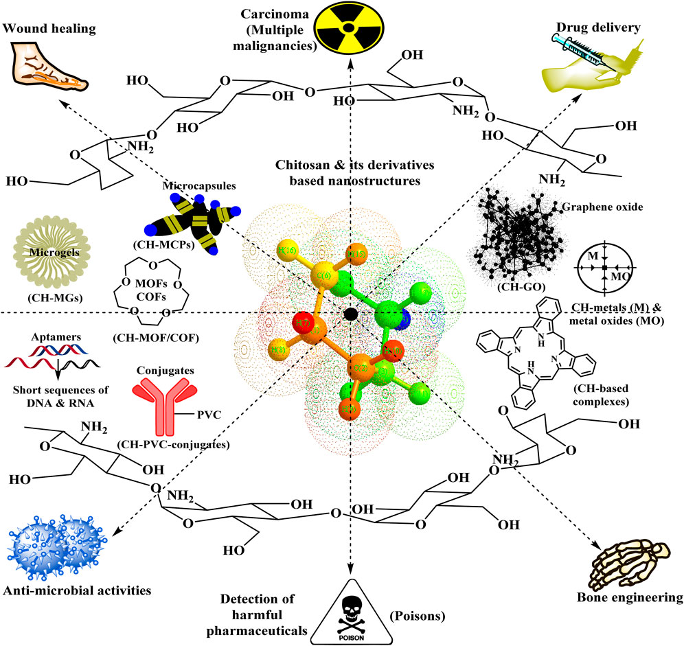
Two key benefits associated with nano-scale dimensions are the vast surface area of nanomaterials (NMs) relative to their volume and their capacity to enter the cytoplasm (increased intracellular assimilation) (Amor et al., 2023a). Both the pharmacokinetics and pharmacodynamics of the therapeutic agent might be altered through the incorporation of therapeutic substances into CH- NPs, a process that will boost drug accessibility and chemical stability while minimizing its toxicological profile (Amor et al., 2023b; Ben Amor et al., 2022). This impact may be reinforced by mixing CH with restorative materials to create chitosan-based nanostructures. Reverse micellization, reverse emulsion (Brunel et al., 2008), desolvation (Kim et al., 2014), emulsified solvent dissemination (El-Shabouri, 2002), or electrostatic complexation processes that lead to chitosan-linked tiny structures (Pelegrino et al., 2017). Based on recent literature, the article provides an overview of the current state of CH applications. It highlights the significance of basic and applied research targeted at expanding the use of chitosan-based biomaterials in various scientific fields. One of the accomplishments of scientific advancement in the search for new promising materials in recent years is the manufacturing, investigating, and implementing CH-related nanostructures in many physiological domains to combat multiple medical conditions and improve the quality of life. This review aims to describe chitosan-nanomaterials’ (CH-NMs) potential utility for biological purposes, encompassing vaccine development, drug transport, tissue restoration, and other wholly disclosed usage.
2 Modifications of chitosan structure/chemistry of chitosan and its derivatives
Chitosan is chemically modified to enhance its ability to dissolve, rheological features, thermal integrity, and oxidation resilience (Huang et al., 2018). Active groups in the CH molecular structure include amino and hydroxyl at C-3 and C-6 positions, respectively. Since there is unconstrained rotation, the NH2-(amino) group is often more reactive than the C-6 primary hydroxyl group (-OH), and the initial hydroxyl group tends to be more reactive than the C-3 secondary -OH moiety (Fatullayeva et al., 2022). To develop N-, O-, or N, O-modified chitosan derivatives, amino, hydroxyl, or both amino and hydroxyl groups can be chemically modified in chitosan (Dimassi et al., 2018; Sahariah and Másson, 2017). Chemical interactions involving OH-groups include etherification, esterification, cross-linkages, graft co-polymerization, complexation with poly-anions, and O-acetylation; on the other hand, NH2- substituents undergo acetylation, quaternization, Schiff’s base responses, and grafting, in addition to chemical, physical, and enzymatic de-polymerization (Fatullayeva et al., 2022; Wang and Liu, 2014), as illustrated in Figures 1–3.
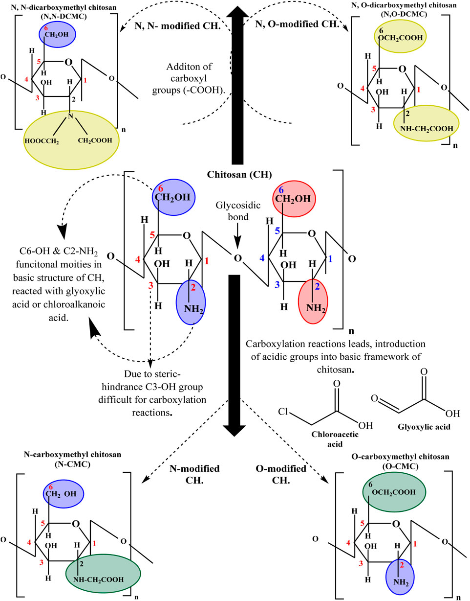
Figure 1. Carboxylation reactions of chitosan to functionalize into -O, -N, -N-O and -N-N- carboxymethyl chitosan derivatives.
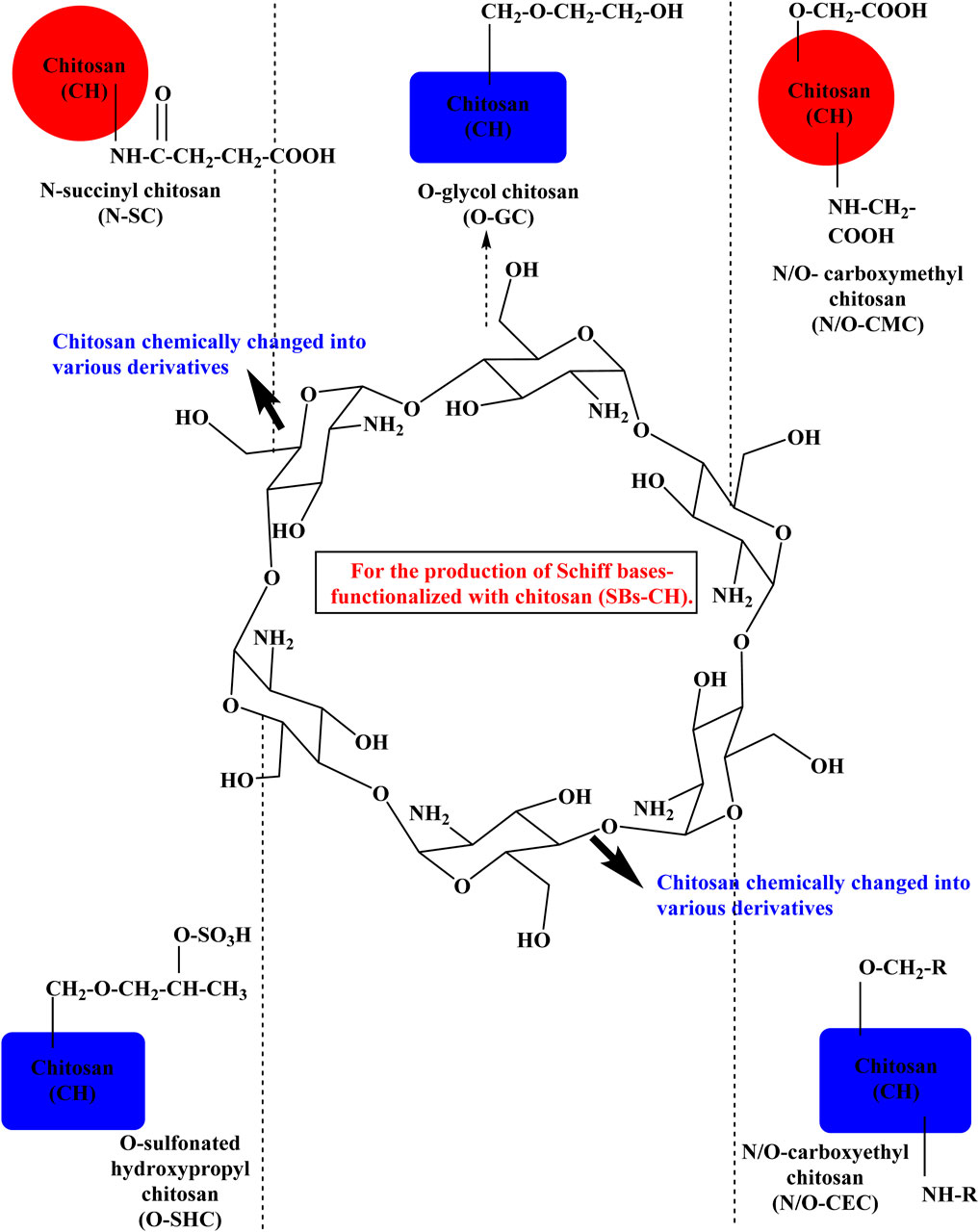
Figure 2. Fabrication of Schiff bases conjugated with chitosan (SBs-CH) to optimize their therapeutic effects.
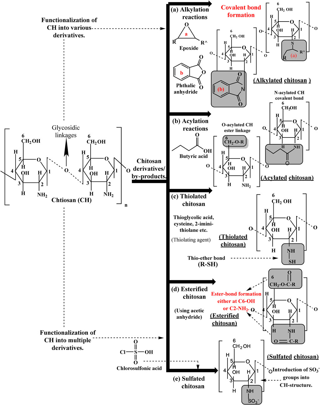
Figure 3. Chemistry of chitosan and its metabolites exhibited multifunctional aspects in bio-medical disciplines.
2.1 Effect of alkylation and acylation reactions on the chitosan structure towards chemical susceptibility
CH bio-polymer transformations can be accomplished via alkylation and acylation operations by utilizing halogen-containing hydrocarbons, anhydrides, and acid halides as acylating agents in a particular reaction media (Prakash and Viswanathamurthi, 2014; Ma et al., 2009). The native crystal structure of CH is altered by synthesized chemical substances, which disrupt the hydrogen bonds that connect the molecules, increasing chitosan permeability and broadening its applications (Ma et al., 2009; Wang and Wang, 2011; Adetunji, 2025; Pawariya et al., 2024). Alkylation introduces alkyl chains—typically alkyl halides like methyl, ethyl, or propyl—to the chitosan molecule. Alkyl groups are often added to the CH chemical framework to substitute hydrogen atoms to attain adhesion (Wang et al., 2020a). Significant nucleophilicity and lone-pair electrons in the chitosan amino group lead to an alkylation reaction on the chitosan molecule’s O or N. Consequently, an N-alkylation reaction is more likely than an O-alkylation reaction (Prakash and Viswanathamurthi, 2014). Generally, an aldehyde and the amino group of chitosan react to form a Schiff base, which is subsequently utilized in a reduction reaction to yield N-alkylation products (Wang et al., 2020a). Increased hydrophobicity, improved chemical endurance, and reduced reactivity in terms of tailored or regulated drug release in numerous drug delivery mechanisms are some of the consequences of an alkylation reaction on CH (Lunkov et al., 2023; Chen W.-C. et al., 2023). Acylation reaction entails adding acyl groups—typically acetic, propionic, or butyric—to the chitosan polymer. This is usually accomplished by reacting molecules with acid chlorides or anhydrides (Sahariah et al., 2023). One of the two possible substrates for the acylation reaction is N (N-acylation) or O (O-acylation), which forms a covalent and ester bond, respectively. Because the CH amino group is more active than its hydroxyl group, the process of acylation reaction preferentially favors the amino group (Kurita et al., 2002; Wan Yusof et al., 2023). Structurally modified CH has several impacts, such as enhanced hydrophilic properties, antibiotic loading, and release characteristics in pharmaceutical formulations frequently altered by acylated CH (Wan Yusof et al., 2023; Tang W. et al., 2023). Acylation also strengthens CH’s mechanical capabilities, which makes it more useful in transplantation and wound rehabilitation treatments (Zhang M. et al., 2023).
2.2 Quaternization reactions modify the basic structure of chitosan in conjunction with multiple processes
Among the many different types of chitosan derivatives is quaternized CH. In various reaction media, the primary NH2-groups quaternize, enhancing CH mucosal adhesion and hydrophilic qualities. Compared to unaltered chitosan, quaternization dramatically improves bioavailability across a much broader pH and variation in concentration by maintaining the positive charge of the chitosan at neutral pH. N, N, N-trimethyl chitosan (TMC) is the most basic type of quaternized chitosan (Frigaard et al., 2022). Quaternization significantly modifies CH bio-polymers, employing a free amino group in the chitosan (Liu et al., 2013). It includes saturating the amino group of the chitosan with small-molecule quaternary ammonium salts or quaternary ammonium groups. Such assemblies exhibit strong steric constraints and, therefore, can sustain a lot of moisture (Ling et al., 2015). Peng et al. exploited chitin and (3-chloro-2-hydroxypropyl) trimethyl-ammonium chloride at room temperature to develop a water-soluble chitin-quaternary ammonium salt. The cationic salt of ammonium has been substituted to a degree of 0.3–0.5 (Peng et al., 2016). The synthesized CH-salt showed excellent antibacterial activities, which also served as a buoyant or lubricant (Wang et al., 2020a).
2.3 Thiolated chitosan
Chemically coupling sulfhydryl-bearing molecules, i.e., glutathione, thioglycolic acid, or cysteine, with the chitosan backbone yields thiolated-CH. Features of adhesion are modified by thiolated-CH mediated assembly of disulfide subunits with polymeric chains and the glycoproteins that are part of the mucus substrate (Chen et al., 2013). Modified CH exhibited exceptional features, notably optimum efficacy in drug delivery systems and other applications in biology and medicine.
Thiol substituents in thiolated CH can combine with additional thiol groups or molecules to generate covalent disulfide (S-S) bonds. Certain circumstances, such as reducing chemicals like glutathione, may trigger disulfide bonds to dissolve (Federer et al., 2020). Thiol groups in mucosal tissues interact with mucin and other molecules containing thiols, enabling thiolated chitosan to stick onto these surfaces (Tekade, 2019). With different biological molecules (DNA, proteins, pharmaceuticals, etc.), thiol structures induce electrostatic interactions and covalent bonding, which can be advantageous for drug encapsulation and gene delivery (Leonard et al., 2023). By cross-linking thiol functional moieties, hydrogels (used in healing injuries, regrowth of tissue, etc.) can be constructed from thiolated CH. The chain-like structure is formed in 3-dimensions via this cross-linking (Noreen and Bernkop-Schnürch, 2023; Liu F. et al., 2023). Cross-linked CH is less cytotoxic than non-cross-linked CH because of the positively charged amino group in the non-cross-linked thiolated chitosan can connect to the negatively charged cell membrane in a cytotoxic way (Noi et al., 2018). The formulation exhibits reduced cytotoxicity due to the neutralization of the positively charged surface in the cross-linked thiolated chitosan (Pistone et al., 2017).
2.4 Carboxylation reactions of chitosan into multiple derivatives
Carboxylation involves introducing acidic substances within CH’s basic structure to increase the product’s soluble content, moisturizing, and film-forming capabilities and diversify its spectrum of applications (Fonseca-Santos and Chorilli, 2017). C6–OH or C2–NH2 CH groups react with glyoxylic acid or chloroalkanoic acid in a carboxylation process, yielding a −COOH group as the end product (Mohammadi et al., 2019), as shown in Figure 1. Carboxy-methylation is the primary method for studying chitosan carboxylation processes (Fonseca-Santos and Chorilli, 2017). Chitosan can be chemically changed into carboxymethyl chitosan (CMC) to increase its affinity for water permeability (Li N. et al., 2022). This can be achieved by incorporating negatively charged carboxyl groups into the NH2 group of glucosamine units or C-6 hydroxyl groups (Yu and Li, 2023; Dumont et al., 2016). Since CMC compounds have both cationic and anionic groups, they have been hypothesized to be poly-ampholytic (Manna et al., 2023). Due to the steric hindrance effect, carboxy-methylation of the C3-OH position in chitosan seems complicated. As a result, the C6-OH group is predominantly involved in the carboxylation mechanism. The C6–OH group exerts a more significant molecular influence on chitosan than the C2–NH2 group might in alkaline conditions (Wang et al., 2020b; Bai et al., 2019). The carboxymethyl substitution cascade is C6-OH > C3-OH > C2-NH2. It is possible to produce chitosan with different reaction parameters and materials after undergoing N-, O-, or N-O-carboxy-methylation (Wang et al., 2020a). O-carboxymethylated chitosan can occur in an ice bath or at room temperature if sodium hydroxide and monochloroacetic acid are added, along with isopropanol/water as a solvent (Jayakumar et al., 2010). N-carboxy-methylation and N, O-carboxy-methylation are primarily induced by elevated temperatures. Via reducing sodium cyanoborohydride and chitosan with glyoxylic acid, N-carboxy-methylation and N, N-carboxy-methylation can be generated. Moreover, direct alkylation may also obtain N-carboxymethylated compounds (Fonseca-Santos and Chorilli, 2017).
2.5 Schiff bases (SBs) reactions with chitosan to alter its functionalities (SBs-CH)
Reacting chemically with Schiff bases (SBs) is one method of altering the structural properties and functionalities of chitosan, a bio-polymer produced from chitin. Chemicals known as SBs have a double bond between nitrogen and carbon. They are commonly known as azomethines or imines. When synthesized using chitosan, SBs can introduce new functional categories or modify the chemical composition of the material. This alteration could provide chitosan analogs with unique benefits for various technologies (Antony et al., 2019). Bio-polymeric amphiphilic SBs anchored in multiple molecular weight chitosan matrices modified with salicylaldehyde and glycidol emerged after interfacing with many metal complexes to increase the biological activity of chitosan for diagnostic or therapeutic purposes in multiple types of tumors (Barbosa et al., 2020).
The standard method to make SBs is to condense the amino groups of chitosan with the carbonyl residues of either ketones or aldehydes while eliminating any water molecules. In 1977, S. Hirano et al. initially synthesized the first SB by treating chitosan using multiple aldehydes in a solvent of acetic acid and methanol (Hirano et al., 1977). Various research groups subsequently documented a large number of SBs. The preferred solvent medium for synthesizing SBs at ambient or refluxing temperature settings is frequently acetic acid, ethanol, methanol, or a mixture of hydrocarbons (Antony et al., 2019). Additionally, SBs have been developed into specialized topologies such as void cylinders, filaments, and microspheres; nevertheless, achieving these architectures might demand an innovative synthetic methodology (Zou et al., 2015; Nada et al., 2014). Occasionally, spin coating is employed for producing SBs-CH (Schiff bases-CH) films (Ren et al., 2017). To produce water-soluble SBs-CH, chitosan can be chemically changed into succinyl chitosan (SC) (Lü et al., 2010), glycol chitosan (GC), carboxymethyl chitosan (CMC) (Baran et al., 2015), carboxyethyl chitosan (CEC) (Qu et al., 2017), and sulfonated hydroxypropyl chitosan (SHPC) (Liu T.-M. et al., 2017), as depicted in Figure 2. Schiff base-modified chitosan SB-CH has several possible alterations and applications, such as enhanced solubility, microbial activity, cross-linking, controlled dissolution systems, and hypo-allergenic surfaces (Antony et al., 2019; Zhang Z. et al., 2023).
2.6 Esterification reactions/esterified-chitosan (ECH) derivatives
The molecular structure of CH consists of -NH2 and -OH groups. Such functional groups and carboxylic acids, i.e., acetic acid (CH3COOH), hydrochloric acid (HCl), perchloric acid (HClO4), etc., are capable of forming ester linkages. Several inorganic acids, especially those that include oxygen, can aid in the esterification of chitosan by acting as reactants or catalysts in the reaction (Santhosh and Bhatt, 2024). During the response, -NH2 attacks the carbonyl carbon of the acid (nucleophilic attack), forming an amide intermediate. The resulting intermediate tends to react further with the hydroxyl group to produce an ester bond (Worch et al., 2021). The insertion of ester groups may influence CH dissolution, charge, and numerous other aspects (Spriano et al., 2023). The degree of esterification, regardless of how significantly CH can be altered, is regulated by tweaking reaction factors, especially the amount of carboxylic acid in the mixture and the overall reaction’s time frame (Bajer, 2023; Huang et al., 2023). Esterified chitosan (ECH) has applications in medicine, including enhancing drug delivery systems and the dispersion of poorly water-soluble therapies (Zhao et al., 2018). ECH is a valuable support material in biotechnology, microbiology, and bioinformatics that can be used to immobilize enzymes and other bio-molecules (Misra and Pathak, 2022; Cheng et al., 2018).
The esterification reaction between chitosan and maleic anhydride is the initial strategy for rendering chitosan readily soluble in water. Resulting derivative represented as (chitosan maleic anhydride) CSM—is permeable. Meanwhile, the CH backbone is reinforced with C=C bonds and carboxylic groups. CSM will retain the majority of CH qualities while also having a few additional intriguing features (Xiao, 2023). Hydrogel composed of polydiacetylene-zinc oxide-carboxymethyl chitosan-hydroxyethyl cellulose (PDA-ZnO-CMCs-HEC) was assessed for its pH responsiveness, colorimetric transitions, temperature profile, crystal structure, functional group modifications upon cross-linking, and microbial inhibitory action (Madivoli et al., 2023).
2.7 Sulfonation/sulfated-chitosan (SCH)
The sulfonation procedure entails adding sulfonate groups to the CH polymer. Usually, to accomplish this, CH reacts with sulfur-trioxide (SO3) or chloro-sulfonic acid, two potent sulfonating agents. In the CH framework, sulfonate residues (-SO3-) substitute the precedence of particular amino groups (-NH2) attributable to a sulfonating reagent (Dimassi et al., 2018; Dehghankar et al., 2023). The extent of sulfonation can be controlled by modulating reaction variables, i.e., duration, humidity, and sulfonating chemical dosage (Hamza et al., 2023). Sulfated-chitosan (SCH) retains the basic glucosamine and N-acetyl-glucosamine repeating units that compose the CH structure. Amino and sulfonate groups link throughout the CH backbone via strong ionic interactions between these groups, i.e., NH2 and SO3− (Chopin et al., 2014; El Sayed, 2023). These groups could be primary or secondary sulfonates depending on the specific reaction conditions. Due to the negative electrostatic charge of sulfonate residues, SCH (sulfated chitosan) is a poly-electrolyte (Dimassi et al., 2018; Ranjani et al., 2019). SCH is bio-compatible, meaning it does not cause any discernible unfavorable reactions in the human body and can be assimilated by it. The inclusion of SO3− bonds raises the water solubility of CH. One beneficial utilization of this characteristic is the development of water-based formulations for medical equipment, such as medicine delivery mechanisms and wound dressings (Dimassi et al., 2018; Campelo et al., 2016). It renders SCH easier to disperse in aqueous solutions. SCH is a viable encapsulation material for therapeutic compounds, including proteins (enzymes, peptides, hormones, antibodies, etc.) and pharmaceuticals (Meyer-Déru et al., 2022; Barbosa et al., 2019). It prolongs the span, and drug transporters remain on these substrates because of their sticky properties, optimizing drug absorption and medicinal efficacy (Dattilo et al., 2023; Mikušová and Mikuš, 2021). It is suitable for an extensive variety of medical applications involving in-vitro or in-vivo pH-controlled release of antibiotics (Mikušová and Mikuš, 2021), immune-antigen assays (Savchenko et al., 2023; Wang W. et al., 2023), regenerative therapies for neurological illnesses, reconstructive surgery, etc. (Wang W. et al., 2023; Budiarso et al., 2023). Heparin and SCH have anticoagulant effects and are often employed as antiviral treatments. Sulfated-chitosan (SCH) has astringent actions because of its strong structural resemblance to heparin (Antil, 2023). A strongly sulfonated chitosan-polyethersulfone heterogeneous matrix membrane is an efficient catalytic reactor for acetic acid esterification (Yahya and Elshaarawy, 2023).
Novel research indicates that sulfated-chitosan (SCH) is the most potent sulfur-containing derivative that stimulates differentiated neuronal cells (Cao et al., 2014a). Because of its capacity to cling to protein growth regulators, SCH is currently used as a route of administration for tissue repair and regeneration (Dimassi et al., 2018; Cao et al., 2014b). Additionally, at a concentration of 100 μg mL−1, SCH promoted the growth of human initial osteoblasts (OB) and the OB-like mesenchymal cellular component of the gigantic cell sarcoma associated with bone (GCSB); at higher concentrations (1,000 μg mL−1). However, activities suppressed it (Han et al., 2020). Sulfated-chitosan (SCH), because it can stimulate bone development and inhibit osteo-malignancies, might eventually be exploited as a biomaterial for the reconstruction of bone (Han et al., 2020).
2.8 Graft co-polymerization (GCP) modulation
Graft co-polymerization (GCP) is one of the chitosan remodeling strategies. CH molecular arrangement is capable of supporting an array of chemicals, such as phenolic molecules, polyethylene ether, aliphatic chains, etc., multiple polymeric materials i.e., polyethylene glycol, poly-lactic acid, poly-pyrrole, collagen, starch and with inorganic materials (bio-active glass, ceramics), because of the -NH2 at position C-2, the main -OH at site C-6, and the secondary -OH at region C-3 (Wang et al., 2020a). CH bio-polymer solubility and cellular activity are enhanced through modulation during the graft GCP procedure (Liu et al., 2017b), employed for producing medicinal products, periodontal disease, biological engineering, gene delivery, and surgical dressing materials (Ito et al., 2013; Kumar et al., 2012). Several redox initiators have been developed to initiate graft co-polymerization, including CAN (ceric ammonium nitrate), APS (ammonium persulfate), PPS (potassium persulfate), TCPB (thiocarbonate-potassium bromate), PDC (potassium di-periodatocuprate), and FAS (ferrous ammonium sulfate) as depicted in Figure 4. Furthermore, radiation from
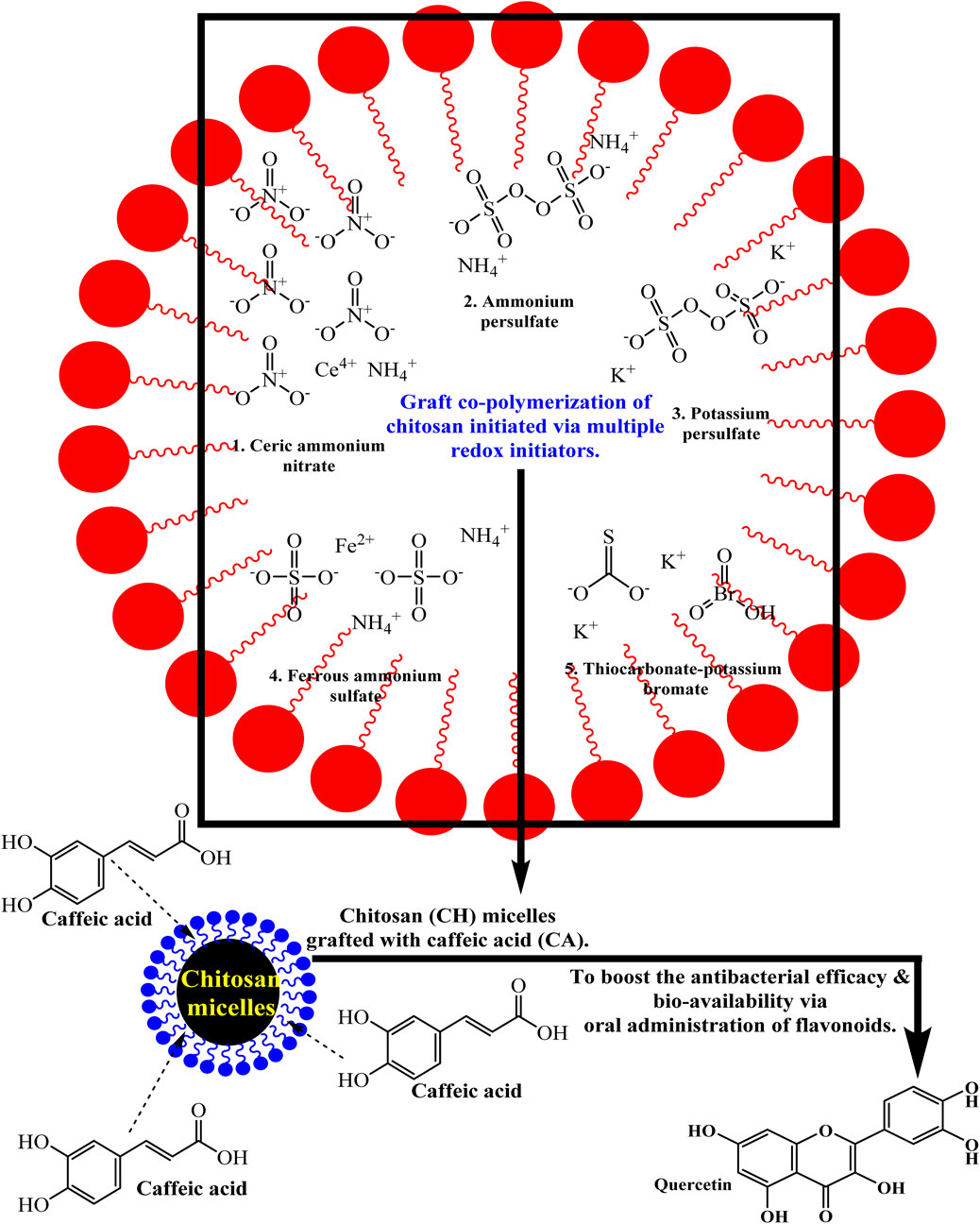
Figure 4. Graft co-polymerization of chitosan exhibited extraordinary antibacterial and drug delivery efficacy.
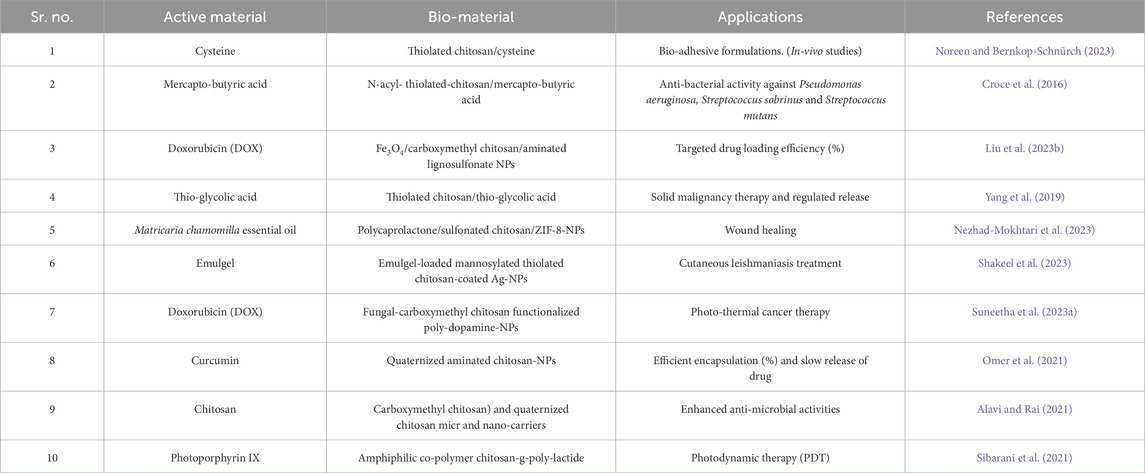
Table 1. Benefits of chitosan and its equivalents in bio-medicine via the addition of various pharmaceutical agents.
3 Chitosan-based nanostructures offer optimal functioning across multiple domains in contrast to un-modified chitosan
3.1 Chitosan based microgels (CH-MGs)
Microgels (MGs) constitute a type of heterogeneous grain or hydrogel composed of a three-dimensional network involving cross-linked polymers disseminated in a solvent, typically water (Li et al., 2023). Dimensions of these microscopic particles typically lie in the range of nanometers to micrometers (Costa et al., 2018; Li et al., 2021). MGs are commonly referred to as “smart” or “responsive” materials because of their ability to endure bi-directional volumetric fluctuations in response to a diversity of external stimuli, such as alterations in temperature, ionic strength, or the presence of certain bio-molecules (Li X. et al., 2022). Vancomycin (VM)-loaded chitosan-polyaniline microgels (CH-PANI MGs), which release VM upon lysozyme activation, have been developed to correspond with the pro-inflammatory gastrointestinal micro-environment. Aniline (ANI) was initially grafted to CH to form CH-PANI polymers, which were subsequently cross-linked with glutaraldehyde (GA) to form CH-PANI MGs, which provide the most significant advantages in treating infestations (Li et al., 2023).
Due to their bioactivity and biocompatibility, polysaccharide-based microgels are valuable components in biological tissue engineering and effective vectors for bio-pharmaceutical administration. Currently, the manufacture of chemically conjugated microgels requires prolonged reaction intervals, limited yield due to minimal water phase percentage consumption, and substantial energy expenditure (Michel et al., 2019). Over the last 10 years, significant research has been conducted on functionalizing MGs based on chitosan by divalent and multivalent metal ions. Bi-metallic chitosan complexes (Zn2+, Cu2+-CH-MGs) showed improved cell survival, angiogenic activity, and antibacterial properties, demonstrating the potential for wound healing (Lončarević et al., 2023; Mutlu et al., 2022). Bi-metallic chitosan fragments have a stronger complexation with zinc (II) ions, but as the ratio of degree of deacetylation (DD) to copper (II) ions increases, their propensity to swell consequently increases. Bi-metallic CH-MGs showed excellent durability during 4 weeks of enzymatic degradation, while bi-metallic systems with a lower concentration of Cu2+ ions showed strong cytocompatibility (Mutlu et al., 2022; Wang Y.-L. et al., 2020). glycol chitosan (GC) and its metabolites have been loaded with photo-thermal treatment agents, fungicides, chemotherapeutic drugs, antimicrobial agents, and specific imaging chemicals, notably gadolinium and iron oxide (Sahiner et al., 2023a).
3.2 Chitosan based microcapsules (CH-MCPs)
Micro-encapsulation (ME) technologies play a crucial role in maintaining the course of the effect and shielding the encapsulated product while modulating its dispersion rates (Valle et al., 2021). Changes applied in terms of the parameters associated with processing throughout the microcapsule manufacturing procedure may impact the MCPs’ functionality, especially their particle size and encapsulating efficacy (Lam et al., 2014). MCPs are particles smaller than a micrometer and can be used to hold chemicals. The main benefits of employing the ME approach are the prolonged and regulated distribution of medications, targeted therapy, extended and controlled distribution of pharmaceuticals, increased shelf life, stabilization, and immobility of microbes and enzymes (Meng et al., 2023). The CH-based microcapsules (CH-MCPs) shielded essential components from abrasive environments. More advanced techniques may be developed in the future to facilitate interactions with the medical and pharmaceutical industries, as well as with the manufacturing and use of MCPs easier (Wani et al., 2023).
Developed a unique smart carrier that is bio-compatible, responsive to many stimuli, and non-immunogenic that can be used for hydrophobic drug transport and triggered release. Using a simple sono-chemical technique, the multi-stimuli responsive smart chitosan-based microcapsules (MSRS-CS-MCs) were successfully synthesized from folic acid (FA) functionalized thiolated chitosan and effectively applied for various diagnostic and therapeutic purposes in bio-medicine (Cui et al., 2017). Polymeric MCPs are being thoroughly studied as drug delivery systems for various uses. Carboxylated chitosan (CCS)/poly (vinyl alcohol) (PVA)-based microcapsules loaded with dexamethasone have been developed to deploy them as an airborne drug or therapy for the cure of bronchial/respiratory diseases (Ameer and Maraie, 2019). Enzymatic and pH double response poly (lactic-co-glycolic acid) @ chitosan @ capsaicin (CAP@CS@PLGA) dual-core shell microcapsules were prepared by a composite emulsion strategy to overcome the limitation associated with the anti-fouling agent capsaicin’s quick discharge. This ensured controlled release and long-lasting antimicrobial effects (Guo Y. et al., 2023). Chitosan-modified microcapsules (CH-MCPs) are paid close attention as a carrier material or substrate for immobilizing enzymes to maximize the catalytic activity and recyclability. Microfluidic production of calcium alginate/chitosan composite microcapsules for peroxidase immobilization with ultrathin shells kept more than 80% of residual activity after six recycle operations (Chen S.-K. et al., 2023).
3.3 Chitosan-based metals (M = Au, Ag, Zn, Cu, etc.) and magnetic nanoparticles (NPs)
Metallic nanoparticles (NPs) have the potential to be used in conjunction with chitosan NPs for the diagnosis and treatment of ailments due to their capacity to convert photon energy into thermal energy, along with their ability to combat bacteria (Jiang M. et al., 2023). The rising prevalence of many diseases, particularly cancer, over the last several decades has culminated in an elevated mortality rate globally (Srivastava, 2024). Medical professionals and researchers have paid significant attention to NPs because of their physicochemical amenities, flexibility to be modified with different molecules, serum stability, diminished auto-immune response, optimal pharmacokinetics, and pharmacodynamics, etc. (Khalaf et al., 2023; Pusta et al., 2023). Furthermore, the incorporation of CH polysaccharide facilitates the manufacturing and transportation of metallic NPs (for instance, magnetic, silver, and gold NPs). In contrast, integrating CH with these metallic NPs can aid in detecting and managing many illnesses (Jiang M. et al., 2023). MNPs features are profoundly affected by their size and configuration. Chitosan has a significant influence on the design and dimensions of MNPs. CH can be used as a core-shell for NPs by encasing metallic particles due to its positive electrical charge. Chitosan has -NH2 and -OH functionalities. These groups hold the charge of the chelating properties of chitosan. A chemical connection forms between Ag+ ion and chitosan (Verma et al., 2021).
Exploring the potential medicinal benefit of chitosan-derived gold NPs for cancer treatment using a multi-spectroscopic method to selectively bind to the parallel G-quadruplex produced by the short telomeric DNA sequence (Khurana et al., 2023). Design of a chitosan-based antimicrobial wound dressing with incorporated ZnO/selenium NPs for adequate recovery and post-operative nursing assistance in pediatrics broken therapy (Ruan et al., 2023). Gold-coated magnetic nanoparticles (NPs) can be employed as a theragnosis agent for magnetic hyperthermia and CT (computed tomography) imaging applications. Magnetic NPs can produce localized heat by applying an alternating magnetic field, which can be used to treat cancer cells selectively in hyperthermia therapy [5a]. MNPs limitations regarding specific purposes: CH-based MNPs may be prohibitive due to the high cost of some metals, such as gold (Jamuna et al., 2023). Although CH is not a conductive material, CH-based metal composites might not be appropriate for use in electrical equipment (Tang W. et al., 2023; Zhang S. et al., 2023). However, metal NPs, especially Ag and AuNPs, pose significant toxicity risks in biological systems due to their ability to generate reactive oxygen species (ROS), which can lead to oxidative stress, mitochondrial dysfunction, and DNA damage. Its small size allows cells in vital organs to absorb and collect it, which can disrupt cellular homeostasis and produce inflammatory responses (Min et al., 2023). Furthermore, extended exposure can alter gene expression and protein function, which raises concerns about long-term biosafety in clinical settings (He et al., 2024). Green manufacturing techniques, dose optimization, and surface modification with bio-compatible polymers are used to mitigate these problems by improving biostability and reducing unfavorable biological interactions (Petrovic et al., 2024).
3.4 Chitosan (poly-vinyl) PVC-conjugates (CH-PVC-conjugates)
Like crabs and prawns, crustacean shells contain chitin, which is converted into the biodegradable polymer chitosan. Polyvinyl chloride, or PVC, is a common synthetic polymer (Rabie et al., 2023). Chemical interactions linked CH and PVC during their conjugation process. Once CH is activated, it can undergo various chemical processes to conjugate with PVC (Abdel-Monem et al., 2022). The free radical-mediated graft co-polymerization procedure is one such approach (Visakh and Darie-Nita, 2022). Radical precursors are used to start CH grafting onto PVC chains (Ţurcanu, 2022). Chloro-acetyl chloride was first used to react with amino-PVC to synthesize 2-chloro-N-(2-aminoethyl) acetamide-functionalized PVC (PVC-AcCl). After that, chitosan was conjugated, resulting in an alternative known as CH-AcPVC, which is PVC chitosan functionalized with acetamide. For the production of CH-PVC-conjugates, this procedure was carried out four times (Abdel-Monem et al., 2022). At a concentration of 500 mg/L, the synthesized conjugates showed excellent bacterial inhibition (five-log10 reduction) after 30 min and nearly complete inhibition after 120 min of incubation against two Gram-negative (Escherichia coli and Salmonella typhimurium) and Gram-positive (Staphylococcus aureus and Listeria monocytogenes) microbial strains (Abdel-Monem et al., 2022). Incorporating Ag-NPs into a CH/PVC blended matrix, resulting in a distinctive CH-PVC/Ag as an antiseptic self-sterilizing nanocomposite bio-material, highlights another achievement in this area of research. Due to a substitution reaction, CH and PVC were covalently bound (Gaballah et al., 2019).
Compared to pure PVC, CH-PVC conjugates can show better mechanical qualities, higher biodegradability, and a variety of potential uses. These sorts of uses might encompass the development of recyclable materials for wrapping and medical devices (El-Naggar et al., 2023). The final product’s intended characteristics and specific usage will determine the precise conditions and reactants employed in the conjugation process (Altinkok et al., 2023). Figure 5 displays beneficial aspects of CH-based nanostructures in treating varied death-causing infections.
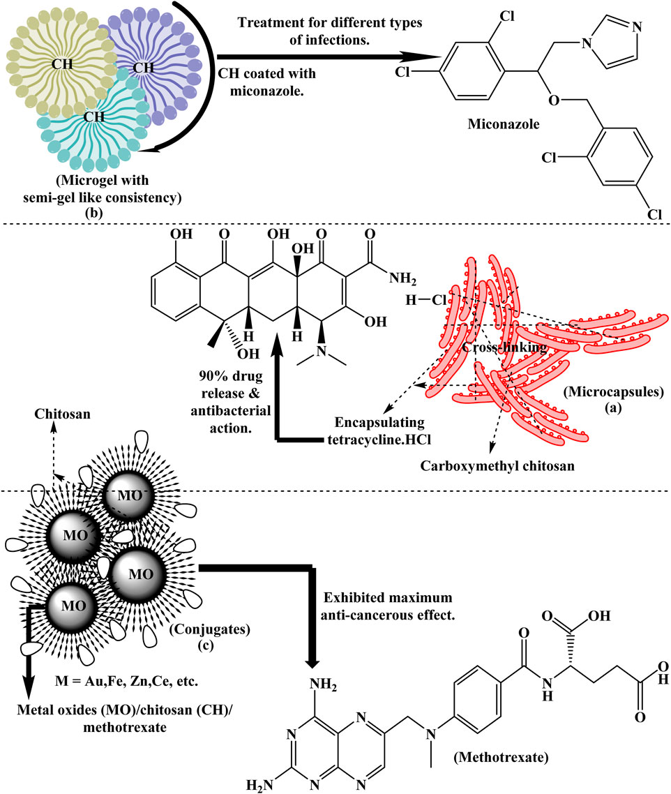
Figure 5. Chitosan-coated microgels, microcapsules and metal oxides are effectively employed in the bio-medical field for multiple advantageous purposes.
3.5 Chitosan graphene oxide nanocomposite (CH-GO)
Chitosan (CH) and graphene oxide (GO) nanocomposites have drawn a lot of interest in the fields of biology and medicine, as shown in Table 2, due to the synergistic effect between GO superior physical-chemical, mechanical, and optoelectronic capabilities or CH extraordinary biological traits. Films, hydrogels, frameworks, micron-sized particles and tiny fibers can be synthesized from CH and GO-based nanocomposites (Feng and Wang, 2022). GO is a kind of extensively oxidized and chemically altered nanosheet material with an abundance of oxygen-polar moieties on its surface, like hydroxyl, epoxy, and carboxyl (Yang et al., 2020). GO has excellent water dispersal properties and can interact physically or chemically with various polymers (Zhang et al., 2021). It is a superb matrix or filler material to obtain usable materials with better tensile and additional operational features (Zhang et al., 2017). GO photo-thermal efficiency enables it to display exceptional biological features, including antiseptic and cancer-fighting properties (Gu et al., 2019). Due to this, GO has tremendous promise in various therapeutic sectors such as drug delivery, tissue engineering, bio-sensing, and medical imaging and is widely used in stretchable semiconductors, photovoltaic cells, and probes (Maiti et al., 2019). GO applications in humans are restricted because numerous studies have shown that it does not show discernible impact at low dosages. For this, the inclusion of the versatile polymer CH may neutralize the harmful effects of GO (Gurunathan and Kim, 2016).
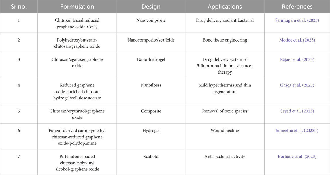
Table 2. Graphene oxide (GO) and chitosan-based nano-formulations demonstrated significant advantageous characteristics in medicine.
CH and GO interact chemically in a variety of ways. (i) Amino groups (-NH2) and hydroxyl groups (-OH) found in CH can interact with the functional groups on the surface of GO that contain oxygen. Dispersion of GO in CH solutions is aided by the–NH2 residues of CH forming hydrogen bonds with the oxygen-containing groups on GO (Sharif and Tavakoli, 2023). (ii) CH is a positively charged polymer because of its amino groups, whereas GO has negatively charged carboxyl groups (-COOH). The negatively charged GO and the positively charged CH may interact electrostatically, increasing the composite’s stability (Ahmed et al., 2023; Motiee et al., 2023). (iii) GO delocalized π-electron system enables π-π stacking interactions between graphene sheets. Nanocomposite formation can be aided by these π-π stacking interactions that can take place between the sheets of GO and via pertinent aromatic rings or conjugated structures in CH or other organic molecules (Ahmed et al., 2023; Yang H. et al., 2023). (iv) CH-GO materials may occasionally require chemical functionalization to fuse the two components covalently. GO is hydrophilic and dielectric because it typically comprises functional molecules that contain oxygen (Motiee et al., 2023; Sanmugam et al., 2023). (v) Within the CH matrix, reduction techniques can be employed to transform GO to reduced graphene oxide (rGO). Graphene component electrical permeability is restored through this reduction procedure. However also eliminates a portion of the oxygen units (Sanmugam et al., 2023).
3.6 Aptamer-modified chitosan
Single-stranded oligonucleotides called aptamers can fold into specific shapes and adhere to the appropriate macromolecules. They are primarily used as targeted mediators in illness diagnosis tests, therapy plans, and biosensors. They can precisely target pathogens, regulate the growth of microbes, transport antibiotics, and contrast chemicals into cancer cells and tissues (Taghavi et al., 2017). Aptamer-conjugated chitosan (CH) NPs can counteract the increased capillary storage action and considerably elevate the effectiveness of traditional therapies while minimizing their adverse effects on normal tissues (Fathi-Karkan et al., 2023). Furthermore, aptamer-conjugated carbohydrate-based nano bio-polymers can be used to develop innovative biosensors to monitor poisons, antibiotics, and other metabolites effectively. Such polymers have demonstrated exceptional antibacterial and antiviral activities (Fathi-Karkan et al., 2023; Jiang and Wu, 2019).
Extraction technological advances yield good productivity at a comparatively low cost. Due to their exceptional longevity and low hypersensitivity, aptamers are excellent substrates for functionalizing nanostructures (Golichenari et al., 2019). Due to their unique orientation, they additionally display a very high degree of segregation among hundreds of nucleotides. Aptamers still possess multiple drawbacks, like excessive renal secretion and nuclease-mediated destruction (Liu et al., 2022). Aptamer hybridization with NMs can potentially mitigate degradation and prolong circulation duration in this regard (Li et al., 2017). The blend of gold NPs modified with carboxymethyl chitosan and aptamer yields optimum consequences for Salmonella typhimurium colorimetric detection (Yi et al., 2019). Chitosan-capped, mesoporous silica NPs modified with aptamer for concomitant administration of daunorubicin and cytarabine in leukemia patients (Heydari et al., 2023).
3.7 Chitosan-based metal-organic frameworks (MOFs)
A novel family of porous crystalline materials made of metal and organic material is called metal-organic frameworks (MOFs). MOFs possess many intriguing features, i.e., excessive porosity, massive specific surface area, and exquisite tunability. MOFs are extensively employed in the administration of drugs, biological sensors, rejuvenation, healthcare technology, ecological preservation, and cell therapies (Valizadeh Harzand et al., 2023). Numerous remarkable traits of MOFs for use in biological processes, notably drug delivery and detection, have recently been documented in recent investigations (Gatou et al., 2023). Conversely, MOFs have drawbacks such as inadequate binding precision and poor stability. By expanding the surface volume and fostering the prospect for MOF-ligand interaction, MOF-based surfaces strengthen the long-term viability and selectivity of traditional MOFs, while conjugated films substantially broaden the area of active functional groups. Their unique characteristic renders them fascinating to pharmaceuticals and as bio-sensing agents (Chen X. et al., 2023).
MOFs have been modified with polymers to increase their therapeutic distribution properties; CH modification of MOFs offers them a pH-sensitive characteristic and provides an atmosphere for prolonged release of DOX (doxorubicin) in cancer treatment (Ashrafizadeh et al., 2023). Moreover, folic acid (FA) can functionalize CH to target malignant cells that overexpress the folate receptor in a targeted manner. Afterwards, CH-modified MOFs with a high drug loading capacity (1.63 g) can be used to load DOX. Interestingly, MOFs can provide imaging by encasing carbon dots (Chowdhuri et al., 2016). MOFs based on CH offer advantageous combined chemotherapy and bio-imaging for the treatment of cervical cancer. CH coatings on the outermost layer of MOFs would collapse and multiply when exposed to the moderately acidic pH of the micro-environment surrounding the tumor, causing DOX to be released at the tumor (Abazari et al., 2018). PVA/chitosan/hyaluronic acid interfaces loaded into an electrospun zinc-based MOF to produce an antibacterial composite nanofiber scaffold for bone regeneration (Salim et al., 2022). A novel hydrogel containing metal-organic polyhedrons/enzymes based on chitosan and has antibacterial properties to aid in wound recovery (Song J. et al., 2022). Metal-organic framework (Al-MOF/GO) was manufactured using a sustainable method based on aluminum succinic acid and graphene oxide (GO). Doxorubicin (DOX) and 5-fluorouracil (5-Fu) injection rates into the artificial Al-MOF/GO (DOX@5-Fu@Al-MOF/GO) were around 78.4% and 65.7%, respectively, as shown in Figure 6. The utilization of chitosan (CH) to encapsulate DOX@5-Fu@Al-MOF/GO resulted in a pH-sensitive and permeable coating (CH/DOX@5-Fu@Al-MOF/GO) with notable advantages across multiple biological domains (Asl et al., 2023). Metal-organic frameworks (MOFs) offer excellent potential for biomedical applications, but their clinical efficacy is limited via toxicity problems caused by metal ion leaching, framework instability, and reactive oxygen species (ROS) generation. Chitosan coating of MOFs significantly reduces these risks by providing a biocompatible, biodegradable barrier that stabilises the framework and limits direct cellular exposure to metal cores (Wiśniewska et al., 2023).
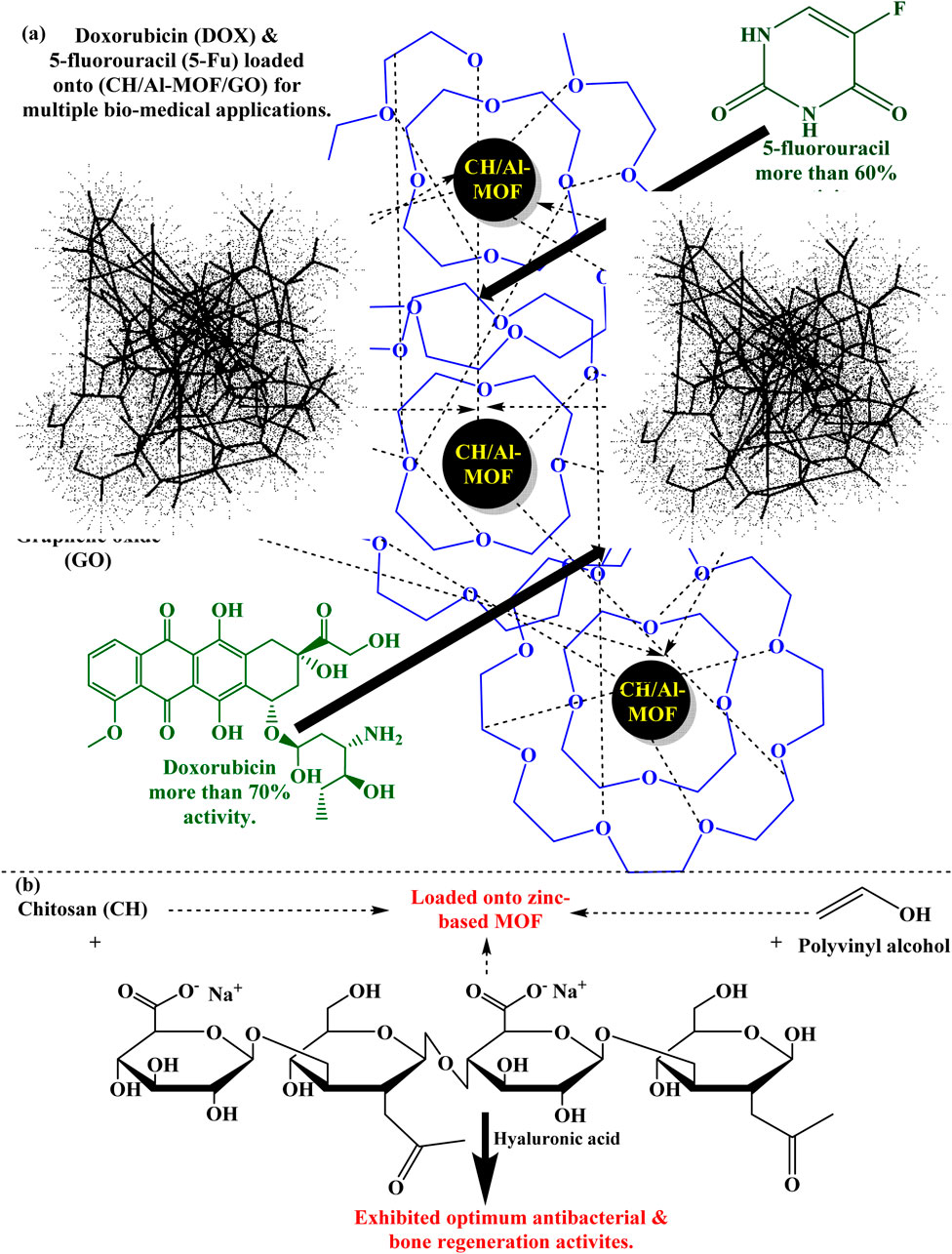
Figure 6. Several drugs loaded onto (a) CH/Al-MOF/GO and (b) CH/Zn-MOF for drug delivery against diverse antibacterial activity and remodeling of bones.
3.8 Chitosan modified complexes
Natural bio-polymer-based transition metal complexes with Schiff base ligands have impacted a new era of possibilities. Complexes exhibit metal-ion-encompassing rectilinear octahedral geometry (Vadivel et al., 2020). Complexation is a prominent characteristic of transition metals that finds extensive application in various fields, including oxidative polymerization, dynamic ophthalmology, and life sciences (Dayan et al., 2014). Transition-metallic complexes have drawn much attention in different commercial applications, such as light-emitting devices, antioxidant chemical manufacturing processes, experimental synthesis, and enzymatic activity in natural sources conversions (Si et al., 2007). Recently, there has been a lot of interest in supramolecular coordination chemistry. This field deals with molecular assemblies based on molecules’ interactions, like metal coordination, hydrogen bonding, - stacking, and host-guest complexation (Song M. et al., 2022). Because of their high stability, the production of supramolecular structures has garnered much interest in chemical materials, medications, sensors, etc. The lone electron pair on the free amino groups and hydroxyl groups of CTS can donate their electrons into the empty orbitals of electron-deficient metal cations because of the presence of strong chelating groups, forming a chelate complex through coordinated interactions (Song M. et al., 2022; Xiao et al., 2022). To produce rhodium (III) complexes, N and O donor atom-containing CH Schiff base (imine) ligands were synthesized. Such bi-dentate ligand donor atoms easily form coordination compounds, encapsulating the metal center in a coordination matrix via binding with transition metal ions. Transition metals to lower and higher oxidation states are typically confirmed in transition metal complexes, such as Schiff base and NO donor (Vadivel et al., 2020). Table 3 shows the significant effects of chitosan and its derivatives-based, synthetically-made nano-formulations.
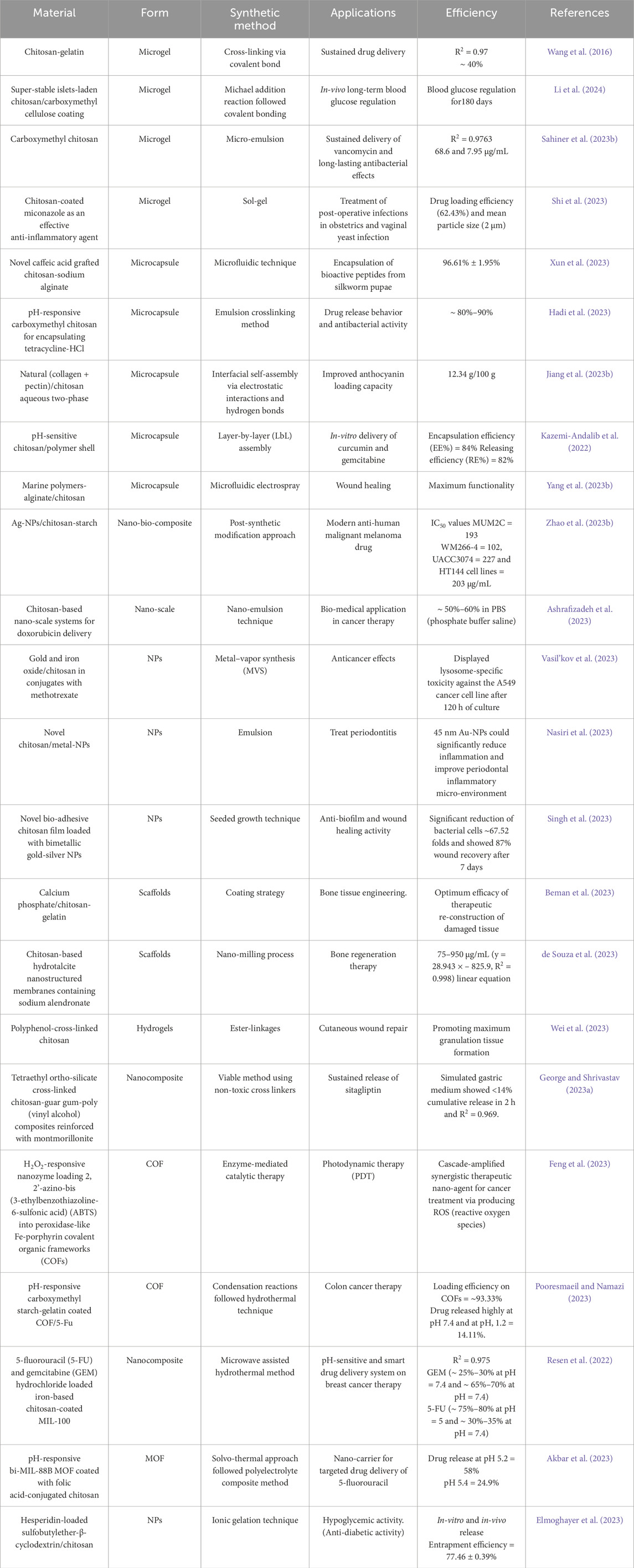
Table 3. Different nano-carriers derived from chitosan and its analogs showed maximum drug release levels, both in-vitro and in-vivo.
3.9 Co-bio-polymeric (C-BP) material-based nanostructures
Bio-polymers (BPs) are artificial polymers derived from petroleum-based products, bio-based monomers, and renewable feedstock. Additionally, they may originate from living organisms such as bacteria, plants, or animals. BPs can be categorized into three groups related to the single-stranded subunit of the polymeric material: complex sugars, polypeptides, and polynucleotides (Sreena and Nathanael, 2023). In bio-medical science, they are widely used for therapies such as tissue manipulation, intracellular image processing, and pharmacological and gene administration for wound recovery (Yadav et al., 2015), as illustrated in Table 4. BPs typically offer a means of mitigating the deleterious impacts that synthetic materials have on the environment, in addition to the issues pertaining to deterioration and survival. Surface charge, magnitude, stiffness, cell integration, and degradation are the main factors that majorly impact BPs production (Verma et al., 2020). C-BPs, i.e., two or more polymers, are the most appropriate material for tailored nanostructures because of their low immunogenicity and minimal cytotoxicity (Sreena and Nathanael, 2023). Thus far, research has demonstrated the ability to combine various polysaccharides with various synthetic polymers and other moieties and proteins. Studies on combinations of carrageenan, mucilage, chitosan (CH), hyaluronic acid (HA), cellulose, sodium alginate (SA), starch, and many more additives can be identified in the scientific literature (Tuwalska et al., 2022).
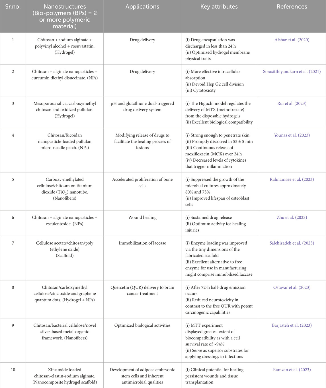
Table 4. Co-bio-polymers (C-BP) revolutionizing bio-medicine through their role in advanced medicine and regenerative therapies.
3.9.1 Chitosan-silk fibroin (CH-SFn)
Cocoons of Bombyx mori yield silk fibroin, a naturally occurring biopolymer of 5,507 amino acid sequences. SFn has exceptional endurance, extraordinary bio-compatibility with humans, and advantageous disposal attributes (Sreena and Nathanael, 2023; Tuwalska et al., 2022; Lujerdean et al., 2022). Additionally, it aids in the stimulation of cellular growth and adherence. SFn can be exploited as films, hydrogels, filaments, cylinders, sponges, scaffolds, and other morphologies extensively utilized in various bio-medical fields (Zhao et al., 2012). SFn is a protein with primary and secondary structures. Blends or mixtures of bio-polymeric (BP) materials, i.e., chitosan (CH) and silk fibroin (SFn) exploited as novel substrates (Tuwalska et al., 2022).
CH-SFn together can be utilized to make membranes, prosthetic skin, bandages, and implanted devices that disperse functionally active materials. Combinations of CH and SFn that contain inorganic particles or employ nanomaterials (NMs) as grafts for both flexible and rigid tissues are conceivable (Xing et al., 2023). It has been determined that the mixtures containing CH and SFn were miscible across 10%–50% by mass CH (Ray et al., 2023). As a potential material for sustained drug release, CH-SFn hydrogels (Xu et al., 2019), three-dimensional frameworks (Vishwanath et al., 2016), microfibers (Lai et al., 2014), films (Li et al., 2018). Tiny particles were synthesized and interconnected with EDC/NHS. These materials had porous structures, stable mechanical strengths, stimuli-responsive swelling performance, and drug-release behaviors (Tuwalska et al., 2022). A polymeric matrix containing inorganic NPs can intercalate to form various nanostructured materials. Hydroxyapatite, zirconia, carbonates of calcium, aragonite, quartz, TiO2, metal/metal oxides (M/MO), and bioactive mineral glass hybrids are examples of inorganic particles that are blended with CH-SFn (Sionkowska and Kozłowska, 2010; Liu et al., 2017c; Zhang et al., 2019).
Research on CH-SFn blends is being conducted to build a new nano bio-composite for therapeutic and thermogenic purposes via magnetized chitosan hydrogel and silk fibroin reinforced with PVA (poly-vinyl alcohol) (Eivazzadeh-Keihan et al., 2023). Silk fibroin/poly-dopamine NPs combined with carboxymethyl chitosan, sodium carboxymethyl cellulose, and agarose hydrogel dressings to deliver antibiotics (Karimi T. et al., 2023). Fabrication of chitosan/silk fibroin/ZnO antibacterial bio-composite hydrogel sponges wrapped with tetracycline hydrochloride for wound treatment (Kaptan et al., 2023).
3.9.2 Chitosan-pullulan (CH-PLn)
Another naturally occurring linear, non-branched exopolysaccharide biopolymer substrate is designated as pullulan (PLn). It is constructed from up of repeated maltotriose units that are coupled with one another by a (1 → 6) glycosidic hyperlink or maltotriose units that are repeatedly interconnected by a (1 → 4) glycosidic bonding (Elangwe et al., 2023). PLn is becoming more prevalent in many bio-medical research owing to its rapid dissolution in fluids, non-toxicity, sustainability, and non-oncogenic capabilities (Gasti et al., 2022). CH-restricted dissolution at neutral pH hinders its use in biomedicine despite its incredible advancements. Combining or modifying CH hydrophilic and hydrophobic strands has been proposed to constitute among the most effective methods for increasing CH lubricity facilitating the production of microgel particulates, hence optimizing the potency of drug encapsulation (Zidan, 2022). Here, CH and PLn, a microbial aqueous homo-polysaccharide, have been blended to produce a synergistic microgel network. PLn is a safe, toxic-free, disposable bio-polymer with various prospective applications in scientific domains (Santamaría et al., 2023).
Interactions between fluorescent polystyrene nanoparticles (PS-NPs), heparin, polycaprolactone (PCL), polyvinyl pyrrolidone (PVP), carboxyl pullulan, and chitosan result in the development of characteristics like robustness, pH resistance, chemical resistivity, mechanical and thermal insulation. Over 80 percent of the studies examined the effects of the PLn content on the properties of the solution and the final nanostructure design (Manivannan et al., 2023). Development of drug-loaded nano-emulsions based on CH, PLn, and alginate as a possible medium for delivering aggressive melanoma (Fard et al., 2022). PLn-nanoparticle loaded thermo-reversible hydrogels comprised of chitosan/guar gum for improved drug delivery across the nasal cavity to the cerebral cortex (Sohail et al., 2022). Manufacturing and evaluation of PLn succinate films fused with hyaluronic acid and chitosan for cutaneous wound regeneration (Wang et al., 2022).
3.9.3 Chitosan-alginate (CH-ALG)
Two of the most researched natural polymers that have garnered attention for their numerous applications in nano form are chitosan (CH) and alginate (ALG) (Feng et al., 2020). One of the industries most benefited by the advancement of nanotechnology is the biomedical field, wherein increasing emphasis is currently focused on advancing the development of vaccine adjuvants, antimicrobial substances, and hypoallergenic systems for administration derived from CH and ALG (Niculescu and Grumezescu, 2022). ALG is among the most feasible organic polymers for integrating with CH, especially because its anionic properties counter-balance the cationic CH backbone to yield a more resilient nanomaterial. Attractive thermodynamic and biological traits are additionally linked with ALG (Bakil, 2020). In formulations based on ALG, CH can improve absorption by softening the intracellular tightly bound junction, thus extending the duration until the active substances are in interface with the epidermis (Lang et al., 2020). Additionally, the combined effects of ALG and CH shield the loaded biomolecules from oxidation, hydrolysis, and proteolytic disintegration, ensuring their safe and effective delivery to the targeted tissues or systems (Loquercio et al., 2015).
Insulin comprised an active component in chitosan and alginate-coated monomethoxy polyethylene glycol poly (lactic-co-glycolic acid) (mPEG-b-PLGA) NPs, which were synthesized via the optimized double emulsion process and are utilized in the medical management of hyperglycemia (Chen et al., 2019). To build for secure ingestion of 5-FU (5-fluorouracil) and BDC (bis-demethoxy-curcumin), a novel pH-sensitive bioactive amine-containing mesoporous silica–alginate/folic acid conjugated o-carboxymethyl-chitosan–gelatin (AMSN-Alg/FA-CMCT-Gel) nanocomposite system has been developed. This approach supports in navigating the constraints of colon cancer (Anirudhan and Nair, 2019). A synthetic mixture of Ag-NPs, chitosan, and alginate showed tremendous potential in microbiological filtration and chemotherapy (Venkatesan et al., 2017). Layer-by-layer fabrication of chitosan/alginate-based platelet-mimicking nano-capsules enhances the lethality of doxorubicin (DOX) against breast cancer cells (Ibrahim et al., 2023). Compared to distinct materials, a CH-ALG blend can have synergistic effects that increase structural, thermal, or dielectric features and diversity. When exposed to rigorous environmental and response conditions, particular problems lead to prohibited stability and intricacy (San et al., 2022).
3.9.4 Chitosan-carrageenan (CH-CAR)
Carrageenan (CAR) is an extraordinarily dense polymeric material that occurs naturally. It is generally non-hazardous and tranquilizing, making it suitable for medical operations such as drug administration, emulsifying agents, persistent absorption of pharmaceuticals with extended retention, and tissue re-growth (Alnaief et al., 2019). Additionally, by hastening their disintegration and mobility, CAR increases the efficacy of medical drugs that are poorly water-soluble (Li et al., 2014). This is so that CAR’s negatively charged anionic sulfate groups may effectively interact with positively charged polymers via electrostatic interactions, hydrogen bonding and ionic complexation mechanisms (Alavi and Mortazavi, 2018). Ionic coagulation may yield NPs at lower concentrations of CH and CAR; the NPs charge on their surface is contingent upon the mass-to-weight ratio and polymer incorporation sequence (Ćirić et al., 2020). Highly positively charged NPs (+60 mV) were produced by progressively infusing low-dosage carrageenan dispersion into chitosan emulsion. Based on L929 fibroblast culture, they demonstrated promising potential as drug transporters with little cytotoxicity at rates up to 3 mg/mL (Lim et al., 2023; Grenha et al., 2010). Contrasting with relatively limited challenges, such as intricate output, regulatory challenges, expenditures and fluctuating qualities, are certain advantageous aspects of CH-CAR mixture, such as tunable properties, enhanced stability, and scalability for commercial endeavors (Koirala et al., 2023; El Idrissi El Hassani, 2023).
Using curcumin (CUR) as a model drug, design a pH-responsive, eco-friendly, non-cytotoxic drug transporter for lipophilic pharmaceuticals that target intestinal delivery using N,N,N-tri-methyl-chitosan chloride/carrageenan (CAR/TMC, 1:1) hydrogel with different molar ratios of Ag-NPs i.e. 1%, 3%, and 5% Ag (El-Maadawy et al., 2022). CH-CAR nanocapsules are designed to encase biologically active substances such as nutritional supplements, antioxidants, and essential oily substances (Ebrahimzadeh et al., 2023). Recent research has examined the physiological assimilation of CH-CAR combinations with varying morphologies that have been substantially absorbed with the maximum percentage of efficacy. pH-sensitive release of doxorubicin through a chitosan core-shell carrier incorporates magnetic laponite and κ-carrageenan (Mahdavinia et al., 2023). Fast hemostasis using a polysaccharide-based (kappa-carrageenan/carboxymethyl chitosan) nanofibrous membrane covered with an antifibrinolytic drug for an in-vitro and in-vivo assessment (Salmasi et al., 2023). Formulation and evaluation of chitosan-alginate/carrageenan-based hydrogel for administering drugs in treating diabetes (George and Shrivastav, 2023b).
4 Optimum positive attributes of chitosan and its analogs in terms of physicochemical, structural and operational aspects, along with minor downsides
Chitosan and its derivatives have drawn considerable interest from various fields because of their distinctive physical, structural, and operational features (Wang et al., 2020b). Here are a few of the most notable advantages of CH and its substitutes and a few minor limitations. Researchers are still investigating and developing technologies to maximize these materials’ potential and mitigate their drawbacks (Aranaz et al., 2021).
4.1 Beneficial/advantageous features correlated with chitosan (CH)
Considering a chemical, structural, and functional perspective, chitosan (CH) and its derivatives provide several advantageous properties for therapeutic application (Ding et al., 2021). Their unique polysaccharide structure, originating from chitin through deacetylation, confers enhanced biological compatibility, thereby rendering them highly suitable for utilization in the medical field (Wang et al., 2020b). Due to the presence of the–NH2 groups in their structure, they are cationic, which makes them able to engage electrostatically with negatively charged macromolecules to facilitate gene treatment and drug administration (Shokati Eshkiki et al., 2024; Dhlamini et al., 2024) Furthermore, the presence of–OH substituents allows for covalent modification to provide specific properties, including improved solubility at physiological pH (Tamer et al., 2023). Particularly in mucosal drug delivery systems, the mucoadhesive qualities of CH promote better drug absorption and retention (Samiraninezhad et al., 2023). Moreover, their intrinsic antibacterial properties benefit wound healing and infection control (Sun et al., 2024).
4.2 Drawbacks/challenges
Although CH and its derivatives have many therapeutic uses, several disadvantages are associated with their mechanical and thermodynamic characteristics (Yuan et al., 2024). One significant disadvantage that might prohibit CH from being utilized in medicinal formulations is its limited dissolution at neutral pH (Almajidi et al., 2024; Azmana et al., 2021). This solubility problem is caused by -NH2 residues on the CH polymeric chain, as when protonated at lower pH levels, it results in diminished dispersion and increased molecular aggregation (Choi et al., 2024). Quaternization or grafting with hydrophilic polymer molecules are popular chemistry modifications proposed to overcome this challenge and boost solubility and lifespan under physiological conditions (Pathak et al., 2021). Another drawback is the potential for CH to trigger allergies. Despite such rare experiences, specific individuals might develop allergic responses when CH or its counterparts are used in medical treatments (Gericke et al., 2024). Thus, it is crucial to assess patient sensitivity and keep a careful eye on adverse occurrences in therapeutic settings (Vargas-Ortíz et al., 2024).
Furthermore, batch-to-batch heterogeneity in CH may make maintaining consistent quality and performance challenging, particularly in pharmacological and biomedical areas (Abolhassani et al., 2024; Khatami et al., 2024). CH traits can vary depending on their source, extraction method, and subsequent processing steps (Kou et al., 2021). The development of standardized medical products may be exceedingly complicated due to this variation; consequently, stringent quality assurance procedures may be required (Milliken et al., 2024).
5 Chitosan (CH) as a potential drug carrier
Researchers in medical engineering are drawn to chitosan-based bio-materials as prospective carriers owing to their superior chemical, cellular, and mechanical properties that facilitate cell adhesion, growth, and division (Supernak et al., 2023). Abundant in free amino groups, the primary molecular structure of CH can be modified physically and chemically to enhance its diverse carrier properties, offering new opportunities for anatomical and medical technology (Wang L. et al., 2023). Additionally, more research on CH-based frameworks across different organs associated with transplantation and regenerative therapies is still to be discovered. Its disintegration products are immune-modulating, ecologically benign, and are unlikely to build up inside the circulatory system. Their benefits include antiviral, anti-inflammatory, hemostatic, wound exudation-reduction, as well as restoration and healing-promoting actions (Pathak et al., 2023).
5.1 Tissue engineering/regeneration
Tissue engineering integrates cells, materials, and bio-chemical components to enhance or replenish biological processes. It provides advantages for various connective tissues, such as veins, urinary tissues, muscle, skin, cartilage, and bone (Medeiros Borsagli, 2023). Since repairing damaged tissue constitutes a significant challenge, better and more resilient bio-materials should be designed for tissue engineering applications. Alginate (ALG), hydroxyapatite, collagen, chitosan (CH), and other natural and synthetic materials are currently used for tissue engineering purposes (Saravanan et al., 2016). CH-modified nanocomposites have been employed recently to advance the science of tissue engineering while enhancing tissue viability. The manipulation of tissues provides enhanced physiological and structural stability (Butnariu, 2023). To promote granulation and tissue repair, CH tiny materials hasten the activation of peritoneal fibroblasts and pronuclear white blood cells (Yadav et al., 2023).
Damage to the axons in the brain and spinal cord causes neurodegenerative diseases and disorders affecting the central nervous system (CNS). CNS has a limited capacity to regenerate itself, so it experiences difficulties mending injured tissue (Pchitskaya et al., 2018). Recently, tissue engineering has been employed as a multimodal way to replace or repair damaged brain tissues. In tissue engineering, the CH-NPs operate as a growth medium for cells, facilitating the development of a particular variety of tissue with predetermined characteristics (Rajabian et al., 2019). Inductive conditions for brain rejuvenation have been designed using both artificial and endogenous biological materials (Amani et al., 2019). 3D-bi-directional multi-functional hydrogels constructed from chitosan and carboxymethyl cellulose are infused with nano-curcumin for synergistically diabetes-related wound therapy. Solvent casting is used in this study to produce intravenous hydrogels using chitosan-CMC-g-PF127. Injectable hydrogels curcumin (Cur)/chitosan-CMC-g-PF127 exhibit a monitored release profile and excellent dilation capabilities with elastomeric performance (Shah et al., 2023). Histopathology outcomes indicate an intriguing propensity for re-growth of tissues, in conjunction with a spike in fibroblasts, keratin cells, and accumulation of collagen, all of which lead to the stimulation of the transdermal interface (Shah et al., 2023; Shah et al., 2019). Injectable hydrogels encapsulating chitosan-CMC-g-PF127 loaded Cur demonstrated rapid wound healing potential by promoting cell migration and proliferation at the exact location of trauma by offering hydrophobic molecules as a persistent drug delivery vehicle (Taheriazam et al., 2023). Immobilization of cellulase on hydrogel composites of chitosan and cellulose nanofiber for tissue engineering active materials, which deteriorate via activated enzymes (Tamo et al., 2022). Zinc oxide NPs (ZnO) and β-cyclodextrin (βCD) doping yielded pseudo-polyrotaxane (Chs-NAI/βCD), Chs-NAI/ZnO-NPs, and Chs-NAI/βCD/ZnO NP composite materials demonstrating the maximum efficacy in bone tissue engineering (Dardeer et al., 2022). The most prevalent causes of death in the modern world and a significant threat to public health include heart disease and stroke. Due to the cardiac muscles severely diminished innate capacity for regeneration, catastrophic coronary artery disease like infarction of the myocardium (MI) currently lacks an appropriate therapy. Heart disorders have been extensively treated with injectable conductive nanocomposite hydrogels (carbon and metal-based nanostructures) for cardiac tissue engineering (Pournemati et al., 2022).
In modern orthopedic surgery, repairing cartilage injured by pathology (osteoarthritis), trauma, or aging is essential. Self-generated chondrocyte organ transplantation, mosaic-plasty, micro-fracture treatment, and natural materials insertion are a few options for re-constructing cartilage (Bashir et al., 2022).
5.2 Therapies for varied malignancies (cancer treatment)
CH serves a purpose in cancer therapy in many ways, such as administering genetic material, chemotherapy drugs, and vaccine adjuvants (Prabaharan, 2015). CH-NPs have become one of the most promising delivery systems for cancer chemotherapy and diagnosis because of the unique characteristics they possess, i.e., decomposition, remarkable cell-membrane penetration, high drug-carrying capacity, pH-dependent therapeutic releasing, versatility, and extended residence duration within the blood-stream (Bashir et al., 2022). Targeted drug delivery has been achieved by combining ligands specific to tumors with CH-based nanostructures, as depicted in Figure 7. CH bio-materials are used in receptor-targeted chemotherapy to target surface receptors that are overexpressed in cancer cells specifically. Receptor-mediated endocytosis is the process by which targeted nanoparticles create other nanoparticles when they come into contact with cell surface receptors (Ghaz-Jahanian et al., 2015). Because different types of malignancies have different expression levels for these receptors, understanding receptor levels and cell types is necessary for developing customized drug carrier systems. Furthermore, CH-NPs release the drug into the cytoplasm by dissolving the assembly at the endo-lysosome’s acidic pH with ligands specific to the tumor via pH-cleavable bonds (Xu et al., 2013). Chitosan-based nanostructures can be designed to interact with cancer cell receptors and stop tumor growth through several different mechanisms of action, as depicted in Table 5. The technique employed can vary depending on the type of receptors targeted, the therapeutic compounds incorporated into the nanostructures, and the architectural layout of the nano-crystals (Virmani et al., 2023). Several standard modes of action include receptor-mediated targeting (Lohiya and Katti, 2022), intrinsic drug distribution (Yee Kuen and Masarudin, 2022), cellular uptake and delivery (Baghani et al., 2022), pH-responsive pharmacological discharge (Dongsar et al., 2023), optimized drug accumulation and retention (Herdiana et al., 2023), focused immune system modulation (Karimi K. et al., 2023), and mixed therapies (Salmasi, 2024; Kurczewska, 2023). In order to develop remedies for cancer that are significantly more effective and feature fewer adverse effects, researchers are constantly investigating and up-grading these routes of action (Bashir et al., 2022).
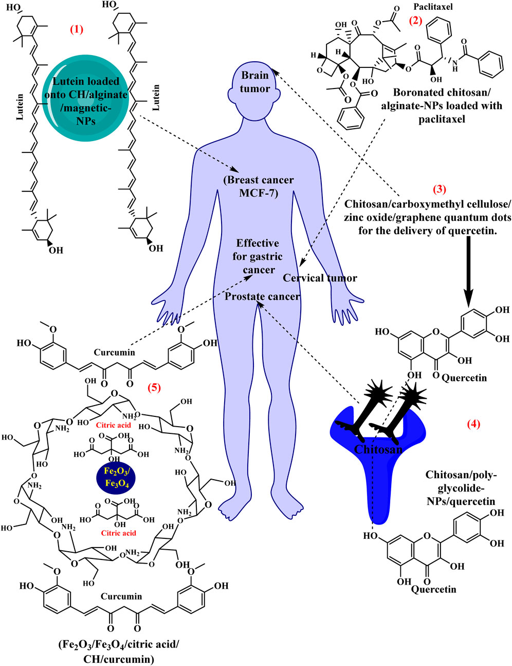
Figure 7. Anti-cancerous protective effects of CH-modified bio-materials against various devastating malignancies.
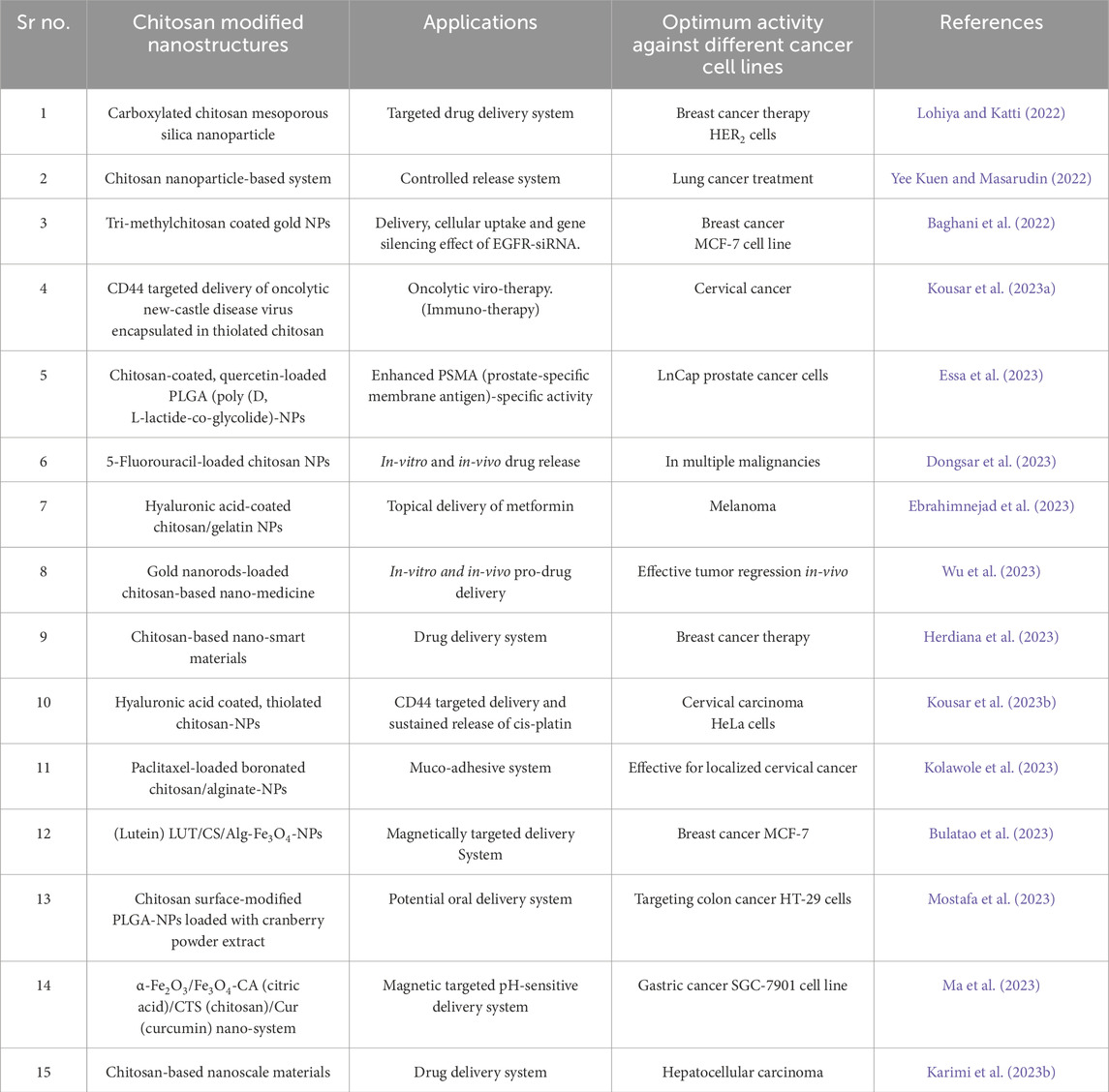
Table 5. Based on preclinical and clinical studies, comparative analysis of several chitosan derivatives in cancer therapy for tumor growth suppression.
5.3 Drug delivery/herbal drug delivery system (in-vitro or in-vivo pharmaco-dynamics and pharmaco-kinetics profiles)
An herbal medicine delivery system is a method or device designed to administer botanical medications—also known as phytomedicines or herbal drugs—to the body in a controlled and effective manner. Herbal remedies are pharmaceutical substances derived from plants, plant extracts or plant-based components (Talemy, 2023). Traditional medical systems, such as Ayurveda, ancient Chinese medicine, and Native American medicine, have been employing these drugs for decades due to their medicinal properties. Due to differences in plant sources, growth environments, and processing techniques, natural drugs might differ in composition and strength (Che, 2024; Ahirwar, 2023). Pharmaceutical delivery devices can make Herbal therapy dosages more consistent and dependable. Targeted administration can minimize the adverse effects of herbal medications by concentrating the active ingredients at the intended site of action and diminishing exposure to other tissues (Che, 2024; Liu H. et al., 2023). Delivery of herbal drugs is made more convenient for patients by dosage forms such as capsules that are transparent pills and transdermal patches, which can enhance adherence to treatment regimens. Herbal medicine has been used for thousands of years. Indigenous herbal medicine is estimated to offer primary healthcare for 80% of the world’s population (Prabhakar et al., 2023).
Considering the long-term viability of botanical products or other bio-active compounds is essential while building CH-based nanostructures. Factors such as pH, temperature, and corrosion propensity should be addressed to sustain the biological function of the loaded chemicals or synthetic substances. A naturally occurring drug identified as camptothecin (CPT) is found in the bark and stem of the Camptotheca acuminata tree species, which is mostly grown in China and Tibet. Despite its ability to cause the death of cancer cells, its limited solubility in water renders its application un-feasible (Mateti et al., 2021). Therefore, CH-modified nano-fibers are employed to overcome this problem (Zaher et al., 2022). Emblica Officinalis, sometimes known as Indian Gooseberry or Amla, is an herb that is widely used in current medicine and is recognized as one of the most prominent medicinal herbs in Ayurveda, the ancient medical system of India. It is manufactured naturally using plant extracts, has tremendous therapeutic properties, and has minimal adverse consequences. Its optimal performance is attributable to its inhibitory activities against strains of human breast cancer cells (Baliga and Dsouza, 2011). CH-based nanostructures are employed for multiple types of cancer for in-vitro and in-vivo release of herbal pharmaceuticals (ND, 2022). Plant flavonol fisetin belongs to the flavonoid class of polyphenols. It functions as a yellow pigmentation reagent in a wide variety of plants. Therapy of breast cancer cell lines with fisetin-loaded chitosan NPs inhibits the expression of miR-96 and miR-183 significantly with excellent outcomes (Habibi Khoei, 2023). Figure 8 illustrates some herbal drugs extracted from natural sources exhibited optimum efficiency against certain malignant tumors.
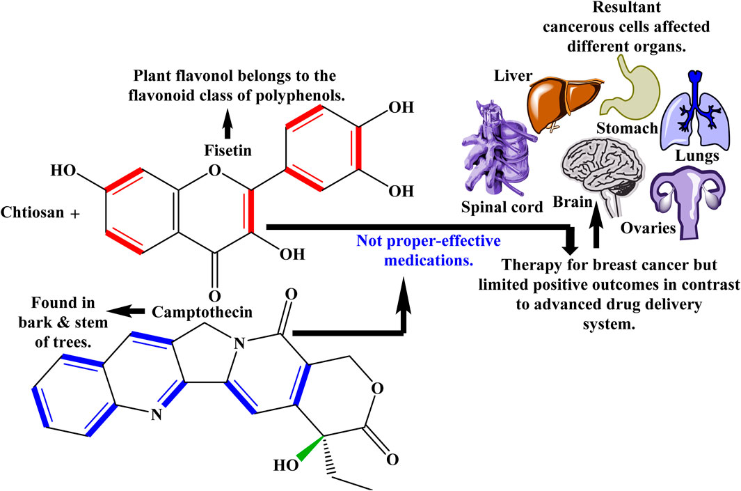
Figure 8. Fisetin and camptothecin against breast cancer with toxicological constrains related to other organs and tissues.
One of the drawbacks of using natural drug delivery systems is that herbal ingredients are vulnerable to temperature, light, and oxygen fluctuations. Moreover, in-sufficient empirical evidence, protection issues, disparities in potency, limited solubility, expensive and absence of regulations, herbal products with a short shell lifespan may contain toxins such as heavy metals (mercury, lead, etc.), herbicides as well as pathogens (Kalachaveedu et al., 2023; Thakkar et al., 2020). Despite the herbal drug delivery system, the novel drug delivery method leverages more functionalized and novel strategies to release the drug promptly with maximal therapeutic efficacy, as illustrated in Figure 9.
Figure 9. Novel in-vitro and in-vivo drug delivery technology to treat a variety of diabetes-related ailments, inhibit the formation of malignant cells and avert several other serious diseases.
5.4 3D-printing/bio-printing
Three-dimensional (3D) printing is an additive manufacturing process that has become a versatile and beneficial infrastructure to solve problems for various medicinal applications, including pharmaceuticals, general medical care, and quickly needed items during a viral pandemic (Zhang et al., 2022). The primary advantage of 3D-printing over traditional manufacturing techniques is its speedy development of intricate, precisely customized structures for particular patients (Giannopoulos et al., 2016). Living human cells and tissues can be researched and synthesized in the medical profession using 3D-printing technology for tissue design and regenerative medicine applications (Capel et al., 2018). 3D-printing also helps pharmaceutical companies construct disease models, medicinal equipment, and drug-containing scaffolds. Ultimately, during the coronavirus disease 2019 (COVID-19) pandemic, 3D-printing technology has provided a cost-effective response to fabricating a wide range of building materials, from diagnostic tools to personal protective equipment (Choong et al., 2020). A variety of medical implants have been successfully developed through the use of popular 3D-printing techniques, such as fused deposition modeling (FDM), semi-solid extrusion (SSE), stereolithography (SLA), selective laser sintering (SLS), and inkjet-based 3D-printing (Zhang et al., 2022). Though this technology has great promise, some problems have been found with existing 3D-printed bio-medical devices, including poor mechanical qualities, a lack of raw materials for organ and tissue fabrication, and a substantial infection rate following transplantation (Shahrubudin et al., 2020).
According to Shirazi 3D-printed bio-medical items provide unique operational and technological problems due to biocompatible substances and clinical testing (both in-vitro and in-vivo), which sets them apart from other printed commodities (Shirazi et al., 2015). It is now possible to create a generic 3D-skin structure at a low cost by utilizing 3D-printing technology. This skin, which is 3D-printed, can be used to evaluate synthetic goods, cosmetics, and drugs. New 3D human skin models may eventually replace animal testing to assess cutaneous sensitivity to a medical design. It will, therefore, make it possible for experts to obtain accurate outcomes from repeated printing trials (Zarrintaj et al., 2018). In situ homogeneously mixed alginate, chitosan, and kaolin bio-ink serve as the substrate in 3D-biological printing of complex tissue scaffolds for skin tissue restoration using an innovative portable bio-pen (Bhattacharyya et al., 2023). Bone cartilage is a very dynamic and varied tissue in shape and function. In this instance, bone abnormalities brought on by tumor removal, trauma, injury, or inflammation can have spaces filled with tissues printed using 3D-printing technology (Shahrubudin et al., 2020). This unique therapy offers an alternative to auto-unions and transplants to preserve health or improve the in-vivo potential. Products made with a 3D approach include bio-fired inlays, intracranial parts, bone architectures, etc. (Velioglu et al., 2019). For bone tissue engineering (BTE), scaffolds based on chitosan/alginate/hardystonite (Cs/Alg/HD) were invented, and they showed the highest yield strength (1.38 MPa) and elastic modulus (125.71 MPa) (Mohandesnezhad et al., 2023).
Drug delivery using 3D technology is popular for both in-vitro and in-vivo pharmacological release. Meanwhile, chemotherapy and antimicrobial activities can be tailored based on the patient’s anatomical features and clinical manifestations via three-dimensional printing (Weisman et al., 2019). Fabrication and characterization of bio-compatible scaffolds based on chitosan/collagen/hydroxyapatite loaded with crocin exhibit pharmacological drug release kinetics for bone tissue manipulation (Jirofti et al., 2023). CH-based printed bio-inks have favorable immunological responses following implantation, good cell/matrix interactions, tissue shape mimicking, and a medium to facilitate nourishment and circulating oxygen. CH in bio-ink formulations may impart high antibacterial activity. Bacteria can pass through the negative shell membrane of CH due to the electrostatic interaction between electrons and ammonium ion, which either kills the bacteria or stops them from growing. Moreover, CH-based printed platforms showed biological molecule adaptability, robust susceptibility, and biological activity even after a range of post-printing modifications (Taghizadeh et al., 2022).
Bio-inks with chitosan-based nanostructures are extensively employed in various medical disciplines. (i) Deployment of chitosan-based materials as appropriate inks for 3D/4D bio-printing and their potential for developing tissue-engineered constructions and cutting-edge drug delivery systems (Agarwal et al., 2023). (ii) As a potential scaffold for tissue engineering, levofloxacin (LEV) was added to three-dimensional printed chitosan (CS), poly (vinyl alcohol) (PVA), or gelatin (Gel) hydrogel. Its in-vitro release continued for 48 h (Koumentakou et al., 2023). (iii) Employing bismuth ferrite (BFO)-NPs, which have ferroelectric and magnetic properties and chitosan as a bio-ink to develop and assemble a 3D-printed wound dressing that can release amoxicillin (AMX) in response to modest electrical stimulation (Baykara et al., 2023). The most promising bio-ink for transplantation operations was discovered to be a ChiMA/GelMA nano-hybrid superstructure with 5% LDHs (ChiMA-G5). Methacrylic anhydride was used in this study to chemically modify chitosan and gelatin to develop methacrylate chitosan (ChiMA) and methacrylate gelatin (GelMA), respectively, to construct bridging defects on the visco-elastic skeleton. Layered double hydroxide nanoparticles (LDHs) were used in the chemical composition of the suitable bio-inks (Yuce-Erarslan et al., 2023). This study is significant for the bio-medical manufacturing industry because it provides information about using 3D printing technology for producing bio-medical products in developing nations. Management industry, human resources, and new manufacturers may find this study helpful (Shahrubudin et al., 2020).
5.5 Wound healing
Wound healing has seen a surge in interest due to the many properties of chitosan-based nanostructures that facilitate tissue development and repair. Repairing damaged tissue and healing the wound to stop infection and additional harm are the main objectives of wound healing. Its goal is to restore the damaged tissue’s structural and functional integrity promptly. Scar tissue may occur during the surgical process, and the repaired tissue might not consistently look precisely comparable to the original tissue. Remodeling, swelling, and inflammation are just a few steps of the intricate wound healing process. As a result, scar tissue, also known as fibrotic tissue, usually forms to cover the wound site. Chemical modification of chitosan-based nanostructures can improve their ability to heal wounds and interact with particular ligands, substances, or biomolecules. The therapeutic potential of chitosan-based nanostructures for wound healing can be influenced by the chemical reactions and changes that are applied to them. Here are some shared chemical changes and reactions, i.e., (i) CH-based nanostructures, such as hydrogels or NPs, can be functionalized by surface-attaching different ligands like growth stimulants, peptides, or analgesics. Covalent bonding mechanisms can accomplish this via amides or esters synthesis. (ii) Materials based on CH can be conjugated with chemically reactive accelerators such as glutaraldehyde or genipin. Cross-linking promotes CH hydrogels and scaffolds to be more resilient and hydraulically advantageous, making them appropriate for wound dressings. (iii) Various chemical techniques are capable of being utilized to load drugs or therapeutic substances into CH-bio-materials. For instance, depending on the specific features of the drug, encapsulation can be achieved by morphological encasing, co-acervation, or ionic gelation. (iv) CH undergoes different reactions, i.e., oxidation, amidation, thiolation, and carboxylation, to generate chitosan derivatives that are more operationally active in wound healing.
To avert bacterial invasion and hasten the healing process of wounds, wound dressings with the ability to sterilize pathogenic microbes and scavenge antioxidants become vital. Comparing quaternized chitosan and sodium carboxymethyl cellulose microgels/baicalein NPs to traditional dressing methodologies or surfaces, they can speed up the healing of infected wounds (Guo J. et al., 2023). A folic acid-cross-linked chitosan hydrogel film laden with amikacin facilitates wound healing by releasing antibiotics in a controlled and effective manner (Mehmood et al., 2023). Effective wound management greatly depends on promptly controlling bleeding or hemorrhage and fostering healing. An innovative reagent for blood clotting and wound closure, an incredibly absorbent bio-sponge comprised of carboxymethyl chitosan, poly-γ-glutamic acid, and platelet-enriched plasma. Blood coagulation was hastened by the sponge’s ability to cling to and squeeze red blood cells. Sponge managed to release vascular endothelial and epidermal growth factors, according to research conducted both in-vitro and in-vivo (Tang J. et al., 2023).
5.6 Vaccine adjuvants (VAs)
Adjuvants are substances or blends mixed into vaccinations to enhance the body’s defense against the target antigen, i.e., the pathogenic component of the vaccine. Vaccines contain adjuvants integrated into them to make them more potent. Adjuvants specifically can boost the immune response, lower the quantity of antigen required, increase the effectiveness of the vaccine in specific individuals, and overall require fewer administered doses (Carmona-Ribeiro and Pérez-Betancourt, 2020). Antigens, which are proteins or other compounds that elicit an immunological response, can be encapsulated or adsorbed by CH-NPs (Bhattacharjee et al., 2022). Thus protects the antigens sheltered from deterioration and facilitates their effective passage to immune cells—like myeloid cells—essential for triggering an immune response (Goharshadi et al., 2022). The material known as CH inherently exhibits immune-stimulating activities (Rodelo et al., 2022). CH can activate cells from the immune system, particularly neutrophils, dendrites, and phagocytes, whenever employed as a VA (Bhattacharjee et al., 2023; Torres et al., 2019). Numerous chemical messengers (i.e., cyto-kines and chemo-kines) are secreted due to this stimulation, supporting an active coordinated defense against infection (Torres et al., 2019). CH-adjuvants are very beneficial for designing vaccines targeting ailments affecting mucosal surfaces, such as the intestinal and pulmonary systems. CH can be mixed with other compounds or adjuvants to achieve synergistic effects, such as toll-reminiscent receptor antagonists (Carmona-Ribeiro and Pérez-Betancourt, 2020; Herdiana et al., 2022; Asgher et al., 2021). Researchers are still investigating and improving these nanostructures to develop safer and more effective immunizations for various infectious diseases (Carmona-Ribeiro and Pérez-Betancourt, 2020).
5.7 Electrochemical sensors based on chitosan (CH-ECSs) for pharmaceutical detection
Pharmaceutical compounds must be found and quantified in various matrices to protect the environment, human health, and drug therapies and guarantee the quality of pharmaceutical products. Developing novel detection techniques that are sensitive, selective, and economical is necessary to fulfill present requirements and ensure the appropriate handling of pharmaceutical materials in modern culture (Bounegru and Bounegru, 2023). High-performance liquid chromatography (HPLC) (Lim and Lord, 2002), gas chromatography-mass spectrometry (GCMS) (Deng et al., 2003), capillary electrophoresis (CE) (Krait et al., 2021), liquid chromatography-mass spectrometry (LC-MS) (Seger, 1946), and immunoassays are examples of traditional analytical methods for pharmaceutical detection that are well-established and have benefits like high compound separation efficiency, sensitivity, and low sample volume requirements (Soleymani and Golsanamluo, 2021). Although these techniques have proven helpful in the field, they can also come with drawbacks, including expense, laboriousness, and the requirement for professional experts and supplies, especially in less spacious or poorly equipped laboratories (Raja, 2020).
Responsiveness, effectiveness, and resilience of ECSs are improved when CH is combined with other NMs. To boost electro-catalytic activity and increase the rate of electron transfer, for instance, carbon NMs like graphene and carbon nanotubes (CNTs) have been added to CH-based ECSs (Shukla et al., 2018; Yusoff, 2019). In addition to CH, metallic-NPs, i.e., gold and silver, can improve the sensitivity and selectivity of sensors (Santos et al., 2018). Additionally, to increase the durability and accuracy of sensors, conductive polymers, including poly-pyrrole and poly-aniline are being coupled with CH (Zahed et al., 2018). A variety of applications have demonstrated the versatility of CH-ECSs. Due to the prevalence of allergic illnesses, they have been successfully used to identify anti-allergic drugs such as cetirizine, phenylephrine, dextromethorphan, and chlorpheniramine maleate (Rajpurohit and Srivastava, 2019). These types of detectors are beneficial in the early detection of cancer. For example, apta-sensors can detect human epidermal growth factor receptor two proteins, an essential predictive diagnostic for breast cancer, and nano-sensors can detect sarcosine, a bio-marker for prostate cancer, using composite films of CNTs and CH (Bounegru and Bounegru, 2023; Rizwan et al., 2018; Pannell et al., 2018). Monitoring of psychoactive substances and their by-products, notably cocaine and heroin, morphine, and methamphetamine, has also been done using CH-based bio-sensors (Bounegru and Bounegru, 2023). Measuring drug levels in patient bodily fluids has become increasingly crucial as the number of patients with Parkinson’s disease rises, especially in the case of clozapine (Clz). CH-functionalized with multi-walled carbon nanotubes (MWCNTs) and nafion showed good sensitivity (1.295 μA/μM) and detection limit (83 nM), with a decent linear dynamic range of Clz (0.1–5 μM), consistency, and recycling (Senel, 2023). The rise in inflammatory disorders has recently spurred interest in developing treatments and diagnostic techniques. Acetylsalicylic acid (ASA), also known as aspirin, is commonly used to treat various disorders at therapeutic levels. However, ASA overdose may culminate in adverse effects such as diarrhea, gastritis etc. ECS, which was designed by self-assembling chitosan coated with gold-NPs (Cs/Au-NPs) on a screen-printed carbon electrode (SPCE), could serve as a useful instrument for analytical aspirin identification in human blood-stream fluids and capsules (Regression co-efficient = 0.99) (Diouf et al., 2020).
There are several benefits to CH-ECSs; nevertheless, it is crucial to recognize their limitations as well. Increasing specificity in intricate sample matrices, scaling, commercialization, and technology integration are among the difficulties (Bounegru and Bounegru, 2023). Figure 10 demonstrates the detection of different pharmaceuticals using CH-based nanostructures.
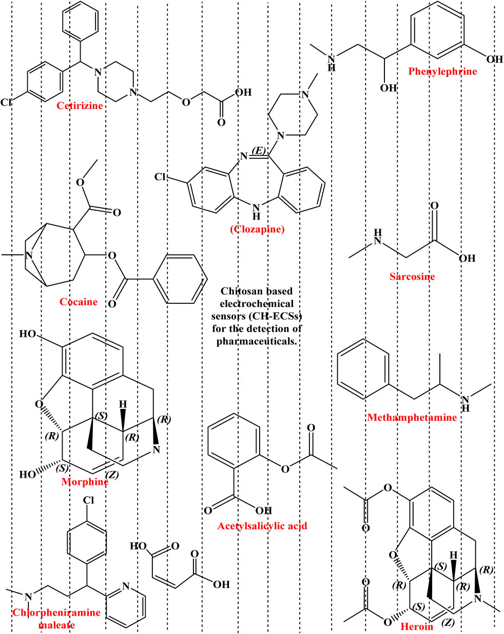
Figure 10. Electrochemical sensors based on CH, employed within the medical field to detect pharmaceuticals and monitor their potency to prevent multiple illnesses or mortality.
6 Conclusion and future perspectives
The projected growth of chitosan-based nanostructures in the medical field holds great promise for revolutionary developments in diagnosis, counseling, and numerous other areas of medicine. The distinctive attributes of CH and the diversity of nanotechnologies have made these small structures essential tools in combating severe medical conditions. Tweaking of CH-nanostructures for personalized healthcare has shifted the field while making it possible to provide trained professionals with remedies that are tailored to the particular requirements of each patient. With a focus on customized and responsive pharmaceuticals, ongoing advancements in drug delivery systems provide new avenues for more effective therapy with fewer side effects. Moreover, the integration of point-of-care diagnostics with contemporary imaging agents demonstrates the potential of chitosan-based nanostructures to revolutionize sickness diagnosis and monitoring. With continued development, these nanostructures have the potential to completely redefine the regulations for medical care by providing individualized treatments that take into account the particular details of every patient’s medical path. The continued investigation of chitosan-based nanostructures is evidence of the bio-medical research communities’ unwavering determination to achieve excellence and their potential to make revolutionary advances in global healthcare.
Bio-engineered chitosan-based nanostructures hold tremendous potential for revolutionizing disease treatment in various ways. Their therapeutic potential must be fully explored by tailoring these nanostructures to address particular problems in multiple illnesses. Utilizing modification techniques on chitosan-functionalized nanostructures presents promising opportunities for improving their adaptability, efficiency, and significance for different clinical applications in bio-medicine. Target cells, tissues, or macromolecules could be selectively linked with tiny structures by adopting molecular engineering techniques to bind particular ligands, peptides, or targeting moieties. Subsequent approaches could concentrate on building responsive chitosan-based nanostructures that can adjust according to variations in their surroundings. It is possible to construct pH-, temperature-, or stimuli-responsive nanostructures that would respond dynamically to the unique conditions of disease locations and maximize therapeutic outcomes while delivering regulated drug release. Advanced analytical techniques, spectroscopy, and high-resolution imaging will illuminate these nanostructures’ structural dynamics and functionality, enabling more intelligent design decisions. Future approaches for chitosan-based nanostructures will heavily rely on computational methods and in-vivo modeling. Virtual design and optimization of nanostructures can be simplified by predictive modeling, which enables researchers to effectively explore ample design space and find combinations with the best qualities for particular medicinal applications. As the area develops, techniques will shift towards an interdisciplinary and collaborative approach, incorporating engineering, chemistry, biology, and materials science knowledge. Combining expertise from several domains can quicken the development of novel chitosan-based nanostructures with a wide range of bio-medical uses. Subsequent approaches will deal with issues about clinical translation and regulatory compliance.
The engineering of chitosan-based nanostructures aids in combination therapy to hold numerous medicinal ingredients. This strategy shows great promise in treating complicated disorders like carcinoma, where the synergistic impact of multiple drugs may boost treatment outcomes and overcome drug resistance. Precision medicine and targeted therapies, innovative drug release systems, advanced imaging modalities, immunotherapy enhancement, and theranostics integration were the subsequent innovations in biomedicine.
Author contributions
AA: Writing – original draft, Writing – review and editing. MI: Conceptualization, Supervision, Writing – original draft, Writing – review and editing. MFW: Writing – original draft, Writing – review and editing. IA: Writing – original draft, Writing – review and editing. RP-S: Writing – original draft, Writing – review and editing. GG-S: Writing – original draft, Writing – review and editing. HI: Conceptualization, Supervision, Writing – original draft, Writing – review and editing.
Funding
The author(s) declare that no financial support was received for the research and/or publication of this article.
Acknowledgments
The listed co-authors of this comprehensive review extend their genuine gratefulness to their corresponding associated institutes/universities for their valuable support in providing access to the required literature sources. This literature assistance was instrumental in the productive finishing point of this scholarly review work.
Conflict of interest
The authors declare that the research was conducted in the absence of any commercial or financial relationships that could be construed as a potential conflict of interest.
The author(s) declared that they were an editorial board member of Frontiers, at the time of submission. This had no impact on the peer review process and the final decision.
Generative AI statement
The author(s) declare that no Generative AI was used in the creation of this manuscript.
Publisher’s note
All claims expressed in this article are solely those of the authors and do not necessarily represent those of their affiliated organizations, or those of the publisher, the editors and the reviewers. Any product that may be evaluated in this article, or claim that may be made by its manufacturer, is not guaranteed or endorsed by the publisher.
References
Abazari, R., Mahjoub, A. R., Ataei, F., Morsali, A., Carpenter-Warren, C. L., Mehdizadeh, K., et al. (2018). Chitosan immobilization on bio-MOF nanostructures: a biocompatible pH-responsive nanocarrier for doxorubicin release on MCF-7 cell lines of human breast cancer. Inorg. Chem. 57 (21), 13364–13379. doi:10.1021/acs.inorgchem.8b01955
Abd Elgadir, M., Uddin, M., Ferdosh, S., Adam, A., Chowdhury, A. J. K., and Sarker, M. I. (2015). Impact of chitosan composites and chitosan nanoparticle composites on various drug delivery systems: a review. J. food drug analysis 23 (4), 619–629. doi:10.1016/j.jfda.2014.10.008
Abdel-Monem, R. A., Rabie, S. T., El-Liethy, M. A., Hemdan, B. A., El-Nazer, H. A., and Gaballah, S. T. (2022). Chitosan-PVC conjugates/metal nanoparticles for biomedical applications. Polym. Adv. Technol. 33 (2), 514–523. doi:10.1002/pat.5533
Abolhassani, H., Eskandari, A., Saremi Poor, A., Zarrabi, A., Khodadadi, B., Karimifard, S., et al. (2024). Nanobiotechnological approaches for breast cancer Management: drug delivery systems and 3D In-Vitro models. Coord. Chem. Rev. 508, 215754. doi:10.1016/j.ccr.2024.215754
Adetunji, J. B. (2025). “Application of chitosan-based nanoparticles for the treatment of liver diseases,” in Chitosan-based nanoparticles for biomedical applications (Elsevier), 275–287.
Afshar, M., Dini, G., Vaezifar, S., Mehdikhani, M., and Movahedi, B. (2020). Preparation and characterization of sodium alginate/polyvinyl alcohol hydrogel containing drug-loaded chitosan nanoparticles as a drug delivery system. J. Drug Deliv. Sci. Technol. 56, 101530. doi:10.1016/j.jddst.2020.101530
Agarwal, T., Chiesa, I., Costantini, M., Lopamarda, A., Tirelli, M. C., Borra, O. P., et al. (2023). Chitosan and its derivatives in 3D/4D (bio) printing for tissue engineering and drug delivery applications. Int. J. Biol. Macromol. 246, 125669. doi:10.1016/j.ijbiomac.2023.125669
Ahirwar, R. K. (2023). An overview of medicinal plants: drugs of tomorrow. Stress-responsive Factors Mol. Farming Med. Plants, 1–16. doi:10.1007/978-981-99-4480-4_1
Ahmed, M. A., Ahmed, M. A., and Mohamed, A. A. (2023). Synthesis, characterization and application of chitosan/graphene oxide/copper ferrite nanocomposite for the adsorptive removal of anionic and cationic dyes from wastewater. RSC Adv. 13 (8), 5337–5352. doi:10.1039/d2ra07883j
Ahmed, S., and Ikram, S. (2016). Chitosan based scaffolds and their applications in wound healing. Achiev. life Sci. 10 (1), 27–37. doi:10.1016/j.als.2016.04.001
Akbar, M. U., Khattak, S., Khan, M. I., Saddozai, U. A. K., Ali, N., AlAsmari, A. F., et al. (2023). A pH-responsive bi-MIL-88B MOF coated with folic acid-conjugated chitosan as a promising nanocarrier for targeted drug delivery of 5-Fluorouracil. Front. Pharmacol. 14, 1265440. doi:10.3389/fphar.2023.1265440
Alavi, M., and Rai, M. (2021). “Antibacterial and wound healing activities of micro/nanocarriers based on carboxymethyl and quaternized chitosan derivatives,” in Biopolymer-based nano films (Elsevier), 191–201.
Alavi, S., and Mortazavi, S. A. (2018). Freeze-dried k-carrageenan/chitosan polyelectrolyte complex-based insert: a novel intranasal delivery system for sumatriptan succinate. Iran. J. Pharm. Res. IJPR 17 (4), 1172–1181.
Almajidi, Y. Q., Ponnusankar, S., Chaitanya, M., Marisetti, A. L., Hsu, C. Y., Dhiaa, A. M., et al. (2024). Chitosan-based nanofibrous scaffolds for biomedical and pharmaceutical applications: a comprehensive review. Int. J. Biol. Macromol. 264, 130683. doi:10.1016/j.ijbiomac.2024.130683
Alnaief, M., Obaidat, R., and Mashaqbeh, H. (2019). Loading and evaluation of meloxicam and atorvastatin in carrageenan microspherical aerogels particles. J. Appl. Pharm. Sci. 9 (1), 083–088. doi:10.7324/JAPS.2019.90112
Altinkok, C., Sagdic, G., Daglar, O., Ercan Ayra, M., Yuksel Durmaz, Y., Durmaz, H., et al. (2023). A new strategy for direct solution electrospinning of phosphorylated poly (vinyl chloride)/polyethyleneimine blend in alcohol media. Eur. Polym. J. 183, 111750. doi:10.1016/j.eurpolymj.2022.111750
Amani, H., Kazerooni, H., Hassanpoor, H., Akbarzadeh, A., and Pazoki-Toroudi, H. (2019). Tailoring synthetic polymeric biomaterials towards nerve tissue engineering: a review. Artif. cells, nanomedicine, Biotechnol. 47 (1), 3524–3539. doi:10.1080/21691401.2019.1639723
Ameer, M. W. A., and Maraie, N. K. (2019). Preparation and in vitro evaluation of implantable film containing microencapsulated sustained release dexamethasone sodium phosphate. Int. J. Pharm. Res. 11 (3). doi:10.31838/ijpr/2019.11.03.037
Amor, I. B., Emran, T. B., Hemmami, H., Zeghoud, S., and Laouini, S. E. (2023a). Nanomaterials based on chitosan for skin regeneration: an update. Int. J. Surg. 109 (3), 594–596. doi:10.1097/js9.0000000000000181
Amor, I. B., Hemmami, H., Laouini, S. E., Temam, H. B., Zaoui, H., and Barhoum, A. (2023b). Biosynthesis MgO and ZnO nanoparticles using chitosan extracted from Pimelia Payraudi Latreille for antibacterial applications. World J. Microbiol. Biotechnol. 39 (1), 19. doi:10.1007/s11274-022-03464-5
Anirudhan, T., and Nair, S. S. (2019). Polyelectrolyte complexes of carboxymethyl chitosan/alginate based drug carrier for targeted and controlled release of dual drug. J. Drug Deliv. Sci. Technol. 51, 569–582. doi:10.1016/j.jddst.2019.03.036
Anitha, A., Sowmya, S., Kumar, P. S., Deepthi, S., Chennazhi, K., Ehrlich, H., et al. (2014). Chitin and chitosan in selected biomedical applications. Prog. Polym. Sci. 39 (9), 1644–1667. doi:10.1016/j.progpolymsci.2014.02.008
Antil, R. (2023). “Chitosan-based biomaterials in biomedical applications,” in Advanced applications of biobased materials (Elsevier), 363–378.
Antony, R., Arun, T., and Manickam, S. T. D. (2019). A review on applications of chitosan-based Schiff bases. Int. J. Biol. Macromol. 129, 615–633. doi:10.1016/j.ijbiomac.2019.02.047
Aranaz, I., Alcántara, A. R., Civera, M. C., Arias, C., Elorza, B., Heras Caballero, A., et al. (2021). Chitosan: an overview of its properties and applications. Polymers 13 (19), 3256. doi:10.3390/polym13193256
Aranaz, I., Harris, R., and Heras, A. (2010). Chitosan amphiphilic derivatives. Chemistry and applications. Curr. Org. Chem. 14 (3), 308–330. doi:10.2174/138527210790231919
Asgher, M., Qamar, S. A., and Iqbal, H. M. (2021). Microbial exopolysaccharide-based nano-carriers with unique multi-functionalities for biomedical sectors. Biologia 76, 673–685. doi:10.2478/s11756-020-00588-7
Ashrafizadeh, M., Hushmandi, K., Mirzaei, S., Bokaie, S., Bigham, A., Makvandi, P., et al. (2023). Chitosan-based nanoscale systems for doxorubicin delivery: exploring biomedical application in cancer therapy. Bioeng. and Transl. Med. 8 (1), e10325. doi:10.1002/btm2.10325
Asl, E. A., Pooresmaeil, M., and Namazi, H. (2023). Chitosan coated MOF/GO nanohybrid as a co-anticancer drug delivery vehicle: synthesis, characterization, and drug delivery application. Mater. Chem. Phys. 293, 126933. doi:10.1016/j.matchemphys.2022.126933
Azmana, M., Mahmood, S., Hilles, A. R., Rahman, A., Arifin, M. A. B., and Ahmed, S. (2021). A review on chitosan and chitosan-based bionanocomposites: promising material for combatting global issues and its applications. Int. J. Biol. Macromol. 185, 832–848. doi:10.1016/j.ijbiomac.2021.07.023
Baghani, L., Noroozi Heris, N., Khonsari, F., Dinarvand, S., Dinarvand, M., and Atyabi, F. (2022). Trimethyl-chitosan coated gold nanoparticles enhance delivery, cellular uptake and gene silencing effect of EGFR-siRNA in breast cancer cells. Front. Mol. Biosci. 9, 871541. doi:10.3389/fmolb.2022.871541
Bai, R., Zhang, X., Yong, H., Wang, X., Liu, Y., and Liu, J. (2019). Development and characterization of antioxidant active packaging and intelligent Al3+-sensing films based on carboxymethyl chitosan and quercetin. Int. J. Biol. Macromol. 126, 1074–1084. doi:10.1016/j.ijbiomac.2018.12.264
Bajer, D. (2023). Hypophosphite cross-linked starch succinate/chitosan membranes as alternative for packaging and pharmaceutical application. Int. J. Biol. Macromol. 249, 126103. doi:10.1016/j.ijbiomac.2023.126103
Bakil, S. N. A. (2020). Sodium alginate-zinc oxide nanocomposite film for antibacterial wound healing applications. Biointerface Res. Appl. Chem. 10 (2), 6245–6252. doi:10.33263/BRIAC105.62456252
Baliga, M. S., and Dsouza, J. J. (2011). Amla (Emblica officinalis Gaertn), a wonder berry in the treatment and prevention of cancer. Eur. J. Cancer Prev. 20 (3), 225–239. doi:10.1097/cej.0b013e32834473f4
Baran, T., Menteş, A., and Arslan, H. (2015). Synthesis and characterization of water soluble O-carboxymethyl chitosan Schiff bases and Cu (II) complexes. Int. J. Biol. Macromol. 72, 94–103. doi:10.1016/j.ijbiomac.2014.07.029
Barbosa, A. I., Coutinho, A. J., Costa Lima, S. A., and Reis, S. (2019). Marine polysaccharides in pharmaceutical applications: fucoidan and chitosan as key players in the drug delivery match field. Mar. Drugs 17 (12), 654. doi:10.3390/md17120654
Barbosa, H. F. G., Attjioui, M., Ferreira, A. P. G., Moerschbacher, B. M., and Cavalheiro, É. T. G. (2020). New series of metal complexes by amphiphilic biopolymeric Schiff bases from modified chitosans: preparation, characterization and effect of molecular weight on its biological applications. Int. J. Biol. Macromol. 145, 417–428. doi:10.1016/j.ijbiomac.2019.12.153
Barjasteh, M., Dehnavi, S. M., Ahmadi Seyedkhani, S., Rahnamaee, S. Y., and Golizadeh, M. (2023). Improved biological activities of dual nanofibrous chitosan/bacterial cellulose wound dressing by a novel silver-based metal-organic framework. Surfaces Interfaces 36, 102631. doi:10.1016/j.surfin.2023.102631
Bashir, S. M., Ahmed Rather, G., Patrício, A., Haq, Z., Sheikh, A. A., Shah, M. Z. u. H., et al. (2022). Chitosan nanoparticles: a versatile platform for biomedical applications. Materials 15 (19), 6521. doi:10.3390/ma15196521
Baykara, D., Pilavci, E., Ulag, S., Valentine Okoro, O., Nie, L., Shavandi, A., et al. (2023). In vitro electrically controlled amoxicillin release from 3D-printed chitosan/bismuth ferrite scaffolds. Eur. Polym. J. 193, 112105. doi:10.1016/j.eurpolymj.2023.112105
Beman, E., Borhan, S., and Hesaraki, S. (2023). Macroporous nanostructured calcium phosphate/chitosan-gelatin composite bone tissue engineering scaffold. Curr. Mater. Sci. Former. Recent Pat. Mater. Sci. 16 (4), 443–452. doi:10.2174/2666145416666230111104341
Ben Amor, I., Hemmami, H., Laouini, S. E., Mahboub, M. S., and Barhoum, A. (2022). Sol-gel synthesis of ZnO nanoparticles using different chitosan sources: effects on antibacterial activity and photocatalytic degradation of AZO Dye. Catalysts 12 (12), 1611. doi:10.3390/catal12121611
Bhattacharjee, R., Dubey, A. K., Ganguly, A., Bhattacharya, B., Mishra, Y. K., Mostafavi, E., et al. (2022). State-of-art high-performance Nano-systems for mutated coronavirus infection management: from Lab to Clinic. OpenNano 8, 100078. doi:10.1016/j.onano.2022.100078
Bhattacharjee, R., Negi, A., Bhattacharya, B., Dey, T., Mitra, P., Preetam, S., et al. (2023). Nanotheranostics to target antibiotic-resistant bacteria: strategies and applications. OpenNano 11, 100138. doi:10.1016/j.onano.2023.100138
Bhattacharyya, A., Ham, H. w., Sonh, J., Gunbayar, M., Jeffy, R., Nagarajan, R., et al. (2023). 3D bioprinting of complex tissue scaffolds with in situ homogeneously mixed alginate-chitosan-kaolin bioink using advanced portable biopen. Carbohydr. Polym. 317, 121046. doi:10.1016/j.carbpol.2023.121046
Borhade, D. D., Nangare, S. N., Patil, D. A., Patil, P. O., Patil, G. S., and Patil, G. B. (2023). Preparation of pirfenidone loaded chitosan-polyvinyl alcohol-graphene oxide-based scaffold: spectroscopical characterizations and antibacterial activity. J. Drug Deliv. Sci. Technol. 82, 104325. doi:10.1016/j.jddst.2023.104325
Bounegru, A. V., and Bounegru, I. (2023). Chitosan-based electrochemical sensors for pharmaceuticals and clinical applications. Polymers 15 (17), 3539. doi:10.3390/polym15173539
Brunel, F., Véron, L., David, L., Domard, A., and Delair, T. (2008). A novel synthesis of chitosan nanoparticles in reverse emulsion. Langmuir 24 (20), 11370–11377. doi:10.1021/la801917a
Budiarso, I. J., Rini, N. D., Tsalsabila, A., Birowosuto, M. D., and Wibowo, A. (2023). Chitosan-based smart biomaterials for biomedical applications: Progress and perspectives. ACS Biomater. Sci. Eng. 9 (6), 3084–3115. doi:10.1021/acsbiomaterials.3c00216
Bulatao, B. P., Nalinratana, N., Jantaratana, P., Vajragupta, O., Rojsitthisak, P., and Rojsitthisak, P. (2023). Lutein-loaded chitosan/alginate-coated Fe3O4 nanoparticles as effective targeted carriers for breast cancer treatment. Int. J. Biol. Macromol. 242, 124673. doi:10.1016/j.ijbiomac.2023.124673
Busilacchi, A., Gigante, A., Mattioli-Belmonte, M., Manzotti, S., and Muzzarelli, R. A. (2013). Chitosan stabilizes platelet growth factors and modulates stem cell differentiation toward tissue regeneration. Carbohydr. Polym. 98 (1), 665–676. doi:10.1016/j.carbpol.2013.06.044
Butnariu, M. (2023). “Biological and chemical aspects of chitosan,” in Chitosan nanocomposites: bionanomechanical applications (Springer), 27–54.
Campelo, C. S., Lima, L. D., Rebêlo, L. M., Mantovani, D., Beppu, M. M., and Vieira, R. S. (2016). In vitro evaluation of anti-calcification and anti-coagulation on sulfonated chitosan and carrageenan surfaces. Mater. Sci. Eng. C 59, 241–248. doi:10.1016/j.msec.2015.10.020
Cao, L., Wang, J., Hou, J., Xing, W., and Liu, C. (2014a). Vascularization and bone regeneration in a critical sized defect using 2-N, 6-O-sulfated chitosan nanoparticles incorporating BMP-2. Biomaterials 35 (2), 684–698. doi:10.1016/j.biomaterials.2013.10.005
Cao, L., Werkmeister, J. A., Wang, J., Glattauer, V., McLean, K. M., and Liu, C. (2014b). Bone regeneration using photocrosslinked hydrogel incorporating rhBMP-2 loaded 2-N, 6-O-sulfated chitosan nanoparticles. Biomaterials 35 (9), 2730–2742. doi:10.1016/j.biomaterials.2013.12.028
Capel, A. J., Rimington, R. P., Lewis, M. P., and Christie, S. D. R. (2018). 3D printing for chemical, pharmaceutical and biological applications. Nat. Rev. Chem. 2 (12), 422–436. doi:10.1038/s41570-018-0058-y
Carmona-Ribeiro, A. M., and Pérez-Betancourt, Y. (2020). Cationic nanostructures for vaccines design. Biomimetics 5 (3), 32. doi:10.3390/biomimetics5030032
Chattopadhyay, D., and Inamdar, M. S. (2010). Aqueous behaviour of chitosan. Int. J. Polym. Sci. 2010, 1–7. doi:10.1155/2010/939536
Chen, M.-C., Mi, F. L., Liao, Z. X., Hsiao, C. W., Sonaje, K., Chung, M. F., et al. (2013). Recent advances in chitosan-based nanoparticles for oral delivery of macromolecules. Adv. drug Deliv. Rev. 65 (6), 865–879. doi:10.1016/j.addr.2012.10.010
Chen, S.-K., Pan, D. W., Liu, Z., Wang, W., Xie, R., Mu, X. T., et al. (2023b). Microfluidic fabrication of calcium alginate/chitosan composite microcapsules with ultrathin shells for peroxidase immobilization. Industrial and Eng. Chem. Res. 62, 10101–10111. doi:10.1021/acs.iecr.3c01062
Chen, T., Zhu, W., Liang, Z., and Zeng, Q. (2019). Self-assembly pH-sensitive chitosan/alginate coated polyelectrolyte complexes for oral delivery of insulin. J. Microencapsul. 36 (1), 96–107. doi:10.1080/02652048.2019.1604846
Chen, W.-C., Ou, S. F., Zheng, N. C., and Chien, H. W. (2023a). Chitosan coating fabricated through nanotopography and alkylation for the prevention of bacterial attachment and corrosion. Prog. Org. Coatings 174, 107281. doi:10.1016/j.porgcoat.2022.107281
Chen, X., Zhang, S. L., Xiao, S. H., Li, Z. F., and Li, G. (2023c). Ultrahigh proton conductivities of postmodified Hf (IV) metal–organic frameworks and related chitosan-based composite membranes. ACS Appl. Mater. and Interfaces 15 (29), 35128–35139. doi:10.1021/acsami.3c08007
Cheng, F., Wang, B., and Xia, Y. (2018). Synthesis and characterization of O-acetyl-chitosan acetic ester. Int. J. Polym. Sci. 2018, 1–8. doi:10.1155/2018/4960416
Choi, J., Hwang, D. S., Lim, C., and Lee, D. W. (2024). Interaction mechanism between low molecular weight chitosan nanofilm and functionalized surfaces in aqueous solutions. Carbohydr. Polym. 324, 121504. doi:10.1016/j.carbpol.2023.121504
Choong, Y. Y. C., Tan, H. W., Patel, D. C., Choong, W. T. N., Chen, C. H., Low, H. Y., et al. (2020). The global rise of 3D printing during the COVID-19 pandemic. Nat. Rev. Mater. 5 (9), 637–639. doi:10.1038/s41578-020-00234-3
Chopin, N., Guillory, X., Weiss, P., Bideau, J., and Colliec-Jouault, S. (2014). Design polysaccharides of marine origin: chemical modifications to reach advanced versatile compounds. Curr. Org. Chem. 18 (7), 867–895. doi:10.2174/138527281807140515152334
Chowdhuri, A. R., Singh, T., Ghosh, S. K., and Sahu, S. K. (2016). Carbon dots embedded magnetic nanoparticles@ chitosan@ metal organic framework as a nanoprobe for pH sensitive targeted anticancer drug delivery. ACS Appl. Mater. and interfaces 8 (26), 16573–16583. doi:10.1021/acsami.6b03988
Ćirić, A., Krajišnik, D., Čalija, B., and Đekić, L. (2020). Biocompatible non-covalent complexes of chitosan and different polymers: characteristics and application in drug delivery. Arh. za Farm. 70 (4), 173–197. doi:10.5937/arhfarm2004173q
Costa, S. A., Simon, J. R., Amiram, M., Tang, L., Zauscher, S., Brustad, E. M., et al. (2018). Photo-Crosslinkable unnatural amino acids enable facile synthesis of thermoresponsive nano-to microgels of intrinsically disordered polypeptides. Adv. Mater. 30 (5), 1704878. doi:10.1002/adma.201704878
Croce, M., Conti, S., Maake, C., and Patzke, G. R. (2016). Synthesis and screening of N-acyl thiolated chitosans for antibacterial applications. Carbohydr. Polym. 151, 1184–1192. doi:10.1016/j.carbpol.2016.06.014
Cui, X., Guan, X., Zhong, S., Chen, J., Zhu, H., Li, Z., et al. (2017). Multi-stimuli responsive smart chitosan-based microcapsules for targeted drug delivery and triggered drug release. Ultrason. sonochemistry 38, 145–153. doi:10.1016/j.ultsonch.2017.03.011
Dardeer, H. M., Taha, A. G., Abouzeid, R. E., and Aly, M. F. (2022). Novel chitosan-acetyl isatin polymer derivatives: synthesis, characterization, and applications in bone tissue engineering. Biomass Convers. Biorefinery 14, 12427–12440. doi:10.1007/s13399-022-03176-8
Dattilo, M., Patitucci, F., Prete, S., Parisi, O. I., and Puoci, F. (2023). Polysaccharide-based hydrogels and their application as drug delivery systems in cancer treatment: a review. J. Funct. Biomaterials 14 (2), 55. doi:10.3390/jfb14020055
Dayan, S., Ozpozan, N. K., Özdemir, N., and Dayan, O. (2014). Synthesis of some ruthenium (II)–Schiff base complexes bearing sulfonamide fragment: new catalysts for transfer hydrogenation of ketones. J. Organomet. Chem. 770, 21–28. doi:10.1016/j.jorganchem.2014.08.002
Dehghankar, M., Hmtshirazi, R., Mohammadi, T., and Tofighy, M. A. (2023). Synthesis and modification methods of metal-organic frameworks and their application in modification of polymeric ultrafiltration membranes: a review. J. Environ. Chem. Eng. 11, 109954. doi:10.1016/j.jece.2023.109954
Deng, A., Himmelsbach, M., Zhu, Q. Z., Frey, S., Sengl, M., Buchberger, W., et al. (2003). Residue analysis of the pharmaceutical diclofenac in different water types using ELISA and GC− MS. Environ. Sci. and Technol. 37 (15), 3422–3429. doi:10.1021/es0341945
de Souza, F. L. C., Simon, A., Barcelos D’Ornellas, E. L., Silva, R. F. d., Covas, A. C. M., Rodrigues, C. R., et al. (2023). Chitosan-based hydrotalcite nanostructured membranes containing sodium alendronate for guided bone regeneration therapy. J. Appl. Pharm. Sci. 13 (8), 113–124. doi:10.7324/japs.2023.40064
Dhlamini, K. S., Selepe, C. T., Ramalapa, B., Tshweu, L., and Ray, S. S. (2024). Reimagining chitosan-based antimicrobial biomaterials to mitigate antibiotic resistance and alleviate antibiotic overuse: a review. Macromol. Mater. Eng. 309, 2400018. doi:10.1002/mame.202400018
Dimassi, S., Tabary, N., Chai, F., Blanchemain, N., and Martel, B. (2018). Sulfonated and sulfated chitosan derivatives for biomedical applications: a review. Carbohydr. Polym. 202, 382–396. doi:10.1016/j.carbpol.2018.09.011
Ding, S., Wang, Y., Li, J., and Chen, S. (2021). Progress and prospects in chitosan derivatives: modification strategies and medical applications. J. Mater. Sci. and Technol. 89, 209–224. doi:10.1016/j.jmst.2020.12.008
Diouf, A., Moufid, M., Bouyahya, D., Österlund, L., El Bari, N., and Bouchikhi, B. (2020). An electrochemical sensor based on chitosan capped with gold nanoparticles combined with a voltammetric electronic tongue for quantitative aspirin detection in human physiological fluids and tablets. Mater. Sci. Eng. C 110, 110665. doi:10.1016/j.msec.2020.110665
Dongsar, T. T., Gupta, N., Almalki, W. H., Sahebkar, A., and Kesharwani, P. (2023). Emerging potential of 5-Fluorouracil-loaded chitosan nanoparticles in cancer therapy. J. Drug Deliv. Sci. Technol. 82, 104371. doi:10.1016/j.jddst.2023.104371
Dumont, V. C., Mansur, A. A., Carvalho, S. M., Medeiros Borsagli, F. G., Pereira, M. M., and Mansur, H. S. (2016). Chitosan and carboxymethyl-chitosan capping ligands: effects on the nucleation and growth of hydroxyapatite nanoparticles for producing biocomposite membranes. Mater. Sci. Eng. C 59, 265–277. doi:10.1016/j.msec.2015.10.018
Ebrahimnejad, P., Rezaeiroshan, A., Babaei, A., Khanali, A., Aghajanshakeri, S., Farmoudeh, A., et al. (2023). Hyaluronic acid-coated chitosan/gelatin nanoparticles as a new strategy for topical delivery of metformin in melanoma. BioMed Res. Int. 2023, 3304105. doi:10.1155/2023/3304105
Ebrahimzadeh, S., Biswas, D., Roy, S., and McClements, D. J. (2023). Incorporation of essential oils in edible seaweed-based films: a comprehensive review. Trends Food Sci. and Technol. 135, 43–56. doi:10.1016/j.tifs.2023.03.015
Eivazzadeh-Keihan, R., Pajoum, Z., Aliabadi, H. A. M., Mohammadi, A., Kashtiaray, A., Bani, M. S., et al. (2023). Magnetized chitosan hydrogel and silk fibroin, reinforced with PVA: a novel nanobiocomposite for biomedical and hyperthermia applications. RSC Adv. 13 (13), 8540–8550. doi:10.1039/d3ra00612c
Elangwe, C. N., Morozkina, S. N., Olekhnovich, R. O., Polyakova, V. O., Krasichkov, A., Yablonskiy, P. K., et al. (2023). Pullulan-based hydrogels in wound healing and skin tissue engineering applications: a review. Int. J. Mol. Sci. 24 (5), 4962. doi:10.3390/ijms24054962
El Idrissi El Hassani, C. (2023). “Biomedical applications of chitosan-based nanostructured composite materials,” in Chitosan nanocomposites: bionanomechanical applications (Springer), 81–107.
El-Maadawy, M. W., Mohamed, R. R., and Hanna, D. H. (2022). Preparation of carrageenan/chitosan-based N, N, N-trimeth yl chitosan chloride silver nanocomposites as pH sensitive carrier for effective controlled curcumin delivery in cancer cells. OpenNano 7, 100050. doi:10.1016/j.onano.2022.100050
Elmoghayer, M. E., Saleh, N. M., and Abu Hashim, I. I. (2023). Enhanced oral delivery of hesperidin-loaded sulfobutylether-β-cyclodextrin/chitosan nanoparticles for augmenting its hypoglycemic activity: in vitro-in vivo assessment study. Drug Deliv. Transl. Res. 14, 895–917. doi:10.1007/s13346-023-01440-6
El-Naggar, M. E., Abdelgawad, A. M., Abdel-Sattar, R., Gibriel, A. A., and Hemdan, B. A. (2023). Potential antimicrobial and antibiofilm efficacy of essential oil nanoemulsion loaded polycaprolactone nanofibrous dermal patches. Eur. Polym. J. 184, 111782. doi:10.1016/j.eurpolymj.2022.111782
El Sayed, M. M. (2023). Production of polymer hydrogel composites and their applications. J. Polym. Environ. 31, 2855–2879. doi:10.1007/s10924-023-02796-z
El-Shabouri, M. H. (2002). Positively charged nanoparticles for improving the oral bioavailability of cyclosporin-A. Int. J. Pharm. 249 (1-2), 101–108. doi:10.1016/s0378-5173(02)00461-1
Essa, D., Kondiah, P. P. D., Kumar, P., and Choonara, Y. E. (2023). Design of chitosan-coated, quercetin-loaded PLGA nanoparticles for enhanced PSMA-specific activity on LnCap prostate cancer cells. Biomedicines 11 (4), 1201. doi:10.3390/biomedicines11041201
Fard, G. H., Moinipoor, Z., Anastasova-Ivanova, S., Iqbal, H. M., Dwek, M. V., Getting, S., et al. (2022). Development of chitosan, pullulan, and alginate based drug-loaded nano-emulsions as a potential malignant melanoma delivery platform. Carbohydr. Polym. Technol. Appl. 4, 100250. doi:10.1016/j.carpta.2022.100250
Faria, R. R., Guerra, R. F., de Sousa Neto, L. R., Motta, L. F., and Franca, E. d. F. (2016). Computational study of polymorphic structures of α-and β-chitin and chitosan in aqueous solution. J. Mol. Graph. Model. 63, 78–84. doi:10.1016/j.jmgm.2015.11.001
Fathi-Karkan, S., Mirinejad, S., Ulucan-Karnak, F., Mukhtar, M., Ghahramani Almanghadim, H., Sargazi, S., et al. (2023). Biomedical applications of aptamer-modified chitosan nanomaterials: an updated review. Int. J. Biol. Macromol. 238, 124103. doi:10.1016/j.ijbiomac.2023.124103
Fatullayeva, S., Tagiyev, D., Zeynalov, N., Mammadova, S., and Aliyeva, E. (2022). Recent advances of chitosan-based polymers in biomedical applications and environmental protection. J. Polym. Res. 29 (7), 259. doi:10.1007/s10965-022-03121-3
Federer, C., Kurpiers, M., and Bernkop-Schnürch, A. (2020). Thiolated chitosans: a multi-talented class of polymers for various applications. Biomacromolecules 22 (1), 24–56. doi:10.1021/acs.biomac.0c00663
Feng, J., Kong, F., Yue, W. S., Yu, H., He, Z. L., Zhai, Y. N., et al. (2023). Covalent organic framework-based nanozyme for cascade-amplified synergistic cancer therapy. Sci. China Mater. 66, 4079–4089. doi:10.1007/s40843-023-2560-8
Feng, R., Wang, L., Zhou, P., Luo, Z., Li, X., and Gao, L. (2020). Development of the pH responsive chitosan-alginate based microgel for encapsulation of Jughans regia L. polyphenols under simulated gastrointestinal digestion in vitro. Carbohydr. Polym. 250, 116917. doi:10.1016/j.carbpol.2020.116917
Feng, W., and Wang, Z. (2022). Biomedical applications of chitosan-graphene oxide nanocomposites. Iscience 25 (1), 103629. doi:10.1016/j.isci.2021.103629
Fonseca-Santos, B., and Chorilli, M. (2017). An overview of carboxymethyl derivatives of chitosan: their use as biomaterials and drug delivery systems. Mater. Sci. Eng. C 77, 1349–1362. doi:10.1016/j.msec.2017.03.198
Frigaard, J., Jensen, J. L., Galtung, H. K., and Hiorth, M. (2022). The potential of chitosan in nanomedicine: an overview of the cytotoxicity of chitosan based nanoparticles. Front. Pharmacol. 13, 880377. doi:10.3389/fphar.2022.880377
Gaballah, S. T., El-Nazer, H. A., Abdel-Monem, R. A., El-Liethy, M. A., Hemdan, B. A., and Rabie, S. T. (2019). Synthesis of novel chitosan-PVC conjugates encompassing Ag nanoparticles as antibacterial polymers for biomedical applications. Int. J. Biol. Macromol. 121, 707–717. doi:10.1016/j.ijbiomac.2018.10.085
Gasti, T., Dixit, S., Hiremani, V. D., Chougale, R. B., Masti, S. P., Vootla, S. K., et al. (2022). Chitosan/pullulan based films incorporated with clove essential oil loaded chitosan-ZnO hybrid nanoparticles for active food packaging. Carbohydr. Polym. 277, 118866. doi:10.1016/j.carbpol.2021.118866
Gatou, M.-A., Vagena, I. A., Lagopati, N., Pippa, N., Gazouli, M., and Pavlatou, E. A. (2023). Functional MOF-based materials for environmental and biomedical applications: a critical review. Nanomaterials 13 (15), 2224. doi:10.3390/nano13152224
George, A., and Shrivastav, P. S. (2023a). Preparation and optimization of tetraethyl orthosilicate cross-linked chitosan-guar gum-poly (vinyl alcohol) composites reinforced with montmorillonite for sustained release of sitagliptin. Int. J. Biol. Macromol. 229, 51–61. doi:10.1016/j.ijbiomac.2022.12.302
George, A., and Shrivastav, P. S. (2023b). Preparation and evaluation of chitosan-alginate/carrageenan hydrogel for oral drug delivery in the treatment of diabetes. J. Bioact. Compatible Polym. 38 (5), 368–386. doi:10.1177/08839115231183487
Gericke, M., Amaral, A. J., Budtova, T., De Wever, P., Groth, T., Heinze, T., et al. (2024). The European Polysaccharide Network of Excellence (EPNOE) research roadmap 2040: advanced strategies for exploiting the vast potential of polysaccharides as renewable bioresources. Carbohydr. Polym. 326, 121633. doi:10.1016/j.carbpol.2023.121633
Ghaz-Jahanian, M. A., Abbaspour-Aghdam, F., Anarjan, N., Berenjian, A., and Jafarizadeh-Malmiri, H. (2015). Application of chitosan-based nanocarriers in tumor-targeted drug delivery. Mol. Biotechnol. 57, 201–218. doi:10.1007/s12033-014-9816-3
Giannopoulos, A. A., Mitsouras, D., Yoo, S. J., Liu, P. P., Chatzizisis, Y. S., and Rybicki, F. J. (2016). Applications of 3D printing in cardiovascular diseases. Nat. Rev. Cardiol. 13 (12), 701–718. doi:10.1038/nrcardio.2016.170
Goharshadi, E. K., Goharshadi, K., and Moghayedi, M. (2022). The use of nanotechnology in the fight against viruses: a critical review. Coord. Chem. Rev. 464, 214559. doi:10.1016/j.ccr.2022.214559
Golichenari, B., Nosrati, R., Farokhi-Fard, A., Faal Maleki, M., Gheibi Hayat, S. M., Ghazvini, K., et al. (2019). Electrochemical-based biosensors for detection of Mycobacterium tuberculosis and tuberculosis biomarkers. Crit. Rev. Biotechnol. 39 (8), 1056–1077. doi:10.1080/07388551.2019.1668348
Graça, M. F., Melo, B. L., Lima-Sousa, R., Ferreira, P., Moreira, A. F., and Correia, I. J. (2023). Reduced graphene oxide-enriched chitosan hydrogel/cellulose acetate-based nanofibers application in mild hyperthermia and skin regeneration. Int. J. Biol. Macromol. 229, 224–235. doi:10.1016/j.ijbiomac.2022.12.291
Grenha, A., Gomes, M. E., Rodrigues, M., Santo, V. E., Mano, J. F., Neves, N. M., et al. (2010). Development of new chitosan/carrageenan nanoparticles for drug delivery applications. J. Biomed. Mater. Res. Part A 92 (4), 1265–1272. doi:10.1002/jbm.a.32466
Gu, Z., Zhu, S., Yan, L., Zhao, F., and Zhao, Y. (2019). Graphene-based smart platforms for combined Cancer therapy. Adv. Mater. 31 (9), 1800662. doi:10.1002/adma.201800662
Guo, J., Lv, A., Wu, J., Sun, E., Zhu, Y., Zhang, X., et al. (2023b). Bandage modified with antibacterial films of quaternized chitosan and sodium carboxymethyl cellulose microgels/baicalein nanoparticles for accelerating infected wound healing. Int. J. Biol. Macromol. 250, 126274. doi:10.1016/j.ijbiomac.2023.126274
Guo, Y., Feng, H., Li, W., Wang, W., Yu, M., and Chen, S. (2023a). Enzyme and pH dual-responsive CAP@ CS@ PLGA microcapsules for controlled release antibacterial application. Biochem. Eng. J. 196, 108956. doi:10.1016/j.bej.2023.108956
Gurunathan, S., and Kim, J.-H. (2016). Synthesis, toxicity, biocompatibility, and biomedical applications of graphene and graphene-related materials. Int. J. nanomedicine 11, 1927–1945. doi:10.2147/ijn.s105264
Habibi Khoei, Z. (2023). Inhibition of miR-96 and miR-183 expression in treatment with fisetin-loaded chitosan nanoparticles in breast cancer cell lines. Nanomedicine Res. J. 8 (3), 290–300. doi:10.22034/nmrj.2023.03.008
Hadi, Z., Navarchian, A. H., and Rafienia, M. (2023). Synthesis of pH-responsive carboxymethyl chitosan for encapsulating tetracycline-HCl: morphology, drug release behavior and antibacterial activity of microcapsules. J. Drug Deliv. Sci. Technol. 84, 104462. doi:10.1016/j.jddst.2023.104462
Hamza, M. F., Guibal, E., Wei, Y., and Fouda, A. (2023). Magnetic amino-sulfonic dual sorbent for uranyl sorption from aqueous solutions–Influence of light irradiation on sorption properties. Chem. Eng. J. 456, 141099. doi:10.1016/j.cej.2022.141099
Han, G., Liu, S., Pan, Z., Lin, Y., Ding, S., Li, L., et al. (2020). Sulfonated chitosan and phosphorylated chitosan coated polylactide membrane by polydopamine-assisting for the growth and osteogenic differentiation of MC3T3-E1s. Carbohydr. Polym. 229, 115517. doi:10.1016/j.carbpol.2019.115517
Hanna, D. H., Hamed, A. A., and Saad, G. R. (2023). Synthesis and characterization of poly (3-hydroxybutyrate)/chitosan-graft poly (acrylic acid) conjugate hyaluronate for targeted delivery of methotrexate drug to colon cancer cells. Int. J. Biol. Macromol. 240, 124396. doi:10.1016/j.ijbiomac.2023.124396
He, J., Ma, Y., Niu, X., Pei, J., Yan, R., Xu, F., et al. (2024). Silver nanoparticles induce endothelial cytotoxicity through ROS-mediated mitochondria-lysosome damage and autophagy perturbation: the protective role of N-acetylcysteine. Toxicology 502, 153734. doi:10.1016/j.tox.2024.153734
Herdiana, Y., Wathoni, N., Gozali, D., Shamsuddin, S., and Muchtaridi, M. (2023). Chitosan-based nano-smart drug delivery system in breast cancer therapy. Pharmaceutics 15 (3), 879. doi:10.3390/pharmaceutics15030879
Herdiana, Y., Wathoni, N., Shamsuddin, S., and Muchtaridi, M. (2022). Drug release study of the chitosan-based nanoparticles. Heliyon 8, e08674. doi:10.1016/j.heliyon.2021.e08674
Heydari, S. R., Ghahremani, M. H., Atyabi, F., Bafkary, R., Jaafari, M. R., and Dinarvand, R. (2023). Aptamer-modified chitosan-capped mesoporous silica nanoparticles for co-delivery of cytarabine and daunorubicin in leukemia. Int. J. Pharm. 646, 123495. doi:10.1016/j.ijpharm.2023.123495
Hirano, S., Yamaguchi, R., Matsuda, N., Miura, O., and Kondo, Y. (1977). Chitosan-aldehyde gel a novel polysaccharide gel produced from chitosan and aldehydes. Agric. Biol. Chem. 41 (8), 1547–1548. doi:10.1271/bbb1961.41.1547
Huang, W., Wang, Y., Huang, Z., Wang, X., Chen, L., Zhang, Y., et al. (2018). On-demand dissolvable self-healing hydrogel based on carboxymethyl chitosan and cellulose nanocrystal for deep partial thickness burn wound healing. ACS Appl. Mater. and interfaces 10 (48), 41076–41088. doi:10.1021/acsami.8b14526
Huang, Y.-y., Yao, Q. b., Jia, X. z., Chen, B. r., Abdul, R., Wang, L. h., et al. (2023). Characterization and application in yogurt of genipin-crosslinked chitosan microcapsules encapsulating with Lactiplantibacillus plantarum DMDL 9010. Int. J. Biol. Macromol. 248, 125871. doi:10.1016/j.ijbiomac.2023.125871
Ibrahim, A., Khalil, I. A., Mahmoud, M. Y., Bakr, A. F., Ghoniem, M. G., Al-Farraj, E. S., et al. (2023). Layer-by-layer development of chitosan/alginate-based platelet-mimicking nanocapsules for augmenting doxorubicin cytotoxicity against breast cancer. Int. J. Biol. Macromol. 225, 503–517. doi:10.1016/j.ijbiomac.2022.11.107
Ito, T., Yoshida, C., and Murakami, Y. (2013). Design of novel sheet-shaped chitosan hydrogel for wound healing: a hybrid biomaterial consisting of both PEG-grafted chitosan and crosslinkable polymeric micelles acting as drug containers. Mater. Sci. Eng. C 33 (7), 3697–3703. doi:10.1016/j.msec.2013.04.056
Jafernik, K., Ładniak, A., Blicharska, E., Czarnek, K., Ekiert, H., Wiącek, A. E., et al. (2023). Chitosan-based nanoparticles as effective drug delivery systems—a review. Molecules 28 (4), 1963. doi:10.3390/molecules28041963
Jamuna, G., Yasodha, S., Thamarai, P., Vickram, A., Swaminaathan, P., Saravanan, A., et al. (2023). Design strategies, utilization and applications of nano-engineered biomaterials for the enhancement of bioenergy: a sustainable approach. Biochem. Eng. J. 200, 109104. doi:10.1016/j.bej.2023.109104
Jang, M. K., Kong, B., Jeong, Y., Lee, C. H., and Nah, J. (2004). Physicochemical characterization of α-chitin, β-chitin, and γ-chitin separated from natural resources. J. Polym. Sci. Part A Polym. Chem. 42 (14), 3423–3432. doi:10.1002/pola.20176
Jayakumar, R., Prabaharan, M., Nair, S., Tokura, S., Tamura, H., and Selvamurugan, N. (2010). Novel carboxymethyl derivatives of chitin and chitosan materials and their biomedical applications. Prog. Mater. Sci. 55 (7), 675–709. doi:10.1016/j.pmatsci.2010.03.001
Jiang, M., Althomali, R. H., Ansari, S. A., Saleh, E. A. M., Gupta, J., Kambarov, K. D., et al. (2023a). Advances in preparation, biomedical, and pharmaceutical applications of chitosan-based gold, silver, and magnetic nanoparticles: a review. Int. J. Biol. Macromol. 251, 126390. doi:10.1016/j.ijbiomac.2023.126390
Jiang, Y., and Wu, J. (2019). Recent development in chitosan nanocomposites for surface-based biosensor applications. Electrophoresis 40 (16-17), 2084–2097. doi:10.1002/elps.201900066
Jiang, Z., Zhao, S., Fan, Z., Zhao, C., Zhang, L., Liu, D., et al. (2023b). A novel all-natural (collagen+ pectin)/chitosan aqueous two-phase microcapsule with improved anthocyanin loading capacity. Food Hydrocoll. 134, 107984. doi:10.1016/j.foodhyd.2022.107984
Jirofti, N., Hashemi, M., Moradi, A., and Kalalinia, F. (2023). Fabrication and characterization of 3D printing biocompatible crocin-loaded chitosan/collagen/hydroxyapatite-based scaffolds for bone tissue engineering applications. Int. J. Biol. Macromol. 252, 126279. doi:10.1016/j.ijbiomac.2023.126279
Kalachaveedu, M., Senthil, R., Azhagiyamanavalan, S., Ravi, R., Meenakshisundaram, H., and Dharmarajan, A. (2023). Traditional medicine herbs as natural product matrices in cancer chemoprevention: a trans pharmacological perspective (scoping review). Phytotherapy Res. 37 (4), 1539–1573. doi:10.1002/ptr.7747
Kaptan, Y., Karal-Yilmaz, O., Izbudak, B., Giray, B., Yilmaz, B., and Bal-Ozturk, A. (2023). Preparation of tetracycline hydrochloride loaded chitosan/silk fibroin/ZnO antibacterial biocomposite hydrogel sponges for wound healing application. J. Polym. Res. 30 (2), 49. doi:10.1007/s10965-022-03435-2
Karimi, K., Mojtabavi, S., Tehrany, P. M., Nejad, M. M., Rezaee, A., Mohtashamian, S., et al. (2023b). Chitosan-based nanoscale delivery systems in hepatocellular carcinoma: versatile bio-platform with theranostic application. Int. J. Biol. Macromol. 242, 124935. doi:10.1016/j.ijbiomac.2023.124935
Karimi, T., Mottaghitalab, F., Keshvari, H., and Farokhi, M. (2023a). Carboxymethyl chitosan/sodium carboxymethyl cellulose/agarose hydrogel dressings containing silk fibroin/polydopamine nanoparticles for antibiotic delivery. J. Drug Deliv. Sci. Technol. 80, 104134. doi:10.1016/j.jddst.2022.104134
Kaya, M., Mujtaba, M., Ehrlich, H., Salaberria, A. M., Baran, T., Amemiya, C. T., et al. (2017). On chemistry of γ-chitin. Carbohydr. Polym. 176, 177–186. doi:10.1016/j.carbpol.2017.08.076
Kazemi-Andalib, F., Mohammadikish, M., Divsalar, A., and Sahebi, U. (2022). Hollow microcapsule with pH-sensitive chitosan/polymer shell for in vitro delivery of curcumin and gemcitabine. Eur. Polym. J. 162, 110887. doi:10.1016/j.eurpolymj.2021.110887
Khalaf, E. M., Abood, N. A., Atta, R. Z., Ramírez-Coronel, A. A., Alazragi, R., Parra, R. M. R., et al. (2023). Recent progressions in biomedical and pharmaceutical applications of chitosan nanoparticles: a comprehensive review. Int. J. Biol. Macromol. 231, 123354. doi:10.1016/j.ijbiomac.2023.123354
Khatami, N., Guerrero, P., Martín, P., Quintela, E., Ramos, V., Saa, L., et al. (2024). Valorization of biological waste from insect-based food industry: assessment of chitin and chitosan potential. Carbohydr. Polym. 324, 121529. doi:10.1016/j.carbpol.2023.121529
Khubiev, O. M., Egorov, A. R., Kirichuk, A. A., Khrustalev, V. N., Tskhovrebov, A. G., and Kritchenkov, A. S. (2023). Chitosan-based antibacterial films for biomedical and food applications. Int. J. Mol. Sci. 24 (13), 10738. doi:10.3390/ijms241310738
Khurana, S., Kukreti, S., and Kaushik, M. (2023). Prospecting the cancer therapeutic edge of chitosan-based gold nanoparticles through conformation selective binding to the parallel G-quadruplex formed by short telomeric DNA sequence: a multi-spectroscopic approach. Int. J. Biol. Macromol. 253, 126835. doi:10.1016/j.ijbiomac.2023.126835
Kim, H.-J., Zhang, K., Moore, L., and Ho, D. (2014). Diamond nanogel-embedded contact lenses mediate lysozyme-dependent therapeutic release. ACS nano 8 (3), 2998–3005. doi:10.1021/nn5002968
Klicova, M., Rosendorf, J., Erben, J., and Horakova, J. (2023). Antiadhesive nanofibrous materials for medicine: preventing undesirable tissue adhesions. ACS omega 8, 20152–20162. doi:10.1021/acsomega.3c00341
Koirala, P., Nirmal, N. P., Woraprayote, W., Visessanguan, W., Bhandari, Y., Karim, N. U., et al. (2023). Nano-engineered edible films and coatings for seafood products. Food Packag. Shelf Life 38, 101135. doi:10.1016/j.fpsl.2023.101135
Kolawole, O. M., Ifeanafor, A. R., Ifade, W. A., Akinleye, M. O., Patrojanasophon, P., Silva, B. O., et al. (2023). Formulation and evaluation of paclitaxel-loaded boronated chitosan/alginate nanoparticles as a mucoadhesive system for localized cervical cancer drug delivery. J. Drug Deliv. Sci. Technol. 87, 104810. doi:10.1016/j.jddst.2023.104810
Kou, S. G., Peters, L. M., and Mucalo, M. R. (2021). Chitosan: a review of sources and preparation methods. Int. J. Biol. Macromol. 169, 85–94. doi:10.1016/j.ijbiomac.2020.12.005
Koumentakou, I., Noordam, M. J., Michopoulou, A., Terzopoulou, Z., and Bikiaris, D. N. (2023). 3D-Printed chitosan-based hydrogels loaded with levofloxacin for tissue engineering applications. Biomacromolecules 24 (9), 4019–4032. doi:10.1021/acs.biomac.3c00362
Kousar, K., Naseer, F., Abduh, M. S., Anjum, S., and Ahmad, T. (2023a). CD44 targeted delivery of oncolytic Newcastle disease virus encapsulated in thiolated chitosan for sustained release in cervical cancer: a targeted immunotherapy approach. Front. Immunol. 14, 1175535. doi:10.3389/fimmu.2023.1175535
Kousar, K., Naseer, F., Abduh, M. S., Kakar, S., Gul, R., Anjum, S., et al. (2023b). Green synthesis of hyaluronic acid coated, thiolated chitosan nanoparticles for CD44 targeted delivery and sustained release of Cisplatin in cervical carcinoma. Front. Pharmacol. 13, 1073004. doi:10.3389/fphar.2022.1073004
Krait, S., Konjaria, M. L., and Scriba, G. K. (2021). Advances of capillary electrophoresis enantioseparations in pharmaceutical analysis (2017–2020). Electrophoresis 42 (17-18), 1709–1725. doi:10.1002/elps.202000359
Kravanja, G., Primožič, M., Knez, Ž., and Leitgeb, M. (2019). Chitosan-based (Nano) materials for novel biomedical applications. Molecules 24 (10), 1960. doi:10.3390/molecules24101960
Kumar, D., Gihar, S., Shrivash, M. K., Kumar, P., and Kundu, P. P. (2020). A review on the synthesis of graft copolymers of chitosan and their potential applications. Int. J. Biol. Macromol. 163, 2097–2112. doi:10.1016/j.ijbiomac.2020.09.060
Kumar, P., Choonara, Y. E., Toit, L. C. d., Modi, G., Naidoo, D., and Pillay, V. (2012). Novel high-viscosity polyacrylamidated chitosan for neural tissue engineering: fabrication of anisotropic neurodurable scaffold via molecular disposition of persulfate-mediated polymer slicing and complexation. Int. J. Mol. Sci. 13 (11), 13966–13984. doi:10.3390/ijms131113966
Kumar, S., Deepak, V., Kumari, M., and Dutta, P. (2016). Antibacterial activity of diisocyanate-modified chitosan for biomedical applications. Int. J. Biol. Macromol. 84, 349–353. doi:10.1016/j.ijbiomac.2015.12.027
Kurczewska, J. (2023). Chitosan-based nanoparticles with optimized parameters for targeted delivery of a specific anticancer drug—a comprehensive review. Pharmaceutics 15 (2), 503. doi:10.3390/pharmaceutics15020503
Kurita, K., Ikeda, H., Yoshida, Y., Shimojoh, M., and Harata, M. (2002). Chemoselective protection of the amino groups of chitosan by controlled phthaloylation: facile preparation of a precursor useful for chemical modifications. Biomacromolecules 3 (1), 1–4. doi:10.1021/bm0101163
Lai, G.-J., Shalumon, K., Chen, S. H., and Chen, J. P. (2014). Composite chitosan/silk fibroin nanofibers for modulation of osteogenic differentiation and proliferation of human mesenchymal stem cells. Carbohydr. Polym. 111, 288–297. doi:10.1016/j.carbpol.2014.04.094
Lam, P.-L., Lee, K. K. H., Ho, Y. W., Wong, R. S. M., Tong, S. W., Cheng, C. H., et al. (2014). The development of chitosan based microcapsules as delivery vehicles for orally administered daunorubicin. RSC Adv. 4 (27), 14109–14114. doi:10.1039/c4ra00195h
Lang, X., Wang, T., Sun, M., Chen, X., and Liu, Y. (2020). Advances and applications of chitosan-based nanomaterials as oral delivery carriers: a review. Int. J. Biol. Macromol. 154, 433–445. doi:10.1016/j.ijbiomac.2020.03.148
Laskar, K., and Rauf, A. (2017). Chitosan based nanoparticles towards biomedical applications. J. Nanomed Res. 5 (2), 00112. doi:10.15406/jnmr.2017.05.00112
Leonard, T. E., Liko, A. F., Gustiananda, M., Putra, A. B. N., Juanssilfero, A. B., and Hartrianti, P. (2023). Thiolated pectin-chitosan composites: potential mucoadhesive drug delivery system with selective cytotoxicity towards colorectal cancer. Int. J. Biol. Macromol. 225, 1–12. doi:10.1016/j.ijbiomac.2022.12.012
Li, D.-W., He, J., He, F. L., Liu, Y. L., Liu, Y. Y., Ye, Y. J., et al. (2018). Silk fibroin/chitosan thin film promotes osteogenic and adipogenic differentiation of rat bone marrow-derived mesenchymal stem cells. J. biomaterials Appl. 32 (9), 1164–1173. doi:10.1177/0885328218757767
Li, F., Mei, H., Xie, X., Zhang, H., Liu, J., Lv, T., et al. (2017). Aptamer-conjugated chitosan-anchored liposomal complexes for targeted delivery of erlotinib to EGFR-mutated lung cancer cells. AAPS J. 19, 814–826. doi:10.1208/s12248-017-0057-9
Li, H., He, W., Feng, Q., Chen, J., Xu, X., Lv, C., et al. (2024). Engineering superstable islets-laden chitosan microgels with carboxymethyl cellulose coating for long-term blood glucose regulation in vivo. Carbohydr. Polym. 323, 121425. doi:10.1016/j.carbpol.2023.121425
Li, L., Ni, R., Shao, Y., and Mao, S. (2014). Carrageenan and its applications in drug delivery. Carbohydr. Polym. 103, 1–11. doi:10.1016/j.carbpol.2013.12.008
Li, N., Wang, Z., and Wang, J. (2022a). Water-swollen carboxymethyl chitosan (CMC)/polyamide (PA) membranes with octopus-branched nanostructures for CO2 capture. J. Membr. Sci. 642, 119946. doi:10.1016/j.memsci.2021.119946
Li, X., Hetjens, L., Wolter, N., Li, H., Shi, X., and Pich, A. (2023). Charge-reversible and biodegradable chitosan-based microgels for lysozyme-triggered release of vancomycin. J. Adv. Res. 43, 87–96. doi:10.1016/j.jare.2022.02.014
Li, X., Kong, L., Hu, W., Zhang, C., Pich, A., Shi, X., et al. (2022b). Safe and efficient 2D molybdenum disulfide platform for cooperative imaging-guided photothermal-selective chemotherapy: a preclinical study. J. Adv. Res. 37, 255–266. doi:10.1016/j.jare.2021.08.004
Li, X., Sun, H., Li, H., Hu, C., Luo, Y., Shi, X., et al. (2021). Multi-responsive biodegradable cationic nanogels for highly efficient treatment of tumors. Adv. Funct. Mater. 31 (26), 2100227. doi:10.1002/adfm.202100227
Lim, C., Hwang, D. S., and Lee, D. W. (2021). Intermolecular interactions of chitosan: degree of acetylation and molecular weight. Carbohydr. Polym. 259, 117782. doi:10.1016/j.carbpol.2021.117782
Lim, C.-K., and Lord, G. (2002). Current developments in LC-MS for pharmaceutical analysis. Biol. Pharm. Bull. 25 (5), 547–557. doi:10.1248/bpb.25.547
Lim, H.-P., Ng, S. S. D., Dasa, D. B., Adnan, S. A., Tey, B. T., Chan, E. S., et al. (2023). Dual (pH and thermal) stimuli-responsive Pickering emulsion stabilized by chitosan-carrageenan composite microgels. Int. J. Biol. Macromol. 232, 123461. doi:10.1016/j.ijbiomac.2023.123461
Ling, Y., Li, X., Zhou, S., Wang, X., and Sun, R. (2015). Multifunctional cellulosic paper based on quaternized chitosan and gold nanoparticle–reduced graphene oxide via electrostatic self-assembly. J. Mater. Chem. A 3 (14), 7422–7428. doi:10.1039/c4ta07160c
Liu, B., Shen, S., Luo, J., Wang, X., and Sun, R. (2013). One-pot green synthesis and antimicrobial activity of exfoliated Ag NP-loaded quaternized chitosan/clay nanocomposites. RSC Adv. 3 (25), 9714–9722. doi:10.1039/c3ra41270a
Liu, F., Wang, L., Zhai, X., Ji, S., Ye, J., Zhu, Z., et al. (2023a). A multi-functional double cross-linked chitosan hydrogel with tunable mechanical and antibacterial properties for skin wound dressing. Carbohydr. Polym. 322, 121344. doi:10.1016/j.carbpol.2023.121344
Liu, H., Bai, Y., Huang, C., Wang, Y., Ji, Y., Du, Y., et al. (2023c). Recent progress of electrospun herbal medicine nanofibers. Biomolecules 13 (1), 184. doi:10.3390/biom13010184
Liu, J., Pu, H., Liu, S., Kan, J., and Jin, C. (2017b). Synthesis, characterization, bioactivity and potential application of phenolic acid grafted chitosan: a review. Carbohydr. Polym. 174, 999–1017. doi:10.1016/j.carbpol.2017.07.014
Liu, J., Qian, Z., Shi, Q., Yang, S., Wang, Q., Liu, B., et al. (2017c). An asymmetric wettable chitosan–silk fibroin composite dressing with fixed silver nanoparticles for infected wound repair: in vitro and in vivo evaluation. RSC Adv. 7 (69), 43909–43920. doi:10.1039/c7ra07588j
Liu, M., Wang, L., Lo, Y., Shiu, S. C. C., Kinghorn, A. B., and Tanner, J. A. (2022). Aptamer-enabled nanomaterials for therapeutics, drug targeting and imaging. Cells 11 (1), 159. doi:10.3390/cells11010159
Liu, Q., Tan, Z., Zheng, D., and Qiu, X. (2023b). pH-responsive magnetic Fe3O4/carboxymethyl chitosan/aminated lignosulfonate nanoparticles with uniform size for targeted drug loading. Int. J. Biol. Macromol. 225, 1182–1192. doi:10.1016/j.ijbiomac.2022.11.179
Liu, T.-M., Xu, J.-J., and Qiu, Y.-R. (2017a). A novel kind of polysulfone material with excellent biocompatibility modified by the sulfonated hydroxypropyl chitosan. Mater. Sci. Eng. C 79, 570–580. doi:10.1016/j.msec.2017.05.103
Lohiya, G., and Katti, D. S. (2022). Carboxylated chitosan-mediated improved efficacy of mesoporous silica nanoparticle-based targeted drug delivery system for breast cancer therapy. Carbohydr. Polym. 277, 118822. doi:10.1016/j.carbpol.2021.118822
Lončarević, A., Ostojić, K., Urlić, I., and Rogina, A. (2023). Preparation and properties of bimetallic chitosan spherical microgels. Polymers 15 (6), 1480. doi:10.3390/polym15061480
Loquercio, A., Castell-Perez, E., Gomes, C., and Moreira, R. G. (2015). Preparation of chitosan-alginate nanoparticles for trans-cinnamaldehyde entrapment. J. Food Sci. 80 (10), N2305–N2315. doi:10.1111/1750-3841.12997
Lü, S., Liu, M., and Ni, B. (2010). An injectable oxidized carboxymethylcellulose/N-succinyl-chitosan hydrogel system for protein delivery. Chem. Eng. J. 160 (2), 779–787. doi:10.1016/j.cej.2010.03.072
Lujerdean, C., Baci, G. M., Cucu, A. A., and Dezmirean, D. S. (2022). The contribution of silk fibroin in biomedical engineering. Insects 13 (3), 286. doi:10.3390/insects13030286
Lunkov, A., Zubareva, A., Varlamov, V., Nechaeva, A., and Drozd, N. (2023). Chemical modification of chitosan for developing of new hemostatic materials: a review. Int. J. Biol. Macromol. 253, 127608. doi:10.1016/j.ijbiomac.2023.127608
Ma, G., Yang, D., Kennedy, J. F., and Nie, J. (2009). Synthesize and characterization of organic-soluble acylated chitosan. Carbohydr. Polym. 75 (3), 390–394. doi:10.1016/j.carbpol.2008.07.035
Ma, M., Lv, Z., Li, Y., Zhu, Z., Ling, C., et al. (2023). Enhanced anti-cancer effects of magnetic targeted pH-sensitive curcumin delivery system based on heterogeneous magnetic α-Fe2O3/Fe3O4 nanoparticles on gastric cancer SGC-7901 cells. Arabian J. Chem. 16 (12), 105352. doi:10.1016/j.arabjc.2023.105352
Madivoli, E. S., Schwarte, J. V., Kareru, P. G., Gachanja, A. N., and Fromm, K. M. (2023). Stimuli-responsive and antibacterial cellulose-chitosan hydrogels containing polydiacetylene nanosheets. Polymers 15 (5), 1062. doi:10.3390/polym15051062
Mahdavinia, G. R., Hoseinzadeh, H., Labib, P., Jabbari, P., Mohebbi, A., Barzeger, S., et al. (2023). (Magnetic laponite/κ-carrageenan)@ chitosan core–shell carrier for pH-sensitive release of doxorubicin. Polym. Bull. 80, 12923–12943. doi:10.1007/s00289-023-04688-7
Maiti, D., Tong, X., Mou, X., and Yang, K. (2019). Carbon-based nanomaterials for biomedical applications: a recent study. Front. Pharmacol. 9, 1401. doi:10.3389/fphar.2018.01401
Manivannan, M., Nathan, S. S., Sasikumar, P., Ramkumar, L., Navaneethan, D., Prabu, P., et al. (2023). Review on applications of Pullulan in bone tissue engineering: blends and composites with natural and synthetic polymers. Polym. Polym. Compos. 31, 09673911231192810. doi:10.1177/09673911231192810
Manna, S., Seth, A., Gupta, P., Nandi, G., Dutta, R., Jana, S., et al. (2023). Chitosan derivatives as carriers for drug delivery and biomedical applications. ACS Biomaterials Sci. and Eng. 9 (5), 2181–2202. doi:10.1021/acsbiomaterials.2c01297
Mateti, T., Aswath, S., Vatti, A. K., Kamath, A., and Laha, A. (2021). A review on allopathic and herbal nanofibrous drug delivery vehicles for cancer treatments. Biotechnol. Rep. 31, e00663. doi:10.1016/j.btre.2021.e00663
Medeiros Borsagli, F. G. (2023). Carboxymethyl chitosan and its derivatives in tissue engineering. Springer.
Mehmood, Y., Shahid, H., Arshad, N., Rasul, A., Jamshaid, T., Jamshaid, M., et al. (2023). Amikacin-loaded chitosan hydrogel film cross-linked with folic acid for wound healing application. Gels 9 (7), 551. doi:10.3390/gels9070551
Meng, Q., Zhong, S., Wang, J., Gao, Y., and Cui, X. (2023). Advances in chitosan-based microcapsules and their applications. Carbohydr. Polym. 300, 120265. doi:10.1016/j.carbpol.2022.120265
Meyer-Déru, L., David, G., and Auvergne, R. (2022). Chitosan chemistry review for living organisms encapsulation. Carbohydr. Polym. 295, 119877. doi:10.1016/j.carbpol.2022.119877
Michel, S. E., Dutertre, F., Denbow, M. L., Galan, M. C., and Briscoe, W. H. (2019). Facile synthesis of chitosan-based hydrogels and microgels through thiol–ene photoclick cross-linking. ACS Appl. Bio Mater. 2 (8), 3257–3268. doi:10.1021/acsabm.9b00218
Mikušová, V., and Mikuš, P. (2021). Advances in chitosan-based nanoparticles for drug delivery. Int. J. Mol. Sci. 22 (17), 9652. doi:10.3390/ijms22179652
Milliken, R. L., Quinten, T., Andersen, S. K., and Lamprou, D. A. (2024). Application of 3D printing in early phase development of pharmaceutical solid dosage forms. Int. J. Pharm. 653, 123902. doi:10.1016/j.ijpharm.2024.123902
Min, Y., Suminda, G. G. D., Heo, Y., Kim, M., Ghosh, M., and Son, Y. O. (2023). Metal-based nanoparticles and their relevant consequences on cytotoxicity cascade and induced oxidative stress. Antioxidants 12 (3), 703. doi:10.3390/antiox12030703
Misra, S. K., and Pathak, K. (2022). “Microscale and nanoscale chitosan-based particles for biomedical use,” in Chitosan in biomedical applications (Elsevier), 37–73.
Mohammadi, E., Daraei, H., Ghanbari, R., Dehestani Athar, S., Zandsalimi, Y., Ziaee, A., et al. (2019). Synthesis of carboxylated chitosan modified with ferromagnetic nanoparticles for adsorptive removal of fluoride, nitrate, and phosphate anions from aqueous solutions. J. Mol. Liq. 273, 116–124. doi:10.1016/j.molliq.2018.10.019
Mohandesnezhad, S., Monfared, M. H., Samani, S., Farzin, A., Poursamar, S. A., Ai, J., et al. (2023). 3D-printed bioactive Chitosan/Alginate/Hardystonite scaffold for bone tissue engineering: synthesis and characterization. J. Non-Crystalline Solids 609, 122261. doi:10.1016/j.jnoncrysol.2023.122261
Mohite, P., Shah, S. R., Singh, S., Rajput, T., Munde, S., Ade, N., et al. (2023). Chitosan and chito-oligosaccharide: a versatile biopolymer with endless grafting possibilities for multifarious applications. Front. Bioeng. Biotechnol. 11, 1190879. doi:10.3389/fbioe.2023.1190879
Moore, M. J., Lam, Y. T., Santos, M., Tan, R. P., Yang, N., Hung, J., et al. (2023). Evaluation of the immune response to chitosan-graft-poly(caprolactone) biopolymer scaffolds. ACS Biomaterials Sci. and Eng. 9 (6), 3320–3334. doi:10.1021/acsbiomaterials.3c00553
Mostafa, K. (2023). Fabrication of poly (AA)-chitosan nanoparticles graft copolymer via microwave irradiation system for enhancing water solubility and antimicrobial properties. Pigment and Resin Technol. 52 (4), 431–438. doi:10.1108/prt-12-2021-0137
Mostafa, M. M., Amin, M. M., Zakaria, M. Y., Hussein, M. A., Shamaa, M. M., and Abd El-Halim, S. M. (2023). Chitosan surface-modified PLGA nanoparticles loaded with cranberry powder extract as a potential oral delivery platform for targeting colon cancer cells. Pharmaceutics 15 (2), 606. doi:10.3390/pharmaceutics15020606
Motiee, E.-S., Karbasi, S., Bidram, E., and Sheikholeslam, M. (2023). Investigation of physical, mechanical and biological properties of polyhydroxybutyrate-chitosan/graphene oxide nanocomposite scaffolds for bone tissue engineering applications. Int. J. Biol. Macromol. 247, 125593. doi:10.1016/j.ijbiomac.2023.125593
Mutlu, N., Liverani, L., Kurtuldu, F., Galusek, D., and Boccaccini, A. R. (2022). Zinc improves antibacterial, anti-inflammatory and cell motility activity of chitosan for wound healing applications. Int. J. Biol. Macromol. 213, 845–857. doi:10.1016/j.ijbiomac.2022.05.199
Nada, A. A., James, R., Shelke, N. B., Harmon, M. D., Awad, H. M., Nagarale, R. K., et al. (2014). A smart methodology to fabricate electrospun chitosan nanofiber matrices for regenerative engineering applications. Polym. Adv. Technol. 25 (5), 507–515. doi:10.1002/pat.3292
Nasiri, K., Masoumi, S. M., Amini, S., Goudarzi, M., Tafreshi, S. M., Bagheri, A., et al. (2023). Recent advances in metal nanoparticles to treat periodontitis. J. Nanobiotechnology 21 (1), 283. doi:10.1186/s12951-023-02042-7
Nd, C.-C. (2022). Generalities of Rambutan and Extraction of Ellagitannins Generalidades del Rambután y Extracción de Elagitaninos. Rev. Científica la Univ. Autónoma Coahuila 14 (28).
Nezhad-Mokhtari, P., Kazeminava, F., Abdollahi, B., Gholizadeh, P., Heydari, A., Elmi, F., et al. (2023). Matricaria chamomilla essential oil-loaded hybrid electrospun nanofibers based on polycaprolactone/sulfonated chitosan/ZIF-8 nanoparticles for wound healing acceleration. Int. J. Biol. Macromol. 247, 125718. doi:10.1016/j.ijbiomac.2023.125718
Niculescu, A.-G., and Grumezescu, A. M. (2022). Applications of chitosan-alginate-based nanoparticles—an up-to-date review. Nanomaterials 12 (2), 186. doi:10.3390/nano12020186
Noi, I., Schlachet, I., Kumarasamy, M., and Sosnik, A. (2018). Permeability of novel chitosan-g-poly (methyl methacrylate) amphiphilic nanoparticles in a model of small intestine in vitro. Polymers 10 (5), 478. doi:10.3390/polym10050478
Noreen, S., and Bernkop-Schnürch, A. (2023). Thiolated poly-and oligosaccharide-based hydrogels for tissue engineering and wound healing. Adv. Funct. Mater. 34, 2310129. doi:10.1002/adfm.202310129
Okamoto, Y., Yano, R., Miyatake, K., Tomohiro, I., Shigemasa, Y., and Minami, S. (2003). Effects of chitin and chitosan on blood coagulation. Carbohydr. Polym. 53 (3), 337–342. doi:10.1016/s0144-8617(03)00076-6
Omer, A. M., Ziora, Z. M., Tamer, T. M., Khalifa, R. E., Hassan, M. A., Mohy-Eldin, M. S., et al. (2021). Formulation of quaternized aminated chitosan nanoparticles for efficient encapsulation and slow release of curcumin. Molecules 26 (2), 449. doi:10.3390/molecules26020449
Ostovar, S., Pourmadadi, M., and Zaker, M. A. (2023). Co-biopolymer of chitosan/carboxymethyl cellulose hydrogel improved by zinc oxide and graphene quantum dots nanoparticles as pH-sensitive nanocomposite for quercetin delivery to brain cancer treatment. Int. J. Biol. Macromol. 253, 127091. doi:10.1016/j.ijbiomac.2023.127091
Panahi, M., Rahbari-Sisakht, M., and Faramarzi, M. (2023). Conjugation of folic acid with poly (NVCL-co-PEGMA)-grafted chitosan as a new doxorubicin delivery system. Int. J. Biol. Macromol. 236, 123933. doi:10.1016/j.ijbiomac.2023.123933
Pang, Y., Qin, A., Lin, X., Yang, L., Wang, Q., Wang, Z., et al. (2017). Biodegradable and biocompatible high elastic chitosan scaffold is cell-friendly both in vitro and in vivo. Oncotarget 8 (22), 35583–35591. doi:10.18632/oncotarget.14709
Pannell, M. J., Doll, E. E., Labban, N., Wayu, M. B., Pollock, J. A., and Leopold, M. C. (2018). Versatile sarcosine and creatinine biosensing schemes utilizing layer-by-layer construction of carbon nanotube-chitosan composite films. J. Electroanal. Chem. 814, 20–30. doi:10.1016/j.jelechem.2018.02.023
Pathak, K., Misra, S. K., Sehgal, A., Singh, S., Bungau, S., Najda, A., et al. (2021). Biomedical applications of quaternized chitosan. Polymers 13 (15), 2514. doi:10.3390/polym13152514
Pathak, R., Bhatt, S., Punetha, V. D., and Punetha, M. (2023). Chitosan nanoparticles and based composites as a biocompatible vehicle for drug delivery: a review. Int. J. Biol. Macromol. 253, 127369. doi:10.1016/j.ijbiomac.2023.127369
Pawariya, V., De, S., and Dutta, J. (2024). Chitosan-based Schiff bases: promising materials for biomedical and industrial applications. Carbohydr. Polym. 323, 121395. doi:10.1016/j.carbpol.2023.121395
Pchitskaya, E., Popugaeva, E., and Bezprozvanny, I. (2018). Calcium signaling and molecular mechanisms underlying neurodegenerative diseases. Cell calcium 70, 87–94. doi:10.1016/j.ceca.2017.06.008
Pelegrino, M. T., Silva, L. C., Watashi, C. M., Haddad, P. S., Rodrigues, T., and Seabra, A. B. (2017). Nitric oxide-releasing nanoparticles: synthesis, characterization, and cytotoxicity to tumorigenic cells. J. Nanoparticle Res. 19, 57–15. doi:10.1007/s11051-017-3747-4
Peng, N., Ai, Z., Fang, Z., Wang, Y., Xia, Z., Zhong, Z., et al. (2016). Homogeneous synthesis of quaternized chitin in NaOH/urea aqueous solution as a potential gene vector. Carbohydr. Polym. 150, 180–186. doi:10.1016/j.carbpol.2016.04.110
Petrovic, S., Bita, B., and Barbinta-Patrascu, M.-E. (2024). Nanoformulations in pharmaceutical and biomedical applications: green perspectives. Int. J. Mol. Sci. 25 (11), 5842. doi:10.3390/ijms25115842
Pistone, S., Goycoolea, F. M., Young, A., Smistad, G., and Hiorth, M. (2017). Formulation of polysaccharide-based nanoparticles for local administration into the oral cavity. Eur. J. Pharm. Sci. 96, 381–389. doi:10.1016/j.ejps.2016.10.012
Pooresmaeil, M., and Namazi, H. (2023). pH-sensitive carboxymethyl starch-gelatin coated COF/5-Fu for colon cancer therapy. Industrial Crops Prod. 202, 117102. doi:10.1016/j.indcrop.2023.117102
Pournemati, B., Tabesh, H., Jenabi, A., Mehdinavaz Aghdam, R., Hossein Rezayan, A., Poorkhalil, A., et al. (2022). Injectable conductive nanocomposite hydrogels for cardiac tissue engineering: focusing on carbon and metal-based nanostructures. Eur. Polym. J. 174, 111336. doi:10.1016/j.eurpolymj.2022.111336
Prabaharan, M. (2015). Chitosan-based nanoparticles for tumor-targeted drug delivery. Int. J. Biol. Macromol. 72, 1313–1322. doi:10.1016/j.ijbiomac.2014.10.052
Prabhakar, P. K., Anand, K., Bala, I., Shakya, R., Massaon, H. K., Suwalka, A., et al. (2023). Revolutionizing herbal medicine: exploring nano drug delivery systems. Sumatera Med. J. 6 (3), 210–226. doi:10.32734/sumej.v6i3.12799
Prakash, G., and Viswanathamurthi, P. (2014). New ruthenium (II) carbonyl complexes bearing disulfide Schiff base ligands and their applications as catalyst for some organic transformations. Spectrochimica Acta Part A Mol. Biomol. Spectrosc. 129, 352–358. doi:10.1016/j.saa.2014.03.086
Pusta, A., Tertis, M., Crăciunescu, I., Turcu, R., Mirel, S., and Cristea, C. (2023). Recent advances in the development of drug delivery applications of magnetic nanomaterials. Pharmaceutics 15 (7), 1872. doi:10.3390/pharmaceutics15071872
Qu, J., Zhao, X., Ma, P. X., and Guo, B. (2017). pH-responsive self-healing injectable hydrogel based on N-carboxyethyl chitosan for hepatocellular carcinoma therapy. Acta Biomater. 58, 168–180. doi:10.1016/j.actbio.2017.06.001
Rabie, S. T., Abdel-Monem, R. A., Darwesh, O. M., and Gaballah, S. T. (2023). Synthesis and characterization of functionalized modified PVC-chitosan as antimicrobial polymeric biomaterial. Polym. Bull. 80 (8), 8899–8918. doi:10.1007/s00289-022-04478-7
Rahnamaee, S. Y., Dehnavi, S. M., Bagheri, R., Barjasteh, M., Golizadeh, M., Zamani, H., et al. (2023). Boosting bone cell growth using nanofibrous carboxymethylated cellulose and chitosan on titanium dioxide nanotube array with dual surface charges as a novel multifunctional bioimplant surface. Int. J. Biol. Macromol. 228, 570–581. doi:10.1016/j.ijbiomac.2022.12.159
Raja, A. N. (2020). Recent development in chitosan-based electrochemical sensors and its sensing application. Int. J. Biol. Macromol. 164, 4231–4244. doi:10.1016/j.ijbiomac.2020.09.012
Rajabian, A., Rameshrad, M., and Hosseinzadeh, H. (2019). Therapeutic potential of Panax ginseng and its constituents, ginsenosides and gintonin, in neurological and neurodegenerative disorders: a patent review. Expert Opin. Ther. Pat. 29 (1), 55–72. doi:10.1080/13543776.2019.1556258
Rajaei, M., Rashedi, H., Yazdian, F., Navaei-Nigjeh, M., Rahdar, A., and Díez-Pascual, A. M. (2023). Chitosan/agarose/graphene oxide nanohydrogel as drug delivery system of 5-fluorouracil in breast cancer therapy. J. Drug Deliv. Sci. Technol. 82, 104307. doi:10.1016/j.jddst.2023.104307
Rajpurohit, A. S., and Srivastava, A. K. (2019). Simultaneous electrochemical sensing of three prevalent anti-allergic drugs utilizing nanostructured manganese hexacyanoferrate/chitosan modified screen printed electrode. Sensors Actuators B Chem. 294, 231–244. doi:10.1016/j.snb.2019.05.046
Ramzan, A., Mehmood, A., Ashfaq, R., Andleeb, A., Butt, H., Zulfiqar, S., et al. (2023). Zinc oxide loaded chitosan-elastin-sodium alginate nanocomposite gel using freeze gelation for enhanced adipose stem cell proliferation and antibacterial properties. Int. J. Biol. Macromol. 233, 123519. doi:10.1016/j.ijbiomac.2023.123519
Ranjani, M., Pannipara, M., Al-Sehemi, A. G., Vignesh, A., and kumar, G. G. (2019). Chitosan/sulfonated graphene oxide/silica nanocomposite membranes for direct methanol fuel cells. Solid State Ionics 338, 153–160. doi:10.1016/j.ssi.2019.05.010
Ray, S., Nandi, S. K., and Dasgupta, S. (2023). Enhanced bone regeneration using Antheraea mylitta silk fibroin and chitosan based scaffold: in-vivo and in-vitro study. Biomed. Mater. 18 (5), 055019. doi:10.1088/1748-605x/acee3c
Ren, J., Xuan, H., and Ge, L. (2017). Colorful self-healing polyelectrolyte nano-film based on Schiff base linkage capable of sensing. Eur. Polym. J. 93, 521–529. doi:10.1016/j.eurpolymj.2017.06.007
Ren, X., Ren, J., Li, Y., Yuan, S., and Wang, G. (2023). Preparation of caffeic acid grafted chitosan self-assembled micelles to enhance oral bioavailability and antibacterial activity of quercetin. Front. Veterinary Sci. 10, 1218025. doi:10.3389/fvets.2023.1218025
Resen, A. K., Atiroğlu, A., Atiroğlu, V., Guney Eskiler, G., Aziz, I. H., Kaleli, S., et al. (2022). Effectiveness of 5-Fluorouracil and gemcitabine hydrochloride loaded iron-based chitosan-coated MIL-100 composite as an advanced, biocompatible, pH-sensitive and smart drug delivery system on breast cancer therapy. Int. J. Biol. Macromol. 198, 175–186. doi:10.1016/j.ijbiomac.2021.12.130
Rizwan, M., Elma, S., Lim, S. A., and Ahmed, M. U. (2018). AuNPs/CNOs/SWCNTs/chitosan-nanocomposite modified electrochemical sensor for the label-free detection of carcinoembryonic antigen. Biosens. Bioelectron. 107, 211–217. doi:10.1016/j.bios.2018.02.037
Rodelo, C. G., Salinas, R. A., Armenta Jaime, E., Armenta, S., Galdámez-Martínez, A., Castillo-Blum, S. E., et al. (2022). Zinc associated nanomaterials and their intervention in emerging respiratory viruses: journey to the field of biomedicine and biomaterials. Coord. Chem. Rev. 457, 214402. doi:10.1016/j.ccr.2021.214402
Ruan, Q., Yuan, L., Gao, S., Ji, X., Shao, W., Ma, J., et al. (2023). Development of ZnO/selenium nanoparticles embedded chitosan-based anti-bacterial wound dressing for potential healing ability and nursing care after paediatric fracture surgery. Int. Wound J. 20, 1819–1831. doi:10.1111/iwj.13947
Rui, Q., Gao, J., Yin, Z. Z., Li, J., Cai, W., Wu, D., et al. (2023). A biodegradable pH and glutathione dual-triggered drug delivery system based on mesoporous silica, carboxymethyl chitosan and oxidized pullulan. Int. J. Biol. Macromol. 224, 1294–1302. doi:10.1016/j.ijbiomac.2022.10.215
Sahariah, P., Kontogianni, G. I., Scoulica, E., Sigurjonsson, O. E., and Chatzinikolaidou, M. (2023). Structure-activity relationship for antibacterial chitosan carrying cationic and hydrophobic moieties. Carbohydr. Polym. 312, 120796. doi:10.1016/j.carbpol.2023.120796
Sahariah, P., and Másson, M. (2017). Antimicrobial chitosan and chitosan derivatives: a review of the structure–activity relationship. Biomacromolecules 18 (11), 3846–3868. doi:10.1021/acs.biomac.7b01058
Sahiner, M., Yilmaz, A. S., Ayyala, R. S., and Sahiner, N. (2023a). Biocompatible glycol chitosan microgels as effective drug carriers. Gels 9 (5), 398. doi:10.3390/gels9050398
Sahiner, M., Yilmaz, A. S., Ayyala, R. S., and Sahiner, N. (2023b). Carboxymethyl chitosan microgels for sustained delivery of vancomycin and long-lasting antibacterial effects. Gels 9 (9), 708. doi:10.3390/gels9090708
Salehizadeh, P., Emam-Djomeh, Z., Aliabbasi, N., Hajikhani, M., and Kennedy, J. F. (2023). Fabrication of cellulose acetate/chitosan/poly (ethylene oxide) scaffold as an efficient surface area substrate for immobilization of laccase. Carbohydr. Polym. Technol. Appl. 6, 100356. doi:10.1016/j.carpta.2023.100356
Salim, S. A., Taha, A. A., Khozemy, E. E., El-Moslamy, S. H., and Kamoun, E. A. (2022). Electrospun zinc-based metal organic framework loaded-PVA/chitosan/hyaluronic acid interfaces in antimicrobial composite nanofibers scaffold for bone regeneration applications. J. Drug Deliv. Sci. Technol. 76, 103823. doi:10.1016/j.jddst.2022.103823
Salmasi, S. S., Ehsani, M., Zandi, M., Saeed, M., and Sabeti, M. (2023). Polysaccharide-based (kappa carrageenan/carboxymethyl chitosan) nanofibrous membrane loaded with antifibrinolytic drug for rapid hemostasis-in vitro and in vivo evaluation. Int. J. Biol. Macromol. 247, 125786. doi:10.1016/j.ijbiomac.2023.125786
Salmasi, Z. (2024). “Different combination therapies pertaining to pancreatic cancer,” in Recent advances in nanocarriers for pancreatic cancer therapy (Elsevier), 15–34.
Samiraninezhad, N., Asadi, K., Rezazadeh, H., and Gholami, A. (2023). Using chitosan, hyaluronic acid, alginate, and gelatin-based smart biological hydrogels for drug delivery in oral mucosal lesions: a review. Int. J. Biol. Macromol. 252, 126573. doi:10.1016/j.ijbiomac.2023.126573
San, H. H. M., Alcantara, K. P., Bulatao, B. P. I., Chaichompoo, W., Nalinratana, N., Suksamrarn, A., et al. (2022). Development of turmeric oil—loaded chitosan/alginate nanocapsules for cytotoxicity enhancement against breast cancer. Polymers 14 (9), 1835. doi:10.3390/polym14091835
Sanmugam, A., Abbishek, S., Kumar, S. L., Sairam, A. B., Palem, V. V., Kumar, R. S., et al. (2023). Synthesis of chitosan based reduced graphene oxide-CeO2 nanocomposites for drug delivery and antibacterial applications. J. Mech. Behav. Biomed. Mater. 145, 106033. doi:10.1016/j.jmbbm.2023.106033
Santamaría, E., Anjinho de Barros, L., González, C., and Maestro, A. (2023). Rheological study of the formation of pullulan hydrogels and their use as carvacrol-loaded nanoemulsion delivery systems. Gels 9 (8), 644. doi:10.3390/gels9080644
Santhosh, G., and Bhatt, A. S. (2024). “Biopolymer sustainable films for food industries: properties and application based on chitosan,” in Tailored functional materials for clean and sustainable development (Apple Academic Press), 121–138.
Santos, A. M., Wong, A., and Fatibello-Filho, O. (2018). Simultaneous determination of salbutamol and propranolol in biological fluid samples using an electrochemical sensor based on functionalized-graphene, ionic liquid and silver nanoparticles. J. Electroanal. Chem. 824, 1–8. doi:10.1016/j.jelechem.2018.07.018
Saravanan, S., Leena, R., and Selvamurugan, N. (2016). Chitosan based biocomposite scaffolds for bone tissue engineering. Int. J. Biol. Macromol. 93, 1354–1365. doi:10.1016/j.ijbiomac.2016.01.112
Savchenko, I. V., Zlotnikov, I. D., and Kudryashova, E. V. (2023). Biomimetic Systems 1556 Involving Macrophages and Their Potential for Targeted Drug Delivery. Biomimetics, 8(7), 543. doi:10.3390/biomimetics8070543
Sayed, A., Mazrouaa, A. M., Mohamed, M. G., and Abdel-Raouf, M. E. S. (2023). Green synthesis of chitosan/erythritol/graphene oxide composites for simultaneous removal of some toxic species from simulated solution. Environ. Sci. Pollut. Res. 30 (10), 25903–25919. doi:10.1007/s11356-022-23951-4
Seger, C. (1946)2012). Usage and limitations of liquid chromatography-tandem mass spectrometry (LC-MS/MS) in clinical routine laboratories. Wien. Med. Wochenschr. 162 (21-22), 499–504. doi:10.1007/s10354-012-0147-3
Senel, M. (2023). Electrochemistry test strip as platform for in situ detection of blood levels of antipsychotic clozapine in finger-pricked sample volume. Biosensors 13 (3), 346. doi:10.3390/bios13030346
Seyedkhani, S. A., Dehnavi, S. M., and Barjasteh, M. (2023). A comprehensive study on a novel chitosan/Ag-MOFs nanocomposite coatings for bone implants: physico-chemical, biological and electrochemical properties. Mater. Chem. Phys. 308, 128268. doi:10.1016/j.matchemphys.2023.128268
Shah, S. A., Sohail, M., Karperien, M., Johnbosco, C., Mahmood, A., and Kousar, M. (2023). Chitosan and carboxymethyl cellulose-based 3D multifunctional bioactive hydrogels loaded with nano-curcumin for synergistic diabetic wound repair. Int. J. Biol. Macromol. 227, 1203–1220. doi:10.1016/j.ijbiomac.2022.11.307
Shah, S. A., Sohail, M., Khan, S., Minhas, M. U., de Matas, M., Sikstone, V., et al. (2019). Biopolymer-based biomaterials for accelerated diabetic wound healing: a critical review. Int. J. Biol. Macromol. 139, 975–993. doi:10.1016/j.ijbiomac.2019.08.007
Shahrubudin, N., Koshy, P., Alipal, J., Kadir, M., and Lee, T. (2020). Challenges of 3D printing technology for manufacturing biomedical products: a case study of Malaysian manufacturing firms. Heliyon 6 (4), e03734. doi:10.1016/j.heliyon.2020.e03734
Shakeel, M., Kiani, M. H., Sarwar, H. S., Akhtar, S., Rauf, A., Ibrahim, I. M., et al. (2023). Emulgel-loaded mannosylated thiolated chitosan-coated silver nanoparticles for the treatment of cutaneous leishmaniasis. Int. J. Biol. Macromol. 227, 1293–1304. doi:10.1016/j.ijbiomac.2022.11.326
Sharif, M., and Tavakoli, S. (2023). Biodegradable chitosan-graphene oxide as an affective green filler for improving of properties in epoxy nanocomposites. Int. J. Biol. Macromol. 233, 123550. doi:10.1016/j.ijbiomac.2023.123550
Shi, L., Xu, S., Zhu, Q., and Wei, Y. (2023). Chitosan-coated miconazole as an effective anti-inflammatory agent for the treatment of postoperative infections in obstetrics and vaginal yeast infection control on in vitro evaluations. Microb. Pathog. 184, 106312. doi:10.1016/j.micpath.2023.106312
Shirazi, S. F. S., Gharehkhani, S., Mehrali, M., Yarmand, H., Metselaar, H. S. C., Adib Kadri, N., et al. (2015). A review on powder-based additive manufacturing for tissue engineering: selective laser sintering and inkjet 3D printing. Sci. Technol. Adv. Mater. 16, 033502. doi:10.1088/1468-6996/16/3/033502
Shokati Eshkiki, Z., Mansouri, F., Karamzadeh, A. R., Namazi, A., Heydari, H., Akhtari, J., et al. (2024). Chitosan and its derivative-based nanoparticles in gastrointestinal cancers: molecular mechanisms of action and promising anticancer strategies. J. Clin. Pharm. Ther. 2024. doi:10.1155/2024/1239661
Shukla, S. K., Lavon, A., Shmulevich, O., and Ben-Yoav, H. (2018). The effect of loading carbon nanotubes onto chitosan films on electrochemical dopamine sensing in the presence of biological interference. Talanta 181, 57–64. doi:10.1016/j.talanta.2017.12.081
Si, Z., Li, J., Li, B., Zhao, F., Liu, S., and Li, W. (2007). Synthesis, structural characterization, and electrophosphorescent properties of rhenium (I) complexes containing carrier-transporting groups. Inorg. Chem. 46 (15), 6155–6163. doi:10.1021/ic061645o
Sibarani, J., Sirait, S. H., Widihati, I. A. G., and Manurung, M. (2021). Positively charged nanomicelles in water of amphiphilic copolymer chitosan-g-polylactide as drug carrier of photoporphyrin IX for photodynamic therapy. J. Appl. Polym. Sci. 138 (30), 50729. doi:10.1002/app.50729
Singh, C., Mehata, A. K., Vikas, , Tiwari, P., Setia, A., Malik, A. K., et al. (2023). Design of novel bioadhesive chitosan film loaded with bimetallic gold-silver nanoparticles for antibiofilm and wound healing activity. Biomed. Mater. 18 (2), 025014. doi:10.1088/1748-605x/acb89b
Sionkowska, A., and Kozłowska, J. (2010). Characterization of collagen/hydroxyapatite composite sponges as a potential bone substitute. Int. J. Biol. Macromol. 47 (4), 483–487. doi:10.1016/j.ijbiomac.2010.07.002
Sohail, M., Khan, S. A., Minhas, M. U., Mahmood, A., Shah, S. A., et al. (2022). Chitosan/guar gum-based thermoreversible hydrogels loaded with pullulan nanoparticles for enhanced nose-to-brain drug delivery. Int. J. Biol. Macromol. 215, 579–595. doi:10.1016/j.ijbiomac.2022.06.161
Soleymani, J., and Golsanamluo, Z. (2021). Advanced materials for immunosensing of pharmaceutical and drug compounds. ImmunoAnalysis 1 (1), 5. doi:10.34172/ia.2021.05
Song, J., Zhang, C., Kong, S., Liu, F., Hu, W., Su, F., et al. (2022a). Novel chitosan based metal-organic polyhedrons/enzyme hybrid hydrogel with antibacterial activity to promote wound healing. Carbohydr. Polym. 291, 119522. doi:10.1016/j.carbpol.2022.119522
Song, M., Wang, Y., Xiao, T., Cai, Z., Zou, W., He, J., et al. (2022b). A resonance Rayleigh scattering method for sensitive detection of chitosan based on supramolecular complex and mechanism study. Spectrochimica Acta Part A Mol. Biomol. Spectrosc. 270, 120797. doi:10.1016/j.saa.2021.120797
Sorasitthiyanukarn, F. N., Muangnoi, C., Rojsitthisak, P., and Rojsitthisak, P. (2021). Chitosan-alginate nanoparticles as effective oral carriers to improve the stability, bioavailability, and cytotoxicity of curcumin diethyl disuccinate. Carbohydr. Polym. 256, 117426. doi:10.1016/j.carbpol.2020.117426
Spriano, S., Riccucci, G., Örlygsson, G., Ng, C., Vernè, E., Sehn, F., et al. (2023). Coating of bioactive glasses with chitosan: the effects of the glass composition and coating method on the surface properties, including preliminary in vitro results. Surf. Coatings Technol. 470, 129824. doi:10.1016/j.surfcoat.2023.129824
Sreena, R., and Nathanael, A. J. (2023). Biodegradable biopolymeric nanoparticles for biomedical applications-challenges and future outlook. Materials 16 (6), 2364. doi:10.3390/ma16062364
Srivastava, M. (2024). Advances in magnetic-nanoparticle-based cancer therapy through hyperthermia: basic Understanding and history, in engineered biomaterials: Progress and prospects. World Sci., 469–545. doi:10.1142/9789811272011_0012
Subhapradha, N., and Shanmugam, A. (2017). Fabrication of β-chitosan nanoparticles and its anticancer potential against human hepatoma cells. Int. J. Biol. Macromol. 94, 194–201. doi:10.1016/j.ijbiomac.2016.10.016
Sun, Z., Hu, K., Wang, T., Chen, X., Meng, N., Peng, X., et al. (2024). Enhanced physiochemical, antibacterial, and hemostatic performance of collagen-quaternized chitosan-graphene oxide sponges for promoting infectious wound healing. Int. J. Biol. Macromol. 266, 131277. doi:10.1016/j.ijbiomac.2024.131277
Suneetha, M., Kim, H., and Han, S. S. (2023a). Doxorubicin-loaded fungal-carboxymethyl chitosan functionalized polydopamine nanoparticles for photothermal cancer therapy. Pharmaceutics 15 (4), 1281. doi:10.3390/pharmaceutics15041281
Suneetha, M., Zo, S., Choi, S. M., and Han, S. S. (2023b). Antibacterial, biocompatible, hemostatic, and tissue adhesive hydrogels based on fungal-derived carboxymethyl chitosan-reduced graphene oxide-polydopamine for wound healing applications. Int. J. Biol. Macromol. 241, 124641. doi:10.1016/j.ijbiomac.2023.124641
Supernak, M., Makurat-Kasprolewicz, B., Kaczmarek-Szczepańska, B., Pałubicka, A., Sakowicz-Burkiewicz, M., Ronowska, A., et al. (2023). Chitosan-based membranes as gentamicin carriers for biomedical applications—influence of chitosan molecular weight. Membranes 13 (6), 542. doi:10.3390/membranes13060542
Taghavi, S., Ramezani, M., Alibolandi, M., Abnous, K., and Taghdisi, S. M. (2017). Chitosan-modified PLGA nanoparticles tagged with 5TR1 aptamer for in vivo tumor-targeted drug delivery. Cancer Lett. 400, 1–8. doi:10.1016/j.canlet.2017.04.008
Taghizadeh, M., Taghizadeh, A., Yazdi, M. K., Zarrintaj, P., Stadler, F. J., Ramsey, J. D., et al. (2022). Chitosan-based inks for 3D printing and bioprinting. Green Chem. 24 (1), 62–101. doi:10.1039/d1gc01799c
Taheriazam, A., Entezari, M., Firouz, Z. M., Hajimazdarany, S., Hossein Heydargoy, M., Amin Moghadassi, A. H., et al. (2023). Eco-friendly chitosan-based nanostructures in diabetes mellitus therapy: promising bioplatforms with versatile therapeutic perspectives. Environ. Res. 228, 115912. doi:10.1016/j.envres.2023.115912
Talemy, F. P. (2023). “Product development and formulations from dried herbs, spices, and medicinal plant,” in Drying of herbs, spices, and medicinal plants (CRC Press). (Boca Raton: CRC Press Taylor & Francis Group), 125–156. doi:10.1201/9781003269250-7
Tamer, T. M., ElTantawy, M. M., Brussevich, A., Nebalueva, A., Novikov, A., Moskalenko, I. V., et al. (2023). Functionalization of chitosan with poly aromatic hydroxyl molecules for improving its antibacterial and antioxidant properties: practical and theoretical studies. Int. J. Biol. Macromol. 234, 123687. doi:10.1016/j.ijbiomac.2023.123687
Tamo, A. K., Tran, T. A., Doench, I., Jahangir, S., Lall, A., David, L., et al. (2022). 3D printing of cellulase-laden cellulose nanofiber/chitosan hydrogel composites: towards tissue engineering functional biomaterials with enzyme-mediated biodegradation. Materials 15 (17), 6039. doi:10.3390/ma15176039
Tang, J., Yi, W., Yan, J., Chen, Z., Fan, H., Zaldivar-Silva, D., et al. (2023b). Highly absorbent bio-sponge based on carboxymethyl chitosan/poly-γ-glutamic acid/platelet-rich plasma for hemostasis and wound healing. Int. J. Biol. Macromol. 247, 125754. doi:10.1016/j.ijbiomac.2023.125754
Tang, W., Wang, J., Hou, H., Li, Y., Wang, J., Fu, J., et al. (2023a). Review: application of chitosan and its derivatives in medical materials. Int. J. Biol. Macromol. 240, 124398. doi:10.1016/j.ijbiomac.2023.124398
Tekade, M. (2019). “Thiolated-chitosan: a novel mucoadhesive polymer for better-targeted drug delivery,” in Biomaterials and bionanotechnology (Elsevier), 459–493.
Thakkar, S., Anklam, E., Xu, A., Ulberth, F., Li, J., Li, B., et al. (2020). Regulatory landscape of dietary supplements and herbal medicines from a global perspective. Regul. Toxicol. Pharmacol. 114, 104647. doi:10.1016/j.yrtph.2020.104647
Torres, F. G., Troncoso, O. P., Pisani, A., Gatto, F., and Bardi, G. (2019). Natural polysaccharide nanomaterials: an overview of their immunological properties. Int. J. Mol. Sci. 20 (20), 5092. doi:10.3390/ijms20205092
Ţurcanu, A. A. (2022). Interface modification and compatibilization of polyvinylchloride (PVC) nano-micro-and macro-blends. Polyvinylchloride-based Blends Prep. Charact. Appl., 111–135. doi:10.1007/978-3-030-78455-3_6
Tuwalska, A., Grabska-Zielińska, S., and Sionkowska, A. (2022). Chitosan/silk fibroin materials for biomedical applications—a review. Polymers 14 (7), 1343. doi:10.3390/polym14071343
Vadivel, T., Dhamodaran, M., Kulathooran, S., Kavitha, S., Amirthaganesan, K., Chandrasekaran, S., et al. (2020). Rhodium (III) complexes derived from complexation of metal with azomethine linkage of chitosan biopolymer Schiff base ligand: spectral, thermal, morphological and electrochemical studies. Carbohydr. Res. 487, 107878. doi:10.1016/j.carres.2019.107878
Valizadeh Harzand, F., Mousavi Nejad, S. N., Babapoor, A., Mousavi, S. M., Hashemi, S. A., Gholami, A., et al. (2023). Recent advances in metal-organic framework (MOF) asymmetric membranes/composites for biomedical applications. Symmetry 15 (2), 403. doi:10.3390/sym15020403
Valle, J. A. B., Valle, R. d. C. S. C., Bierhalz, A. C. K., Bezerra, F. M., Hernandez, A. L., and Lis Arias, M. J. (2021). Chitosan microcapsules: methods of the production and use in the textile finishing. J. Appl. Polym. Sci. 138 (21), 50482. doi:10.1002/app.50482
Vargas-Ortíz, J. R., Gonzalez, C., and Esquivel, K. (2024). “The magnetic nanoparticle actions: a subtle border between biomedicine and toxicology,” in Nanomaterials for biomedical and bioengineering applications (Springer), 27–56.
Vasil’kov, A., Voronova, A., Batsalova, T., Moten, D., Naumkin, A., Shtykova, E., et al. (2023). Evolution of gold and iron oxide nanoparticles in conjugates with methotrexate: synthesis and anticancer effects. Materials 16 (8), 3238. doi:10.3390/ma16083238
Velioglu, Z. B., Pulat, D., Demirbakan, B., Ozcan, B., Bayrak, E., and Erisken, C. (2019). 3D-printed poly (lactic acid) scaffolds for trabecular bone repair and regeneration: scaffold and native bone characterization. Connect. tissue Res. 60 (3), 274–282. doi:10.1080/03008207.2018.1499732
Venkatesan, J., Lee, J. Y., Kang, D. S., Anil, S., Kim, S. K., Shim, M. S., et al. (2017). Antimicrobial and anticancer activities of porous chitosan-alginate biosynthesized silver nanoparticles. Int. J. Biol. Macromol. 98, 515–525. doi:10.1016/j.ijbiomac.2017.01.120
Verma, D. K., Malik, R., Meena, J., and Rameshwari, R. (2021). Synthesis, characterization and applications of chitosan based metallic nanoparticles: a review. J. Appl. Nat. Sci. 13 (2), 544–551. doi:10.31018/jans.v13i2.2635
Verma, M. L., Dhanya, B., Sukriti, , Rani, V., Thakur, M., Jeslin, J., et al. (2020). Carbohydrate and protein based biopolymeric nanoparticles: current status and biotechnological applications. Int. J. Biol. Macromol. 154, 390–412. doi:10.1016/j.ijbiomac.2020.03.105
Virmani, T., Kumar, G., Sharma, A., Pathak, K., Akhtar, M. S., Afzal, O., et al. (2023). Amelioration of cancer employing chitosan, its derivatives, and chitosan-based nanoparticles: recent updates. Polymers 15 (13), 2928. doi:10.3390/polym15132928
Visakh, P., and Darie-Nita, R. N. (2022). Polyvinylchloride (PVC)-Based Blends: State of Art, New Challenges and Opportunities. In: Polyvinylchloride-based Blends. P. M. V., Darie-Nita, R. N. Springer, Cham. Springer Series on Polymer and Composite Materials 1–17. doi:10.1007/978-3-030-78455-3_1
Vishwanath, V., Pramanik, K., and Biswas, A. (2016). Optimization and evaluation of silk fibroin-chitosan freeze-dried porous scaffolds for cartilage tissue engineering application. J. Biomaterials Sci. 27 (7), 657–674. doi:10.1080/09205063.2016.1148303
Wang, J., and Wang, H. (2011). Preparation of soluble p-aminobenzoyl chitosan ester by Schiff's base and antibacterial activity of the derivatives. Int. J. Biol. Macromol. 48 (3), 523–529. doi:10.1016/j.ijbiomac.2011.01.016
Wang, K., Lin, S., Nune, K. C., and Misra, R. D. K. (2016). Chitosan-gelatin-based microgel for sustained drug delivery. J. Biomaterials Sci. 27 (5), 441–453. doi:10.1080/09205063.2016.1143673
Wang, K., and Liu, Q. (2014). Chemical structure analyses of phosphorylated chitosan. Carbohydr. Res. 386, 48–56. doi:10.1016/j.carres.2013.12.021
Wang, L., Xu, Z., Zhang, H., and Yao, C. (2023c). A review on chitosan-based biomaterial as carrier in tissue engineering and medical applications. Eur. Polym. J. 191, 112059. doi:10.1016/j.eurpolymj.2023.112059
Wang, T., Cao, W., Wang, Y., Qu, C., Xu, Y., and Li, H. (2023b). Surface modification of quartz sand: a review of its progress and its effect on heavy metal adsorption. Ecotoxicol. Environ. Saf. 262, 115179. doi:10.1016/j.ecoenv.2023.115179
Wang, W., Meng, Q., Li, Q., Liu, J., Zhou, M., Jin, Z., et al. (2020b). Chitosan derivatives and their application in biomedicine. Int. J. Mol. Sci. 21 (2), 487. doi:10.3390/ijms21020487
Wang, W., Xue, C., and Mao, X. (2020a). Chitosan: structural modification, biological activity and application. Int. J. Biol. Macromol. 164, 4532–4546. doi:10.1016/j.ijbiomac.2020.09.042
Wang, W., Yu, W., Li, G., Huang, H., Song, X., Yu, L., et al. (2023a). Engineering versatile nano-bacteria hybrids for efficient tumor therapy. Coord. Chem. Rev. 488, 215178. doi:10.1016/j.ccr.2023.215178
Wang, Y.-L., Zhou, Y. N., Li, X. Y., Huang, J., Wahid, F., Zhong, C., et al. (2020c). Continuous production of antibacterial carboxymethyl chitosan-zinc supramolecular hydrogel fiber using a double-syringe injection device. Int. J. Biol. Macromol. 156, 252–261. doi:10.1016/j.ijbiomac.2020.04.073
Wang, Z., Li, K., Xu, Q., Fu, G., and Yang, W. (2022). Preparation and evaluation of chitosan-and hyaluronic acid-grafted pullulan succinate films for skin wound healing. Int. J. Biol. Macromol. 223, 1432–1442. doi:10.1016/j.ijbiomac.2022.11.100
Wani, S. U. D., Ali, M., Mehdi, S., Masoodi, M. H., Zargar, M. I., and Shakeel, F. (2023). A review on chitosan and alginate-based microcapsules: mechanism and applications in drug delivery systems. Int. J. Biol. Macromol. 248, 125875. doi:10.1016/j.ijbiomac.2023.125875
Wan Yusof, W. R., Awang, N. Y. F., Azhar Laile, M. A., Azizi, J., Awang Husaini, A. A. S., Seeni, A., et al. (2023). Chemically modified water-soluble chitosan derivatives: modification strategies, biological activities, and applications. Polymer-Plastics Technol. Mater. 62 (16), 2182–2220. doi:10.1080/25740881.2023.2249979
Ways, M., Lau, W. M., and Khutoryanskiy, V. V. (2018). Chitosan and its derivatives for application in mucoadhesive drug delivery systems. Polymers 10 (3), 267. doi:10.3390/polym10030267
Wei, Q., Zhao, Y., Wei, Y., Wang, Y., Jin, Z., Ma, G., et al. (2023). Facile preparation of polyphenol-crosslinked chitosan-based hydrogels for cutaneous wound repair. Int. J. Biol. Macromol. 228, 99–110. doi:10.1016/j.ijbiomac.2022.12.215
Weisman, J. A., Ballard, D. H., Jammalamadaka, U., Tappa, K., Sumerel, J., D'Agostino, H. B., et al. (2019). 3D printed antibiotic and chemotherapeutic eluting catheters for potential use in interventional radiology: in vitro proof of concept study. Acad. Radiol. 26 (2), 270–274. doi:10.1016/j.acra.2018.03.022
Wiśniewska, P., Haponiuk, J., Saeb, M. R., Rabiee, N., and Bencherif, S. A. (2023). Mitigating metal-organic framework (MOF) toxicity for biomedical applications. Chem. Eng. J. 471, 144400. doi:10.1016/j.cej.2023.144400
Worch, J. C., Stubbs, C. J., Price, M. J., and Dove, A. P. (2021). Click nucleophilic conjugate additions to activated alkynes: exploring thiol-yne, amino-yne, and hydroxyl-yne reactions from (bio) organic to polymer chemistry. Chem. Rev. 121 (12), 6744–6776. doi:10.1021/acs.chemrev.0c01076
Wu, D., Zhang, W., Li, Y., Zhao, Z., Ji, W., Liu, H., et al. (2023). Gold nanorods-loaded chitosan-based nanomedicine platform enabling an effective tumor regression in vivo. Int. J. Pharm. 632, 122561. doi:10.1016/j.ijpharm.2022.122561
Xiao, C. (2023). Rational development of a unique family of renewable polymers. Front. Mater. Sci. 17 (1), 230629. doi:10.1007/s11706-023-0629-9
Xiao, T., Wang, Y., Hao, Y., Cai, Z., Song, M., He, J., et al. (2022). Resonance Rayleigh scattering method for highly sensitive detection of copper ions in water based on salicylaldeoxime-copper (Ⅱ)-2-methylimidazole supramolecular. Microchem. J. 181, 107744. doi:10.1016/j.microc.2022.107744
Xing, L., Fan, Y. T., Zhou, T. J., Gong, J. H., Cui, L. H., Cho, K. H., et al. (2018). Chemical modification of chitosan for efficient vaccine delivery. Molecules 23 (2), 229. doi:10.3390/molecules23020229
Xing, X., Han, Y., and Cheng, H. (2023). Biomedical applications of chitosan/silk fibroin composites: a review. Int. J. Biol. Macromol. 240, 124407. doi:10.1016/j.ijbiomac.2023.124407
Xu, S., Olenyuk, B. Z., Okamoto, C. T., and Hamm-Alvarez, S. F. (2013). Targeting receptor-mediated endocytotic pathways with nanoparticles: rationale and advances. Adv. drug Deliv. Rev. 65 (1), 121–138. doi:10.1016/j.addr.2012.09.041
Xu, Z., Tang, E., and Zhao, H. (2019). An environmentally sensitive silk fibroin/chitosan hydrogel and its drug release behaviors. Polymers 11 (12), 1980. doi:10.3390/polym11121980
Xun, X.-M., Zhang, Z. A., Yuan, Z. X., Tuhong, K., Yan, C. H., Zhan, Y. F., et al. (2023). Novel caffeic acid grafted chitosan-sodium alginate microcapsules generated by microfluidic technique for the encapsulation of bioactive peptides from silkworm pupae. Sustain. Chem. Pharm. 32, 100974. doi:10.1016/j.scp.2023.100974
Yadav, M., Kaushik, B., Rao, G. K., Srivastava, C. M., and Vaya, D. (2023). Advances and challenges in the use of chitosan and its derivatives in biomedical fields: a Review. Carbohydr. Polym. Technol. Appl. 5, 100323. doi:10.1016/j.carpta.2023.100323
Yadav, P., Yadav, H., Shah, V. G., Shah, G., and Dhaka, G. (2015). Biomedical biopolymers, their origin and evolution in biomedical sciences: a systematic review. J. Clin. diagnostic Res. JCDR 9 (9), ZE21–ZE25. doi:10.7860/JCDR/2015/13907.6565
Yahya, R., and Elshaarawy, R. F. (2023). Highly sulfonated chitosan-polyethersulfone mixed matrix membrane as an effective catalytic reactor for esterification of acetic acid. Catal. Commun. 173, 106557. doi:10.1016/j.catcom.2022.106557
Yang, C., Ding, X., Shang, L., and Zhao, Y. (2023b). Marine polymers-alginate/chitosan composited microcapsules for wound healing. Chem. Eng. J. 456, 140886. doi:10.1016/j.cej.2022.140886
Yang, C., Yan, Z., Lian, Y., Wang, J., and Zhang, K. (2020). Graphene oxide coated shell-core structured chitosan/PLLA nanofibrous scaffolds for wound dressing. J. Biomaterials Sci. 31 (5), 622–641. doi:10.1080/09205063.2019.1706149
Yang, H., Zhang, Z., Zhou, X., Binbr Abe Menen, N., and Rouhi, O. (2023a). Achieving enhanced sensitivity and accuracy in carcinoembryonic antigen (CEA) detection as an indicator of cancer monitoring using thionine/chitosan/graphene oxide nanocomposite-modified electrochemical immunosensor. Environ. Res. 238, 117163. doi:10.1016/j.envres.2023.117163
Yang, N., Wang, Y., Zhang, Q., Chen, L., and Zhao, Y. (2019). In situ formation of poly (thiolated chitosan-co-alkylated β-cyclodextrin) hydrogels using click cross-linking for sustained drug release. J. Mater. Sci. 54 (2), 1677–1691. doi:10.1007/s10853-018-2910-3
Yee Kuen, C., and Masarudin, M. J. (2022). Chitosan nanoparticle-based system: a new insight into the promising controlled release system for lung cancer treatment. Molecules 27 (2), 473. doi:10.3390/molecules27020473
Yi, J., Wu, P., Li, G., Xiao, W., Li, L., He, Y., et al. (2019). A composite prepared from carboxymethyl chitosan and aptamer-modified gold nanoparticles for the colorimetric determination of Salmonella typhimurium. Microchim. Acta 186, 711–718. doi:10.1007/s00604-019-3827-5
Younas, A., Dong, Z., Hou, Z., Asad, M., Li, M., and Zhang, N. (2023). A chitosan/fucoidan nanoparticle-loaded pullulan microneedle patch for differential drug release to promote wound healing. Carbohydr. Polym. 306, 120593. doi:10.1016/j.carbpol.2023.120593
Yu, R., and Li, S. (2023). Synthesis, properties, and applications of carboxymethyl chitosan-based hydrogels. Springer.
Yuan, J., Wang, S., Yang, J., Schneider, K. H., Xie, M., Chen, Y., et al. (2024). Recent advances in harnessing biological macromolecules for wound management: a review. Int. J. Biol. Macromol. 266, 130989. doi:10.1016/j.ijbiomac.2024.130989
Yuce-Erarslan, E., Tutar, R., İzbudak, B., Alarçin, E., Kocaaga, B., Guner, F. S., et al. (2023). Photo-crosslinkable chitosan and gelatin-based nanohybrid bioinks for extrusion-based 3D-bioprinting. Int. J. Polym. Mater. Polym. Biomaterials 72 (1), 1–12. doi:10.1080/00914037.2021.1981322
Yusoff, N. (2019). “Graphene–polymer modified electrochemical sensors,” in Graphene-based electrochemical sensors for biomolecules (Elsevier), 155–186.
Zahed, F. M., Hatamluyi, B., Lorestani, F., and Es’haghi, Z. (2018). Silver nanoparticles decorated polyaniline nanocomposite based electrochemical sensor for the determination of anticancer drug 5-fluorouracil. J. Pharm. Biomed. Analysis 161, 12–19. doi:10.1016/j.jpba.2018.08.004
Zaher, S., Soliman, M. E., Elsabahy, M., and Hathout, R. M. (2022). Protein nanoparticles as natural drugs carriers for cancer therapy. Adv. Traditional Med. 23, 1035–1064. doi:10.1007/s13596-022-00668-w
Zarrintaj, P., Manouchehri, S., Ahmadi, Z., Saeb, M. R., Urbanska, A. M., Kaplan, D. L., et al. (2018). Agarose-based biomaterials for tissue engineering. Carbohydr. Polym. 187, 66–84. doi:10.1016/j.carbpol.2018.01.060
Zhang, H., Yan, T., Xu, S., Feng, S., Huang, D., Fujita, M., et al. (2017). Graphene oxide-chitosan nanocomposites for intracellular delivery of immunostimulatory CpG oligodeoxynucleotides. Mater. Sci. Eng. C 73, 144–151. doi:10.1016/j.msec.2016.12.072
Zhang, L., Forgham, H., Shen, A., Wang, J., Zhu, J., Huang, X., et al. (2022). Nanomaterial integrated 3D printing for biomedical applications. J. Mater. Chem. B 10 (37), 7473–7490. doi:10.1039/d2tb00931e
Zhang, L., Li, X., Shi, C., Ran, G., Peng, Y., Zeng, S., et al. (2021). Biocompatibility and angiogenic effect of chitosan/graphene oxide hydrogel scaffolds on EPCs. Stem Cells Int. 2021, 1–17. doi:10.1155/2021/5594370
Zhang, M., An, H., Zhang, F., Jiang, H., Wan, T., Wen, Y., et al. (2023a). Prospects of using chitosan-based biopolymers in the treatment of peripheral nerve injuries. Int. J. Mol. Sci. 24 (16), 12956. doi:10.3390/ijms241612956
Zhang, S., Liao, Y., Lu, K., Wang, Z., Wang, J., Lai, L., et al. (2023c). Chitosan/silica hybrid aerogels with synergistic capability for superior hydrophobicity and mechanical robustness. Carbohydr. Polym. 320, 121245. doi:10.1016/j.carbpol.2023.121245
Zhang, X.-y., Chen, Y. p., Han, J., Mo, J., Dong, P. f., Zhuo, Y. h., et al. (2019). Biocompatiable silk fibroin/carboxymethyl chitosan/strontium substituted hydroxyapatite/cellulose nanocrystal composite scaffolds for bone tissue engineering. Int. J. Biol. Macromol. 136, 1247–1257. doi:10.1016/j.ijbiomac.2019.06.172
Zhang, Z., Song, Q., Jin, Y., Feng, Y., Li, J., and Zhang, K. (2023b). Advances in Schiff base and its coating on metal biomaterials—a review. Metals 13 (2), 386. doi:10.3390/met13020386
Zhao, D., Yu, S., Sun, B., Gao, S., Guo, S., and Zhao, K. (2018). Biomedical applications of chitosan and its derivative nanoparticles. Polymers 10 (4), 462. doi:10.3390/polym10040462
Zhao, Q., Wu, H., Shen, W., Han, X., Zheng, B., and Wang, Y. (2023a). Dielectric barrier discharge plasma-modified chitosan flocculant and its flocculation performance. Int. J. Biol. Macromol. 251, 126364. doi:10.1016/j.ijbiomac.2023.126364
Zhao, Z., Chen, A., Li, Y., Hu, J., Liu, X., Li, J., et al. (2012). Fabrication of silk fibroin nanoparticles for controlled drug delivery. J. Nanoparticle Res. 14, 736–10. doi:10.1007/s11051-012-0736-5
Zhao, Z., Fang, L., Lv, D., Chen, L., Zhang, B., and Wu, D. (2023b). Design and synthesis of Ag NPs/chitosan-starch nano-biocomposite as a modern anti-human malignant melanoma drug. Int. J. Biol. Macromol. 236, 123823. doi:10.1016/j.ijbiomac.2023.123823
Zhu, Z., He, F., Shao, H., Shao, J., Li, Q., Wang, X., et al. (2023). Chitosan/alginate nanoparticles with sustained release of esculentoside A for burn wound healing. ACS Appl. Nano Mater. 6 (1), 573–587. doi:10.1021/acsanm.2c04714
Zidan, M. M. (2022). Electro-microgellation of CS/PL spheres self-embedded with in situ AgNPs formation as a losartan delivery system.
Keywords: chitosan, multi-functional nano-carriers, bio-responsive structures, nano bio-interfaces, surface functionalization, inter-disciplinary research, theranostic platform
Citation: Anwar A, Imran M, Warsi MF, Alsafari IA, Parra-Saldívar R, Gutiérrez-Soto G and Iqbal HMN (2025) Chitosan and its derivatives-based nanostructures in conjunction with their versatile applications in bio-medicine for alleviating contiguous diseases. Front. Mater. 12:1588627. doi: 10.3389/fmats.2025.1588627
Received: 06 March 2025; Accepted: 04 June 2025;
Published: 03 July 2025.
Edited by:
Ahmed A. Abdala, Hamad Bin Khalifa University, QatarReviewed by:
Anna Smola-Dmochowska, Polish Academy of Sciences, PolandBhavani Patel, Medline Industries, LP, United States
Copyright © 2025 Anwar, Imran, Warsi, Alsafari, Parra-Saldívar, Gutiérrez-Soto and Iqbal. This is an open-access article distributed under the terms of the Creative Commons Attribution License (CC BY). The use, distribution or reproduction in other forums is permitted, provided the original author(s) and the copyright owner(s) are credited and that the original publication in this journal is cited, in accordance with accepted academic practice. No use, distribution or reproduction is permitted which does not comply with these terms.
*Correspondence: Muhammad Imran, bXVoYW1tYWQuaW1yYW5AaXViLmVkdS5waw==; Hafiz M. N. Iqbal, aGFmaXouaXFiYWx4QHVhbmwuZWR1Lm14