 Irina Gheorghe1,2
 Ionela Avram3
 Viorica Maria Corbu2,3
 Luminita Măruţescu1,2Marcela Popa2
 Irina Balotescu2Ion Blăjan4Venus Mateescu4
 Daniela Zaharia5Andreea Ştefania Dumbravă1Octavia Emilia Zetu1Ionut Pecete6Violeta Corina Cristea6
 Dan Batalu7
 Mihai Alexandru Grigoroscuta8Mihail Burdusel8Gheorghe Virgil Aldica8Petre Badica8Adina Daniela Datcu9Nicoleta Ianovici9
 Coralia Bleotu10
 Veronica Lazar1,2
 Lia Mara Diţu1,2*
 Mariana Carmen Chifiriuc1,2,11
- 1Department of Microbiology and Immunology, Faculty of Biology, University of Bucharest, Bucharest, Romania
- 2Research Institute of the University of Bucharest, University of Bucharest, Bucharest, Romania
- 3Department of Genetics, Faculty of Biology, University of Bucharest, Bucharest, Romania
- 4Romanian Peasant Museum, Bucharest, Romania
- 5Faculty of History, University of Bucharest, Bucharest, Romania
- 6Central Reference Synevo-Medicover Laboratory, Bucharest, Romania
- 7Metallic Materials Science, Physical Metallurgic University Politehnica of Bucharest, Bucharest, Romania
- 8National Institute of Materials Physics, Magurele, Romania
- 9West University of Timisoara, Timisoara, Romania
- 10Ştefan S Nicolau Institute of Virology, Romanian Academy, Bucharest, Romania
- 11Academy of Romanian Scientist, Bucharest, Romania
The 17th–19th century wooden and stone churches are an iconic symbol for the Romanian national heritage, raising urgent needs for the development of efficient and ecofriendly restoration and preservation solutions. Nanotechnology has a great but largely unexplored potential in this field, providing new tools and methods to achieve higher consolidation and protection efficiency, mainly due to the ability of nanoparticles to inhibit the growth and metabolic activity of different biodeteriorating agents, including fungi. The purpose of the present study was to report for the first time on the efficiency of MgB2 materials, mainly prized for their practical superconducting properties, against a large collection of filamentous fungal strains recently isolated from biodeteriorated wooden and stone heritage objects. Four types of MgB2 powders, with a crystallite size of 42–113 nm, were tested by qualitative (on 149 strains) and quantitative (on 87 strains) assays. The cytotoxicity was evaluated by the microscopic analysis of SiHa cells morphology and Hep2 cell cycle analysis and the ecotoxicity by the Allium test. The tested filamentous fungal strains belonged to 11 different genera, and those isolated from mural paintings and wooden objects exhibited the best capacity to colonize the inert substratum. All MgB2 powders exhibited similar and relatively low minimal inhibitory concentrations (MIC) values against the Aspergillus and Penicillium isolates, which were predominated among isolates. From the tested powders, PVZ and CERAC proved to be more efficient against the strains isolated from stone and wood materials, while LTS was active against the fungal strains colonizing the mural paintings and museum objects. The cytotoxicity results indicated that the tested powders are toxic for the human cells at concentrations higher than 50 µg/ml, but, however, the very short lifetime of these NPs prevents their accumulation in the natural environment and, thus, the occurrence of toxic effects. The tested powders proved to be ecofriendly at the active antifungal concentrations, as suggested by the phytotoxicity test results. Taken together, our results suggest the potential of the MgB2 materials for the development of environmentally safe antifungal substances, which can be used in the control of the material cultural heritage biodeterioration process.
Introduction
Romania hosts the territory of a valuable heritage of a few hundreds of churches, classified according to their national or local importance as monuments of category A or B, respectively (Angelescu, 1957; Nicu and Stoleriu, 2019). The 17th–19th century wooden and stone churches are an iconic symbol for the Romanian national heritage, representing important places not only for the spiritual but also for secular individual and collective events of the founding communities (Bratu, 1982; Săsăran et al., 2020). The first endeavors toward conserving and valuing Romanian old buildings which would later be called historical monuments were recorded historically in 17th–19th centuries. Starting with 1955 (legislative act HCM nr. 661/April 22, 1955), most historical monuments passed in state’s property, which started campaigns for saving them (Cezara, 1992). Many of the Romanian heritage churches of national importance (category A) are in different stages of degradation, being, over time, subjected to extremely different conservation, protection, restoration, and capitalization procedures (Georgescu et al., 2017).
Despite the major ecological role in the biogeochemical cycles, however, microorganisms could have deleterious effects when colonizing the wood and stones parts of historic buildings and monuments, as well as cultural heritage objects made of natural materials (e.g., paper, wood, and textiles). The biodeterioration process is facilitated by the capacity of different microorganisms, particularly of filamentous fungi, to form biofilms on different substrates, from stone to paper and to alter them directly or following the release metabolic products (e.g., organic acids, enzymes, mycotoxins, and pigments) (Sterflinger and Piñar, 2013). Different fungal genera (e.g., Cladosporium, Penicillium, Fusarium, and Aspergillus) produce a large arsenal of lignocellulolytic enzymes (polyphenol oxidases, hemicellulases, cellulases, cellobiohydrolases, endoglucanases, β-glycosidases, xylanases, β-xylosidases, and mannanases), pectinases, proteases, amylases, phospholipases, chelating organic acids, such as gluconic, citric, and oxalic acids, and pigments favoring the colonization of cultural heritage building and objects (Chen, 2005; Sinsabaugh, 2010; Pinzari and Montanari, 2011; Biswas et al., 2013; Shen et al., 2013; Schneider et al., 2016; Sharma et al., 2016; Fierascu et al., 2017). In order to develop effective strategies to prevent biodeterioration and allow restoration and conservation of heritage monuments, one must know the type and metabolic activity of microorganisms which are involved in the biodeterioration in different geographical regions, with different climate (Negi and Sarethy, 2019).
The spectacular progress of research in the nanotechnology field led to the achievement of important knowledge of materials at the atomic and molecular scale and the extent of the use of nanoparticles in all fields of human activity. Nanomaterials, by comparison with larger-scale materials, harbor unique physical, chemical, mechanical, and optical properties. Among these, their larger surface area allows their better reactivity and interaction with desired targets (Pop et al., 2015). Nanotechnology has a great but largely unexplored potential in the field of preservation and restoration of cultural heritage monuments, providing new tools and methods to achieve higher consolidation and protection efficiency, mainly due to the ability of nanoparticles to inhibit the growth and metabolic activity of different biodeteriorating agents, including microorganisms. Metal and metal oxide nanomaterials such as TiO2 (De Filpo et al., 2013; Harandi et al., 2016), MgO, CaO, and ZnO bare nanoparticles (NPs) included in polymeric matrix (Nations et al., 2011; Aldosari et al., 2017; Aldosari et al., 2019) proved a promising potential as stone protective active or photoactive nanomaterials due to their time stability and large spectrum of antimicrobial activity (including against fungi). The application of MgO- and Zn-doped MgO NPs as protective coatings on calcareous stones showed important antifungal properties, successfully inhibiting the epilithic and endolithic fungal colonization especially belonging to Aspergillus and Penicillium spp. (Sierra-Fernandez et al., 2017). MgO NPs, Ag NPs, and ZnO NPs prevented the fungal degradation of paper objects due to their antibiofilm and cellulase-inhibitory activity (Fouda et al., 2019; Franco Castillo et al., 2019). Moreover, MgO is acting as a paper deacidification agent and does not produce side effects on treated paper materials (Baglioni and Giorgi, 2006). In addition, the fungicidal activity of MgO NPs was also demonstrated against phytopathogenic fungi highlighting their promising potential for use in agricultural applications (Chen et al., 2020). Till present, there are few information regarding the antimicrobial activity of other Mg based NPs against filamentous fungi from cultural heritage buildings.
Recently, MgB2, mainly prized for its practical superconducting properties, emerged as a novel biocompatible and biodegradable material with antibacterial activity with potential applications for the biomedical field (Batalu et al., 2014; Badica et al., 2018). In this study, we expand the potential range of antimicrobial applications of this compound to the field of preservation and restoration of cultural heritage monuments. Our report is on the efficiency of MgB2 NPs against a large collection of filamentous fungal strains isolated from biodeteriorated wooden and stone Romanian heritage objects. In addition, for a preliminary MgB2 material safety evaluation, both cytotoxicity and ecotoxicity assessment data are reported.
Materials and Methods
Inorganic Nanoparticles Based on Halogenated Magnesium Salts
The black raw powders of MgB2 were supplied by Pavezyum (PVZ), LTS, Alfa Aesar (AA), and CERAC. The powders contain MgB2 as the main phase and MgB4, MgO, and Mg phases as secondary phases. They were characterized by X-ray diffraction (Bruker AXS D8 Advance diffractometer, CuKα radiation). According to the decreasing amount of MgB2 and Mg and increasing amount of MgB4 and MgO, the four powders have been ordered as follows: LTS (97 wt.% MgB2), Pavezyum (94.5 wt.% MgB2), Alfa Aesar (87.9 wt.% MgB2), and CERAC (80.3 wt.% MgB2). No MgB4 and Mg were detected in LTS and CERAC, respectively. The crystallite size of MgB2 was relatively similar for LTS (113 nm), Alfa Aesar (113 nm), and CERAC (105 nm) and significantly lower for MgB2 (42 nm). Structural analysis indicates that powders are different. They also present other very different details in respect to morphological, agglomeration, granulometric, and surface state features that will be presented elsewhere. The differences will produce different environments for the mycromycetes strains. For example, the alkalinity of the environment plays an important role in the biological process. When the four investigated powders are immersed in water (pH = 7.5), the pH of solutions (measured with a sensor resolution of ±0.005 and for a slow stirring with a crossed blade impeller at 19 rpm) stabilizes with different kinetics at values in the range of pH = 9.9–10.1. Namely, the highest rate of alkalinity increase was measured for LTS followed by PVZ, AA, and CERAC.
Filamentous Fungal Strains and Their Source of Provenance
The in vitro evaluation of the antimicrobial efficiency of MgB2 powders has been performed in two steps: (i) qualitative screening on 149 and (ii) quantitative assay on 87 filamentous fungal strains, belonging to 11 different genera. The samples were taken with cotton sterile swabs, from a total number of 13 wooden and stone churches. The tested strains have been recently isolated (2018 and 2019) from different heritage buildings and objects located in different geographical areas of Romania, namely, the wooden churches such as the Holy Three Hierarchs church—Troas, Savarsin, Arad; the Lord’s greeting church—Barzava, Grosii Noi, Arad; the entrance of the Virgin Mary into the church—Julita, Arad; the Assumption of the Virgin Mary church—Lunca Motilor, Hunedoara (Supplementary Figure S1B); and the Saint Hierarch Nicolae from the Bucharest Romanian Peasant Museum (Supplementary Figure S1A) and the stone churches such as the Virgin Mary church–Strei, Hunedoara; the Descent of the Holy Spirit church—Ostrov, Hunedoara; Sântămărie church—Orlea, Hunedoara; Prislop Monastery—Silvasu de Sus; Hunedoara; the Descent of the Holy Spirit church—Paros, Hunedoara; St. Nicholas Church—Densuş, Hunedoara; the Ascension of the Lord church–Nucsoara, Hunedoara; and the Saint George church—Sinpetru, Hunedoara (Supplemetary Figure S1C). Strains were collected also from the heritage objects of the Romanian Peasant Museum in Bucharest and Museum of History and Archeology in Tulcea (Supplementary Figure S1A). The churches in our research were selected according to the following criteria: age (XII–XVII centuries in the case of stone churches and, respectively, XVII and XVIII centuries in the case of wooden churches), diversity of construction materials (wood (oak wood, beech wood, softwood, specific to the area, which involves relatively easy handling/sculpting/cutting; in addition to these, in Alba county, pinewood is used; mural paintings in oak wood); stone (mountain rock and construction gravel from the area); paper objects; textiles (raw material represented by linen, cotton, hemp, or wool and processed using loom; hand sewing by area-specific embroidery or sewing techniques attested by the National Inventory of Intangible Cultural Heritage), and mural paintings (in general, the mural is founded inside the churches, it has no chromatic shades, the color palettes used are specific to the ermines of Byzantine painting but without multiple tones, and outside there are no special techniques for treating masonry as in the south part of the country (the area of Oltenia and Muntenia) where the climate is harsher in winter and affects the quality of the painting, ceramics, stage of deterioration (≥80%), cultural representation, and nationally and locally, the ability to reconfigure as a landmark in the ethnographic region they belong to)]. In the area of Ţara Haţegului, our research involved taking samples both from the outer and the inner environments (wood, stone, both of various types, some being subjected to extensive and professional conservation procedures, and others being in an advanced stage of deterioration and less recoverable). A number of 22 severely affected objects (paper, textiles, wood, and ceramics) from the above-cited museums were also selected as relevant for research and conservation. The samples were taken from areas of about 10 cm2 of the walls of the investigated churches (from the narthex, nave, and altar) and museum objects to which aeromicrobiota samples were added (adapted after Trovão et al., 2019). A microbial attack was suspected as the majority of the isolation sources of the investigated churches presented visible alterations, colored spots, discolored areas, deposits, or patina on the surfaces; for this reason, several samples were taken (Supplemetary Figure S1D). Areas without visible changes were considered as negative controls (Gheorghe et al., 2020 accepted for publication). After sampling, cotton swabs were submerged in 1 ml of sterile distilled water solution for spore suspension, and dilutions were performed and then plated onto two different generalist media: PDA (potato dextrose agar) supplemented with chloramphenicol (0.05 g/l) and Rose Bengal (Liofilchem) incubated at 22 ± 1°C for 5–7 days. The fungal isolates were identified to the genus or species level using biometric parameters and microscopic features. Automated identification was performed using MALDI-TOF mass spectrometry (Wilkendorf et al., 2020). All fungal strains have been included in the Microbial Collection of Research Institute of University of Bucharest.
Semi-Quantitative Evaluation of the Fungal Adherence to the Inert Substratum
The microtiter broth method was performed for the assessment of biofilm development on the inert substrate. Fungal suspensions with a final density of 0.4–5 × 10⁴ CFU/ml (colony forming units) were prepared in potato dextrose broth medium (PDB) and added in 96 multiwell plates which were incubated at 22°C for 5–7 days. After incubation, the wells were discarded, washed three times with phosphate buffered saline (PBS), and the fungal cells adhered to the plastic walls were stained with 1% violet crystal solution for 15 min. The colored biofilm mass was thereafter fixed with cold methanol for 5 min and resuspended by 33% acetic acid solution. The absorbance at 490 nm of the colored suspension was measured using the BioTek Synergy-HTX ELISA multimode reader, the obtained values being proportional with the number of the adhered fungal cells. The negative control was represented by the sterile PDB medium. All tests were performed in triplicate.
Qualitative Screening of the Extracellular Cellulase Production
Filamentous fungi screening was performed on agar plates containing carboxymethylcellulose (CMC) as substrate and Congo red (Johnsen and Krause, 2014). For this purpose, the agarized medium was inoculated with fungal culture for 5–7 days at 26–28°C. The evaluation of the enzymatic activity was performed by measuring the diameter of the transparent area formed around the fungal colony.
Qualitative Screening of the Antifungal Activity of the Tested NPs
The qualitative screening of the antifungal activity was performed by an adapted diffusion method, on PDA medium inoculated with standard fungal cell suspensions. Subsequently, 10 μl of NPs solution (10 mg/ml) encoded 1 = PVZ, 2 = LTS, 3 = AA, and 4 = CERAC, as already presented in the Inorganic Nanoparticles Based on Halogenated Magnesium Salts section, prepared in dimethyl sulfoxide (DMSO) were spotted over. The plates were incubated for 5–7 days at 22°C; then, the growth inhibition diameter zone was measured, and the values were converted in arbitrary units using the following convention: 0 value for no inhibition zone, 1 value for a growth inhibition diameter zone up to 10 mm, and 2 value for a growth inhibition diameter zone of 11–20 mm.
Quantitative Evaluation of the Antifungal Activity of the Tested NPs
The quantitative evaluation of the antifungal activity was performed in RPMI (Roswell Park Memorial Institute) 1640 medium using the microdilution method in 96 multiwell plates (Najjee et al., 2018). The serial two-fold microdilutions of the compounds were achieved in 100 μl of RPMI 1640 medium seeded with the standard fungal inoculum of 0.4–5 × 10⁴ CFU/ml. After incubation for 5–7 days at 22°C, the minimum inhibitory concentration (MIC) values were established as corresponding to the lowest concentration at which the tested compounds inhibited the growth of the microbial cultures. The final results were calculated and graphically expressed after the DMSO inhibitory effect was eliminated.
Cytotoxicity Assays
Microscopic Analysis
The eukaryotic cells belonging to SiHa cell line were inoculated on 24-well cell culture plates in a final density of 1 × 10⁵ cells/well Dulbecco (DMEM:F12) (Sigma, United States) culture medium supplemented with 10% heat inactivated fetal bovine serum (Sigma, United States) for 24 h at 37°C, in a humid atmosphere with 5% CO2. Subsequently, the cells were treated with binary serial dilutions of the tested NPs, starting with 100 µg/ml and incubated for another 16–24 h in the same conditions described above. After this treatment, cells were stained with 100 µg/ml fluorescein diacetate and 50 µg/ml propidium iodide for 5 min at 37°C. The culture plates were visualized using the Observer D1 Zeiss microscope (λ = 546 nm).
Cell Cycle Analysis
The eukaryotic cells belonging to Hep2 cell line (human epithelioma) were inoculated in 96-well multiwell plates and maintained in DMEM (Dulbecco’s modified Eagle medium: F12 medium) (Gibco), supplemented with 2% fetal bovine serum, 1% glutamine, 1% penicillin-streptomycin, and 1% fungizone at 37°C, in a humid atmosphere with 5% CO2, until the monolayer has reached a 80–100% confluence. The cells treated with 50 µg/ml NPs solutions were maintained at 37°C in a humid atmosphere with 5% CO2 for 16 h. At the end of the incubation time, cells were trypsinized, washed with PBS, and fixed with 1 ml 70% cold ethanol for at least 30 min at −20°C. After fixation, the cells were washed with PBS, resuspended in 100 μl PBS, treated with RNAase (final concentration of 1 mg/ml) at 37°C for 30 min, and stained with propidium iodide (final concentration of 100 μg/ml) for 30 min at 37°C, with periodic agitation. The results were determined using the Beckman Coulter XLM flow cytometer and analyzed using FlowJo software.
Ecotoxicity
The ecotoxicity of MgB2 powders was evaluated by the Allium test. For this, Allium cepa bulbs were used, with an average weight of 1.9093 g. These were divided in 12 sets and the control set. The healthy bulbs were maintained in tap water for 2 days to form roots at room temperature. Then, a volume of 10 μl of MgB2 DMSO solutions of three different concentrations (1, 10, and 20 mg/ml) was added, and the bulbs were analyzed after 24 h of contact. For the fresh biomass determination, the bulbs were weighted using an analytical balance. The final growth rate (FGR) was calculated using the following formula (Mangalampalli et al., 2017; Datcu et al., 2020):
where ln represents the natural logarithm, and W1 and W2 are the values of fresh biomass weighted at times t1 (before adding the compounds) and t2 (after 24 h of contact).
As the obtained values, according to the Shapiro–Wilk test, did not have a normal distribution, we have chosen for the statistical analysis the Kruskal–Wallis one-way analysis of variance performed with PAST software. p values below 0.05 were considered significant.
Results
Fungal Strains Selection
In order to evaluate the antifungal activity of MgB2 NPs, different filamentous fungal strains belonging to 11 different genera with a large species richness of species (Alternaria, two species of Arthrinium, at least 11 species of Aspergillus, at least two species of Cladosporium, Byssochlamys, and Curvularia, two species of Fusarium and Mucor, at least 14 species of Penicillium, and two species of Rhizopus and Purpureocillium) were selected, and the tested strains belonged predominantly to Penicillium and Aspergillus species. The fungal species and the number of strains from each genus, used in the qualitative screening and quantitative MIC assay, respectively, are listed in Supplementary Tables S1, S2. The initial qualitative screening was performed on a larger number of strains, and the most susceptible ones were further tested by the quantitative assay, in order to establish the MIC values of the tested NPs.
The selected filamentous fungal strains have been isolated from different biodeteriorated materials, the most frequent isolation sources being the wooden churches walls and wood objects (33%), followed by stone surfaces (church wall and crosses) (27%) and textile objects (22%) (Figure 1).
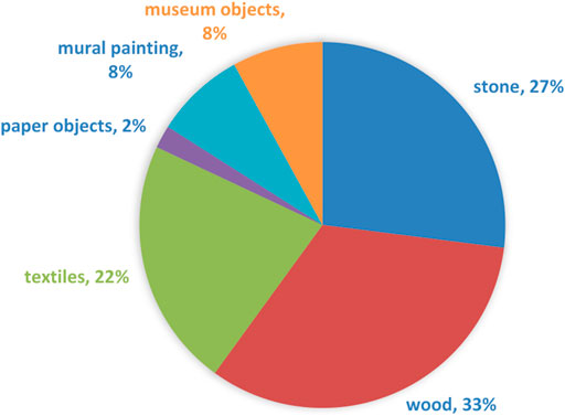
FIGURE 1. Graphic representation of tested fungal strains distribution by isolation sources.
Semiquantitative Evaluation of the Fungal Adherence to Inert Substrata
The assessment of biofilm development on the inert substrate of selected fungal strains was performed using the microtiter broth method. The ability to adhere to inert surfaces and generate biofilms was correlated with the fungal strains source of isolation and genus, as it can be observed in Figures 2-3. The most adherent strains (the highest absorbance average value) are the fungal strains isolated from mural paintings, followed by the strains isolated from wooden objects. The least adherent strains were paper’s isolated strains and those isolated from museum objects (ceramics and wood).
The most adherent strains belonged to Arthrinium and Cladosporium genera, followed by Alternaria, Aspergillus, and Penicillium (Figure 3).
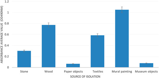
FIGURE 2. Graphic representation of the average absorbance values obtained for fungal strains isolated from different sources.
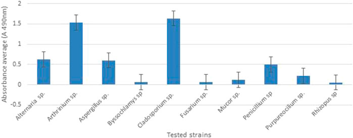
FIGURE 3. Graphic representation of the average absorbance values obtained for the fungal strains belonging to different genera.
Cellulase Production by Filamentous Fungi Species
The distribution of cellulose production by isolation sources revealed that the most potent cellulose producing strains were isolated from paper objects (100%), followed by strains isolated from textiles (89%), wooden (83%), and stone objects (78%), and at last, the encountered cellulose producing strains were isolated from mural paintings (57%) (Figure 4).
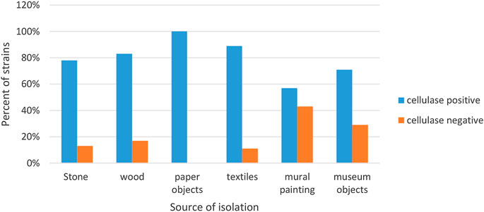
FIGURE 4. Graphic representation of the cellulose-producing strains by isolation sources.
From a total number of 88 strains, the most frequently encountered cellulose producing strains belonged to the Aspergillus (34.09%) and Penicillium (30.68%) genus (Figure 5).

FIGURE 5. Graphic representation of the cellulose-producing strains by genus.
Qualitative Screening of the Antifungal Activity
The qualitative screening of the antifungal activity was performed using the adapted diffusion method, toward 149 fungal strains and allowed the comparison of the antimicrobial efficiency of tested MgB2 NPs and the selection of fungal strains which were tested in the quantitative assay. The diffusion assay revealed a comparable efficiency for the tested NPs, with a slightly better inhibitory activity being observed for LTS and AA (Figure 6 and Supplemetary Figures S2, S3, S4).
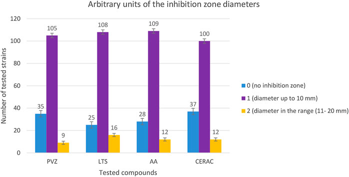
FIGURE 6. Graphic representation of the distribution of diameters zone inhibition arbitrary units.
Quantitative Evaluation of the Antifungal Activity
For the quantitative evaluation of the antifungal efficiency of MgB2 powders, the MIC values that have been determined using the binary serial dilution method, for the most susceptible strains. The results were interpreted depending on the genera of tested strains (Figure 7 and Supplementary Figures S5-S6) and their sources of isolation (Figure 8 and Supplementary Figures S10-S15).
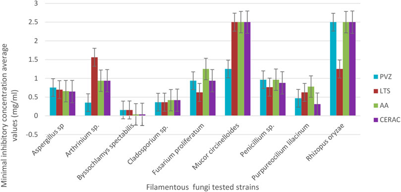
FIGURE 7. Graphic representation of minimal inhibitory concentrations average values obtained for the fungal strains belonging to different genera.
As it can be observed in Figure 7, the most susceptible strain was Byssochlamys spectabilis showing the lowest MIC values for all tested NPs, followed by Cladosporium spp., Purpureocillum lilacinum, Aspergillus spp., and Penicillium spp. strains. The most resistant species were Rhizopus orhizae and Mucor circinelloides, showing the highest MIC values for the majority of tested compounds. It is remarkable that all four types of MgB2 powders exhibited similar and relatively low MIC values against the Aspergillus and Penicillium strains, which were the most frequently isolated in our study from the biodeteriorated heritage religious monuments and objects.
Regarding the distribution of the MIC values depending on the isolation source of the tested strains, it can be observed that the most susceptible strains were those isolated from deteriorated paper (Supplementary Figure S12). The PVZ NPs, followed by CERAC, proved to be more efficient against the strains isolated from stone and wood materials, while the LTS was active against the fungal strains colonizing the mural paintings and museum objects (Figure 8).
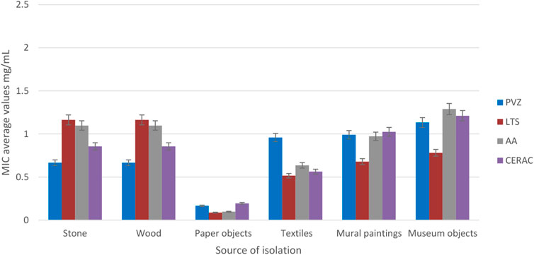
FIGURE 8. Graphic representation of minimal inhibitory concentrations average values obtained for the fungal strains from different isolation sources.
Cytotoxicity of the Tested NPs
The MgB2 NPs at 100 µg/ml concentration induced a cellular mortality rate of 100%, as revealed by the propidium iodide red staining. Propidium iodide is a chemical agent that intercalates between the nitrogenous bases in the DNA of the dead cells. At 50 µg/ml concentration, many cells are still viable, but the morphological aspects suggest significant changes, suggesting cellular damage. Instead, at lower concentrations (10 µg/ml), a lower toxicity is revealed by the predominance of viable cells colored in green by fluorescein diacetate (FDA) (Figure 9).
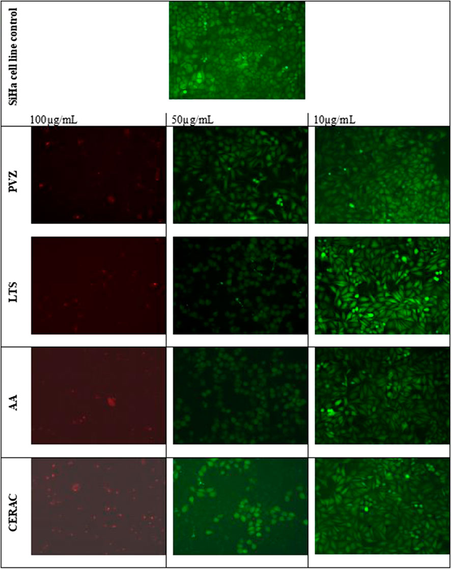
FIGURE 9. Microscopic aspect of SiHa cells after NPs treatment (inverted microscopy images).
The cytotoxicity effects observed after microscopic examination of the cellular monolayers have been further confirmed by the results of the cell cycle analysis on Hep2 cells suggesting that all tested compounds induced the occurrence of an apoptosis peak (visible in the very left of the flow cytometry histogram image, follow the arrow in Figure 8) at the concentration of 50 µg/ml. The proapoptotic effect of the tested NPs was in the following decreasing order: CERAC > PVZ > LTS = AA (Figure 10).
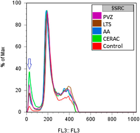
FIGURE 10. The flow cytometry histogram results of the cell cycle analysis.
The Ecotoxicity of MgB2
The results of the ecotoxicity test are shown in Figure 11 and Supplementary Table S3. The growth rate analysis indicates significant differences among the sample sets. After MgB2 administration, the mean values of the instantaneous growth rate, expressed as the ln of the fresh biomass weight (W1 and W2), decreased after treatment with the majority of the tested samples, excepting LTS of 1 mg/ml, AA at all three tested concentrations, and PVZ of 1 mg/ml. The RGR decreased in the presence of the majority of the tested powders, excepting CERAC 10 mg/ml, LTS 1 mg/ml, AA 1 mg/ml, and all PVZ tested samples. However, the recorded differences were not statistically significant, as revealed by p values >0.05 for the great majority of experimental variants (Supplementary Table S3).
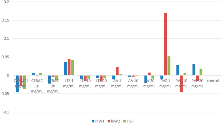
FIGURE 11. Variation of instantaneous growth rate (ln W1 and ln W2) and FGR in the presence of the tested MgB2 powders at different concentrations.
Discussion
The specific geographical and climatic conditions of Romania (different climates, types of reliefs, various soil types, diverse vegetation, and factors with a particularly accentuated dynamics) require careful consideration of the cultural heritage in an integrated, transdisciplinary manner. Our study was conducted in different regions of Romania, but with special emphasis given by the age and cultural value of the objects on Alba-Hunedoara counties, which are part of the region known as Ţara Haţegului (the Land of Hateg) but also in Bucharest, the capital of Romania, at the monumental church translated from its initial location to the National Museum of the Romanian Peasant and also from the History and Archeology Museum of Tulcea, which has an important restoration and conservation laboratory for metal objects, ceramics, textiles, wood, and icons. Some of these churches have changed their functionality overtime, becoming touristic sites or museums, while others still conduct the religious service for the local community. Therefore, the analyzed samples were very diverse from the viewpoint of age, materials, degradation level, and cultural importance, and they were taken from both the outer and the inner environment of the churches. Given the age and cultural importance of the selected monuments, extensive research is required to identify less invasive methods to support their conservation, especially in case of those harboring mural painting, part of it being completely or partially destroyed and, consequently, in variable degrees of recovery.
Our investigation revealed a high taxonomic diversity of the filamentous fungi involved in the biodeterioration of wood and stone churches and of different religious objects, with a clear predominance of Cladosporium, Aspergillus, and Penicillium species. Their predominance could be partially explained by their capacity to adhere and form biofilms on different materials, as revealed by the results of the biofilm development assay study. Our results are very similar with those reported by Pepe et al. (2010); Unković et al. (2015); Trovão et al. (2019); Lupan et al. (2013); and Ilieş et al. (2019). Fungal biofilms are very efficient systems for enzyme production, colonizing the surfaces. They grow within the painting layers of mural paintings (Gorbushina et al., 2004), and in case of some fungi species, the hyphae are able to penetrate the concrete structure (Li et al., 2018). The typical structure of fungi biofilm is represented by hyphae covered by an extracellular matrix (ECM) generally composed of polysaccharides, galactomannan, glucans, and melanin (Beauvais and Latgé, 2015). The polymeric structure acts as a glue that traps dirt and other particulate materials, increasing the destructive mechanisms of the biofilm and making the structure more difficult to clean (Gaylarde and Glyn Morton, 1999). Cellulose is the most abundant organic polymer in nature, and cellulose decomposition has a special significance in the carbon biogeochemicalcycle (Lederberg, 1992). In our study, two of the ten investigated genera, Aspergillus and Penicillium, dominate the spectrum of the main agents involved in the alteration of the color and in the structural biodeterioration of wood due to the presence of cellulolytic enzymes that degrade the wood fibers (Štafura et al., 2017). This genus usually acts in low humidity conditions, such as those found in the investigated churches, causing by dissolving cellulose and hemicellulose, the colorimetric alteration (called foxing-type spots), deposits, or pigment on the surfaces of the wooden surfaces (Blanchette, 2000; Piñar et al., 2015). Our results are partially confirmed by a previous reported study in Romania, the cellulose producing strains being recovered in decreasing frequency from paper objects, textiles, and wood samples (Fierascu et al., 2017) due their lignocellulose hydrolysis capacity in the case of the Penicllium genus and cellulolytic enzymes in the case of A. niger strains (Bergadi et al., 2014; Schneider et al., 2016). Pigment production was recorded for 22% of the tested Aspergillus, Penicillium, and Fusarium strains. Yellow, pink, and brown pigments predominated and were frequently detected among strains isolated from textiles and wooden substrata (data not shown), being well known that they play a crucial role in the deterioration of different materials the cultural heritage objects are made of (Irene Rojas et al., 2012).
In this study, we have studied the efficiency of different types of MgB2 NPs against the filamentous fungal strains isolated from the biodeteriorated heritage culture religious buidings and objects. The results of the qualitative screening have shown that the tested NPs have been active against 108 (for CERAC) to 124 (for LTS) microbial strains from the total of 149 tested ones. According to results addressed in Inorganic Nanoparticles Based on Halogenated Magnesium Salts Section, the highest rate of the pH increase (Inorganic Nanoparticles Based on Halogenated Magnesium Salts Section) is for LTS and the lowest for CERAC promoting the idea that the highest bioactivity is for LTS and the lowest for CERAC. We observed that pH rate and qualitative screening data correlate well, and the pH behavior is identified as one of the key parameters. Furthermore, the results of the quantitative assays evidenced different intensities of the antifungal effect, the MIC concentrations varying from 0.039 to 2.5 mg/ml. The order of efficiency of the four types of MgB2 was PVZ > LTS > CERAC > AA, as evidenced by the average MIC values obtained against all tested strains. This is explained by powders different morphological features, purity, and phase content. Nevertheless, it was not possible to reveal a key parameter specific to powders to control their antimicrobial efficiency. This indicates on possible synergetic and complex effects which should be further investigated in connection with the fungi types. Regarding the efficiency of the tested NPs against molds isolated from different sources, PVZ and CERAC proved to be promising for the development of antifungal products that could be applied outdoors, being more efficient against strains isolated from stone and wood walls. Instead, the LTS powder is promising for indoor antifungal solutions, as they have been more active against strains isolated from the church and museum objects. One observes that the average antifungal efficiency does not follow the order of the pH increase rate. It results that other specific features of the powders and of the strains have to be taken into consideration.
The cytotoxicity results indicated that the at concentrations higher than 50 µg/ml, the tested NPs induce the apoptotic death of the epithelial cells. However, although the noncytotoxic concentrations are much lower than the MIC values required for the inhibition of fungal growth, the short lifetime of these NPs prevents their accumulation in the natural environment and occurrence of toxic effects.
The outdoor use of MgB2 solutions for antifungal solutions applied on heritage objects would require assesment of ecotoxicity. Therefore, we have performed the Allium cepa ecotoxicity test, which allows the noninvasive evaluation of the phytotoxic effects of different substances (Hoffmann and Poorter, 2002). In the presence of water, MgB2 degrades, the reaction products being Mg(OH)2 and H3BO3 (or B2O3). Water degradation of MgB2 prevents its accumulation in the natural environment. According to our ecotoxicity results, the tested powders or their degradation compounds can be considered ecofriendly at concentrations up to 20 mg/ml.
Conclusion
To the best of our knowledge, this is the first report on the efficiency of MgB2 against a large collection of biodeteriorating molds isolated from heritage wood and stone buildings and objects. The obtained results evidenced the promising potential of MgB2 powders to inhibit the biodeteriorating fungi growth, their effects varying not only with their physicochemical properties but also with the taxonomic affiliation and isolation source of the Micromycetes strains. It is to be noticed that all tested powders proved to be active against the Aspergillus and Penicillium strains, which seem to dominate the etiological spectrum of the biodeteriorating fungi in Romania, as these genera were the most frequently isolated from the biodeteriorated Romanian heritage objects. Moreover, the tested powders proved to be ecofriendly at the active antifungal concentrations. Powders’s specific features are important to tailor their antifungal efficiency. Among the important parameters is identified the pH increase rate. This depends in a significant way on the phase amount of MgB2 in the powders, but other parameters are also important (granulometric distribution, morphology, and surface state). The results of this research could have a direct impact on the conservation and, thus, on the safeguarding of monuments by maintaining the carpentry and masonry in a stage that allows the transmission of this tangible and intangible heritage to future generations.
Data Availability Statement
The original contributions presented in the study are included in the article/Supplementary Material, further inquiries can be directed to the corresponding author.
Author Contributions
IG and MCC conceived and designed the experiments and drafted the manuscript, IrB, IoB, and VM performed the sampling campaigns, IG, IA, VMC, LM, and MP performed the isolation of the fungal strains, IP and VC performed the MALDI-TOF identification, AD, OZ, IG, and IA performed the antimicrobial assays and biofilm development assays, DB, GA, MG, MB, and PB provided the MgB2 powders characterization, CB conceived and performed the cytotoxicity experiments, NI and AD conceived and performed the ecotoxicity experiments, VL, LD, and MCC participated to data analysis and interpretation, drafted and corrected the manuscript, DB and PB performed the review and editing of the draft, MCC and IG supervised the experiments, PB and MC assured project administration and funding acquisition. All authors approved the final form of the manuscript.
Conflict of Interest
The authors declare that the research was conducted in the absence of any commercial or financial relationships that could be construed as a potential conflict of interest.
Acknowledgments
This is a short text to acknowledge the contributions of specific colleagues, institutions, or agencies that aided the efforts of the authors. Authors acknowledge the Romanian National Authority for Scientific Research and Innovation, UEFISCDI, for financial support through research projects PDI-PFE-CDI ID 335 “Interdisciplinary Institutional Platform for Excellence in Research, Development, Innovation and Professional Training in Archaeological Sciences (ArchaeoScience # RO),” PCCDI/2018 no. 52, Multidisciplinary and complex platform for the integrative and systematic research of the identity of the tangible and non-tangible cultural heritage of Romania. Subproject 3—“New technologies for preservation, conservation, recovering and restauration of the cultural heritage,” and 74-COFUND-M-ERA.NET II—BIOMB (within PNCDI III).
Supplementary Material
The Supplementary Material for this article can be found online at: https://www.frontiersin.org/articles/10.3389/fmats.2020.601059/full#supplementary-material.
References
Aldosari, M. A., Darwish, S. S., Adam, M. A., Elmarzugi, N. A., and Ahmed, S. M. (2017). Protecting of marble stone facades of historic buildings using multifunctional TiO2 nanocoatings. Sustainability 9, 20021–20115. doi:10.3390/su9112002
Aldosari, M. A., Darwish, S. S., Adam, M. A., Ermazugi, N. A., and Ahmed, S. M. (2019). Using ZnO nanoparticles in fungal inhibition and self-protection of exposed marble columns in historic sites. Archaeol. Anthropol. Sci. 11, 3407–3422. doi:10.1007/s12520-018-0762-z
Angelescu, M. (1957). Romanian cultural heritage Romanian Ministry of Culture. Available at: https://www.fonduri-patrimoniu.ro/Files/lansare.
Badica, P., Batalu, D. N., Grigoroscuta, M. A., Burdusel, M., Aldica, G. V., Popa, M., et al. (2018). Powders, sintered bodies, and coatings based on MgB2 resistant to microbian colonization and efficient against microbian biofilms, and a method of their application. Patent Request OSIM (Romania).2018A/01129.
Baglioni, P., and Giorgi, R. (2006). Soft and hard nanomaterials for restoration and conservation of cultural heritage. Soft Matter 2, 293–303. doi:10.1039/b516442g
Batalu, D., Stanciuc, A. M., Moldovan, L., Aldica, G., and Badica, P. (2014). Evaluation of pristine and Eu ₂O₃-added MgB₂ ceramics for medical applications: hardness, corrosion resistance, cytotoxicity and antibacterial activity. Mater. Sci. Eng. C Mater. Biol. Appl. 42, 350–361. doi:10.1016/j.msec.2014.05.046
Beauvais, A., and Latgé, J. P. (2015). Aspergillus biofilm In vitro and In vivo. Microbiol. Spectr. 3 (4), 1–10. doi:10.1128/microbiolspec. MB-0017-2015
Bergadi, F., Laachari, F., Elabed, S., Mohammed, I. H., and Ibnsouda, S. K. (2014). Cellulolytic potential and filter paper activity of fungi isolated from ancient’s manuscripts from the Medina of Fez. Ann. Microbiol. 64, 815–822. doi:10.1007/s13213-013-0718-6
Biswas, J., Sharma, K., Harris, K. K., and Rajput, Y. (2013). Biodeterioration agents: bacterial and fungal diversity dwelling in or on the pre-historic rock-paints of Kabra-Pahad, India. Iran. J. Microbiol. 5 (3), 309–314.
Blanchette, R. A. (2000). A review of microbial deterioration found in archaeological wood from different environments. Int. Biodeterior. Biodegrad. 46, 189–204. doi:10.1016/S0964-8305(00)00077-9
Bratu, A. (1982). Pictura murală maramureşeană. Meşteri zugravi şi interferenţe stilistice. ACS, Colecţia monografii. 39, 193–194.
Cezara, M. (1992). Legislaţia privind monumentele istorice din România 1892–1992. Studiu comparativ. La legislation des monuments historiques de Roumanie: 1892–1992. Etude comparative the Legislation Concerning the Historical Monuments from Romania 1892–1992, Comparave study. 2. Revista Monumentelor Istorice.
Chen, J., Wu, L., Lu, M., Lu, S., Li, Z., and Ding, W. (2020). Comparative study on the fungicidal activity of metallic MgO nanoparticles and macroscale MgO against soilborne fungal phytopathogens. Front. Microbiol. 11, 1–19. doi:10.3389/fmicb.2020.00365
Datcu, A. D., Ciobanu, D. G., Boros, B. V., Ostafe, V., and Ianovici, N. (2020). A new approach for phytotoxicity testing using Allium cepa bulbs. Rom. Biotechnol. Lett. 25 (2), 1488–1494. doi:10.25083/rbl/25.2/1488.1494
De Filpo, G., Palermo, A. M., Rachiele, F., and Fiore Pasquale, N. (2013). Preventing fungal growth in wood by titanium dioxide nanoparticles. Int. Biodeterior. Biodegrad. 85, 217–222. doi:10.1016/j.ibiod.2013.07.007
Fierascu, I., Avramescu, S. M., Fierascu, R. C., Ortan, A., Vasilievici, G., Cîmpeanu, C., et al. (2017). Micro-analytical and microbiological investigation of selected book papers from the 19th century. J. Therm. Anal. Calorim. 129 (3), 1377–1387. doi:10.1007/s10973-017-6370-9
Fouda, A., Abdel-Maksoud, G., Abdel-Rahman, M. A., Eid, A. M., Barghoth, M., and El-Sadany, M. A. H. (2019). Monitoring the effect of biosynthesized nanoparticles against biodeterioration of cellulose-based materials by Aspergillus niger. Cellulose 26, 6583–6597. doi:10.1007/s10570-019-02574-y
Franco Castillo, I., García Guillén, E., M de la Fuente, J., Silva, F., and Mitchell, S. G. (2019). Preventing fungal growth on heritage paper with antifungal and cellulase inhibiting magnesium oxide nanoparticles. J. Mater. Chem. B. 7, 6412. doi:10.1039/C9TB00992B
Gaylarde, C., and Glyn Morton, L. H. (1999). Deteriogenic biofilms on buildings and their control: a review. Biofouling 14 (1), 59–74. doi:10.1080/0892701990937839
Georgescu, M. S., Ochinciuc, C. V., Georgescu, E. S., and Colda, I. (2017). Heritage and climate changes in Romania: the St. Nicholas church of densus, from degradation to restoration. Energy Proc. 133, 76–85. doi:10.1016/j.egypro.2017.09.374
Gheorghe, I., Sărbu, I., Pecete, I., Blărjan, I., and Balotescu, I. (2020). Multi-level characterization of microbial consortia involved in the biodeterioration of wooden and stone romanian heritage churches. Conserv. Sci. Cult. Heritage. 20.
Gorbushina, A. A., Heyrman, J., Dornieden, T., Gonzalez-Delvalle, M., Krumbein, W. E., Laiz, L., et al. (2004). Bacterial and fungal diversity and biodeterioration problems in mural painting environments of St. Martins church (Greene–Kreiensen, Germany). Int. Biodeterior. Biodegrad. 53 (1), 13–24. doi:10.1016/j.ibiod.2003.07.003
Harandi, D., Ahmadi, H., and Mohammadi Achachluei, M. (2016). Comparison of TiO2 and ZnO nanoparticles for the improvement of consolidated wood with polyvinyl butyral against white rot. Int. Biodeterior. Biodegrad. 108, 142–148. doi:10.1016/j.ibiod.2015.12.017
Hoffmann, W. A., and Poorter, H. (2002). Avoiding bias in calculations of relative growth rate. Ann. Bot. 90 (1), 37–42. doi:10.1093/aob/mcf140
Ilieş, D. C., Oneţ, A., Herman, G., Indrie, L., Ilieş, A., Burtă, L., et al. (2019). Exploring the indoor environment of heritage buildings and it role in the conservation of valuable objects. Environ. Eng. Manag. J. 18, 2579–2586. doi:10.30638/eemj.2019.243
Irene Rojas, T. I., Aira, M. J., Batista, A., Lourdes Cruz, I., and González, S. (2012). Fungal biodeterioration in historic buildings of Havana (Cuba). Grana. 51 (1), 44–51. doi:10.1080/00173134.2011.643920
Johnsen, H. R., and Krause, K. (2014). Cellulase activity screening using pure carboxymethylcellulose: application to soluble cellulolytic samples and to plant tissue prints. Int. J. Mol. Sci. 15 (1), 830–838. doi:10.3390/ijms15010830.15
Li, Q., Zhang, B., Yang, X., and Ge, Q. (2018). Deterioration-associated microbiome of stone monuments: structure, variation, and assembly. Appl. Environ. Microbiol. 84 (7), e02680–17. doi:10.1128/AEM.02680-17
Lupan, I., Ianc, M. B., Kelemen, B. S., Carpa, R., Rosca-CasianChiriac, O. M. T., and Popescu, O. (2013). New and old microbial communities colonizing a seventeenth-century wooden church. Folia Microbiol. 59, 45–51. doi:10.1007/s12223-013-0265-3
Mangalampalli, B., Dumala, N., and Grover, P. (2017). Allium cepa root tip assay in assessment of toxicity of magnesium oxide nanoparticles and microparticles. J. Environ. Sci. 66, 125–137. doi:10.1016/j.jes.2017.05.012
Najjee, H., Kamerzan, C., Măruţescu, L., Gheorghe, I., Popa, M., Grădişteanu, G., et al. (2018). Antifungal activity of some medicinal plant extracts against Candida albicans nosocomial isolates. Rom. Biotechnol. Lett. 23 (6), 14073–14076. doi:10.26327/RBL2018.190
Nations, S., Wages, M., Canas, J. E., Maul, J., Theodorakis, C., and Cobb, G. P. (2011). Acute effects of Fe2O3, TiO2, ZnO and CuO nanomaterials on Xenopus laevis. Chemosphere 83, 1053–1061. doi:10.1016/j.chemosphere.2011.01.061
Negi, A., and Sarethy, I. P. (2019). Microbial biodeterioration of cultural heritage: events, colonization, and analyses. Microb. Ecol. 78, 1014–1029. doi:10.1007/s00248-019-01366-y
Nicu, I. C., and Stoleriu, C. C. (2019). Land use changes and dynamics over the last century around churches of Moldavia, Bukovina, Northern Romania—challenges and future perspectives. Habitat Int. 88, 101979. doi:10.1016/j.habitatint.2019.04.006
Pepe, O., Sannino, L., Palomba, S., Anastasio, M., Blaiotta, G., Villani, F., et al. (2010). Heterotrophic microorganisms in deteriorated medieval wall paintings in southern Italian churches. Microbiol. Res. 165 (1), 21–32. doi:10.1016/j.micres.2008.03.005
Piñar, G., Tafer, H., Sterflinger, K., and Pinzari, F. (2015). Amid the possible causes of a very famous foxing: molecular and microscopic insight into Leonardo da Vinci’s self-portrait. Environ. Microbiol. Rep. 7 (6), 849–859. doi:10.1111/1758-2229.12313
Pinzari, F., and Montanari, M. (2011). “Mould growth on library materials stored in compactus-type shelving units (Chapter 11),” in Sick building syndrome in public buildings and workplaces. Editor S. A. Abdul- Wahab Al-Sulaiman (Burlington, NJ: Elsevier).
Pop, C. S., Hussien, M. D., Popa, M., Mares, A., Grumezescu, A. M., Grigore, R., et al. (2015). Metallic-based micro and nanostructures with antimicrobial activity. Curr. Top. Med. Chem. 15 (16), 1577. doi:10.2174/1568026615666150414125015
Săsăran, P., Ţenter, A., and Jianu, L. D. (2020). Cultural heritage of a three centuries old wooden church. Drăghia viilage, Lăpuş Land, Maramureş county. Intech-Open. http://dx.doi.org/10.5772/intechopen.91483
Schneider, W. D. H., Gonçalves, T. A., Uchima, C. A., Couger, M. B., Prade, R., Squina, F. M., et al. (2016). Penicillium echinulatum secretome analysis reveals the fungi potential for degradation of lignocellulosic biomass. Biotechnol. Biofuels 9, 66. doi:10.1186/s13068-016-0476-3
Sharma, R., Prakash, O., Sonawane, M. S., Nimonkar, Y., Golellu, P. B., and Sharma, R. (2016). Diversity and distribution of phenol oxidase producing fungi from soda lake and description of Curvularia lonarensis sp. nov. Front. Microbiol. 7, 1847. doi:10.3389/fmicb.2016.01847
Shen, Y., Hu, T. J., Zeng, G. M., Huang, D. L., Yin, L., Liu, Y., et al. (2013). [Biodegradation of lignocellulose by Penicillium simplicissimum and characters of lignocellulolytic enzymes]. Huanjing Kexue 34 (2), 781–788.
Sierra-Fernandez, A., De la Rosa-García, S. C., Gomez-Villalba, L. S., Gómez-Cornelio, S., Rabanal, M. E., Fort, R., et al. (2017). Synthesis, photocatalytic, and antifungal properties of MgO, ZnO and Zn/Mg oxide nanoparticles for the protection of calcareous stone heritage. ACS Appl. Mater. Interfaces 9 (29), 24873–24886. doi:10.1021/acsami.7b0613
Sinsabaugh, R. L. (2010). Phenol oxidase, peroxidase and organic matter dynamics of soil. Soil Biol. Biochem. 42 (3), 391–404. doi:10.1016/j.soilbio.2009.10.014
Štafura, A., Nagy, Š., Bučková, M., Puškárová, A., Kraková, L., Čulík, M., et al. (2017). The influence of microfilamentous fungi on wooden organ pipes: one year investigation. Int. Biodeterior. Biodegrad. 121, 139–147. doi:10.1016/j.ibiod.2017.04.006
Sterflinger, K., and Piñar, G. (2013). Microbial deterioration of cultural heritage and works of art—tilting at windmills? Appl. Microbiol. Biotechnol. 97 (22), 9637–9646. doi:10.1007/s00253-013-5283-1
Trovão, J., Portugal, A., Soares, F., Paiva, D. S., Mesquita, N., Coelho, C., et al. (2019). Fungal diversity and distribution across distinct biodeterioration phenomena in limestone walls of the old cathedral of Coimbra, UNESCO World Heritage Site. Int. Biodeterior. Biodegrad. 142, 91–102. doi:10.1016/j.ibiod.2019.05.008
Unković, N., Ljaljević Grbić, M., Subakov Simić, G., Stupar, M., Vukojević, J., and JelikićStanojević, A. D. (2015). Biodeteriogenic and toxigenic agents on 17th century mural paintings and facade of the old church of the Holy Ascension (Veliki Krčimir, Serbia). Indoor Built. Environ. 55, 1–12. doi:10.1177/1420326×15587178.46
Keywords: biodegradation, cultural heritage, fungi, magnesium diboride, ecofriendly, antifungal
Citation: Gheorghe I, Avram I, Maria Corbu V, Măruţescu L, Popa M, Balotescu I, Blăjan I, Mateescu V, Zaharia D, Dumbravă AŞ, Zetu OE, Pecete I, Cristea VC, Batalu D, Grigoroscuta MA, Burdusel M, Aldica GV, Badica P, Datcu AD, Ianovici N, Bleotu C, Lazar V, Diţu LM and Chifiriuc MC (2021) In Vitro Evaluation of MgB2 Powders as Novel Tools to Fight Fungal Biodeterioration of Heritage Buildings and Objects. Front. Mater. 7:601059. doi: 10.3389/fmats.2020.601059
Received: 02 September 2020; Accepted: 02 December 2020;
Published: 13 January 2021.
Edited by:
David K. Mills, Louisiana Tech University, United StatesReviewed by:
Joanna Mystkowska, Bialystok University of Technology, PolandDouglas Boniek, Federal University of Minas Gerais, Brazil
Copyright © 2021 Gheorghe, Avram, Maria Corbu, Măruţescu, Popa, Balotescu, Blăjan, Mateescu, Zaharia, Dumbravă, Zetu, Pecete, Cristea, Batalu, Grigoroscuta, Burdusel, Aldica, Badica, Datcu, Ianovici, Bleotu, Lazar, Diţu and Chifiriuc. This is an open-access article distributed under the terms of the Creative Commons Attribution License (CC BY). The use, distribution or reproduction in other forums is permitted, provided the original author(s) and the copyright owner(s) are credited and that the original publication in this journal is cited, in accordance with accepted academic practice. No use, distribution or reproduction is permitted which does not comply with these terms.
*Correspondence: Lia Mara Diţu, bGlhLW1hcmEuZGl0dUBiaW8udW5pYnVjLnJv