 Ioana Maria Cortea
 Luminiţa Ghervase*
 Lucian Ratoiu
 Roxana Rădvan
- Department of Optoelectronic Methods and Techniques for Artwork Restoration and Conservation, National Institute for Research and Development in Optoelectronics INOE 2000, Măgurele, Romania
The article presents a multi-analytic investigation of a severely degraded Jewish ritual parchment coming from a private collection. The main aim of the study was to obtain key information on the parchment manufacturing technique and original materials used, information that could help understand the historical context of the object. To this aim, a series of noninvasive investigations were carried out by means of multi- and hyperspectral imaging, Fourier transform infrared (FTIR) spectroscopy and X-ray fluorescence (XRF) spectroscopy. Specific degradations and mapping of previous conservation treatments could be highlighted via multispectral imaging. Short-wave infrared images indicated the use of both iron gall and carbon black ink, probably one related to the original writing and the other to a later intervention. To improve the imaging of degraded or partially lost text, a linear spectral unmixing classification of the HSI dataset was proposed that showed promising results, allowing it to be applied to similar objects. XRF analysis offered an in-depth view of the chemical fingerprint of the original iron gall ink and critical findings on the existence of other inorganic compounds originating from the parchment manufacture. Registered FTIR data indicated denaturation of the collagen fibers and the presence of fungal-derived calcium oxalates and zinc carboxylates. In accordance with ancient Jewish parchment preparation techniques, the use of calcium sulfate, vegetable tannins, and oils was also inferred from the registered infrared spectra. The corroborated results offer valuable information on the origin, production technology, and overall degradation state of the parchment manuscript. Not least, the findings could be of great interest for conservators and restorers in the field.
Introduction
Parchment or charta pergamena has played an important role in human history, many ancient and historical documents, manuscripts, and books being recorded on this widely used written support (Avrin 2010; Bicchieri et al., 2019). Obtained from animal hides, parchment manufacturing technique varies according to the geographical area and historical period (Avrin 2010). The European manufacturing methods involved the use of salt and lime baths, while in the Middle East, tannins and mineral compounds were used, as is the case of traditional Jewish parchment preparation (Maor et al., 2020).
Jewish manuscripts written on parchment are not only invaluable documents and artifacts but also sacred objects (Greene 1992). Parchment obtained exclusively from the skin of a kosher animal was used for centuries mainly for the writing of religious texts (Avrin 2010). With the exception of the Dead Sea Scrolls—a large collection of manuscripts that include Books of the Hebrew Bible (except the Book of Esther) and other sacred texts, that have received great attention (Derrick 1991; Wolff et al., 2012; Maor et al., 2020), other Jewish manuscripts have been less studied.
In this article, a Jewish parchment scroll coming from a private collection was investigated using a multi-technique approach. The parchment, significantly degraded, includes chapters from the Book of Esther also known in Hebrew as Megillat Esther (Grossman 1997). The last of the five scrolls in the Hebrew Bible, especially the Book of Esther tells the story of the salvation of the Jewish people during the Achaemenid Persian Empire, and it is one of the most important texts, read during the Jewish holiday of Purim (Patai 2015).
Written scrolls of the Book of Esther were produced since the Talmudic period, the preparation technique following strict traditional rules of ritual scriptural objects (Grossman 1997; Patai 2015). Although no separate technical manuals exist in terms of Hebrew manuscript production, a number of halachic texts include dedicated information on regard of the allowed materials, forms, and methods of writing (Patai 2015). According to the geographical region in which the scrolls were produced, the skin of goats, sheep, or calves were used (Avrin 2010).
Sacred texts had to be written only on one side of the parchment, using square script and black ink. There were also clear rules on regard of the configuration of the written page, for the Book of Esther, for example, a minimum of eleven lines per column being imposed (Patai 2015). Documents with more than one column were contained in scrolls (Wandrey 2017), which were kept in a cylindrical metal or wood case (Avrin 2010).
There are also restrictions in terms of treatment and repair of degraded or damaged scrolls, the only allowed intervention being the use of parchment patch placed on the outside. When the objects were too damaged and could no longer be used, they had to be hidden away or buried (Avrin 2010).
Decoration and illustration of holy texts were also forbidden (Avrin 2010), but over time, some of the restrictions on the construction of sacred scrolls were gradually revised (Grossman 1997). The oldest preserved illuminated Esther scrolls are dated from 16th century Italy where the cultural background of the Italian Renaissance sparked interest among leading artists for the decoration of scrolls. The practice of hand-illuminated parchment scrolls flourished during the 17th and 18th centuries, and declined in the 19th (Patai 2015).
A large number of Esther Scrolls dating from the 14th to the 17th century have been preserved in various collections (Grossman 1997; Richler 2001), with most of the research focused on paleographical and codicological descriptions (Richler 2001; Wandrey 2017), or conservation studies (Grossman 1997). More recent research on radiocarbon analysis (Oliveira et al., 2015) and parchment degradation (Bicchieri et al., 2013; Hajji et al., 2018) were published on various Torah scrolls ranging from the 16th to the 19th century, or historical Yemenite scriptorial fragments.
The main objective of this study was to extract, by means of a noninvasive multi-analytic approach, key information on the parchment manufacturing technique and on the composition of the original materials used and their degradation. Obtaining such data could help not only in understanding the historical context of the manuscript but would also provide valuable data for the best restoration procedure and long-term conservation strategy of this severely damaged object. Not least, the research brings into attention less studied types of artifacts whose conservation and restoration require a different management due to their particular framing as sacred objects (Greene 1992; Grossman 1997).
The potential of an integrated noninvasive study for the characterization of highly degraded parchment artifacts was explored. Multi- and hyperspectral imaging were used to obtain high-resolution imaging documentation of the parchment and study-specific degraded areas and ink typology (Bearman and Spiro 1996; MacDonald et al., 2013; Giacometti et al., 2014; Cohen et al., 2017). Identification of inorganic compounds derived from the parchment manufacturing process (salts and minerals) and characterization of the black ink were carried out by using X-ray fluorescence (XRF) (Hahn et al., 2004; Wolff et al., 2012; Duran et al., 2014). Attenuated total reflection Fourier transform infrared (ATR-FTIR) spectroscopy (Derrick 1991; Hajji et al., 2018; Bicchieri et al., 2019; Maor, Shor, and Aizenshtat 2020) was used to obtain information on the overall state of degradation of the parchment support and investigate the presence of organic tanning agents and other nonproteinaceous materials.
Materials and Methods
Manuscript
The investigated manuscript (Figure 1), in the size of 620 × 520 mm, comes in the form of a scroll, 24 cm in diameter. Regarding the history of the object, not much is known, but the fact that the scroll was intended with a liturgical purpose, and it presents chapters 1, 2, 3, and the beginning of chapter 4 from the Book of Esther. Given that it represents only part of the Book of Esther, it can be assumed that the manuscript had been sown to the rest of the Book; however, the advanced degradation state of the margins made it impossible to detect signs of sewing.
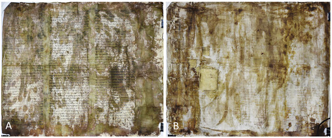
FIGURE 1. Jewish parchment investigated in this study: (A) recto and (B) verso.
The scroll has no hand paint or engraved decorations. The text is written in Hebrew, in square script and black ink, only on one side of the parchment, as indicated for Jewish religious texts (Patai 2015). The text is written on three elongated columns each of 45 even lines. The ink is not very well preserved, which makes the legibility of the text difficult. At the moment of the investigation, the object had no documentation in terms of paleographic or stylistic study.
Visual examination of the manuscript indicates a poor state of conservation, the media support being extremely fragile and rigid. The manuscript has a highly nonhomogenous appearance with mechanical deformations (folds and undulations) and various stains ranging from dark brown to pale olive. The edges are fractured, while on the verso of the scroll, repair interventions can be clearly seen (parchment patches). Ink corrosion and some white powdery deposits were also detected (Figure 2). This state of advanced degradation could be correlated both with the age of the manuscript and with inappropriate storage conditions (Bicchieri et al., 2013).
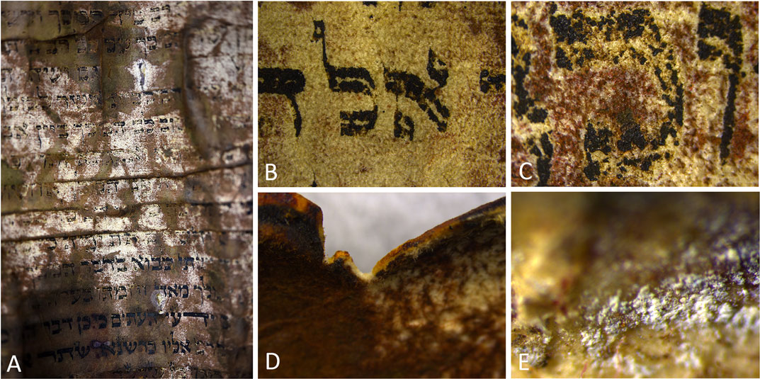
FIGURE 2. Details that highlight various types of degradation: (A) discolorations and staining of the parchment, (B) ink fading, (C) ink corrosion, (D) fractured edges, and (E) white powdery deposits.
Multi- and Hyperspectral Imaging
Multispectral imaging (MSI) of the manuscript was performed using the ARTIST system from Art Innovation. The system, equipped with a CCD progressive scan image sensor, is capable of acquiring spectral images that cover the spectral range from 365 to 1,100 nm. A UV hand lamp from Karl Deutsch with a maximum peak at 365 nm was used for the UV imaging modes, while for the visible and near-infrared acquisitions, halogen lamps were employed.
Hyperspectral imaging (HSI) was performed with a HySpex SWIR-384 system from NEO, which enabled noninvasive recordings in the SWIR (short-wave infrared, spectral range: 954–2,514 nm), on 288 spectral bands. The system, equipped with a state-of-the-art MCT detector, has an FOV (field of view) of 16⁰ across the track and an FPA (focal plane array) cooling system which ensures a constant temperature of 150K, a characteristic that provides low background noise. For the best available resolution, we used the 30-cm lens. HSI data were acquired in push-broom technique. Data processing was carried out in ENVI software package (Harris Geospatial Solutions).
X-Ray Fluorescence
XRF was performed in situ on the parchment, with the portable, energy-dispersive instrument TRACER III (Bruker Elemental). The equipment disposes of a Rh anode and was operated at 40 kV tube power, 11 μA current intensity, 60-s analysis time, no filter, and air atmosphere, allowing a fair detection of all elements between 1 and 40 KeV. Peak identification was performed through standard Bayesian deconvolution with ARTAX software. Data processing was achieved via Origin 8 and Microsoft Excel software. A total of 10 areas were analyzed on different parts of the parchment: six on the front (denoted by numbers 1–6) and four on the verso (denoted by B1, B2, B3, and B4) of the manuscript. The equipment used has a 3 × 4-mm spot size, allowing area analysis; therefore, it is important to understand the exact appearance of each analyzed area, when discussing the results. All six areas are also shown in detail, as captured by the integrated camera of the TRACER-III SD equipment. For better comparison, all XRF data were normalized with respect to the Rh Kα line (O’Meara et al., 2001; Wilke 2016).
Fourier Transform Infrared Spectroscopy
Attenuated total reflectance (ATR) FTIR measurements were performed by means of a PerkinElmer Spectrum Two FTIR spectrometer equipped with a GladiATR accessory from PIKE Technologies. Spectra were acquired at 4 cm−1 spectral resolution and 16 scans within the 4,000–400 cm−1 spectral region. Spectral data are shown in transmission (T%) with baseline correction. Data processing was done using Essential FTIR. FTIR measurements were carried out in situ, in a number of 16 points. Spectra were recorded on areas with various chromatic variations and on the ink inscriptions.
Results and Discussion
Imaging Documentation
Multispectral imaging allowed a detailed documentation regarding the overall conservation state of the parchment manuscript. Several specific degradations could be highlighted such as abrasions and scratches but also signs of repeated mechanical stress, including small cracks and fractures. Images acquired under both UV fluorescence (UVF) and UV reflectance (UVR) acquisition modes indicate that the olive–brown–stained areas of the parchment are characterized by a strong UV absorption (without emission), making these areas appear darker (Figure 3). These black areas could indicate a more recent consolidation intervention performed with an organic material. In general, new retouching or repairs appear dark under UV light, traditional binders such as oils, or natural resins showing fluorescence only after a certain level of degradation have occurred (Rie and René, 1982a; Rie and René, 1982b). However, it is not common to have such irregular interventions carried out on such large areas, unless the object is found in an advanced state of degradation. Another explanation for these highly UV-absorbing areas could be due to the fact that their surface composition is chemically different (and therefore displays different optical characteristics) due to the existence of potential localized degradation, unevenly distributed within the parchment. Various studies carried out on Dead Sea Scrolls fragments have shown that the darker areas exhibit a higher degree of degradation than the light ones, and they were usually associated with a greater conversion to gelatin (Derrick 1991; Maor, Shor, and Aizenshtat 2020).
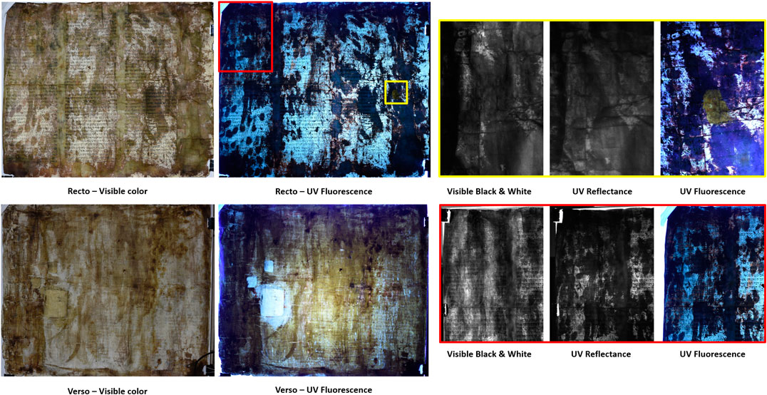
FIGURE 3. UV fluorescence examination of the recto and verso of the parchment. On the left side, visible (black and white), UV reflectance and UV fluorescence comparisons for two significant details located on the recto.
UVF images registered on the verso of the parchment highlight areas with strong yellow–green fluorescence signal, which may indicate the presence of zinc oxide (Cosentino 2014). This yellow UV fluorescence signal was also registered on small areas located on the writing side of the parchment, indicating most probably accidental leaks. Existing literature (Avrin 2010) mentions the use of cooked zinc white for the surface treatment of the animal skin, such practice being still used for Yemenite parchments.
A purple red fluorescence can also be observed, especially on the writing side of the parchment (Figure 3), relatively clustered along the edges of the dark areas. This fluorescent signal could be related to a degraded organic material (associated with a treatment) or to some microbial or fungal contamination. The presence of a biologic attack would also explain the reddish brown spots easily observed on the upper part of the parchment. Red or purple stains associated with localized destruction of the collagen fibers are common biological damage phenomena that severely affect the visual appearance of parchment manuscripts (Piñar et al., 2015; Migliore et al., 2019).
The existence of some scratches on the writing side of the parchment could also be highlighted, best seen within the images recorded at 2,400 nm (Figure 4). This feature could be related to the original parchment preparation or may indicate a mechanical cleaning procedure of the text. Within the final stages of preparation, animal skins were scraped with sharp knives and smoothed by abrasion with pumice stones or other hard materials (Derrick 1991), procedures that may leave the surface with small imperfections. Scratch marks are also characteristic for reused parchments, the so-called palimpsests (palímpsēstos, “again scraped”), whose initial writing has been mechanically removed (Avrin 2010). Given the fact that these scratches were found only on restricted areas, the existence of a palimpsest is unlikely. Moreover, in Jewish tradition, a completely erased sheet was not supposed to be used for a religious text. Letters or words could have been erased and corrected instead (Avrin 2010).

FIGURE 4. Hyperspectral imaging details that highlight the existence of scratches at the surface level of the parchment: (A) 954 nm, (B) 1,600 nm, and (C) 2,400 nm.
In order to gain insights into the ink typology, the behavior of the writing material at different wavelengths of the SWIR region was investigated by means of hyperspectral imaging. As observed within the registered images (Figure 5), the ink inscriptions start to lose opacity toward longer wavelengths, becoming almost transparent after 1,200 nm, a characteristic behavior for iron gall inks (Cohen et al., 2017). Although this behavior is dominant, there are also areas where the ink inscriptions are still visible even at 1800 nm, situation that may indicate the use of a carbon black ink (Cohen et al., 2017). The existence of two distinct types of inks could be a clear indicator that the parchment suffered a conservation intervention at a certain moment in time. Moreover, a close examination of the acquired images highlights that the carbon-based letters are mainly present within the olive–brown–stained areas of the parchment that, as mentioned above, are suspected of having undergone through some type of treatment. Letter corrections penned in carbon ink could also have been made, Jewish sources mentioning the use of both carbon and iron gall ink within the same manuscript (Colini et al., 2018).
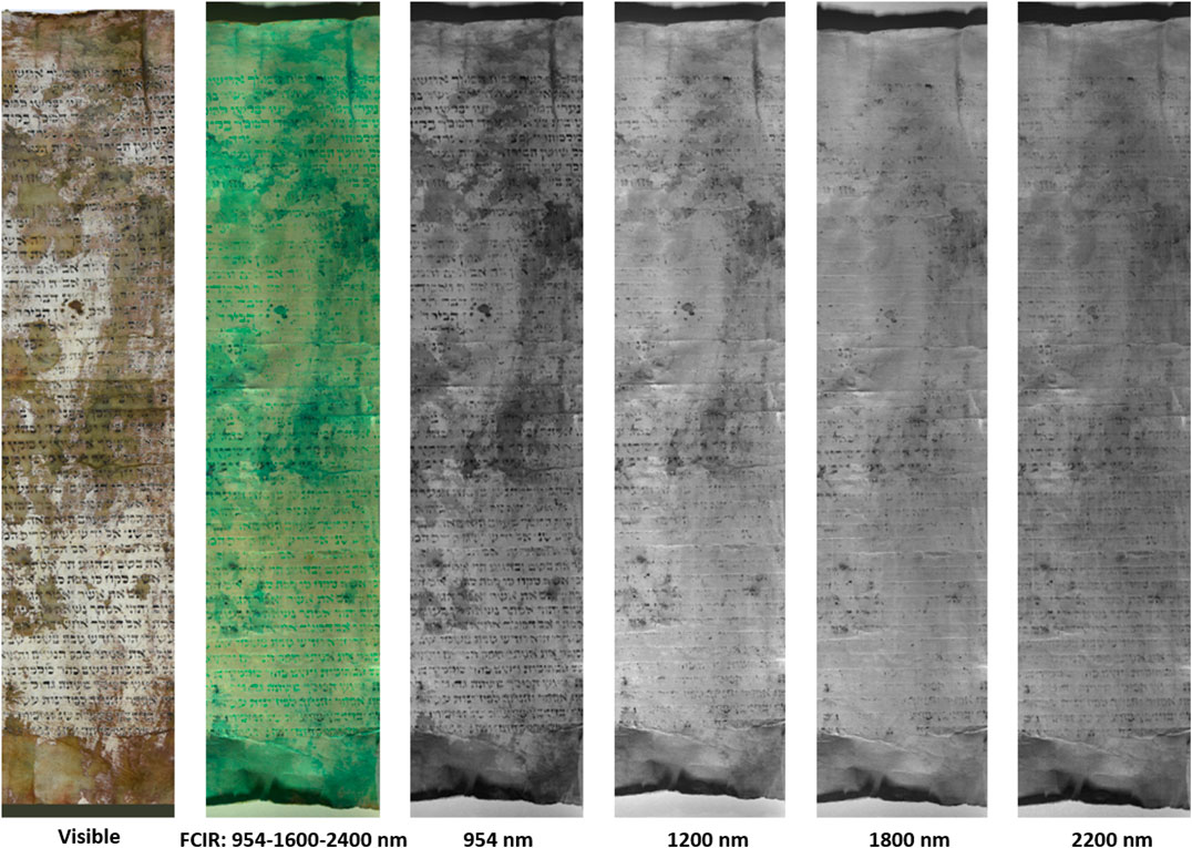
FIGURE 5. Hyperspectral imaging details showing the behavior of the ink components at specific wavelength values.
The ability of the hyperspectral system to characterize all the pixels from an image in terms of spectral profiles was exploited in ENVI, where a series of algorithms have been applied in order to classify and map the various materials present. Based on the variability of the spectral profiles and by using false-color infrared (FCIR) images, in which the differences of the spectral profiles were enhanced, high-accuracy ROI’s (regions of interest) could be selected. Due to the resolution of the hyperspectral camera sensor and also to the physical structure and state of conservation of the aged parchment, the majority of pixels were indicating a mixture of materials. In order to obtain an accurate classification, linear spectral unmixing (LSU) was used (Bioucas-Dias et al., 2012). The output of this algorithm produced five different fractions corresponding to the selected end-members and one corresponding to the root square errors, which represent the uncertainty of the mixing calculations. Based on the results, we were able to display different gray-scale images (Figure 6) the fractions of dark letters, light (faded) letters, parchment support (background), and brown–olive stains. Moreover, the fraction corresponding to dark letters was able to record from traces of material the integral shape of the letters affected in time by repeated abrasions of the surface. This specific feature indicates a powerful documentation tool for noninvasive investigation of historical documents whose text is partially degraded or lost.
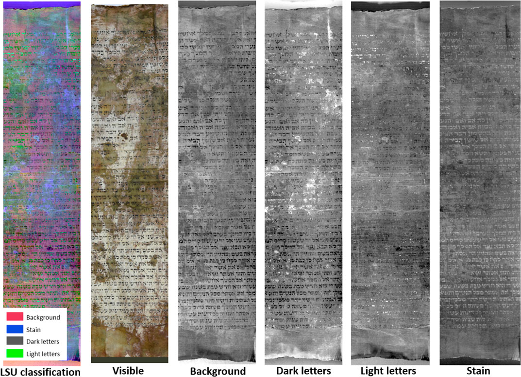
FIGURE 6. Details of the LSU classification. In bright tones are designated the areas with high errors, while with dark grey tones are signaled accurate classified areas based on their spectral profiles.
X-Ray Fluorescence Analysis
The analysis of the acquired XRF spectra allowed the identification of several elements, as listed in Table 1, of which some could be correlated with the ink and the way the parchment was manufactured. Some of the most representative elements are plotted in Figure 7, for all six analysis points.
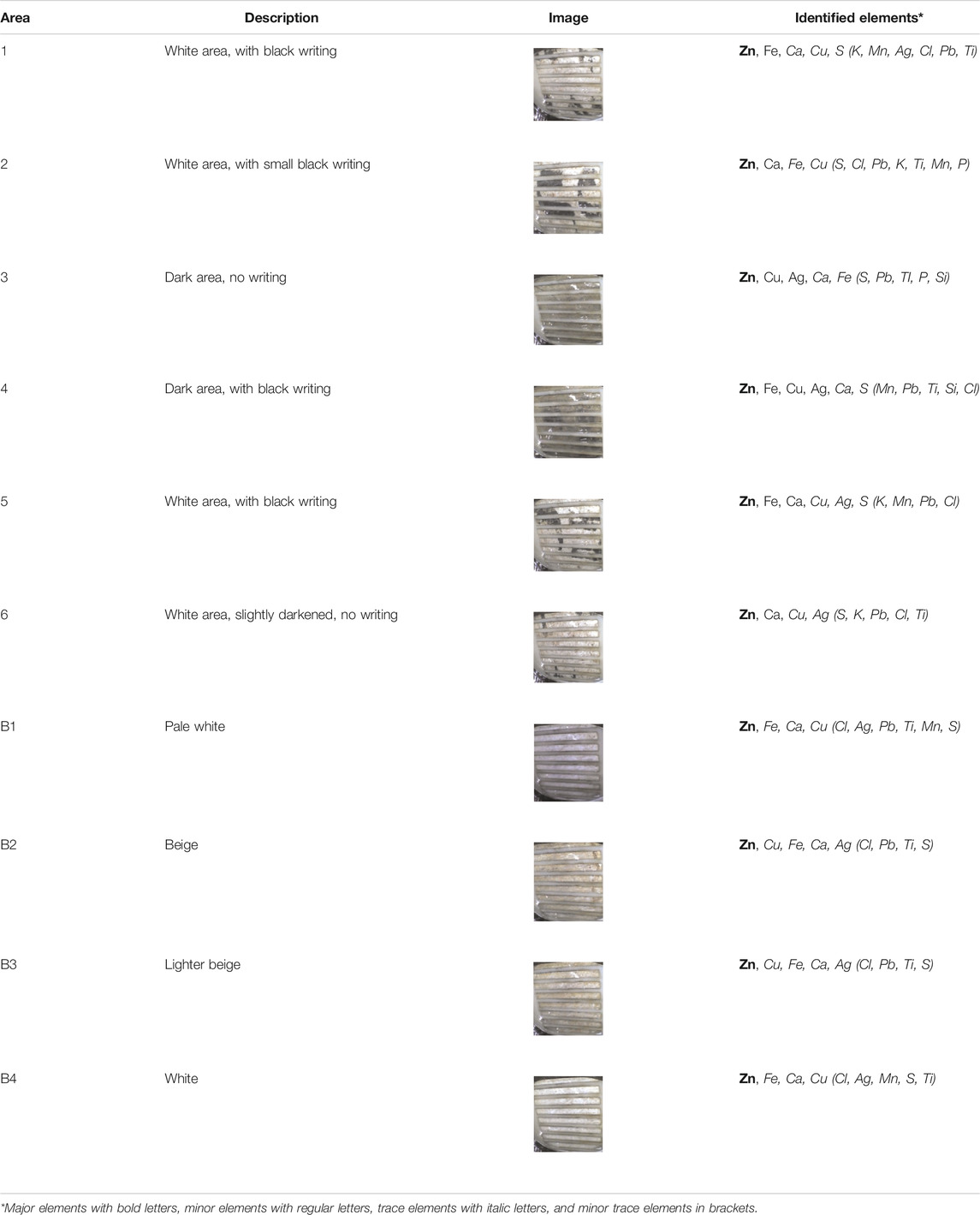
TABLE 1. Results of the XRF analysis.
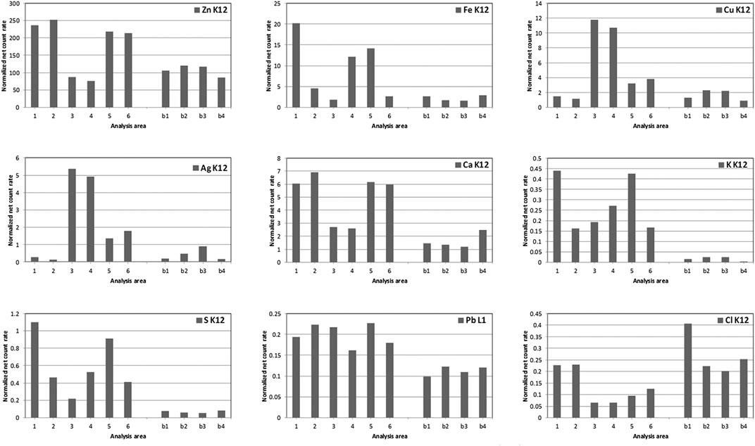
FIGURE 7. Elemental distribution for all analysis points.
As can be seen in the figure, the areas with writings showed higher net counts of Fe, S, and K, elements which showed strong positive correlation with each other, inferring the idea that they come from the same source. Historically, the most encountered iron-based ink was iron gall (Hahn et al., 2004; Creagh et al., 2016). Although many recipes are known, typically iron gall represents a 1:1 mixture of iron (II) sulfate and gallic acid, sometimes with higher iron sulfate ratio (Lee, Mahon and Creagh, 2006). Furthermore, other trace elements detected, such as Mn could be contaminants of the iron sulfate, indicators of the origin of the raw materials and/or of the manufacturing process (Hahn et al., 2004). Such trace elements can be used to identify the origin of an ink and even to compare various inks based on the ratios of trace to major constituents (Hahn et al., 2007).
There are visible Ag lines in some of the analysis points, but at higher intensity in darker areas. Copper, which was also present in all spectra, showed a similar tendency with silver, having higher net counts in darker areas. Silver may be related to a past disinfection procedure (Gutarowska et al., 2012; Pietrzak et al., 2017), but that does not correlate with the fact that areas which appear more degraded (darker areas) have more intense silver lines.
Additionally, even if it is not present as a well-defined peak, due to spectral overlapping with the Rh L lines coming from the X-ray source, chlorine may be present as the Bayesian deconvolution better fits the acquired spectra when Cl is added in the processing. Several possible explanations could be based on the manufacturing process. On the one hand, there are studies reporting the presence of chlorine salts in the case of parchments treated with sea water in areas where fresh water was scarce (Gai 2013). On the other hand, it may also be a residue of the various solutions used in the manufacturing process, such as brine—applied for dehairing the skin, or potassium salt—used as preservative (Hajji et al., 2018), or it may also come from previous restoration/disinfection treatments (Campbell 1994; Hahn et al., 2007). The fact that chlorine levels appear slightly higher on the verso of the manuscript seems to support the idea of a disinfection treatment for the provenance of chlorine.
By far, the most intense lines in all spectra belong to Zn Kα and Kβ lines (Figure 8), situated at approximately 8.63 and 9.57 KeV, respectively. For all points, Zn appears to be the most intense element, with the highest intensity on white areas (1, 2, 5, and 6) and the lowest intensities on the darkest areas (3 and 4), which in contrast, show the highest Ag intensities. The high Zn intensities detected for this parchment could come from the manufacturing process (Avrin 2010). Similar findings were also reported on Moroccan Jewish parchments, the rich zinc content being linked to the parchment bleaching process (Hajji et al., 2018). A zinc-based compound could have also been used for conservation purposes. In the past, restorers had used various additives, including zinc chloride, in order to increase gelatin flexibility (Campbell 1994). Not least, a certain amount of zinc could also come from the ink components, zinc sulfate being frequently used in the composition of the vitriol (a mixture of hydrated metal sulfates) used for the iron gall ink preparation (Cohen et al., 2017).
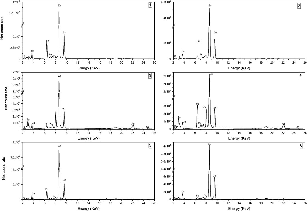
FIGURE 8. XRF spectra registered on the investigated areas located on the front side of the parchment.
Similarly, Ca lines are evident in all spectra, with lower intensity on the two darker areas. Calcium, coming from calcium hydroxide, which in time turns to calcite, may come from the liming process used for depilating the animal skin (Forbes 1996; Duran et al., 2014). The presence of lime could be a possible indicator of the manuscript’s origin as it is known that lime was preferred in the manufacturing process of western parchments, while the eastern parchments were mostly manufactured using enzymatic processes (Bicchieri et al., 2011).
Other trace elements can be linked to the manufacturing process or the storage conditions along the lifetime of the scroll. For instance, according to previous studies, Pb could come from inkwells (Bicchieri et al., 2013). Also, Pb and Cu traces may come from the way the scroll was written: typically a lead brass, called a lead pen plummet, was used to etch the lines on the parchment (Grossman 1997). Cu, however, was attributed as trace element in most of the analyzed areas, showing higher intensity lines only for areas 3 and 4. These are both areas, which have a darker appearance. The literature reports the use of copper only as constituent of the writing ink, either as impurity or intentional addition in the vitriol (Hahn et al., 2007; Cohen et al., 2015; Pronti et al., 2018). For the analyzed areas, however, no direct correlation could be determined between copper levels and areas with writing, which may be due to the heterogeneity caused by mobility of the copper ions and infiltration into the parchment (Cohen et al., 2015).
Fourier Transform Infrared Analysis
FTIR spectra registered on clear (noninked) parchment areas (Figure 9A) displays typical spectra of type I collagen. The main features within the spectra are the amide I and amide II absorptions. Amide I assigned to C=O stretching vibrations with contributions from C–N stretch, and N–H bend is seen around 1,637–1,628 cm−1. Amide II assigned to C–N stretching and N–H bending vibrations is registered around 1,539–1,537 cm−1. The broad absorption at 3,400–3,100 cm−1 and the small band around 3,074 cm−1 correspond to the first and second components of Fermi resonance between N–H stretch and the overtone of amide II. The Amide III absorbance at approximately 1,240–1,235 cm−1 is ascribed to N–H bending and C–N stretching vibrations (Boyatzis et al., 2016; Hajji et al., 2018). The small peak around 1,162 cm−1, observed in most of the registered spectra, can be ascribed to N–H residual modes found in collagen (Bicchieri et al., 2008).
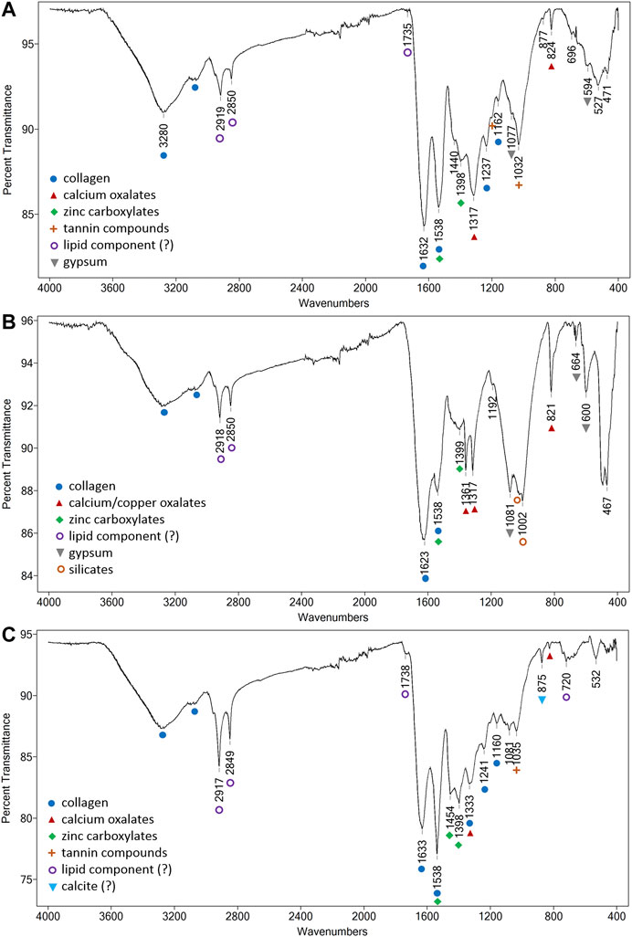
FIGURE 9. FTIR spectra registered on (A) clear (noninked) area, (B) ink inscriptions, and (C) back side of the parchment scroll.
Detailed examination of the amide I and amide II bands shows signs of collagen denaturation, one of the most noticeable changes being observed via the shift of the amide II band position from the initial literature reference value of 1,550 cm−1 toward lower wavenumbers, an indicator of the helix–coil conversion (Derrick 1991). Gelatinization, frequently found in historical parchments, occurs when the triple helix of the collagen molecules becomes structurally disorganized under the influence of external factors such as heat or water.
A series of bands corresponding to nonproteinaceous materials were also found on the surface of the manuscript. Calcium carbonate (CaCO3) could be present as indicated via the weak, but characteristic, band centered around 874 cm−1 (out of plane O–C–O bend) (Bicchieri et al., 2011). However, as important absorption bands associated with the fundamental vibrations of the carbonate ion in calcite (Pronti et al., 2018) are missing, a clear identification is not straightforward.
Calcium-based oxalates, specifically weddellite (CaC2O4.2H2O), can be inferred in most of the spectra via the characteristic peaks observed around 1,320 cm−1 (C=O stretching) and 825 cm−1 (O–C–O bending) (Nevin et al., 2008). These same bands together with a sharp peak around 1,360 cm−1 (C=O stretching), best seen on the ink areas investigated (Figure 9B), can be ascribed to copper oxalates (Buse, Otero, and Melo 2019). Compared to calcium oxalate that shows only one strong C=O stretching vibration around 1,320 cm−1, copper oxalate exhibits 2 C=O stretching vibrations (Nevin et al., 2008), which can be used for identification. As indicated by previous studies (Bicchieri et al., 2013; Pronti et al., 2018), the presence of oxalates can suggest a biological attack due to fungal species that are known to be able to reprecipitate secondary carbonates.
For most of the investigated areas, the amide II absorption displays a very sharp peak, unlike the normally broad appearance typical for proteins. Another uncommon aspect is related to the fact that the amide II peak displays a higher intensity than the amide I band. This uncommon situation may be due to some overlapping signals within this spectral region. In this case, zinc carboxylates are most probably present, situation that may also explain the high zinc level registered within the XRF data. According to the literature (Otero et al., 2014), the main absorption for zinc carboxylates, given by the asymmetric stretch vibration band of the carboxylate group, is characterized by a sharp and strong peak around 1,539 cm−1. As shown in Figure 9C, this sharp peak, ascribed to zinc palmitates and stearates, is more intense on the investigated areas located on the verso of the manuscript. Moreover, for the same areas, other characteristic absorptions for zinc soaps can be clearly observed: sharp peaks around 1,398 cm−1 (COO− stretching) and 1,455 cm−1 (CH2 bending), this last peak being specific for zinc oleates (Otero et al., 2014).
Metal carboxylates or soaps are not commonly found on parchment manuscripts. Previous studies (Vichi, Eliazyan, and Kazarian 2018) carried out on historical leather book covers dated between the 16th and the 19th century found calcium carboxylates that were linked with the presence of an oil emulsion used for the conservation treatment of the objects. Calcium soaps were also identified on the surface of medieval illuminated parchment manuscripts (Vetter, Latini, and Schreiner 2019), likewise correlated with the presence of an organic binder. The presence of zinc soaps could therefore be correlated with the presence of fatty acids (as a result of the interaction of the fatty acid fractions with a zinc compound) originating from an oil- or an egg-based binder that was used to treat the surface of the manuscript. The presence of a lipid component would also explain the strong C–H absorptions observed at 2,920 cm−1 and 2,850 cm−1, moreover as these absorptions appear strongly correlated with the absorptions of the zinc carboxylates (Vetter, Latini, and Schreiner 2019). A small fraction lipid, typically between 0.2 and 3.0% (Možir et al., 2014), could also be due to amino acid residues of the polypeptide chains found in leather, but these are typically characterized by weak C–H stretching modes (Manfredi et al., 2015). The hypothesis of using a lipid material on the parchment surface seems to be sustained by the small carbonyl band observed in some of the registered spectra between 1734 and 1740 cm−1 (Vichi, Eliazyan, and Kazarian 2018). This same band could also be partially ascribed to the presence of carbonyl compounds (esters) as a result of the polypeptide chain oxidation (Derrick 1991).
The possibility of using a lipid binder for the surface treatment of the parchment is supported by historical recipes. According to existing sources (Bicchieri et al., 2011), sacred Jewish parchments were frequently treated with vegetable tannins and oils. Usually obtained by extracting plant galls, tannins were applied by briefly soaking the parchment in the extract solution or by lightly spraying the extract on the hair side (Maor, Shor, and Aizenshtat 2020). Based on the registered FTIR data, evidence of a tanning treatment can also be inferred. The peak at 1,031 cm−1, clearly seen in most of the spectra, can be ascribed to aromatic C–C vibrational modes in tannins and can be considered a marker for tannin processing of parchment (Boyatzis et al., 2016).
The tannic acid treatment should also give rise to characteristic IR absorptions around 1710, 1,327 and 1,205 cm−1 (Bicchieri et al., 2008). Among the abovementioned absorptions, only the band around 1,203 cm−1 (stretching vibrations of the ester –C–O–C– and the carboxylic C–O–) could be observed in all FTIR spectra registered on the writing side (as a small shoulder band). The characteristic tannin carboxylic C=O stretching around 1710 cm−1 is masked by the board amide I band, while the peak around 1,330 cm−1 (CH in plane deformation) overlaps with the calcium oxalate band at 1,320 cm−1, causing a band broadening and a shift of the peak center.
The presence of gypsum (CaSO4·2H2O) could also be inferred as indicated by the strong band around 1,080 cm−1 along the sharp band at 596 cm−1 and the peak at 664 cm−1 due to the S–O stretching and bending vibrations of the SO4 group (Pronti et al., 2018), best seen on the ink area investigated. The O–H stretching and bending vibrations in the 3,550–3,400 cm−1 region and the bands at 1,684 and 1,621 cm−1 of the different water molecules typically found in hydrated Ca sulfates are masked by the collagen contributions. The presence of gypsum is not uncommon, powdered gypsum being sometimes used for the removal of excess fat in animal skin processing (Vichi, Eliazyan, and Kazarian 2018), for the acceleration of the drying process and ink absorption increase (Pronti et al., 2018), or, in illuminated manuscripts as a filler, for improving the mechanical properties and paint application (Melo et al., 2016). Some types of silicates could also be present (Derrick 1991) as indicated by the sharp peak around 1,002 cm−1, the shoulder band around 1,024 cm−1, and the peak around 530 cm−1.
FTIR spectra registered on inked areas are dominated by the contributions given by the parchment and overall by higher concentrations of oxalates—as indicated by the sharp peak at 821 cm−1. In some of the inked areas, the shoulder band observed around 1,440 cm−1 could be assigned to the symmetric stretch of iron (III)–gallate coordination complex (Boyatzis et al., 2016). No other characteristic peaks for iron gall inks could be observed, most probably due to the large overlapping signals with the parchment support.
Conclusions
In this study, a noninvasive multi-analytic approach was considered for the investigation of a severely degraded Jewish manuscript of unknown history. Carried out investigations allowed a comprehensive characterization of the original materials used and provided critical information on the overall conservation state of the object.
Multispectral imaging allowed identification and mapping of previous conservation interventions, as well as highlights regarding specific forms of degradation. The presence of two different types of inks was assessed based on the different behavior within the infrared region of the spectrum. Iron gall ink was found to be originally used, while a carbon-based ink was applied for letter corrections or latter interventions, most probably due to degradation and fading of the initial inscriptions. The use of spectral unmixing algorithms allowed a relatively accurate classification of the characteristic materials present, in accordance with XRF and FTIR data. Moreover, an algorithm was proposed that showed promising results for the reconstruction of degraded letters, thus proving a potential valuable tool for the investigations of parchment manuscripts whose text is hardy visible or partially lost due to extensive degradation.
XRF analysis allowed an in-depth view within the chemical fingerprint of the original iron gall ink as well as other important information on the existence of various inorganic compounds originating from the manufacturing process. Fe, S, K, Mn, Cu, and Zn were related to the vitriol recipe used for the iron gall ink preparation, while in terms of skin preparation technique, a bleaching process with cooked zinc white could have been applied. For the ruling of the manuscript, a lead pen plummet was most probably used as inferred by registered Pb traces.
Results of the FTIR analyses offered an in-depth view regarding the deterioration rate of the collagen material, denaturation of proteins being observed mainly via the amide II band shift toward lower wavenumbers. Fungal-derived calcium oxalates could be identified as well as copper oxalates, the latter mainly associated with ink inscriptions. High concentrations of zinc carboxylates were also highlighted on both faces of the manuscript, possibly due to an inadequate intervention applied at some point. In addition, information regarding the preparation of the parchment itself was obtained, the presence of calcium sulfate (gypsum) and some types of silicates being assessed. Vegetable tannins and oils could also be inferred from the registered infrared spectra, in accordance with ancient Jewish parchments preparation techniques.
Overall results are in good agreement and complement each other, proving the advantages of a multi-technique approach. Selected protocol allowed a thoroughly noninvasive investigation (without sampling), which also allows in situ analysis to be performed for the situations when the objects cannot be moved. The article brings new knowledge regarding a specific type of cultural heritage objects, which have multiple values, from a historical, cultural, and also from a religious point of view, objects which have, surprisingly, not received a lot of attention in the context of analytical investigation. Not least, the findings are of great interest for conservation and restoration practitioners, by offering valuable information for the restoration procedure of this highly degraded Jewish ritual parchment.
Data Availablity Statement
The raw data supporting the conclusions of this article will be made available by the authors, without undue reservation.
Author Contributions
IC designed the research, performed FTIR analysis, and led the writing of the manuscript; LG carried out the XRF measurements and helped with the writing of the original draft; LR performed multi- and hyperspectral investigations and helped with the interpretation of the results; and RR coordinated the data collection and supervised the preparation of the manuscript. All authors read and approved the final manuscript.
Funding
This research was funded by the Romanian Ministry of Research and Innovation under Program 1. Development of the National R-D System, 1.2. Institutional Performance-PFE-CDI no. 19/2018, ProINSTITUTIO, and under the Core Program, grant no. PN 19-18.01.01.
Conflict of Interest
The authors declare that the research was conducted in the absence of any commercial or financial relationships that could be construed as a potential conflict of interest.
References
Bearman, G. H., and Spiro, S. I. (1996). Archaeological applications of advanced imaging techniques. Biblic. Archaeol. 59 (1), 55–66. doi:10.2307/3210537
Bicchieri, M., Monti, M., Piantanida, G., and Sodo, A. (2013). Non-destructive spectroscopic investigation on historic yemenite scriptorial fragments: evidence of different degradation and recipes for iron tannic Inks,. Anal. Bioanal. Chem. 405, 2713–2721. doi:10.1007/s00216-012-6681-4
Bicchieri, M., Biocca, P., Colaizzi, P., and Pinzari, F. (2019). Microscopic observations of paper and parchment: the archaeology of small objects. Heritage Sci. 4, 7. doi:10.1186/s40494-019-0291-9
Bicchieri, M., Monti, M., Piantanida, G., Pinzari, F., and Sodo, A. (2011). Non-destructive spectroscopic characterization of parchment DocumentsVibrational spectroscopy. Vib. Spectrosc. 55 (2), 267–272. doi:10.1016/j.vibspec.2010.12.006
Bicchieri, M., Monti, M., Piantanida, G., Sodo, A., Teresa Tanasi, M., Tre, R., et al. (2008). “Inside the parchment,” in Proceedings of Art2008 9th international conference. On the database and journal of non-destructive Testing, {\copyright} NDT, Jerusalem, Israel, May 2008.
Bioucas-Dias, J. M., José, M., Plaza, A., Nicolas, D., Parente, M., Qian, D., et al. (2012). Hyperspectral unmixing overview: geometrical, statistical, and sparse regression-based approaches. IEEE J. Sel. Top. Appl. Earth Obs. Remote Sens. 5 (2), 354–379. doi:10.1109/JSTARS.2012.2194696
Boyatzis, S. C., Georgia, V., and Ekaterini, M. (2016). A study of the deterioration of aged parchment marked with laboratory iron gall inks using FTIR-ATR spectroscopy and micro hot table. Heritage Sci. 4, 13. doi:10.1186/s40494-016-0083-4
Buse, J., Otero, V., and Melo, M. (2019). “New insights into synthetic copper greens: the search for specific signatures by Raman and infrared spectroscopy for their characterization in medieval artworks.” Heritage 2 (2), 1614–1629. doi:10.3390/heritage2020099
Campbell, Harry. (1994). “The book and paper conservator’s professional association, the American institute for conservation of historic and artistic works. Preservation Issues 13, 2.
Cohen, Z. E. K., Hahn, O., Glaser, L., Łojewski, T., and Rabin, I. (2015). Composition of the primary inks in medieval palimpsests—effects of ink removal. Opuscula Musealia 23, 75–82. doi:10.4467/20843852.OM.15.007.5385
Cohen, Zina., Olszowy-Schlanger, Judith., Hahn, Oliver., and Rabin, Ira. (2017). “Composition analysis of writing materials in geniza fragments.” in Jewish manuscript cultures: new perspectives. (Berlin, Germany: Walter de Gruyter). doi:10.1515/9783110546422-013
Colini, C., Hahn, O., and Bonnerot, O. (2018). The quest for mixed inks. in Proceedings of the Second International Conference on Natural Science and Technology in Manuscript Analysis, manuscript cultures, Hamburg, Germany, December 2018.
Cosentino, A. (2014). “Identification of pigments by multispectral imaging; a flowchart method. Heritage Sci. 2, 8. doi:10.1186/2050-7445-2-8
Creagh, D., Otieno-Alego, V., Treasure, A., Kubik, M., and Hallam, D. (2016). The use of radiation in the study of cultural heritage artefacts. Radiat. Phys. Chem. 137, 221–224. doi:10.1016/j.radphyschem.2016.01.040
Derrick, M. (1991). “Evaluation of the state of degradation of Dead Sea scroll samples using FT-IR spectroscopy.” in AIC Book and Paper Group Annual. (Los Angeles, CA: Getty Conservation Institute).
Duran, A., López-Montes, A., Castaing, J., and Espejo, T. (2014). Analysis of a royal 15th century illuminated parchment using a portable XRF-XRD system and micro-invasive techniques. J. Archaeol. Sci. 45 (1), 52–58. doi:10.1016/j.jas.2014.02.011
Forbes, R. J. (1996). Studies in ancient technology: leather in antiquity - sugar and its substitutes in antiquity—glass. Leiden, Netherlands: Brill Academic Publishers.
P. L. Gai (Editor) (2013). In-Situ microscopy in materials research: leading international research: leading international research in electron and scanning probe microscopies (Berlin, Germany: Springer Science+Business Media, LLC).
Giacometti, A., Campagnolo, A., MacDonald, L., Simon, M., Robson, S., Tim, W., et al. (2014). “Visualising macroscopic degradation of parchment and writing via multispectral images.” in In care and conservation of manuscripts. (Copenhagen, Denmark: Tusculanum Press), 15.
Greene, V. (1992). “Accessories of holiness’: defining jewish sacred objects. J. Am. Inst. Conserv. 31 (1), 31–39. doi:10.1179/019713692806156411
Grossman, A. K. (1997). The gantse magillah: conservation of a 14-15th century parchment esther scroll. The Book & Paper Group Annual, Vol. 16, 21–32.
Gutarowska, B., Skora, J., Zduniak, K., and Rembisz, D. (2012). Analysis of the sensitivity of microorganisms contaminating museums and archives to silver nanoparticles. Int. Biodeterior. Biodegrad. 68, 7–17. doi:10.1016/j.ibiod.2011.12.002
Hahn, O., Malzer, W., Kanngiesser, B., and Beckhoff, B. (2004). Characterization of iron-gall inks in historical manuscripts and music compositions using x-ray fluorescence spectrometry. X Ray Spectrom. 33, 234–239. doi:10.1002/xrs.677
Hahn, O., Wolff, T., Feistel, H.-O., Rabin, I., and Beit-Arié, M. (2007). The erfurt Hebrew giant bible and the experimental XRF analysis of ink and plummet composition. Gazette du Livre Médiéval 51, 16–29. doi:10.3406/galim.2007.1754
Hajji, L., Ghizlane, I. S., Lhassani, A., Talbi, M., El Kouali, M., Latifa Bouamrani, M., et al. (2018). Characterization of natural degradation of historical Moroccan jewish parchments by complementary spectroscopic techniques. Microchem. J. 139, 250–259. doi:10.1016/j.microc.2018.03.006
Lee, Alana. S., Mahon, Peter. J., and Creagh, Dudley. C. (2006). Raman analysis of iron gall inks on parchment. Vib. Spectrosc. 41 (2), 170–175. doi:10.1016/j.vibspec.2005.11.006
MacDonald, L., Giacometti, A., Campagnolo, A., Robson, S., Tim, W., Terras, M., et al. (2013). “Multispectral imaging of degraded parchment.” in In lecture notes in computer science (including subseries lecture notes in artificial intelligence and lecture notes in bioinformatics), Editor S. Tominaga, R. Schettini, A. Trémeauet al. (Berlin, Germany: Springer). doi:10.1007/978-3-642-36700-7_12
Manfredi, M., Bearman, G., France, F., Shor, P., and Marengo, E. (2015). Quantitative multispectral imaging for the detection of parchment ageing caused by light: a comparison with atr-ftir, gc-ms and tga analyses. Int. J. Conserv. Sci. 6 (1), 3–14.
Maor, Y., Shor, P., and Aizenshtat, Z. (2020). Parchment browning and the Dead Sea scrolls—part I: artificial aging.” Polym. Degrad. Stab. 176, 109109. doi:10.1016/j.polymdegradstab.2020.109109
Melo, M. J., Araújo, R., Castro, R., and Casanova, C. (2016). Colour degradation in medieval manuscripts. Microchem. J. 124, 837–844. doi:10.1016/j.microc.2015.10.014
Migliore, L., Nicoletta, P., Mercuri, F., Orlanducci, S., Rubechini, A., and Cristina Thaller, M. (2019). Three ancient documents solve the jigsaw of the parchment purple spot deterioration and validate the microbial succession model. Sci. Rep. 9 (1), 1623. doi:10.1038/s41598-018-37651-y
Možir, A., Irena, K. C., Marinšek, M., and Strlič, M. (2014). Material properties of historic parchment: a reference collection survey. Stud. Conserv. 59 (3), 136–149. doi:10.1179/2047058413Y.0000000100
Nevin, A., Jaana, L. M., Osticioli, I., Gautier, G., and Colombini, M. P. (2008). The identification of copper oxalates in a 16th century Cypriot exterior wall painting using micro FTIR, micro Raman spectroscopy and gas chromatography-mass spectrometry. J. Cult. Herit. 9 (2), 154–161. doi:10.1016/j.culher.2007.10.002
Oliveira, F. M., Araujo, C. A. R., MacArio, K. D., and Cid, A. S. (2015). Radiocarbon analysis of the Torah scrolls from the national museum of Brazil collection. Nucl. Instrum. Meth B 361, 531–534. doi:10.1016/j.nimb.2015.02.014
Otero, V., Sanches, D., Montagner, C., Vilarigues, M., Carlyle, L., Lopes, J. A., et al. (2014). Characterisation of metal carboxylates by Raman and infrared spectroscopy in works of art. J. Raman Spectrosc. 45 (11-12), 1197–1206. doi:10.1002/jrs.4520
O’Meara, J. M., Jimmy BörjessonChettle, D. R., and Mattsson, S. (2001). Normalisation with coherent scatter signal: improvements in the calibration procedure of the 57Co-based in Vivo XRF bone-Pb measurement. Appl. Radiat. Isot. 54 (2), 319–325. doi:10.1016/S0969-8043(99)00279-1
Patai, R. (2015). Encyclopedia of jewish folklore and traditions. Encyclopedia of jewish folklore and traditions. Abingdon, United Kingdom: Routledge. doi:10.4324/9781315704722
Pietrzak, K., Anna, O., Danielewicz, D., Dybka, K., Pangallo, D., Krakova, L., et al. (2017). Disinfection of archival documents using thyme essential oil, silver nanoparticles misting and low temperature plasma. J. Cult. Herit. 24, 69–77. doi:10.1016/j.culher.2016.10.011
Piñar, G., Sterflinger, K., and Pinzari, F. (2015). Unmasking the measles-like parchment discoloration: molecular and microanalytical approach. Environ. Microbiol. 17 (2), 427–443. doi:10.1111/1462-2920.12471
Pronti, L., Perino, M., Cursi, M., Santarelli, M. L., Felici, A. C., and Bracciale, M. P. (2018). Characterization and digital restauration of XIV-XV centuries written parchments by means of nondestructive techniques: three case studies. J. Spectro. 2018, 2081548. doi:10.1155/2018/2081548
B. Richler (2001) in Hebrew manuscripts in the biblioteca palatina in parma: catalogue. (Jerusalem, Isrel: Jewish National and University Library).
Rie, D. L., and René, E. (1982b). Fluorescence of paint and varnish layers (Part II). Stud. Conserv. 27 (2), 65–69. doi:10.1179/sic.1982.27.1.1
Rie, D. L., and René, E. (1982a). Fluorescence of paint and varnish layers (Part I). Stud. Conserv. 27 (1), 1–7. doi:10.1179/sic.1982.27.1.1
Vetter, W., Latini, I., and Schreiner, M. (2019). Azurite in medieval illuminated manuscripts: a reflection-FTIR study concerning the characterization of binding media. Heritage Science 7, 21. doi:10.1186/s40494-019-0262-1
Vichi, A., Eliazyan, G., and Kazarian, S. G. (2018). Study of the degradation and conservation of historical leather book covers with macro attenuated total reflection-fourier transform infrared spectroscopic imaging. ACS Omega 3 (7), 7150–7157. doi:10.1021/acsomega.8b00773
Wandrey, I. (2017). Jewish manuscript cultures: new perspectives. Berlin, Germany: Walter de Gruyter GmbH & Co. KG.
Wilke, D. (2016). Pb correction algorithms for non-destructive provenancing of lead and tin glazed slip wares. Univers. J. Mater. Sci. 4 (6), 125–132. doi:10.13189/ujms.2016.040602
Keywords: parchment, Jewish manuscript, nondestructive analysis, Fourier transform infrared spectroscopy, X-ray fluorescence spectroscopy, hyperspectral imaging
Citation: Cortea IM, Ghervase L, Ratoiu L and Rădvan R (2020) Application of Spectroscopic and Hyperspectral Imaging Techniques for Rapid and Nondestructive Investigation of Jewish Ritual Parchment. Front. Mater. 7:601339. doi: 10.3389/fmats.2020.601339
Received: 31 August 2020; Accepted: 17 November 2020;
Published: 18 December 2020.
Edited by:
Alina Catrinel Ion, Politehnica University of Bucharest, RomaniaReviewed by:
Jude Mary Runge, Independent Researcher, Chicago, IL, United StatesHong Chen, Southern University of Science and Technology, China
Copyright © 2020 Cortea, Ghervase, Ratoiu and Rădvan. This is an open-access article distributed under the terms of the Creative Commons Attribution License (CC BY). The use, distribution or reproduction in other forums is permitted, provided the original author(s) and the copyright owner(s) are credited and that the original publication in this journal is cited, in accordance with accepted academic practice. No use, distribution or reproduction is permitted which does not comply with these terms.
*Correspondence: Luminiţa Ghervase, Z2hlcnZhc2VAaW5vZS5ybw==