 Chang Li
 Shengwei Ma3
 Xiaojuan Zheng
- 1Sanya Nanfan Research Institute, Hainan University, Sanya, China
- 2School of Tropical Agriculture and Forestry, Hainan University, Haikou, China
- 3Yazhouwan National Laboratory, Sanya, China
- 4Wuhan Botanical Garden, Chinese Academy of Sciences, Wuhan, China
Introduction: Mihoutao (Actinidia) has significant nutritional and economic value, with China leading the world in both cultivation area and production volume. However, bacterial canker caused by Pseudomonas syringae pv. actinidiae (Psa) poses a devastating threat to the Mihoutao industry, severely affecting yields. The most effective strategy to combat this disease is through the breeding of resistant varieties. The leucine-rich repeat receptor-like kinase (LRR-RLK) family, a major subclass of plant receptor-like kinases, plays crucial roles in plant growth and stress regulation. However, research on LRR-RLK genes in Mihoutao remains unexplored.
Methods: In this study, we performed a genome-wide identification of the LRR-RLK gene family in Actinidia chinensis cv. Hongyang (HY). Their phylogenetic relationships, gene structures, conserved motif, chromosomal location, gene duplication events and promoter cis-elements were analyzed. In addition, we analyzed the genes of the LRR-RLK gene family that respond to Psa infection based on transcriptomic data and verified their gene expression patterns by qRT-PCR.
Results and discussion: n this study, we identified and manually corrected LRR-RLK genes in two haplotypes of HY, resulting in a nonredundant set of 394 AcLRR-RLK genes. Phylogenetic analysis revealed that these AcLRR-RLK genes are classified into 21 subfamilies distributed across 29 chromosomes. Gene structure analysis revealed high diversity in the LRR domains of AcLRR-RLK genes, while the kinase domains were relatively conserved. Analysis of cis-acting elements further suggested the involvement of AcLRR-RLK genes in critical biological processes such as the light response, hormone response, growth and development, and biotic and abiotic stresses in HY. Furthermore, transcriptomic analysis across different tissues revealed that the majority of AcLRR-RLK genes play a role in five tissues: root, stem, leaf, flower, and fruit. Additionally, transcriptomic analysis under Psa infection indicated that, HY activated its defense response, with an increasing number of AcLRR-RLK genes responding to pathogen invasion. Among these genes, 48 AcLRR-RLK genes were identified as core genes responsive to Psa, and genes from subgroup XII may play a critical role in the defense mechanism against pathogen invasion. Our study provides an in-depth investigation into the characteristics and functions of the AcLRR-RLK gene family in A. chinensis, laying a molecular foundation for further disease-resistant and high quality breeding efforts.
1 Introduction
Pattern recognition receptors (PRRs) identify conserved pathogen-associated molecular patterns (PAMPs) and activate pattern-triggered immunity (PTI) to restrict the pathogenicity of pathogenic bacteria. Receptor-like kinases (RLKs) are among the primary PRRs in plants (Ngou et al., 2022b). RLKs are widely present in plants, among which leucine-rich repeat receptor-like kinases (LRR-RLK) constitute the largest subfamily within the plant receptor-like kinase family (Shiu and Bleecker, 2001). Phylogenetic studies conducted on 33 plant species have revealed that the average number of LRR-RLK genes in angiosperms is approximately 250 per species (Dufayard et al., 2017; Ngou et al., 2022a). The LRR-RLK proteins consist of three typical domains, including an extracellular domain (ECD), a single transmembrane domain (TM), and an intracellular kinase domain (KD). The extracellular LRR domain is responsible for perceiving external signals, whereas the intracellular kinase domain activates kinase activity through autophosphorylation, which subsequently phosphorylates specific substrates to activate downstream signaling (Gou et al., 2010). The kinase domain is a conserved domain within the RLK gene family; therefore, the classification of RLKs relies primarily on phylogenetic analysis based on the kinase domain. The plant RLK family can be subdivided into 60 subfamilies on the basis of kinase phylogeny, with LRR-RLK genes further classified into 21 subfamilies. These subfamilies are named according to the subfamily classification of Arabidopsis thaliana LRR-RLK genes, with Roman numerals used for naming (Shiu et al., 2004). In addition, some LRR-RLK genes contain atypical domains, such as TM_EphA1 and AsmA_2 in the LRR-II, LRR-XI-1, and LRR-Xb subfamilies of Phaseolus vulgaris L. (da Silva Dambroz et al., 2023).
LRR-RLK genes play crucial roles in plant defense against pathogens, with the XII subgroup containing numerous genes involved in pathogen recognition, including FLS2, EFR, and Xa21 (Dufayard et al., 2017; Liu et al., 2017). LRR-RLK genes are primarily responsible for recognizing PAMPs composed of proteins and peptides, such as bacterial flagellin and transcription elongation factors (Chinchilla et al., 2006; Zipfel et al., 2006). For example, in the model plant Arabidopsis thaliana, FLS2 senses 22 amino acid residues at the N-terminus of flagellin (flg22) and subsequently forms a heterodimer with the BAK1 protein, initiating the PTI response (Chinchilla et al., 2006). LRR-RLK genes also participate in responses to abiotic stresses such as drought and salinity. In maize, the overexpression of SbER2-1 enhances water use efficiency and the net photosynthetic rate under drought stress, indicating a significant role of SbER2-1 in the plant drought stress response (Li et al., 2019). Additionally, some LRR-RLK genes are involved in regulating plant growth and development as well as hormone signaling pathways. For example, XIP1/CEPR1 and CEPR2 in Arabidopsis thaliana regulate both lateral root primordium initiation and local lateral root formation (Dimitrov and Tax, 2018).
Actinidia (commonly known as Mihoutao in Chinese) is a deciduous climbing fruit tree belonging to the Actinidiaceae family and is native to China, where it has abundant wild resources and a wide geographical distribution. In this paper, we use the name Mihoutao to refer to A. chinensis, reflecting its Chinese origin. The cultivation area of this industry has been expanding worldwide since the domestication of Mihoutao in the 20th century. According to the latest statistics from the United Nations Food and Agriculture Organization (FAO) in 2024 (https://www.fao.org/statistics/en/), China ranks first in the world in both the area and production of Mihoutao, accounting for 69.58% and 52.44% of the world’s total harvested area and production, respectively. The pathogenic bacterium of bacterial canker in Mihoutao (A. chinensis) is Pseudomonas syringae pv. actinidiae (Psa), a devastating disease that threatens Mihoutao production (Chapman et al., 2012). Currently, cultivating resistant varieties is the most effective way to prevent and control major Mihoutao diseases.
Owing to the importance of LRR-RLK genes in plant growth and development, as well as in response to biotic and abiotic stresses, current studies have identified and characterized the LRR-RLK gene family in various plant species, including Oryza sativa (Sun and Wang, 2011), Arabidopsis thaliana (Man et al., 2020), Saccharum (Ding et al., 2024), Arachis hypogaea L (Wang et al., 2024)., and Liriodendron chinense (Mu et al., 2024), among others. However, it has been shown that the LRR-RLK gene family has significant species specificity. For example, the gene expansion patterns of Arabidopsis and rice differ significantly (Shiu et al., 2004), the LRR-I subfamily of Cruciferae and mosses is specifically amplified (Kileeg et al., 2023), and the structure and expression patterns of some subfamilies in sugarcane and peanut showed uniqueness (Ding et al., 2024; Wang et al., 2024). More critically, there is a specialized mutualistic relationship between Mihoutao and the pathogen Psa, a Mihoutao-specific pathogen whose infection reprograms the gene expression profile. Therefore, it is necessary to analyze and characterize the Mihoutao LRR-RLK gene family. The genome of A. chinensis Hongyang, has been assembled at the haplotype level (Yue et al., 2023), but LRR-RLK family analysis and comparisons at the haplotype level have not been reported. In this study, we identified and classified LRR-RLK genes separately from the HapA and HapP genomes. Additionally, via the use of multiple transcriptomic datasets collected from public databases (Song et al., 2019), we conducted a comprehensive analysis of the expression patterns of the LRR-RLK genes in the roots, stems, leaves, flowers, fruits, and under disease stress conditions. Through this analysis, we explored LRR-RLK genes related to Mihoutao growth, development, and disease resistance, laying a solid foundation for future functional study.
2 Materials and methods
2.1 Annotation and physicochemical properties of the LRR-RLK genes in A. chinensis
The protein sequences of the two haplotypes of Hongyang (Yue et al., 2023) were annotated for LRR-RLK genes. Conserved kinase domains (PFAM PF00069.26 and PF07714.21) were searched in the filtered protein sequences using HMMER v3.4. Sequences containing kinase domains were annotated for transmembrane and LRR domains using TMHMM v2.0 and InterProScan v5.52 (Sonnhammer et al., 1998; Zdobnov and Apweiler, 2001). Protein sequences containing LRR, TM, and kinase domains were identified as LRR-RLK genes. The annotated LRR-RLK genes were manually curated using IGV-GSAman (Robinson et al., 2023) to correct any gene structure errors arising from inaccurate genomic annotations. LRR-RLK gene structures were manually corrected using transcriptome data. We performed comparative analysis of genome-annotated LRR-RLK gene structures with transcriptome-derived structures from RNA-seq alignments. Consistent structures were validated, while discrepancies underwent manual correction based on transcriptomic evidence, followed by structural domain re-annotation for validation. Genes lacking transcriptomic support retained original annotations. The genes that underwent manual curation with IGV-GSAman were renamed accordingly, such as Achv4a03GSAman00001.t1. Among the 394 genes, 20 AcLRR-RLK genes were manually corrected based on RNA-seq evidence. The protein sequences of LRR-RLK genes from different subfamilies in Arabidopsis thaliana were downloaded from the TAIR website (https://www.arabidopsis.org/). The ExPASy ProtParam tool (Wilkins et al., 1999) (http://web.ExPASy.org/protparam/) was used to predict the physicochemical parameters of the proteins. WoLF PSORT (Horton et al., 2007) (https://wolfpsort.hgc.jp/) was employed to generate subcellular localization predictions.
2.2 Phylogenetic and conserved motif analysis of the AcLRR-RLK genes
The kinase sequences were extracted from the AcLRR-RLK sequences and merged with the kinase sequences of Arabidopsis thaliana LRR-RLK. Multiple sequence alignment was performed using MAFFT software (Katoh et al., 2002). The aligned sequences were then utilized by Fasttree software (Price et al., 2010) to construct a phylogenetic tree via maximum likelihood with 500 bootstraps. The phylogenetic tree was visualized and polished through iTOL (Letunic and Bork, 2021) (https://itol.embl.de/).
2.3 Gene structure and conserved motif identification
The protein sequences of AcLRR-RLK genes were analyzed using the MEME suite (Bailey et al., 2009) (http://memesuite) with the following parameters: optimum width, 5-60; several repetitions; and maximum number of motifs, 15, to determine conserved motifs. InterProScan v5.52 software (Zdobnov and Apweiler, 2001) was used to identify the domains containing conserved motifs. To illustrate the structure of the AcLRR-RLK gene, TBtools (Chen et al., 2023) was employed to generate the exon–intron arrangement of each AcLRR-RLK based on the gff file of A. chinensis.
2.4 Synteny, gene duplication and gene chromosomal location analysis
On the basis of the genome annotation of the two haplotypes of Hongyang, chromosomal location information for the AcLRR-RLK gene was obtained. BLAST (Camacho et al., 2009) software and JCVI (Tang et al., 2024) software were used for synteny analysis. To visualize the results, TBtools software was used to locate the LRR-RLK genes on the corresponding chromosomes and illustrate the gene collinearity within each haplotype.
2.5 Cis-acting regulatory element analysis of the AcLRR-RLK genes
The 2000 bp promoter sequence upstream of the start codon of the AcLRR-RLK genes was extracted for further analysis. The cis-acting elements were predicted using the PlantCare website (http://bioinformatics.psb.ugent.be/webtools/plantcare/html/) (Lescot et al., 2002). After summarizing and counting different types of cis-acting elements, visualization was performed using the R package ggplot2 and TBtools software.
2.6 Gene expression analysis of the AcLRR−RLK family
Transcriptomic data from different tissues of Hongyang (PRJNA888809) and from various time points after infection with Psa (PRJNA514180) were collected from NCBI. The transcriptomic data were filtered using the fastp software (Chen et al., 2018). The expression levels of the nonredundant Hongyang genomic cDNA sequences were calculated using the filtered transcriptomic data and salmon software (Patro et al., 2017; Song et al., 2019). To identify expressed genes and minimize background noise, we applied a TPM (Transcripts Per Million) > 1 threshold, consistent with common practice in transcriptomic analyses (Hebenstreit et al., 2011; Hart et al., 2013). The TPM values were normalized via min–max normalization: Xnorm = (X - Xmin)/(Xmax - Xmin), which scales the expression values to a range between 0 and 1. This normalization was used for heatmap visualization only. For statistical testing, raw TPM values were used in edgeR. Three biological replicates per tissue/time point were analyzed. The expression patterns of AcLRR-RLK genes in different tissues were visualized using TBtools. Differentially expressed genes at various time points post-infection were analyzed using the edgeR package in R. Genes with unadjusted p-value < 0.05 and fold-change > 1.5 were considered significantly differentially expressed. Differentially expressed genes were further visualized as volcano plots and heatmaps using the R package ggplot and pheatmap software.
2.7 qRT-PCR validation of the AcLRR−RLK genes
The A. chinensis cv. Hongyang was used for quantitative real-time PCR (qRT-PCR) analysis in this study. One-week-old leaves of cv. Hongyang plants were selected for Psa inoculation and water treatment. On the day of inoculation, a fresh bacterial suspension of Psa at a concentration of 1×10⁶CFU/mL was prepared using sterile water containing 0.01% Tween-20. The abaxial surface of each leaf was gently inoculated with a brush soaked in the bacterial suspension. Six plants were inoculated per treatment group (Psa), and mock-inoculated plants were treated with sterile water containing 0.01% Tween-20 only.
Seven representative AcLRR-RLK genes were selected for qRT-PCR validation. At 0, 12, 24, 48, and 96 hours after inoculation (hai), three containers from each treatment were randomly selected for sampling. Each biological replicate consisted of pooled leaf samples from three plants per container. All samples were immediately flash-frozen in liquid nitrogen and stored at −80°C until RNA extraction. Total RNA was extracted using the Eastep Super Total RNA Extraction Kit (Shanghai Promega, LS1040). Single-stranded cDNA was synthesized using HiScript III 1st Strand cDNA Synthesis Kit (+gDNA wiper) (Vazyme, R312). Reverse transcription quantitative real-time polymerase chain reactions (qRT-PCR) were performed on HiScript II One Step qRT-PCR SYBR Green Kit (Vazyme, Q221). The qRT-PCR reaction mixture consisted of 10 µL SYBR Green, 0.4 µL of each primer, 6.2 µL ddH2O, and 3 µL cDNA, totaling 20 µL. The qRT–PCR reaction conditions were as follows: 30s of pre-denaturation at 95°C, followed by 40 cycles of denaturation at 95°C for 10 s and annealing at 60°C for 30 s. A melting curve analysis was performed immediately after amplification, consisting of denaturation at 95°C for 15 s, annealing at 60°C for 60 s, and a final denaturation at 95°C for 15 s to confirm the specificity of amplification products. Each target gene and sample were set up with 3 technical replicates and 2 biological replicates. The actinidia Actin gene served as the internal reference (Bai, 2023), and primers were designed using Primer 5.0 software (Supplementary Table S7). Relative expression levels were calculated using the 2⁻ΔΔCt method. Data significance analysis was performed using SPSS 27.0 software, and the expression results were visualized using the R package ggplot2.
3 Results
3.1 Identification and manual correction of the A. chinensis LRR-RLK
On the basis of the typical domains of LRR-RLK genes (LRR, TM and kinase domains), the protein sequences of the HapA and HapP genomes of A. chinensis were annotated, and the longest sequence for each transcript of the same gene was selected. To ensure the accuracy of the gene structures, IGV-GSAman software was used to manually correct each LRR-RLK gene on the basis of transcriptomic data and domains. After manual correction, the gene names of the LRR-RLK genes were renamed, and a total of 336 and 351 AcLRR-RLK protein sequences were ultimately retained in the HapA and HapP genomes, respectively (Supplementary Table S1). Through collinearity analysis, the AcLRR-RLK genes that have a collinear relationship between HapA and HapP were identified, and on the basis of this collinear relationship, redundant AcLRR-RLK genes (394 genes) were screened for further analysis (Supplementary Table S1). Structural annotation revealed that 70% of AcLRR-RLK genes started with the LRRNT_2 domain at the N-terminus and that nearly all ended with the Pkinase/PK_Tyr_Ser-Thr domain at the C-terminus. Some AcLRR-RLK genes, in addition to the typical domains, also included various types of atypical domains (Supplementary Figure S1). In A. chinensis, 154 (39%) AcLRR-RLK genes contained atypical domains (Supplementary Table S1; Supplementary Figure S2). The most common atypical domains were TM_ErbB1 and Syndecan (Supplementary Figure S2). Nine genes carried the TM_ErbB1 domain, and 11 genes carried the Syndecan domain. Syndecan is a multifunctional transmembrane heparan sulfate cell surface receptor that may play a role in early development in both plants and animals (Rawson et al., 2005; Rhiner et al., 2005; De Rossi and Whiteford, 2013). TM_ErbB1 belongs to the epidermal growth factor receptor (ErbB) family, a class of receptor tyrosine kinases (RTKs) that play key roles in cell growth, proliferation, and differentiation (Bocharov et al., 2008; Mineev et al., 2010; Bocharov et al., 2012; Endres et al., 2013; Bragin et al., 2016). These results suggest that there are no significant differences in the number or domain types of AcLRR-RLK genes between the two haplotypes. The atypical domains carried by AcLRR-RLK genes are associated primarily with the growth and development of A. chinensis. These results suggest that AcLRR-RLK genes that contain TM_ErbB1 and Syndecan atypical domains may be involved in the growth and development of Actinidia chinensis cv. Hongyang.
3.2 Physicochemical properties of proteins in A. chinensis
The physicochemical properties of the AcLRR-RLK proteins were analyzed via the ExPASy ProtParam tool (Wilkins et al., 1999) (Supplementary Table S1). The protein sequence length of AcLRR-RLK genes varies considerably. The amino acid length is in the range of 255-1646. The molecular weight varies from 27.86 kDa to 178.46 kDa. The proteins exhibited varying levels of hydrophilicity, ranging from -0.42 to 0.249. According to established criteria, a pI less than 7 indicates an acidic protein, and an instability index less than 40 indicates a stable protein. In A. chinensis, the predicted theoretical pI of the proteins was in the range of 4.85–9.73. Among them, 131 AcLRR-RLK genes had a theoretical pI greater than 7, making them positively charged in acidic solutions. The protein instability index was in the range of 24.29–56.66. Among the 394 AcLRR-RLK genes, 208 were stable acidic proteins, 80 were stable basic proteins, 55 were unstable acidic proteins, and 51 were unstable basic proteins.
Furthermore, according to the subcellular localization prediction of WoLF PSORT, 48% of AcLRR-RLK proteins in A. chinensis are present on the plasma membrane (Supplementary Table S1). However, only one AcLRR-RLK is located in the peroxisome.
3.3 Phylogenetic analyses of the AcLRR-RLK gene family
The phylogenetic relationship of the AcLRR-RLK gene family was analyzed on the basis of the classification of the model plant Arabidopsis thaliana. The phylogenetic tree was constructed using multiple sequence alignment results from the kinase domain. The results indicate that AcLRR-RLK genes can be divided into 21 subfamilies (Figure 1; Supplementary Table S1), which is consistent with the findings in Arabidopsis thaliana, and named using Roman numerals on the basis of a published report (Shiu et al., 2004). Among the AcLRR-RLK genes in A. chinensis, Group XIIa and III was the largest subfamily, including 72 and 65 AcLRR-RLK genes, respectively. The LRR-RLK XIIa subfamily, which commonly plays a central role in pathogen recognition and immune response, shows significant gene expansion in many plants (Magalhães et al., 2016). The expansion of XIIa is likely to be driven primarily by coevolutionary selection pressures in a host-pathogen “arms race”. But group III also occupies a high proportion in rice (10.7%) and Arabidopsis (19.6%) (Shiu et al., 2004). It suggests that III is generally conserved in dicotyledons and may be involved in core functions such as organ morphogenesis and hormone signaling. This is followed by groups XI, which contain 54 genes. The remaining groups had no more than 30 AcLRR-RLK genes. The smallest group is Xc, containing only 3 AcLRR-RLK genes. Similarly, Xc is also extremely underrepresented in other plant species (0.8% in Arabidopsis, 0.3% in rice) (Shiu et al., 2004), suggesting that it is functionally highly conserved and likely restricted to the underlying signaling pathway, and that natural selection has not driven its expansion. From the perspective of domains, the extracellular domains of groups I and VIII-2 contain not only the LRR domain but also the Malectin domain (Supplementary Figure S1). In group I, the Malectin domain precedes the LRR domain, whereas in group VIII-2, it follows the LRR domain. These findings suggest that the largest group of AcLRR-RLK genes is group III, whereas the smallest groups are groups VIIb and XIIIb. Additionally, groups I and VIII-2 possess both Malectin and LRR domains in their extracellular domains.
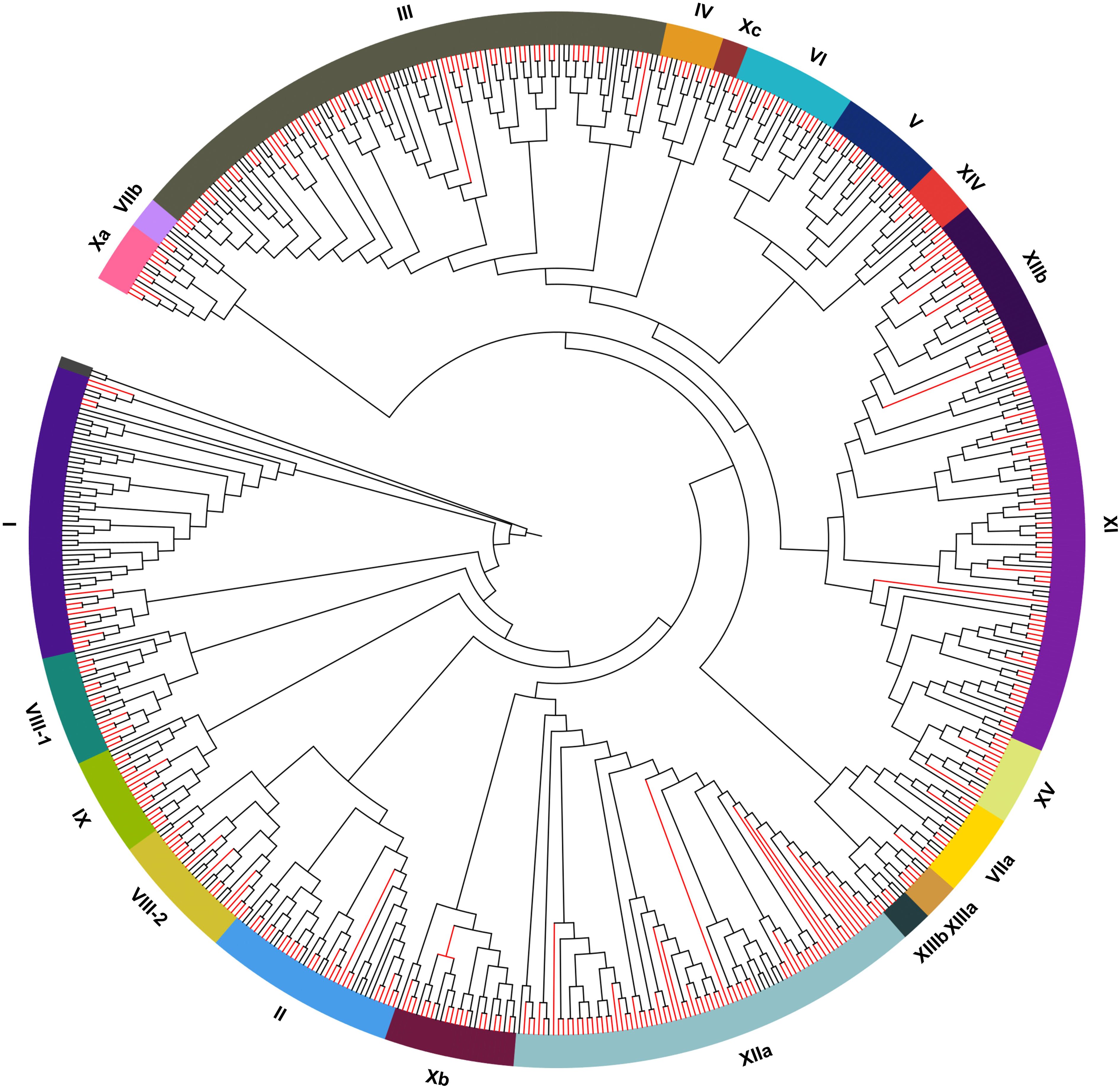
Figure 1. Phylogenetic trees of LRR-RLK genes in A. thaliana and A. chinensis. The red branch represents the LRR-RLK genes in A. chinensis. The black branch represents the LRR-RLK genes in A. thaliana.
3.4 Conserved motif gene structure and domain conservation analyses of AcLRR-RLK genes in A. chinensis HapA and HapP
Conserved motif analysis of AcLRR-RLK genes was performed using the MEME suite, resulting in the identification of 15 motifs (Figure 2). In AcLRR-RLK genes, nine conserved motifs were identified in the kinase domain, including motif 1, motif 2, motif 3, motif 4, motif 6, motif 7, motif 8, motif 9, and motif 14, while the remaining motifs were located in the LRR domain. Notably, motif 6 in the kinase region was present in 97.5% of AcLRR-RLK genes. Motif 5, motif 10, motif 11 and motif 12 in the LRR region were present in more than 95% of the AcLRR-RLK genes. Further observation revealed that some conserved motifs in the LRR region were similar, such as motif 5, motif 10 and motif 11, whereas the conserved motifs in the kinase region were quite different. These results indicate that the kinase domain of AcLRR-RLK genes is highly conserved and that the conserved motifs differ greatly. Although the overall diversity of LRR regions is high, some conserved fragments and conserved regions still have certain repeatability. These results are consistent with reported studies (Kileeg et al., 2023). It is further confirmed that the diversity of LRR domains among subfamilies drives the functional diversification of the LRR-RLK genes, whereas the kinase domains are highly conserved within the family to maintain the core function of signal transduction.
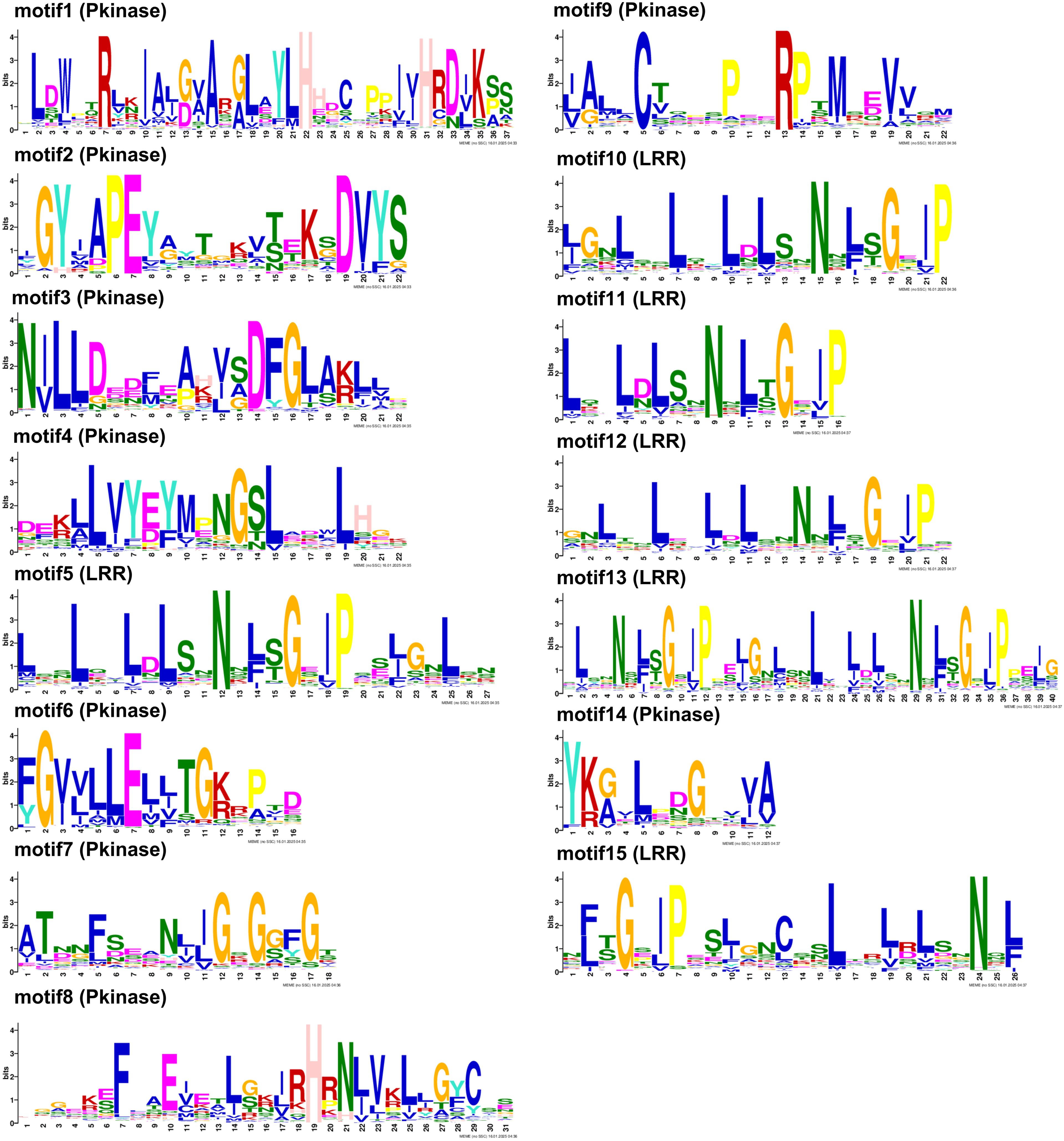
Figure 2. Conserved motifs of AcLRR-RLK genes in A. chinensis. If the bit value of the amino acid at this position is less than 1, it is represented by x; for 2 > bits ≥ 1, it is represented by a lowercase letter; for 3 > bits ≥ 2, it is represented by a capital letter; and for bits ≥ 3, it is represented by a bold capital letter. The text in parentheses indicates the domain in which the motif resides.
We further investigated the conserved motif arrangement and gene structure distributions across different groups (Supplementary File 1). From the perspective of kinase domains, all subfamilies of AcLRR-RLK genes contain motifs 1, 3, 4, 6, 7, 8, and 9, and within the same subfamily, the arrangement of these motifs on the genes is similar. Interestingly, motif 14 is present in all subfamilies except for XV (Figure 3a), and motif 2 is present in all subfamilies except for XIV (Figure 3b). In terms of the LRR domain, the number of conserved motifs carried by different AcLRR-RLK subfamilies is in the range of 1–42. Some larger subfamilies, such as XIIa and XIIb, show significant variation in the number of conserved motifs in the LRR region among their members (Supplementary File 1). Overall, among the 21 subfamilies, members of subfamilies V and I generally carry the fewest conserved motifs, whereas members of subfamily XI carry the most conserved motifs. The number of conserved motifs carried by members within the XIIa and XIIb subfamilies varies significantly, which may reflect greater genetic variation within these two subfamilies. In terms of gene structure, most subfamilies have 2-4 exons, with subfamilies I, II, V, VI, VIII-1, VIII-2, and XIIIb typically having more than 10 exons. In A. chinensis, subfamilies with more exons tend to have elongated exons and larger intron structures. Within the same subfamily, the motif carrying patterns, sequence arrangement, and gene structure of the members are relatively conserved.
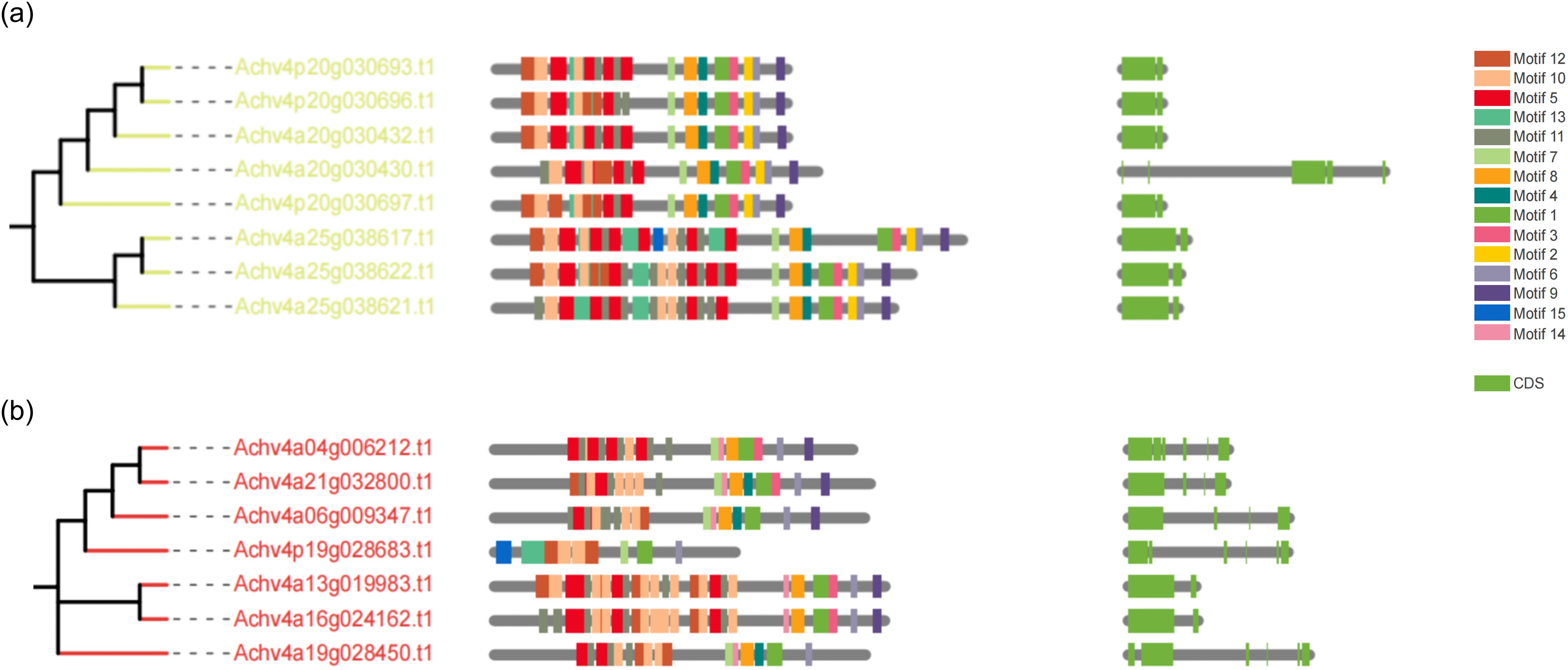
Figure 3. Phylogenetic tree, conserved motifs, and gene structure of group XV and XIV AcLRR-RLK genes in A. chinensis. (a) Phylogenetic tree, conserved motifs, and gene structure of group XV AcLRR-RLK genes in A. chinensis. (b) Phylogenetic tree, conserved motifs, and gene structure of group XIV AcLRR-RLK genes in A. chinensis. The AcLRR-RLK phylogenetic tree was constructed on the basis of kinase sequences. The arrangement of conserved motifs in each AcLRR-RLK was visualized using conserved motif information, with colored boxes representing motifs 1–15. The AcLRR-RLK gene structure in the cds region was visualized, with the green boxes representing the cds regions.
3.5 Analysis of cis-regulatory element distribution in AcLRR-RLK promoters
Cis-acting elements in the promoter region play crucial roles in gene transcription and expression, making their analysis essential for understanding gene function. We utilized the PlantCare website to predict cis-acting elements in the upstream promoter regions of AcLRR-RLK genes. The identified elements were classified into four primary categories: light response, hormone response, stress response, and growth and metabolism (Figure 4). In A. chinensis, the most abundant elements were light-responsive elements present in all the upstream sequences of AcLRR-RLK genes, with G-box and Box 4 elements as the primary conserved motifs. The second most prevalent category was hormone-responsive elements, particularly those related to methyl jasmonate (MeJA) and abscisic acid (ABA) responsiveness, which were abundant in the AcLRR-RLK genes. Among the stress-responsive cis-acting elements, those related to anaerobic induction, low-temperature stress, drought stress, and defense against biotic stress were prominent and occupied a significant portion of the upstream promoter regions of AcLRR-RLK genes. Finally, elements associated with plant growth and metabolism, relatively fewer in number in AcLRR-RLK genes, included conserved motifs such as O2-site, CAT-box, and GCN4_motif.

Figure 4. Cis-element analysis of the LRR-RLK genes in A. chinensis.
Further analysis of the distribution of different types of cis-regulatory elements carried by different subfamilies (Supplementary Figure S3) revealed that all subfamilies contained many light-responsive elements, with group XIIb being the subfamily that included the most light-responsive element-related elements. Group XIIIb contains the most MeJA (methyl jasmonate) response-related cis-regulatory elements, followed by group IV. Additionally, VIIb is the only subfamily that does not contain MeJA response-related cis-regulatory elements. Xc is the only subfamily that lacks abscisic acid response-related cis-regulatory elements. These results suggest that all subfamilies of AcLRR-RLK genes play a role in the light response, but the functions of different subfamilies in other biological processes may differ.
3.6 Gene chromosomal location, duplications, and collinearity
The chromosome localization results indicated that AcLRR-RLK genes are distributed across 29 chromosomes (Supplementary Figure S4). For HapA (Supplementary Figure S4a), the greatest number of genes was observed on Chr03, followed by Chr11 and Chr16, whereas Chr07 had the fewest AcLRR-RLK genes. In HapP (Supplementary Figure S4b), Chr11 had the most genes, closely followed by Chr03 and Chr08, and Chr07 again had the least number of AcLRR-RLK genes. Notably, HapP had a significantly greater number of genes on Chr10 than did HapA. HapP also had more AcLRR-RLK genes than HapA did on Chr04, Chr08, Chr11, Chr17, Chr20, Chr21, Chr23, and Chr26. Conversely, compared with HapP, HapA presented more genes on Chr05, Chr13, Chr15, Chr19, Chr24 and Chr28. AcLRR-RLK genes belonging to the same subfamily were not localized in a few specific chromosomal regions but were instead distributed across multiple chromosomes. Most AcLRR-RLK genes were located toward the chromosomal ends (subtelomeric regions), with relatively similar distribution patterns observed in both haplotypes.
Gene duplication is one of the primary drivers of genome and genetic system evolution (Moore and Purugganan, 2003). There are three main types of gene duplication, including whole genome duplication, tandem duplication, and segmental duplication, all of which can give rise to numerous gene families (Qiao et al., 2019). According to the JCVI collinearity analysis (Supplementary Table S3; Figure 5; Supplementary Figure S5), the gene duplications of AcLRR-RLK genes were all segmental duplications. In HapA and HapP, 135 and 136 pairs of duplicated genes are distributed across different chromosomes, respectively, and these gene pairs are predominantly found in regions with relatively high gene density. The duplicated gene pairs in HapA and HapP included 222 and 225 AcLRR-RLK genes, respectively, accounting for more than half of the Hongyang AcLRR-RLK genes. Analysis of gene collinearity between the two haplotypes revealed that most AcLRR-RLK genes are collinear, with some being haplotype-specific AcLRR-RLK genes. HapA and HapP contain 38 and 27 specific AcLRR-RLK genes, respectively (Supplementary Table S1; Supplementary Figure S4), and these specific genes are distributed across multiple chromosomes (Supplementary Figure S4). From the distribution of AcLRR-RLK genes on chromosomes, some haplotype-specific AcLRR-RLK genes are clustered within specific chromosomal regions. In HapA, the specific AcLRR-RLK clusters are located on chromosomes Chr08, Chr10, Chr13, Chr16, Chr19 and Chr28. However, the specific AcLRR-RLK genes in HapP are more sparsely distributed. These findings suggest that the genes of the AcLRR-RLK family are generated through gene duplication, with segmental duplication playing a major role in the expansion of the AcLRR-RLK gene family. Additionally, some haplotype-specific AcLRR-RLK genes are present between the two haplotypes.
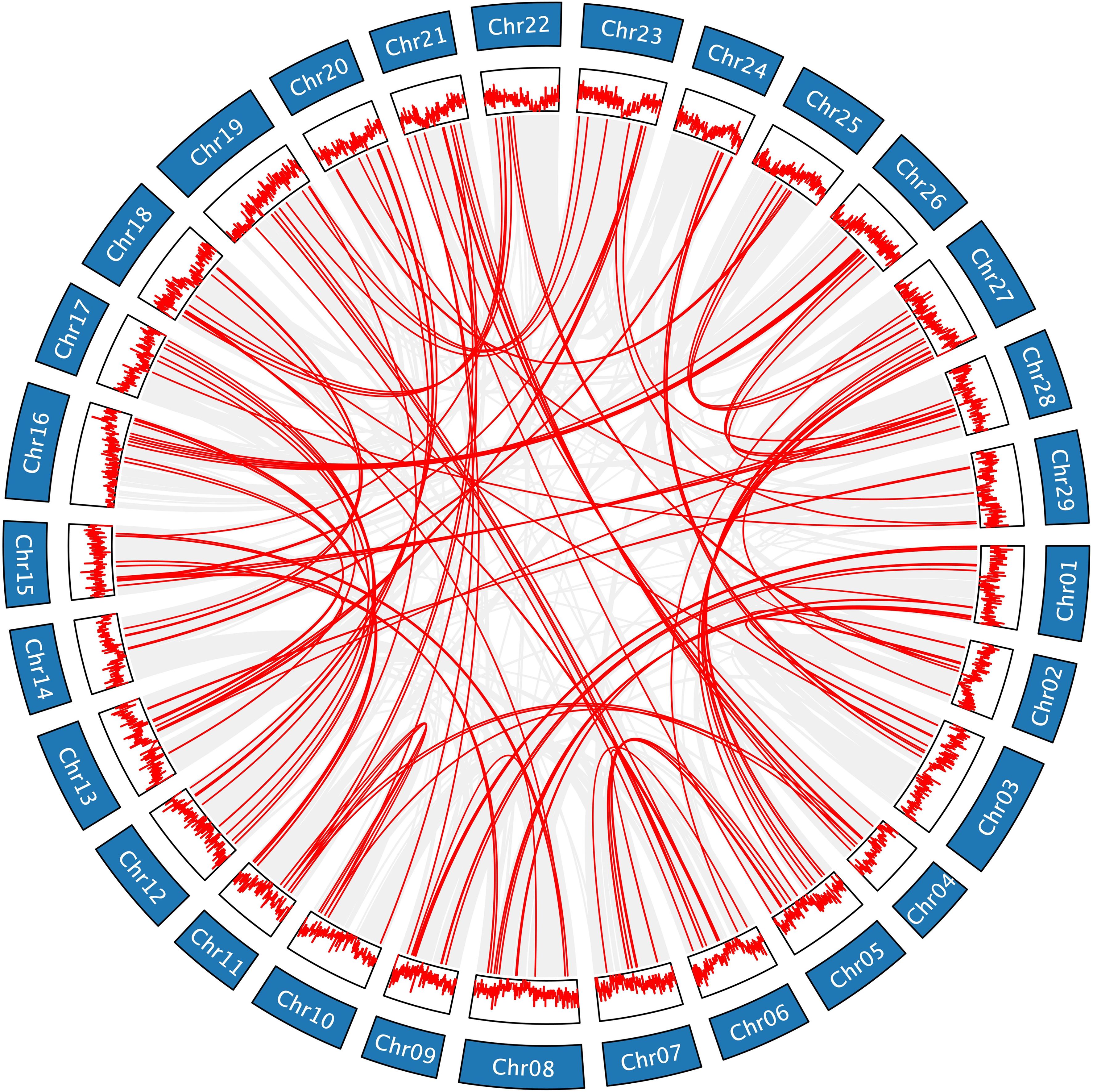
Figure 5. Genomic collinearity of the AcLRR-RLK family in A. chinensis HapA. Distribution and collinearity of the AcLRR-RLK family in the genome. The ring in the middle represents the gene density of each chromosome. The gray background lines represent a collinear background, and the red lines indicate a collinear relationship between AcLRR-RLK members.
3.7 AcLRR-RLK gene expression in different tissues
We analyzed the expression patterns of different groups of AcLRR-RLK genes in various plant tissues using transcriptome data (Supplementary Figure S6; Supplementary Table S5). The expression levels of the TPM were measured. We observed that group IV genes were not expressed in flowers, whereas genes from the other groups were expressed in all the tissues. Groups I, II, III, IX, V, VIIb, VIII-2, Xa, Xb, XI, XIIa, and XIV broadly expressed in all tissues, suggesting that these groups may function in multiple tissues. Further combined with gene duplication analysis (Supplementary Table S3), it was found that 86% of the genes in group III were duplicate gene pairs, and these gene pairs were generally highly expressed in multiple tissues. This result implies that these duplicated gene pairs in III may maintain their original functions and have functional redundancy. Some groups were highly expressed specifically in certain tissues. For example, groups VIIa, VI, VIII-1, and XV showed specific expression in root, stem, leaf, and fruit; XIIb genes were abundant in root, stem, and leaf; and group IV genes were prevalent in stems. Group Xc is highly expressed in flower, stem, leaf, and fruit, and the promoter region of its genes carry MeJA-responsive elements (Supplementary Table S2). MeJA has been reported to be closely associated with floral organ development and opening in a variety of plants, including Arabidopsis thaliana and Brassica napus L. (Heil et al., 2001; Pak et al., 2017; Wang et al., 2024). Therefore, the Xc subpopulation may coordinate floral development through the MeJA signaling pathway. These findings indicate that most groups have broad functions across all tissues, whereas some may have undergone functional differentiation, acting in several specific tissues or through specific signaling pathways. Furthermore, we found that all the genes were highly expressed in stems, implying that the growth and development of stems require the synergistic action of all the AcLRR-RLK subfamilies.
From the genetic level, 30 (79%) HapA-specific genes and 14 (52%) HapP-specific genes were expressed in different tissues without significant tissue specificity (Supplementary Table S5). A greater proportion of HapA-specific genes exhibited expression across multiple tissues, suggesting broader involvement in tissue development.
3.8 Responses of AcLRR-RLK genes under Psa infection in A. chinensis
Pseudomonas syringae pv. actinidiae (Psa) is the pathogenic bacterium responsible for causing bacterial canker in Mihoutao (Cameron and Sarojini, 2014). The key virulence factor of Psa is the protein secretion system involving the type III secretion system (T3SS). This secretion system secretes various toxic effectors capable of disrupting the immune defense responses of the plant, enabling the pathogen to rapidly adapt to the host environment (Jayaraman et al., 2023; Colombi et al., 2024). To investigate the expression patterns of AcLRR-RLK in response to Psa, we analyzed AcLRR-RLK expression levels during Psa infection using transcriptome data from phloem tissues collected at different time points after infection. Differential expression gene analysis comparing various time points after pathogen infection with the 0 hour after inoculation (hai) sample revealed that significantly more AcLRR-RLK genes were downregulated than upregulated at 12 hai (Supplementary Figure S7a). These findings suggest that the early stages of pathogen infection may suppress the expression of certain defense-related AcLRR-RLK genes. However, at 24 hai, a notable increase in the number of upregulated genes was observed (Supplementary Figure S7b), indicating that plants may start activating AcLRR-RLK genes related to disease resistance or the emergency response at this time. As the infection progressed (Supplementary Figures S7c, d), the number of upregulated AcLRR-RLK genes gradually increased. This implies that during early Psa infection, the expression of some AcLRR-RLK genes is suppressed, but as infection persists, the defense response of Mihoutao is activated, and an increasing number of AcLRR-RLK genes begin to respond to pathogen infection.
Heatmaps were generated to illustrate the differential expression of AcLRR-RLK genes at various time points (12 hai, 24 hai, 48 hai, and 96 hai) after Psa infection (Figure 6; Supplementary Table S4). Compared with 0 hai, the expression levels of certain genes significantly increased during the early infection stage (12 hai), indicating that the plants rapidly initiated a preliminary immune response following pathogen invasion. As the infection progressed (24 hai), the expression levels of these genes further increased, although the expression of some initially highly expressed genes significantly decreased compared with that in 0 hai. This suggests that while the immune response of the plant gradually strengthened, the immune response may have simultaneously suppressed genes related to growth and metabolic activities. At 48 hai, the changes in gene expression peaked, with some defense-related genes that began being expressed in the early and middle stages potentially reaching their maximum expression levels, whereas some genes whose expression was downregulated early continued to be downregulated or maintained at low expression levels. During this phase, the plant significantly adjusts gene expression to combat pathogen invasion, likely in an effort to inhibit pathogen spread or repair damaged tissues. At 96 hai, the changes in gene expression stabilized, indicating that the plant may have adapted to the presence of the pathogen and entered a new physiological equilibrium. Of these LRR-RLK genes that responded to Psa infection, 12 were HapA-specific genes, and these were predominantly delayed responses (e.g., Achv4a10g015387.t1 peaked at 96 hai), which may be related to hormonal signaling regulation. Seven were HapP-specific genes, which tended to be rapidly activated (e.g., Achv4p29GSAman00020.t1 peaks at 24 hai).
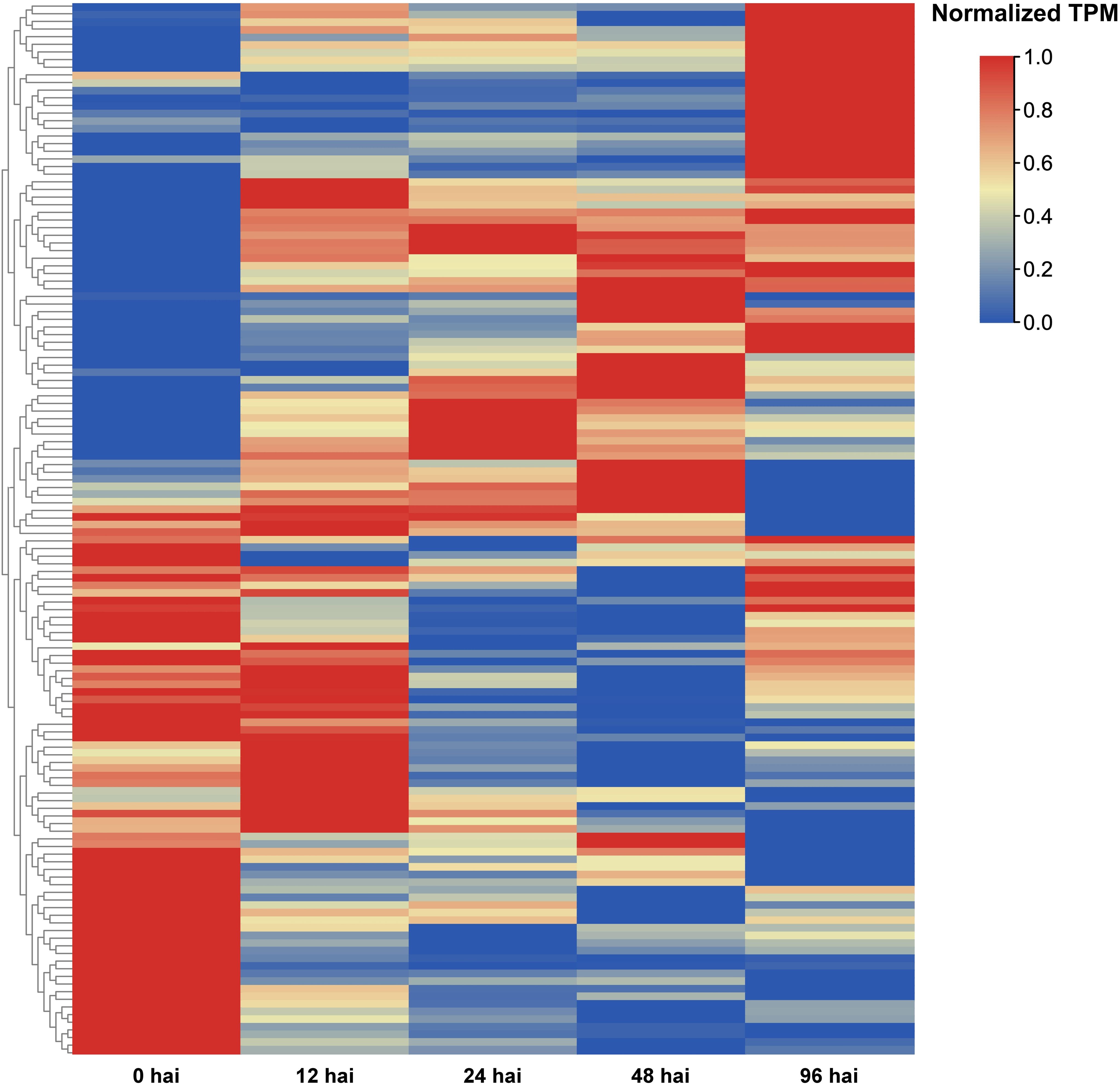
Figure 6. AcLRR-RLK genes were significantly different expressed at different time points after Psa infection compared with 0 hai infection in Actinidia chinensis.
Forty-eight AcLRR-RLK genes showed up-regulation at four time points, 12 hai, 24 hai, 48 hai and 96 hai, after Psa infection, suggesting that they are continuously activated throughout the pathogen invasion (Figure 6; Supplementary Table S4). These 48 AcLRR-RLK genes belong to 14 subfamilies, including I, II, III, IV, V, VI, VIIa, VIII-1, VIII-2, Xb, XI, XIIa, XIIb and XV. Among these, subgroup XIIa contained the greatest number of genes, with 10 AcLRR-RLK genes, followed by the XIIb subfamily, which included 8 AcLRR-RLK genes. Interestingly, we also found that two genes lacking the atypical SKG6 domain, Achv4a22g033382.t1 and Achv4a18g026849.t1, reached peak expression at 96 hours and 48 hours after Psa infection, respectively (Supplementary Table S6). However, three genes containing the atypical SKG6 domain (Achv4a29g045379.t1, Achv4p29GSAman00020.t1, and Achv4a29g045380.t1) showed peak expression as early as 24 hours post-infection. The SKG6 domain is a membrane-associated module exhibiting polarized intracellular localization and includes a highly conserved transmembrane α-helix (Tomishige et al., 2005). This domain may facilitate membrane targeting (Lingwood and Simons, 2010), potentially enabling LRR-RLK receptors to be directed to specific membrane microdomains and to sense external stress more rapidly. This mechanism could explain the early induction of SKG6-containing LRR-RLK genes during the initial stage of Psa infection. Expression analysis suggests these 48 AcLRR-RLK genes are key players in basal defense against Psa infection, as they were significantly and consistently upregulated across all time points. Subgroup XII, in particular, plays a critical role in defending against pathogen invasion. Combined with gene duplication analysis, we found (Supplementary Table S3) that there were 10 duplicated gene pairs in XII, all of which showed significant differences in expression response at different time points of Psa infection, suggesting that they may have undergone functional differentiation in pathogen recognition and defense initiation. This result is the same as those of previously reported studies (Dufayard et al., 2017), further confirming the importance of subgroup XII in plant immune responses. These XII subfamily genes are homologous to known functional genes such as Arabidopsis FLS2 (Chinchilla et al., 2006), EFR (Zipfel et al., 2006), and rice Xa21 (Sun and Wang, 2011), and may serve as potential candidate genes for Psa resistance. In addition, we found that loss/gain of atypical domains may affect the response of LRR-RLK genes to Psa.
To further validate their immune functions, we performed Gene Ontology (GO) enrichment analysis on the 48 core Psa-responsive genes (Supplementary Figure S8). The results showed that these genes were significantly enriched in several plant immunity-related processes, including cell surface receptor kinase signaling pathways, pathogen recognition, hormone stimulus response, and callose deposition. In the MF category, the genes were enriched in molecular functions such as “kinase activity” and “receptor activity”. These enriched terms are highly consistent with the canonical pattern-triggered immunity (PTI) response in plants. Previous studies have shown that LRR-RLK genes are located at the plasma membrane, where they detect pathogen-associated molecular patterns (PAMPs) and activate PTI signaling (Malinovsky et al., 2014; DeFalco and Zipfel, 2021); this is followed by the activation of intracellular signaling pathways such as MAPK cascades, and the deposition of callose and enhancement of lignification in the cell wall, thereby increasing resistance to pathogens (Lloyd et al., 2014; Malinovsky et al., 2014). Therefore, these core AcLRR-RLK genes likely mediate defense against Psa through a typical “PRR recognition-kinase signaling-cell wall reinforcement” mechanism.
3.9 qRT–PCR validation of AcLRR-RLK genes expression under Psa stress
To validate the transcriptome results, we selected seven representative core AcLRR-RLK genes from the XII subfamily responsive to Psa based on the phylogenetic tree (Figure 1) and conducted qRT-PCR assays at 0, 12, 24, 48, and 96 hours after inoculation (hai). The expression trends of the AcLRR-RLK genes after Psa infection aligned with the transcriptomic data (Figure 7). Specifically, compared to baseline (0 hai), the expression level of Achv4p10g015640 reached a peak at 12 hai Psa infection and then subsequently decreased over time. In contrast, the expression levels of Achv4a08g012825, Achv4p10g015639, Achv4p11g016396, and Achv4a19g029045 gradually increased during the infection period. Additionally, Achv4a06g008599 expression was elevated at 24 and 48 hai, followed by a significant decrease at 96 hai. Achv4a27g042182 exhibited an initial increase at 24 hai and then gradually declined over subsequent time points.
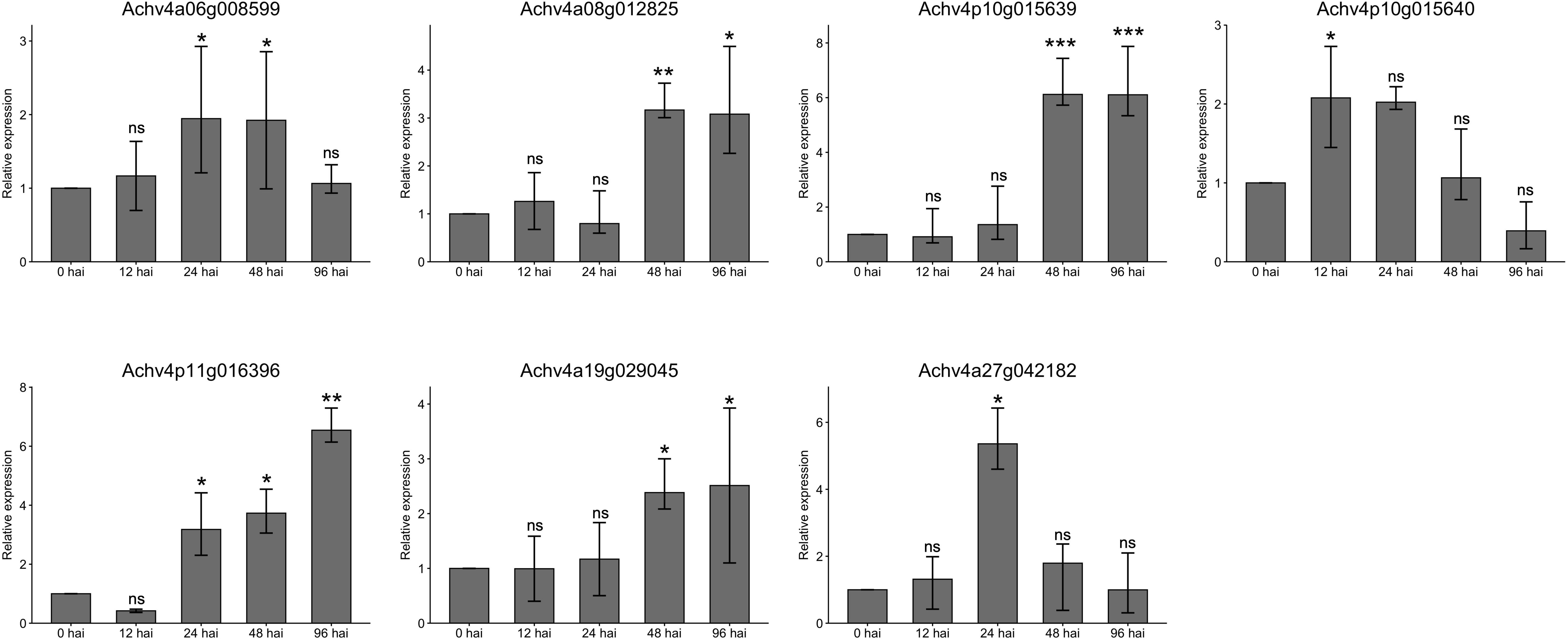
Figure 7. Gene expression of parts of the AcLRR-RLK gene family in Actinidia chinensis at different time points after Psa infection. Seven AcLRR-RLK members were detected by qRT-PCR at no inoculation and after infection for 0, 12, 24, 48 and 96 h. Different symbols indicate significance of differences (ns>0.05, *p≤0.05, **p≤0.01, ***p≤0.001), as determined by one-way ANOVA with SPSS single-factor tests..
4 Discussion
The LRR-RLK gene family, a major class of cell surface receptors, regulates key biological processes including plant growth and development, hormone signaling, and defense responses. In this study, we systematically identified and analyzed the LRR-RLK gene family in A. chinensis cv. Hongyang on the basis of typical domains. All the annotated genes contained at least one LRR domain, a TM domain, and a kinase domain. We identified 336 and 351 LRR-RLK genes in the two haplotypes of A. chinensis (HapA and HapP), respectively, and manually corrected the gene structures to ensure accurate annotation. These genes were classified into 21 subfamilies, which is consistent with the classification of Arabidopsis thaliana and Oryza sativa (Shiu et al., 2004; Sun and Wang, 2011). The kinase domains of the AcLRR-RLK genes were highly conserved, whereas the LRR domains were highly diverse. Structurally, genes within the same subfamily shared similar structures, whereas significant differences were observed among subfamilies. This structural specificity and diversity support the functional differentiation of the LRR-RLK genes (Man et al., 2020). Through the analysis of gene expression patterns, this study further revealed the importance of the AcLRR-RLK gene family in the growth, development, and disease defense of Mihoutao. Most genes were expressed in various tissues, with some genes showing specific high expression in flowers, stems, and fruits, suggesting their potential association with tissue-specific functions in Mihoutao. Additionally, through the prediction and analysis of cis-acting elements, the promoter regions of AcLRR-RLK genes were found to be rich in cis-acting elements related to the light response, hormone regulation (such as ABA and MeJA), abiotic stress (such as drought and low temperature), and disease response, further emphasizing the indispensable role of this gene family in multiple biological processes.
The LRR-RLK gene family has been identified in various plants in recent years. In Phaseolus vulgaris L., 230 LRR-RLK genes distributed across 15 subfamilies have been identified (da Silva Dambroz et al., 2023). In Arachis hypogaea L., 495 LRR-RLK genes have been detected (Wang et al., 2024). Similarly, Saccharum spontaneum harbors 288 LRR-RLK genes (Ding et al., 2024). The model crop A. thaliana has a total of 240 genes belonging to 21 subfamilies of LRR-RLK genes. Compared with the findings in Arabidopsis, the LRR-RLK genes in Mihoutao are also categorized into 21 subfamilies, with group III being the largest, indicating their potentially significant evolutionary role in plant genomes (Shiu et al., 2004; Sun and Wang, 2011). The highly conserved kinase domain and diverse LRR domains of the LRR-RLK genes observed in this study further reinforce their role as key regions for functional differentiation, similar to previous reports (Dufayard et al., 2017). Compared with Phaseolus vulgaris L., A. chinensis harbors a greater proportion of LRR-RLK genes with atypical domains (accounting for 39% of all AcLRR-RLK genes), which are distributed across 19 subfamilies, indicating a broader and more diverse structural complexity. Among these, the most prevalent atypical domains in AcLRR-RLK genes are TM_ErbB1 and Syndecan. AcLRR-RLK genes carrying these domains are distributed primarily in groups I, II, III, IV, IX, V, VIII-2 and XIIa, suggesting that these subfamilies may play roles in growth and development. This hypothesis is further supported by tissue-specific expression analysis of AcLRR-RLK genes, which highlights their potential involvement in developmental processes. For example, the XV subgroups were highly expressed in fruits (Supplementary Figure S5), and the promoter regions of these genes carry hormone-associated elements in response to growth hormone, gibberellin, abscisic acid, and other hormones, which may be involved in fruit growth and ripening. Functional validation of these genes would clarify their roles, providing insights for improving fruit development and ripening.
In A. chinensis, a significant temporal expression pattern of AcLRR-RLK was observed during Psa infection, which is consistent with the expression pattern of LRR-RLK in A. thaliana (Dievart and Clark, 2004). This finding suggests the existence of a conserved immune response pathway for LRR-RLK genes across species. Through the analysis of LRR-RLK gene expression patterns after pathogenic bacterial infection, this study revealed the potential role of these genes in disease resistance. Some genes were suppressed early in the infection process. During the early stages of Psa infection (12 hai), most AcLRR-RLK genes were downregulated, possibly due to the pathogen’s secretion of effector molecules that inhibit the plant’s PTI response (Jayaraman et al., 2023; Colombi et al., 2024). These AcLRR-RLK genes, which are suppressed during the early stages of infection, may be “virulence targets” for pathogen action, host susceptibility factors (S genes) that are utilized by the pathogen to promote infection. As the infection progressed, more LRR-RLK genes were activated, indicating that the plant may gradually initiate mechanisms related to disease defense or the emergency response. Forty-eight AcLRR-RLK genes were consistently up-regulated for expression at four time points after Psa infection. The highest concentration of genes was found in XII subgroup, with 18 of these 48 genes belonging to XII. These subgroup XII genes are homologous to several known functional genes such as FLS2, EFR, and Xa21 (Chinchilla et al., 2006; Zipfel et al., 2006; Sun and Wang, 2011). Functional validation of these genes involved in Psa resistance may provide candidate targets for breeding Actinidiaceae family crops with enhanced resistance.
The function of LRR-RLK genes in disease defense also have been reported in other fruit crops. Among LRR-RLK genes identified in citrus, the expansion and tandem duplication of subgroup XII genes are significantly upregulated upon Xanthomonas citri infection (Magalhães et al., 2016). In grapevine, 42.5% of LRR-RLK genes belong to expanded clades, and multiple genes from the XII, XI, and II subgroups-such as VvSERK3, VaHAESA, and VvFLS2—are involved in PAMP recognition (Tang et al., 2010; Trdá et al., 2014; Liu et al., 2018; Li et al., 2021). In apple, LRR-RLK genes from subgroups II, XI, and XII (e.g., MdBAK1, LRPKm1, and MdFLS2-1) have also been shown to participate in immune responses against various pathogens (Komjanc et al., 1999; Liang et al., 2022; Liu et al., 2022). These studies indicate that subgroups II, XI, and XII are commonly involved in pathogen recognition and immune signaling across multiple fruit species. We found that A. chinensis also harbors a relatively large number of LRR-RLK genes in subgroups II, XI, and XII, with many genes significantly induced during Psa infection. In particular, several subgroup XII genes were continuously upregulated at various stages of infection, suggesting their central role in pathogen detection and defense signal initiation. In contrast (Supplementary Figure S9), citrus contains fewer genes in subgroups II and XI, while grapevine has a notably lower number of subgroup XII genes. These results suggest that the expansion or retention of these key subgroups varies among fruit species, reflecting both functional conservation and species-specific evolutionary divergence.
Integration of transcriptomic, proteomic and metabolomic data can analyze gene functions and regulatory networks from multiple dimensions, which can significantly enhance the understanding of plant disease resistance mechanisms. Transcriptomic data (e.g., TPM values) reveal gene expression levels, but mRNA abundance may not correspond to protein abundance and function. We found that some genes of subgroup XII were significantly up-regulated after Psa infection, and if we further confirm the changes in protein abundance and kinase activity by combining tandem mass tag (TMT) quantitative proteomic and phosphoproteomic data, and observe the accumulation of related disease resistance metabolites by combining the data of targeted and non-targeted metabolomes, it would be helpful to construct the pathway of “LRR-RLK→kinase cascade→metabolic defense”, and to reveal the role of its in the immune signal transduction. This multi-omics strategy can screen the key genes, proteins and metabolites in response to Psa stress at multiple levels, and clarify whether the candidate AcLRR-RLK genes can be converted into functional proteins, avoiding the bias caused by single-omics, and providing more precise targets for downstream functional validation and breeding efforts. In the future, we will combine proteomic and metabolomic data to deeply analyze the function of the AcLRR-RLK gene, construct a regulatory network from signal perception to phenotypic output, and promote the molecular design of disease-resistant varieties.
The expansion of the AcLRR-RLK gene family is achieved primarily through segmental genome duplication, a pattern of gene duplication identical to that of Actinidia eriantha ‘Huate’ (diploid) (Cao et al., 2024), which may represent one of the significant driving forces in the evolution of the diploid Mihoutao genome (Moore and Purugganan, 2003; Qiao et al., 2019). Whereas in Actinidia arguta (tetraploid), the mode of replication of resistance genes is mainly whole genome duplication (WGD) (Kataoka et al., 2010), in Actinidia chinensis var. deliciosa (hexaploid), a combination of WGD and segmental duplication plays a dominant role (Wang et al., 2018; Zhang et al., 2023). Additionally, 38 and 27 haplotype-specific LRR-RLK genes were identified in HapA and HapP, respectively. Transcriptome analysis revealed that HapA-specific LRR-RLK genes were mainly involved in plant development-related processes, reflecting their adaptive evolution in bryophyte morphogenesis. In contrast, HapP-specific genes are more inclined to mediate rapid stress response and enhance defense against pathogens, reflecting their functional bias and adaptive divergence under long-term pathogen selection pressure.
Our study analyzed the LRR-RLK gene family in A. chinensis at the genomic haplotype and transcriptome levels, enriching the understanding of this gene family. We demonstrated the haplotype specificity of the LRR-RLK gene family and identified LRR-RLK genes that respond dynamically to pathogenic infection. These findings provide a new perspective for plant gene family research. Some haplotype-specific LRR-RLK genes with tissue-specific or Psa-induced expression may serve as molecular markers for Mihoutao strain improvement and Psa resistance breeding. For example, the HapP-specific gene Achv4p29GSAman00020.t1 responded rapidly after Psa infection (peaked at 24 hai, Supplementary Table S4) and may activate defense responses by phosphorylating downstream immune signaling proteins. The HapA-specific gene Achv4a16g024156.t1 had the highest level of expression in stems, which may regulate mechanical support of bryophytes. These genes may be candidates for the development of molecular markers for resistance breeding and strain improvement. In addition, LRR-RLK genes that are specifically expressed in tissues and carry elements related to plant growth and development (e.g., VIIb and XV groups) and core LRR-RLK genes that respond to Psa infection can also be used as molecular designed breeding targets for Mihoutao growth and disease resistance. Although we have not conducted in-depth functional studies on these key genes, the multiple disease-response- and growth- related genes identified in this study offer candidate targets for functional validation and molecular breeding of Mihoutao. Our study constructed a comprehensive AcLRR-RLK gene resource database covering 394 manually corrected genes and their expression profiles in different tissues and Psa infection conditions. This database also lays a solid foundation for subsequent functional studies and breeding utilization of disease resistance genes.
5 Conclusions
This study conducted annotation, structural refinement, and systematic analysis of the LRR-RLK genes in the genome of Actinidia chinensis cv. Hongyang, revealing 394 AcLRR-RLK genes that were classified into 21 subfamilies with homologous LRR-RLK genes in Arabidopsis thaliana. In A. chinensis, group XIIa and III had the largest number of genes, whereas group Xc had the fewest genes. Analysis of gene structure and conserved motifs revealed high diversity in the LRR domain, whereas the kinase domain was relatively conserved. Cis-acting element analysis revealed that AcLRR-RLK genes are involved in various important biological processes in plants, demonstrating the functional importance of this family in A. chinensis. Our study revealed that gene expansion in the AcLRR-RLK gene family is due mainly to gene replication, and we identified two haplotype-specific AcLRR-RLK genes. Transcriptome data analysis revealed that most AcLRR-RLK subfamilies play roles in five tissues: root, stem, leaf, flower and fruit. Several subfamilies, such as VIIa, VI, VIII-1, Xc and XV, function in several specific tissues. In addition, analysis of the expression of AcLRR-RLK genes during Psa infection revealed 48 core genes related to the response to this pathogen, among which subgroup XII may play a key role. This study provides valuable insights into the characteristics of the LRR-RLK family in A. chinenses, differences between haplotypes, and responses to biological stress, laying a molecular foundation for the utilization of this gene family and the breeding of varieties resistant to Psa.
Data availability statement
The original contributions presented in the study are included in the article/Supplementary Material. Further inquiries can be directed to the corresponding author.
Author contributions
CL: Data curation, Formal analysis, Investigation, Visualization, Writing – original draft. SM: Conceptualization, Data curation, Supervision, Writing – review & editing. YT: Investigation, Writing – review & editing. XZ: Supervision, Writing – review & editing.
Funding
The author(s) declare financial support was received for the research and/or publication of this article. This research was funded by the Hainan Excellent Talent Team (to J-MZ) and the Yazhouwan National Laboratory Fundamental Research Project (2310JM01).
Acknowledgments
We are very grateful to the Hainan Excellent Talent Team for providing the data analysis platform.
Conflict of interest
The authors declare that the research was conducted in the absence of any commercial or financial relationships that could be construed as a potential conflict of interest.
Generative AI statement
The author(s) declare that no Generative AI was used in the creation of this manuscript.
Any alternative text (alt text) provided alongside figures in this article has been generated by Frontiers with the support of artificial intelligence and reasonable efforts have been made to ensure accuracy, including review by the authors wherever possible. If you identify any issues, please contact us.
Publisher’s note
All claims expressed in this article are solely those of the authors and do not necessarily represent those of their affiliated organizations, or those of the publisher, the editors and the reviewers. Any product that may be evaluated in this article, or claim that may be made by its manufacturer, is not guaranteed or endorsed by the publisher.
Supplementary material
The Supplementary Material for this article can be found online at: https://www.frontiersin.org/articles/10.3389/fpls.2025.1577679/full#supplementary-material
Supplementary File 1 | Phylogenetic tree, conserved motifs, and gene structure of AcLRR-RLK genes in Hongyang. The phylogenetic tree of AcLRR-RLK genes was constructed on the basis of kinase sequences. The arrangement of conserved motifs in each AcLRR-RLK was visualized using conserved motif information, with colored boxes representing motifs 1-15. The gene structures of AcLRR-RLK genes were visualized for the cds region, with the green boxes representing the cds areas.
Supplementary Figure 1 | Domain architectures of the AcLRR-RLK subfamilies. (a) Domain architecture of LRR-I subfamily. (b) Domain architecture of LRR-VIII-2 subfamily. (c) Domain architectures of LRR-Xc and LRR-VIIa subfamilies. (d) Domain architectures of all remaining subfamilies.
Supplementary Figure 2 | Atypical domains carried by AcLRR-RLK genes. The vertical axis represents the types of atypical domains, with only those present in two or more genes specifically labeled. “Other” indicates other atypical domains that occur only in one LRR-RLK. The horizontal axis represents the number of atypical domains.
Supplementary Figure 3 | Analysis of the cis-acting elements of AcLRR-RLK proteins in Hongyang. The heatmap displays the types and numbers of cis-acting elements carried within the 21 AcLRR-RLK subfamilies. The numbers in the boxes represent the average number of cis-acting elements in each subfamily, and different colors indicate the range of cis-acting elements.
Supplementary Figure 4 | Distribution of haplotype-specific LRR-RLK genes on chromosomes in A. chinensis. (a) Locations of HapA AcLRR-RLK genes on chromosomes. (b) Locations of HapP AcLRR-RLK genes on chromosomes. All haplotype-specific AcLRR-RLK genes are marked in blue.
Supplementary Figure 5 | Genomic collinearity of the AcLRR-RLK family in HapP of Hongyang. Distribution and collinearity of the AcLRR-RLK family in the genome. The middle ring represents the gene density of each chromosome. The gray background lines depict a collinear background, whereas the red lines indicate a collinear relationship among AcLRR-RLK members.
Supplementary Figure 6 | Heatmap of the expression of 21 AcLRR-RLK subfamilies in different tissues of A. chinensis. We defined TPM>1 as genes expressed in tissues. Brown and white indicate the expression levels of genes in the heatmap: the stronger the red color is, the higher the gene expression level, whereas the stronger the white color is, the lower the gene expression level. HF: Hongyang fruit. HFR: Hongyang flower. HG: Hongyang root. HL: Hongyang leaf. HST: Hongyang stem.
Supplementary Figure 7 | Volcano plots of AcLRR-RLK genes differentially expressed at different time points after Psa infection compared with 0 hai in Hongyang. (a) Volcano plot of AcLRR-RLK genes differentially expressed at 12 hai compared with 0 hai after Psa infection. (b) Volcano plot of AcLRR-RLK genes differentially expressed at 24 hai compared with 0 hai after Psa infection. (c) Volcano plot of AcLRR-RLK genes differentially expressed at 48 hai compared with 0 hai after Psa infection. (d) Volcano plot of AcLRR-RLK genes differentially expressed at 96 hai compared with 0 hai after Psa infection. Red dots represent significantly upregulated genes. The blue dots represent significantly downregulated genes. The gray dots represent genes whose expression did not significantly differ.
Supplementary Figure 8 | GO enrichment analysis of 48 core Psa-responsive AcLRR-RLK genes. (a) Enriched Biological Process (BP) terms in 48 core Psa-responsive AcLRR-RLK genes. (b) Enriched Molecular Function (MF) terms in 48 core Psa-responsive AcLRR-RLK genes.
Supplementary Figure 9 | Distribution of LRR-RLK genes subfamilies in four fruit crops.
References
Bai, D. F. (2023). Explanation of mechanism underlying the waterlogging tolerance in Actinidia valvata and functional identification of two ethylene responsive factors. Doctoral dissertation. Wuhan, China: Huazhong Agricultural University. CNKI. Available online at: https://link.cnki.net/doi/10.27158/d.cnki.ghznu.2023.000130 (Accessed August 19, 2025).
Bailey, T. L., Boden, M., Buske, F. A., Frith, M., Grant, C. E., Clementi, L., et al. (2009). MEME SUITE: tools for motif discovery and searching. Nucleic Acids Res. 37, W202–W208. doi: 10.1093/nar/gkp335
Bocharov, E. V., Mineev, K. S., Goncharuk, M. V., and Arseniev, A. S. (2012). Structural and thermodynamic insight into the process of “weak” dimerization of the ErbB4 transmembrane domain by solution NMR. Biochim. Et Biophys. Acta-Biomembranes. 1818, 2158–2170. doi: 10.1016/j.bbamem.2012.05.001
Bocharov, E. V., Mineev, K. S., Volynsky, P. E., Ermolyuk, Y. S., Tkach, E. N., Sobol, A. G., et al. (2008). Spatial structure of the dimeric transmembrane domain of the growth factor receptor ErbB2 presumably corresponding to the receptor active state. J. Biol. Chem. 283, 6950–6956. doi: 10.1074/jbc.M709202200
Bragin, P. E., Mineev, K. S., Bocharova, O. V., Volynsky, P. E., Bocharov, E. V., and Arseniev, A. S. (2016). HER2 transmembrane domain dimerization coupled with self-association of membrane-embedded cytoplasmic juxtamembrane regions. J. Mol. Biol. 428, 52–61. doi: 10.1016/j.jmb.2015.11.007
Camacho, C., Coulouris, G., Avagyan, V., Ma, N., Papadopoulos, J., Bealer, K., et al. (2009). BLAST+: architecture and applications. BMC Bioinf. 10, 421. doi: 10.1186/1471-2105-10-421
Cameron, A. and Sarojini, V. (2014). Pseudomonas syringae pv. actinidiae: chemical control, resistance mechanisms and possible alternatives. Plant Pathology. 63, 1–11. doi: 10.1111/ppa.12066
Cao, Y., Zhang, C., Liu, F., Li, D., Zhang, A., Li, L., et al. (2024). Genome-wide identification and expression analysis of kiwifruit leucine-rich repeat receptor-like proteins reveal their roles in biotic and abiotic stress responses. Int. J. Mol. Sci. 25, 16. doi: 10.3390/ijms25084497
Chapman, J. R., Taylor, R. K., Weir, B. S., Romberg, M. K., Vanneste, J. L., Luck, J., et al. (2012). Phylogenetic relationships among global populations of Pseudomonas syringae pv. actinidiae. Phytopathology. 102, 1034–1044. doi: 10.1094/phyto-03-12-0064-r
Chen, C., Wu, Y., Li, J., Wang, X., Zeng, Z., Xu, J., et al. (2023). TBtools-II: A “one for all, all for one” bioinformatics platform for biological big-data mining. Mol. Plant 16, 1733–1742. doi: 10.1016/j.molp.2023.09.010
Chen, S., Zhou, Y., Chen, Y., and Gu, J. (2018). fastp: an ultra-fast all-in-one FASTQ preprocessor. Bioinformatics 34, i884–i890. doi: 10.1093/bioinformatics/bty560
Chinchilla, D., Bauer, Z., Regenass, M., Boller, T., and Felix, G. (2006). The Arabidopsis receptor kinase FLS2 binds flg22 and determines the specificity of flagellin perception. Plant Cell. 18, 465–476. doi: 10.1105/tpc.105.036574
Colombi, E., Bertels, F., Doulcier, G., McConnell, E., Pichugina, T., Sohn, K. H., et al. (2024). Rapid dissemination of host metabolism-manipulating genes via integrative and conjugative elements. Proc. Natl. Acad. Sci. U S A. 121, e2309263121. doi: 10.1073/pnas.2309263121
da Silva Dambroz, C. M., Aono, A. H., de Andrade Silva, E. M., and Pereira, W. A. (2023). Genome-wide analysis and characterization of the LRR-RLK gene family provides insights into anthracnose resistance in common bean. Sci. Rep. 13, 13455. doi: 10.1038/s41598-023-40054-3
DeFalco, T. A. and Zipfel, C. (2021). Molecular mechanisms of early plant pattern-triggered immune signaling. Mol. Cell 81, 3449–3467. doi: 10.5167/uzh-207233
De Rossi, G. and Whiteford, J. R. (2013). Novel insight into the biological functions of syndecan ectodomain core proteins. Biofactors. 39, 374–382. doi: 10.1002/biof.1104
Dievart, A. and Clark, S. E. (2004). LRR-containing receptors regulating plant development and defense. Development. 131, 251–261. doi: 10.1242/dev.00998
Dimitrov, I. and Tax, F. E. (2018). Lateral root growth in Arabidopsis is controlled by short and long distance signaling through the LRR RLKs XIP1/CEPR1 and CEPR2. Plant Signal Behav. 13, e1489667. doi: 10.1080/15592324.2018.1489667
Ding, H., Feng, X., Yuan, Y., Wang, B., Wang, Y., and Zhang, J. (2024). Genomic investigation of duplication, functional conservation, and divergence in the LRR-RLK Family of Saccharum. BMC Genomics 25, 165. doi: 10.1186/s12864-024-10073-z
Dufayard, J. F., Bettembourg, M., Fischer, I., Droc, G., Guiderdoni, E., Perin, C., et al. (2017). New insights on leucine-rich repeats receptor-like kinase orthologous relationships in angiosperms. Front. Plant Sci. 8. doi: 10.3389/fpls.2017.00381
Endres, N. F., Das, R., Smith, A. W., Arkhipov, A., Kovacs, E., Huang, Y., et al. (2013). Conformational coupling across the plasma membrane in activation of the EGF receptor. Cell. 152, 543–556. doi: 10.1016/j.cell.2012.12.032
Gou, X., He, K., Yang, H., Yuan, T., Lin, H., Clouse, S. D., et al. (2010). Genome-wide cloning and sequence analysis of leucine-rich repeat receptor-like protein kinase genes in Arabidopsis thaliana. BMC Genomics 11, 19. doi: 10.1186/1471-2164-11-19
Hart, T., Komori, H. K., LaMere, S., Podshivalova, K., and Salomon, D. R. (2013). Finding the active genes in deep RNA-seq gene expression studies. BMC Genomics 14, 778. doi: 10.1186/1471-2164-14-778
Hebenstreit, D., Fang, M., Gu, M., Charoensawan, V., van Oudenaarden, A., and Teichmann, S. A. (2011). RNA sequencing reveals two major classes of gene expression levels in metazoan cells. Mol. Syst. Biol. 7, 497. doi: 10.1038/msb.2011.28
Heil, M., Koch, T., Hilpert, A., Fiala, B., Linsenmair, K. E., and Boland, W. (2001). Extrafloral nectar production of the ant-associated plant, Macaranga tanarius, is an induced, indirect, defensive response elicited by jasmonic acid. Proc. Natl. Acad. Sci. 98, 1083–1088. doi: 10.1073/pnas.98.3.1083
Horton, P., Park, K. J., Obayashi, T., Fujita, N., Harada, H., Adams-Collier, C. J., et al. (2007). WoLF PSORT: protein localization predictor. Nucleic Acids Res. 35, W585–W587. doi: 10.1093/nar/gkm259
Jayaraman, J., Yoon, M., Hemara, L. M., Bohne, D., Tahir, J., Chen, R. K. Y., et al. (2023). Contrasting effector profiles between bacterial colonisers of kiwifruit reveal redundant roles converging on PTI-suppression and RIN4. New Phytol. 238, 1605–1619. doi: 10.1111/nph.18848
Kataoka, I., Mizugami, T., Kim, J., Beppu, K., Fukuda, T., Sugahara, S., et al. (2010). Ploidy variation of hardy kiwifruit (Actinidia arguta) resources and geographic distribution in Japan. Scientia Horticulturae. 124, 409–414. doi: 10.1016/J.SCIENTA.2010.01.016
Katoh, K., Misawa, K., Kuma, K., and Miyata, T. (2002). MAFFT: a novel method for rapid multiple sequence alignment based on fast Fourier transform. Nucleic Acids Res. 30, 3059–3066. doi: 10.1093/nar/gkf436
Kileeg, Z., Haldar, A., Khan, H., Qamar, A., and Mott, G. (2023). Differential expansion and retention patterns of LRR-RLK genes across plant evolution. Plant Direct 7, e493. doi: 10.1101/2023.07.26.549740
Komjanc, M., Festi, S., Rizzotti, L., Cattivelli, L., Cervone, F., and Lorenzo, G. (1999). A leucine-rich repeat receptor-like protein kinase (LRPKm1) gene is induced in Malus × domestica by Venturia inaequalis infection and salicylic acid treatment. Plant Mol. Biol. 40, 945–957. doi: 10.1023/A:1006275924882
Lescot, M., Déhais, P., Thijs, G., Marchal, K., Moreau, Y., Van de Peer, Y., et al. (2002). PlantCARE, a database of plant cis-acting regulatory elements and a portal to tools for in silico analysis of promoter sequences. Nucleic Acids Res. 30, 325–327. doi: 10.1093/nar/30.1.325
Letunic, I. and Bork, P. (2021). Interactive Tree Of Life (iTOL) v5: an online tool for phylogenetic tree display and annotation. Nucleic Acids Res. 49, W293–w296. doi: 10.1093/nar/gkab301
Li, H., Han, X., Liu, X., Zhou, M., Ren, W., Zhao, B., et al. (2019). A leucine-rich repeat-receptor-like kinase gene SbER2-1 from sorghum (Sorghum bicolor L.) confers drought tolerance in maize. BMC Genomics 20, 737. doi: 10.1186/s12864-019-6143-x
Li, Y., Jiao, C., Wei, Z., Chai, S., Jia, H., Gao, M., et al. (2021). Analysis of grapevine’s somatic embryogenesis receptor kinase (SERK) gene family: VqSERK3/BAK1 overexpression enhances disease resistance. Phytopathology. 112, 1081–1092. doi: 10.1094/PHYTO-04-21-0136-R
Liang, Z., Liu, K., Jiang, C., Yang, A., Yan, J., Han, X., et al. (2022). Insertion of a TRIM-like sequence in MdFLS2-1 promoter is associated with its allele-specific expression in response to Alternaria alternata in apple. Front. Plant Sci. 13. doi: 10.3389/fpls.2022.1090621
Lingwood, D. and Simons, K. (2010). Lipid rafts as a membrane-organizing principle. Science. 327, 46–50. doi: 10.1126/science.1174621
Liu, P.-L., Du, L., Huang, Y., Gao, S.-M., and Yu, M. (2017). Origin and diversification of leucine-rich repeat receptor-like protein kinase (LRR-RLK) genes in plants. BMC Evolutionary Biol. 17, 47. doi: 10.1186/s12862-017-0891-5
Liu, X., Xu, S., Wang, X., Xin, L., Wang, L., Mao, Z., et al. (2022). MdBAK1 overexpression in apple enhanced resistance to replant disease as well as to the causative pathogen Fusarium oxysporum. Plant Physiol. Biochem. 179, 144–157. doi: 10.1016/j.plaphy.2022.03.014
Liu, S., Zhang, C., Chao, N., Lu, J., and Zhang, Y. (2018). Cloning, Characterization, and Functional Investigation of VaHAESA from Vitis amurensis Inoculated with Plasmopara viticola. Int. J. Mol. Sci. 19 (4), 1204. doi: 10.3390/ijms19041204
Lloyd, S. R., Schoonbeek, H. J., Trick, M., Zipfel, C., and Ridout, C. J. (2014). Methods to study PAMP-triggered immunity in Brassica species. Mol. Plant-Microbe Interact. 27, 286–295. doi: 10.1094/MPMI-05-13-0154-FI
Magalhães, D., Scholte, L., Silva, N., Oliveira, G., Zipfel, C., Takita, M., et al. (2016). LRR-RLK family from two Citrus species: genome-wide identification and evolutionary aspects. BMC Genomics 17, 623. doi: 10.1186/s12864-016-2930-9
Malinovsky, F. G., Fangel, J. U., and Willats, W. G. (2014). The role of the cell wall in plant immunity. Front. Plant science. 5. doi: 10.3389/fpls.2014.00178
Man, J., Gallagher, J. P., and Bartlett, M. (2020). Structural evolution drives diversification of the large LRR-RLK gene family. New Phytologist. 226, 1492–1505. doi: 10.1111/nph.16455
Mineev, K. S., Bocharov, E. V., Pustovalova, Y. E., Bocharova, O. V., Chupin, V. V., and Arseniev, A. S. (2010). Spatial structure of the transmembrane domain heterodimer of erbB1 and erbB2 receptor tyrosine kinases. J. Mol. Biol. 400, 231–243. doi: 10.1016/j.jmb.2010.05.016
Moore, R. C. and Purugganan, M. D. (2003). The early stages of duplicate gene evolution. Proc. Natl. Acad. Sci. U S A. 100, 15682–15687. doi: 10.1073/pnas.2535513100
Mu, Z., Xu, M., Manda, T., Chen, J., Yang, L., and Hwarari, D. (2024). Characterization, evolution, and abiotic stress responses of leucine-rich repeat receptor-like protein kinases (LRR-RLK) in Liriodendron chinense. BMC Genomics 25, 748. doi: 10.1186/s12864-024-10560-3
Ngou, B. P. M., Ding, P., and Jones, J. D. G. (2022a). Thirty years of resistance: Zig-zag through the plant immune system. Plant Cell. 34, 1447–1478. doi: 10.1093/plcell/koac041
Ngou, B. P. M., Heal, R., Wyler, M., Schmid, M. W., and Jones, J. D. G. (2022b). Concerted expansion and contraction of immune receptor gene repertoires in plant genomes. Nat. Plants. 8, 1146–1152. doi: 10.1038/s41477-022-01260-5
Pak, H., Li, Y., Kim, H., and Jiang, L. (2017). cDNA-Amplified fragment length polymorphism analysis reveals differential gene expression induced by exogenous MeJA and GA3 in oilseed rape (Brassica napus L.) flowers. J. Integr. Agriculture. 16, 47–56. doi: 10.1016/S2095-3119(16)61407-7
Patro, R., Duggal, G., Love, M. I., Irizarry, R. A., and Kingsford, C. (2017). Salmon provides fast and bias-aware quantification of transcript expression. Nat. Methods 14, 417–419. doi: 10.1038/nmeth.4197
Price, M. N., Dehal, P. S., and Arkin, A. P. (2010). FastTree 2–approximately maximum-likelihood trees for large alignments. PLoS One 5, e9490. doi: 10.1371/journal.pone.0009490
Qiao, X., Li, Q., Yin, H., Qi, K., Li, L., Wang, R., et al. (2019). Gene duplication and evolution in recurring polyploidization-diploidization cycles in plants. Genome Biol. 20, 38. doi: 10.1186/s13059-019-1650-2
Rawson, J. M., Dimitroff, B., Johnson, K. G., Rawson, J. M., Ge, X., Van Vactor, D., et al. (2005). The heparan sulfate proteoglycans Dally-like and Syndecan have distinct functions in axon guidance and visual-system assembly in Drosophila. Curr. Biol. 15, 833–838. doi: 10.1016/j.cub.2005.03.039
Rhiner, C., Gysi, S., Frohli, E., Hengartner, M. O., and Hajnal, A. (2005). Syndecan regulates cell migration and axon guidance in C. elegans. Dev. (Cambridge England). 132, 4621–4633. doi: 10.1242/dev.02042
Robinson, J. T., Thorvaldsdottir, H., Turner, D., and Mesirov, J. P. (2023). igv.js: an embeddable JavaScript implementation of the Integrative Genomics Viewer (IGV). Bioinformatics 39 (1), btac830. doi: 10.1093/bioinformatics/btac830
Shiu, S. H. and Bleecker, A. B. (2001). Receptor-like kinases from Arabidopsis form a monophyletic gene family related to animal receptor kinases. Proc. Natl. Acad. Sci. U.S.A. 98, 10763–10768. doi: 10.1073/pnas.181141598
Shiu, S.-H., Karlowski, W. M., Pan, R., Tzeng, Y.-H., Mayer, K. F. X., and Li, W.-H. (2004). Comparative analysis of the receptor-like kinase family in arabidopsis and rice. Plant Cell 16, 1220–1234. doi: 10.1105/tpc.020834
Song, Y., Sun, L., Lin, M., Chen, J., Qi, X., Hu, C., et al. (2019). Comparative transcriptome analysis of resistant and susceptible kiwifruits in response to Pseudomonas syringae pv. actinidiae during early infection. PLoS One 14, e0211913. doi: 10.1371/journal.pone.0211913
Sonnhammer, E. L., von Heijne, G., and Krogh, A. (1998). A hidden Markov model for predicting transmembrane helices in protein sequences. Proc. Int. Conf Intell. Syst. Mol. Biol. 6, 175–182.
Sun, X. and Wang, G. L. (2011). Genome-wide identification, characterization and phylogenetic analysis of the rice LRR-kinases. PLoS One 6, e16079. doi: 10.1371/journal.pone.0016079
Tang, H., Krishnakumar, V., Zeng, X., Xu, Z., Taranto, A., Lomas, J. S., et al. (2024). JCVI: A versatile toolkit for comparative genomics analysis. Imeta. 3, e211. doi: 10.1002/imt2.211
Tang, P., Zhang, Y., Sun, X., Tian, D., Yang, S., and Ding, J. (2010). Disease resistance signature of the leucine-rich repeat receptor-like kinase genes in four plant species. Plant Sci. 179, 399–406. doi: 10.1016/J.PLANTSCI.2010.06.017
Tomishige, N., Noda, Y., Adachi, H., and Yoda, K. (2005). SKG6, a suppressor gene of synthetic lethality of kex2Δ gas1Δ mutations, encodes a novel membrane protein showing polarized intracellular localization. J. Gen. Appl. Microbiol. 51, 323–326. doi: 10.2323/JGAM.51.323
Trdá, L., Fernandez, O., Boutrot, F., Heloir, M., Kelloniemi, J., Daire, X., et al. (2014). The grapevine flagellin receptor VvFLS2 differentially recognizes flagellin-derived epitopes from the endophytic growth-promoting bacterium Burkholderia phytofirmans and plant pathogenic bacteria. New Phytol. 201 (4), 1371–1384. doi: 10.1111/nph.12592
Wang, J., Yu, J., Li, J., Sun, P., Wang, L., Yuan, J., et al. (2018). Two likely auto-tetraploidization events shaped kiwifruit genome and contributed to establishment of the actinidiaceae family. iScience. 7, 230–240. doi: 10.1016/j.isci.2018.08.003
Wang, C., Zhang, J., Li, J., Xie, J., and Chai, Q. (2024). Exogenous methyl jasmonate regulates endogenous hormone synthesis of soilless cultivated Chinese chive to promote growth physiology and photosynthesis. Scientia Hortic. 327, 112861. doi: 10.1016/j.scienta.2024.112861
Wang, Q., Zhao, X., Sun, Q., Mou, Y., Wang, J., Yan, C., et al. (2024). Genome-wide identification of the LRR-RLK gene family in peanut and functional characterization of AhLRR-RLK265 in salt and drought stresses. Int. J. Biol. Macromolecules 254. doi: 10.1016/j.ijbiomac.2023.127829
Wilkins, M. R., Gasteiger, E., Bairoch, A., Sanchez, J. C., Williams, K. L., Appel, R. D., et al. (1999). Protein identification and analysis tools in the ExPASy server. Methods Mol. Biol. 112, 531–552. doi: 10.1385/1-59259-584-7:531
Yue, J., Chen, Q., Wang, Y., Zhang, L., Ye, C., Wang, X., et al. (2023). Telomere-to-telomere and gap-free reference genome assembly of the kiwifruit Actinidia chinensis. Horticulture Res. 10 (2), uhac264. doi: 10.1093/hr/uhac264
Zdobnov, E. M. and Apweiler, R. (2001). InterProScan–an integration platform for the signature-recognition methods in InterPro. Bioinformatics. 17, 847–848. doi: 10.1093/bioinformatics/17.9.847
Zhang, A., Xiong, Y., Liu, F., and Zhang, X. (2023). A genome-wide analysis of the pentatricopeptide repeat protein gene family in two kiwifruit species with an emphasis on the role of RNA editing in pathogen stress. Int. J. Mol. Sci. 24 (18), 13700. doi: 10.3390/ijms241813700
Keywords: Actinidia chinensis, LRR-RLK, phylogenetic analyses, Pseudomonas syringae pv. actinidiae, abiotic stress response
Citation: Li C, Ma S, Tian Y and Zheng X (2025) Genome-wide identification and expression analyses of the LRR-RLK gene family in Actinidia chinensis. Front. Plant Sci. 16:1577679. doi: 10.3389/fpls.2025.1577679
Received: 16 February 2025; Accepted: 08 August 2025;
Published: 01 September 2025.
Edited by:
Mehar Hasan Asif, National Botanical Research Institute (CSIR), IndiaReviewed by:
Guriqbal Singh Dhillon, University of Idaho, United StatesNeelam Pathak, Dr. Rammanohar Lohia Avadh University, India
Copyright © 2025 Li, Ma, Tian and Zheng. This is an open-access article distributed under the terms of the Creative Commons Attribution License (CC BY). The use, distribution or reproduction in other forums is permitted, provided the original author(s) and the copyright owner(s) are credited and that the original publication in this journal is cited, in accordance with accepted academic practice. No use, distribution or reproduction is permitted which does not comply with these terms.
*Correspondence: Xiaojuan Zheng, emhlbmd4aWFvanVhbjA4MDVAMTYzLmNvbQ==