 Gurudayal Ram Guru
 Pramod W. Ramteke
 Csilla Veres3
 Csaba Vágvölgyi
- 1Centre for Tissue Culture Technology, Jacob Institute of Biotechnology and Bioengineering, Sam Higginbottom University of Agriculture, Technology and Sciences, Prayagraj, Uttar Pradesh, India
- 2Department of Molecular Biology and Genetic Engineering, Rashtrasant Tukadoji Maharaj Nagpur University, Nagpur, Maharashtra, India
- 3Department of Biotechnology and Microbiology, Faculty of Science and Informatics, University of Szeged, Szeged, Hungary
The use of nanoparticles (NPs) in plant tissue culture systems represents a new approach to improve the efficiency of micropropagation. Owing to their nanoscale size, high surface area concomitant with volume, and controllable and targeted release, researchers have tested the experimental benefits of NPs in various ways during each phase of in vitro propagation, which include enhancing surface sterilization to reduce microbial contamination, the targeted uptake of specific macro-and micronutrients, regulating plant hormonal activity to enhance callogenesis, increased shoot multiplication and rooting, and increasing the survival rate during acclimatization. In addition, some situations where NPs are applied can reduce oxidative stress and regulate hormonal pathways, which will stabilize the physiological state of the plant and support better developmental integrity of the regenerating plantlets. In moving forward with the application of nanoparticles, the major limiting factors are nanotoxicity, persistence in the environment, species specificity, and the lack of an established regulatory framework. In this review, the recent published successes in NP-mediated micropropagation are summarized, how they impart their effects in plant science at the cellular and molecular levels are explained, and potential future innovations such as green-synthesized nanomaterials and new smart delivery platforms are also identified. Realizing the full potential of nanotechnologies for application with micropropagation will be critical for developing scalable, sustainable, and precision agricultural production systems.
1 Introduction
The process of plant micropropagation has quickly become an essential technology in the fields of agriculture, horticulture, forestry, and conservation biology since it allows for rapid multiplication of genetically identical and pathogen-free high-throughput plant material without the risk of contamination (Engelmann and Dussert, 2000 ; Cassells, 2002 ; El-Esawi, 2016). Plant micropropagation is a tissue culture process in vitro and is broadly defined as the process of obtaining plant material from plant tissues under controlled laboratory conditions. In its brief evolution from concept to application, micropropagation has advanced significantly over the past several decades in various aspects of media optimization, phytohormone use, and culture systems (Gupta et al., 2020a). However, hurdles still exist in micropropagation, such as microbial contamination, nutrient use inefficiencies, hormonal imbalances, oxidative stress, and suboptimal processes for acclimatization (Abdalla et al., 2022). In response to these issues, the continued development of nanotechnology, particularly the use of nanoparticles, signifies a breakthrough opportunity to overcome some of the challenges in micropropagation and increase its efficiency (Bhandari et al., 2022).
As a new field for creating new materials that exploit manipulation of matter at the nanoscale of 1–100 nanometers, nanotechnology has exciting potential applications in the life sciences as a whole (Tawade and Wasewar, 2023; Singh, 2023; Singh et al., 2024a, Singh et al., 2024b). In plant biotechnology, exciting investigations using NPs are beginning to demonstrate their highly unique physicochemical properties, such as an increased ratio of surface area to volume, enhanced reactivity and surface chemistry, which can be easily tuned and modified, with controlled release and targeted delivery abilities. These properties put NPs in an excellent position for multiple purposes in micropropagation, such as (but not limited to) antimicrobial sterilization, cation and hormone delivery, activating or steering morphogenic pathways, changing plant responses to oxidative stress, and improving plant metabolism, especially in sensitive growth and development phases (Newsom, 2022).
One of the first uses, and perhaps one of the most researched uses, of nanoparticles in micropropagation is their antimicrobial properties (Karimi, 2019). Standard tissue culture systems are often highly vulnerable to bacterial and fungal contamination, arising from either endogenous or exogenous sources and ultimately resulting in a meaningful loss of significant germplasm (Dangariya et al., 2020). Numerous metallic nanoparticles, such as silver (AgNPs), zinc oxide (ZnO NPs), copper oxide (CuO NPs), and titanium dioxide (TiO2 NPs), have ambiguous but strong, broad-spectrum antimicrobial effects on a variety of plant pathogens (Mandal and Sahu, 2021). There are multiple potential mechanisms by which pathogen mechanisms can interfere; these include altering microbial membranes, generating reactive oxygen species (ROS), and disrupting function at the levels of reaction and genetics (Mostafa et al., 2018). The use of nanoparticles offers a high probability of reducing or even eliminating toxic chemical sterilants that pose potential hazards to explant viability and environmental health (Tariq et al., 2020).
NPs may have the ability to serve as smart carriers of nutrients and phytohormones (López-Valdez et al., 2018) due to the challenges of precipitation, slow nutrient uptake, and hormone degradation present in tissue culture. Nanofertilizers and nanoelicitors may have potential to enable the precision of specific control of plant growth regulators by stage, reducing variability and simultaneously improving the reliability and reproducibility of plant tissue culture (Javed et al., 2022; Nagargade et al, 2022).
Notably, nanoparticles may also be useful for addressing oxidative stress, an intrinsic negative side effect in vitro that affects cell viability and morphogenic responses. Some nanoparticles, such as ZnO, TiO2, and SiNPs, activate antioxidant defense systems by increasing the levels of antioxidants, such as superoxide dismutase (SOD), catalase (CAT), and peroxidase (POD). They help maintain the redox balance of the cells, ultimately reducing physiological stress, the amount of tissue browning, the survival of explants, and the ability to support plant regeneration. Additionally, these nanoparticles interact directly with plant signal transduction networks, leading to the upregulation or downregulation of gene expression and thus creating biocompatible devices for lucrative and ultimately establishing vigorous plant growth and resilience during exposure to in vitro conditions (Jalil and Ansari, 2021).
Owing to the promising nature of these findings, the incorporation of nanoparticles in micropropagation is fraught with significant scientific, economic, and regulatory issues. Perhaps the most important issue is nanoparticle toxicity, which can be distinguished in three areas, i.e., cytotoxicity, genotoxicity, or developmental abnormality, depending on the type, concentration, and time of exposure to the nanoparticles. High levels of Ag and CuO nanoparticles lead to oxidative damage, membrane disruption, and other specific types of inhibition of cell division. Second, because there is no protocol for the synthesis, characterization, and use of nanoparticles, it is difficult to replicate and compare them to one another since the variability hinders any efforts to establish best practices and therefore limits the expansion or scale-up of nanoparticle-assisted micropropagation systems (Athar et al., 2022).
Environmental considerations regarding potential nanoparticle bioaccumulation, leaching into soils and water systems, and probable impacts on non-target organisms increase the need for comprehensive risk assessment. Current research addresses the fate of nanoparticles after they have been explanted, what the nanoparticles are doing with the beneficial soil microbiota, and whether any of these materials are entering the food chain after propagation. Therefore, there are significant regulatory gaps associated with the use of engineered nanomaterials in agriculture (including tissue culture). There are limited regulatory frameworks associated with safety assessment, labeling, and disposal, thus, there is considerable ambiguity with respect to commercial adoption and large-scale implementation (Singh and Gurjar, 2022).
In addition, the economic feasibility of using nanoparticles in micropropagation also plays an important role. There are efficacy data for micropropagation using microgram-level doses in laboratory-scale studies, but for propagation on an industrial scale, bioprofessionals will require grams to kilograms of the materials for production efficiency, leading to questions of cost-effectiveness and decision-making related to logistics and quality control. In addition to the synthesized engineered nanoparticles, it is possible to use green synthesis methods with plant extracts or microbial systems to generate nanoparticles of lower environmental concern and possibly lower economic cost, but these methods require optimization to yield the same level of reproducibility and functional capacity that has been defined from chemically synthesized nanoparticles (Khanna et al., 2023).
Taking all these matters into account, the scope of this review is to address the current knowledge of the use of nanoparticles in micropropagation by discussing their mechanistic role, advantages, and challenges. Recent advances in the forms of nanoparticle-mediated antimicrobial agents, nutrient- or hormone-expressing particles, redox regulation, and metabolic pathway modulation are emphasized. However, we also look to address the issues of dose versus response, safety, environmental concerns, regulations, and costs associated with the in vitro use of nanoparticles. In addition to the features of nanoparticles currently found in the literature, we also dissect the future opportunities of biodegradable and smart nanoparticles and the integration of nanoparticle application with precision agriculture and ‘omics’ science along with the greater impact on sustainable plant biotechnology.
By offering a systematic examination of both the advantages and the limitations of integrating nanoparticles into micropropagation, we seek to provide a foundational understanding for researchers, technologists, and policy-makers. Ultimately, in light of rapidly advancing knowledge in nanotechnology, we seek to build a roadmap for rational and safe design and scalable application of innovative solutions to improve the efficiency, reliability, and sustainability of plant micropropagation systems in the 21st century.
2 Effects of nanoparticles on micropropagation efficiency
Recent advancements in the field of science associated with nanotechnology offer a relatively new technology to ameliorate some of the issues in in vitro plant culture. The physicochemical properties of nanoparticles enable them to be highly useful in enhancing the stimulation of micropropagation at all major stages (Figure 1). Various investigations have been conducted in recent years on important crops to assess the effects of nanoparticles at various major stages of micropropagation, as discussed below:
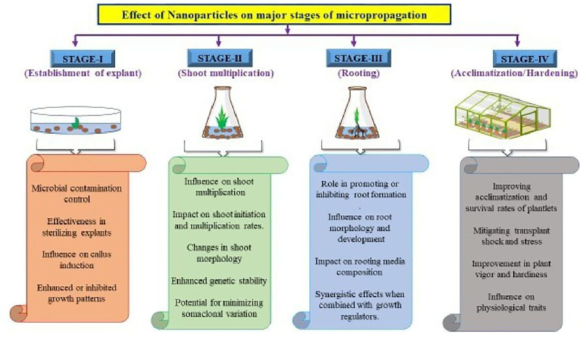
Figure 1. Effect of nanoparticles on major stages of micropagantion.
2.1 Initiation/establishment stage
The initiation stage of micropropagation could be enhanced by the application of nanoparticles such as silver and copper oxide nanoparticles, which, as a consequence, not only improve explant viability but also reduce contamination and enhance morphogenic potential to develop successful plant tissue culture (Table 1). Microbial contamination is widely perceived as the main bottleneck of plant tissue culture, which limits the efficacy of the micropropagation method. Common sterilization practices and techniques used in plant tissue culture can also damage plant tissues and/or produce toxic waste when not performed properly through the use of sodium hypochlorite, ethanol, or mercury chloride. However, nanoparticles, particularly silver nanoparticles, have gained attention as eco-friendly and effective alternatives for sterilization in tissue culture (Salisu et al., 2014).
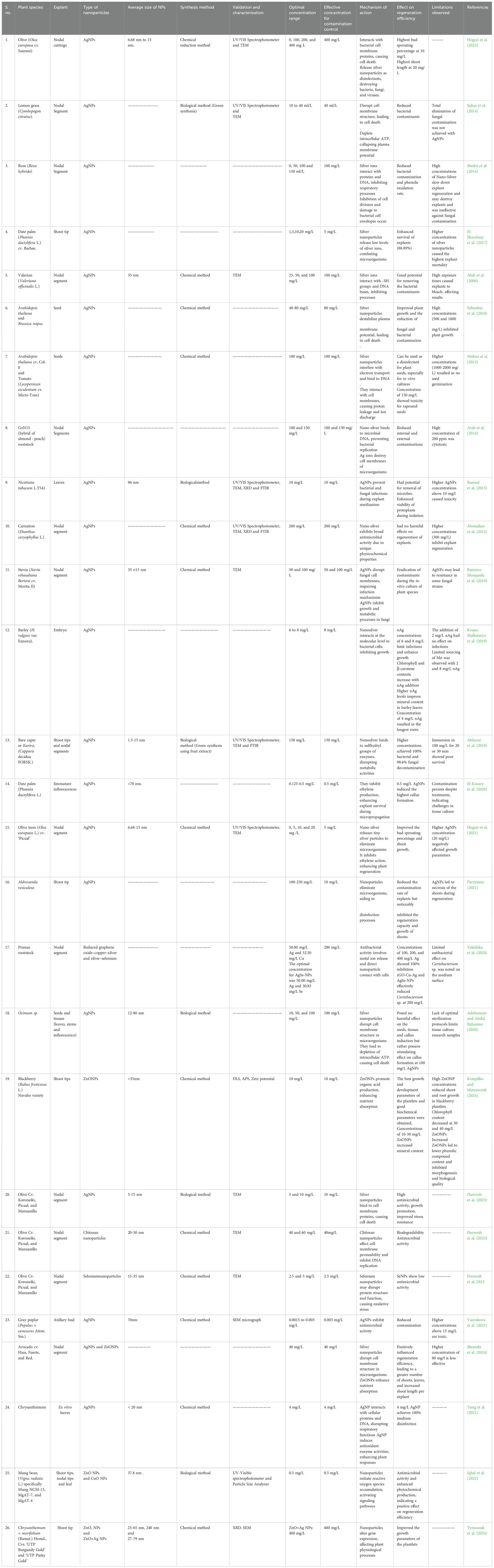
Table 1. Effect of nanoparticles on initiation/establishment stage of micropropagation.
Silver nanoparticles exhibit potent antimicrobial activity against a wide variety of bacteria, fungi and viruses. Their principal mechanisms result from the loss of structural integrity in microbial cell membranes and the production of reactive oxygen species. Other mechanisms of action include the disruption of microbial cell membrane structures, impairment of the electron transport chain, impairment of metabolic pathways for microbial growth, and induction of DNA replication interference, which ultimately leads to cell death (Dakal et al., 2016). These nanoparticles are different from traditional sterilization agents in that they work at low concentrations and do not damage plant tissues (Tung et al., 2021). In several reports, AgNPs have previously achieved high percentages of culture media sterilization and a reduction in microbial contamination (Munkager et al., 2020; Piecková et al., 2021; Wen et al., 2022; Alavi and Ashengroph, 2023).
Nanoparticles such as zinc oxide and titanium dioxide also have antimicrobial activity, which provides more options for sterilization (Abd-elsalam, 2013; Younis et al., 2022). Copper oxide nanoparticle usage also resulted in a 15–25% reduction in the explant infection rate during the initiation phase (Evlakov et al., 2020). Silver nanoparticles had a positive influence on SE and improved plantlet growth, which resulted in improved survival rates under greenhouse conditions (Cuong et al., 2021). There was improved branching of root systems in birch microplants with the introduction of CuO nanoparticles, demonstrating improved adaptive potential. However, some studies have also indicated possible negative effects associated with the introduction of nanoparticles during later stages, such as reduced viability and poor shoot and leaf development (Evlakov et al., 2020). Callus induction is the first step in the regeneration of a plant. This cell calling depends on the availability of important nutrients and growth regulators in the medium. NPs have been shown to increase callus induction via increased nutrient assimilation, altered hormonal activity and reduced oxidative stress (Irum et al., 2020; Malik et al., 2021).
Zinc oxide nanoparticles are essential for stimulating callus formation. These nanoparticles enhance cell division and proliferation by providing more macronutrients and micronutrients to the culture medium (Mousavi Kouhi and Lahouti, 2018). These nanoparticles are also treated as signaling molecules that regulate the synthesis of auxins and cytokinins, which are the two major hormones needed for callus initiation (Khan et al., 2022; Lee et al., 2023). ZnO nanoparticles have been demonstrated to improve callus induction in wheat (Triticum aestivum L.) by assisting in the uptake of important nutrients and facilitating cell activities that are important for plant growth and development Czyz˙owska and Barbasz, 2019).
NPs also strongly influence the morphogenic capabilities of explants during the initiation stage of micropropagation because of their enhanced growth characteristics, the fate of cell differentiation pathways, and increased pathogen resistance. Their unique properties allow for tailored applications that can optimize in vitro conditions for various plant species. In some cases, the presence of specific concentrations of nanoparticles in the nutrient medium, compared with controls, can yield enhanced morphogenic responses, and different plant species have been shown to have a range of optimal nutrient media concentrations to allow enhanced growth (Sichanova et al., 2023).
Gold nanoparticles affect SE by not allowing explant cells to follow a typical differentiation pathway and instead induce the formation of organ-like structures instead of somatic embryos. When the explants are exposed to nanoparticles, the tinkering of their chemical composition of the cell wall occurs, which further affects the developmental pathway (Godel-Je˛drychowska et al., 2023). The advent of nanoparticles into the culture media has decreased the occurrence of infections compared with that of control explants, increasing the survival rates of explants and thereby increasing their adaptability to stressful conditions during the micropropagation process (Evlakov et al., 2020; Pathak et al., 2023). While patterns that emerge from the emergence of nanoparticles may increase morphogenic potential in plants, notable concerns with their level of toxicity and whether nanoparticles have adverse long-term effects on plant health should remain a focus of debate (Pathak et al., 2023).
2.2 Shoot proliferation and multiplication stages
The shoot proliferation stage of micropropagation is a crucial phase that occurs simultaneously with shoot formation in cultured explants. The shoot proliferation stage is critical for calculating the overall multiplication rate and productivity of the process. The use of nanoparticles at the shoot proliferation stage of micropropagation has received much research attention, given their potential to facilitate plant growth and development (Table 2).
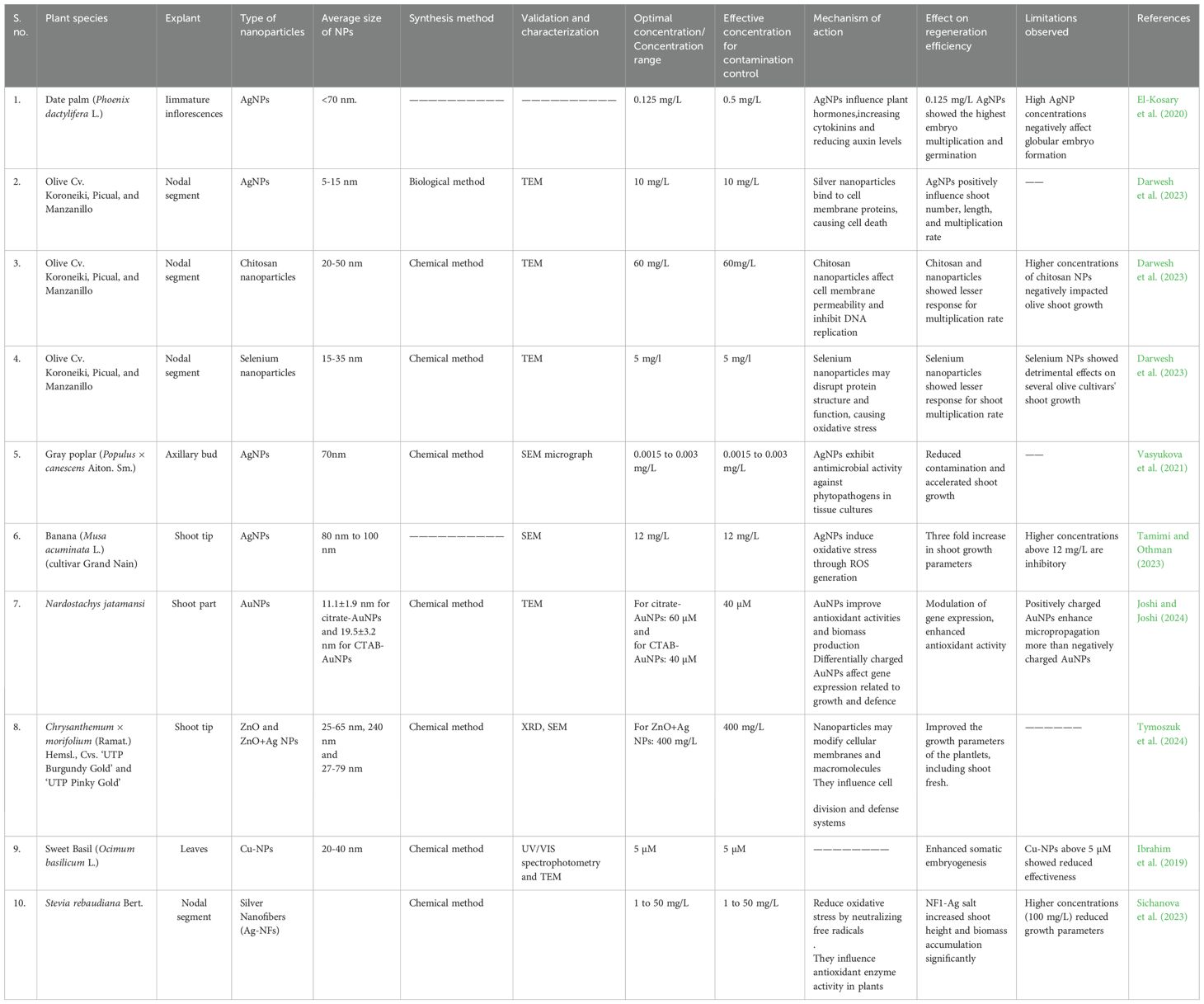
Table 2. Effect of nanoparticles on multiplication stage of micropropagation.
Silver nanoparticles have been shown to increase the mean number of fresh shoots per explant and the number of explants that produce shoots due to an ethylene blocking mechanism (Aghdaei et al., 2012). Copper oxide and silver nanoparticles reduce the infection rate of explants and increase their morphogenic ability after explants are added to the culture medium (Evlakov et al., 2020). Potassium nanoparticles (K-NPs) working at optimal concentrations significantly increase the number of microtubers formed in potato cultivars (Farrag et al., 2024).
Nanoparticles, which primarily include gold (Au), silicon (Si), and carbon-based nanomaterials, have been shown to significantly increase shoot proliferation because of their ability to increase phytohormone levels and increase nutrient transport (Kim et al., 2017; Talebi, 2018). Gold nanoparticles have been shown to stimulate the activity of cytokinin, a hormone that facilitates shoot development. Research has shown that when these nanoparticles are introduced into a culture medium, the quantity and length of shoots increase significantly (Jadczak et al., 2019; Joshi et al., 2022). In addition, silicon nanoparticles can reduce oxidative stress by instigating antioxidant enzymes, which can help promote an environment for shoot proliferation (Mukarram et al., 2022). Carbon-based nanoparticles, such as graphene oxide (GO) and carbon nanotubes (CNTs), have shown potential as agents of stimulation for shoots. These factors are known to improve water and nutrient absorption, cell wall elasticity, and the transport of growth regulators, thus allowing for increased shoot multiplication (Mathew et al., 2021).
High doses of nanoparticles may inhibit plant viability and growth, such as in shoots and leaves, which limits the number of conversions (Evlakov et al., 2020). Additionally, a high dose of nanoparticles could stimulate auxin-influenced branching and root development despite negatively affecting health (Tymoszuk et al., 2024).
NPs can enhance shoot proliferation and overall plant quality; however, it is necessary to recognize that the effects of NPs are concentration-specific and that their overuse can lead to detrimental side effects. This duality emphasizes that optimization is necessary in micropropagation protocols. However, even after initial optimization, there are potential nanotoxicity risks and biosafety concerns associated with the use of nanoparticles in agricultural applications (Pathak et al., 2023).
2.3 Rooting stage
Rooting is another significant step in micropropagation. An established root system leads to good plantlets. This phase often requires auxin-rich media; however, roots can be formed more efficiently via nanoparticles (Talebi, 2018). The authors of studies on various species have shown that nanoparticles can greatly increase plant growth and rooting efficiency during the rooting stage of micropropagation (Table 3). The use of silver nanoparticles at the optimal concentration (12 mg/L), significantly increased both root count and length during banana micropropagation experiments (Tamimi and Othman, 2023). In strawberry micropropagation, the use of AgNPs decreased the level of ethylene accumulation, suggesting an improvement in plantlet quality that would facilitate root development and improve survival (Tung et al., 2021). Silver nanoparticles additionally improved the disinfection of strawberry explants along with the reduction of pathogen infestation and disease, and therefore, importantly, contributed to the establishment of a healthy and robust root system for woody species such as gray poplar (Vasyukova et al., 2021). Zinc oxide nanoparticles in combination with auxins greatly increased rooting percentages in seedling apple microcuttings and resulted in increased root length and decreased callus formation (Alizadeh and Dumanoglu, 2022). Iron oxide nanoparticles, silicon nanoparticles and graphene oxide have all joined the ranks of previously mentioned nanoparticles that have been widely studied for root growth promotion (Triphati et al., 2021; Zhao et al., 2022), whereas iron oxide nanoparticles are particularly influential on root induction and improve nutrient availability, auxin perception and signaling, and activate antioxidant defense pathways leading to increased root formation (Rai et al., 2022). Silicon nanoparticles have a complementary function in root development. They improve phosphorus and potassium assimilation, two nutrients that are directly related to root development, and improve root tissue structural integrity (El-Kady et al., 2017). More recently, silicon nanoparticles have been shown to protect from abiotic stress related to root formation. These NPs have been shown to effectively increase the growth and biomass of strawberry roots (Sener et al., 2023). Graphene oxide is a carbon-based nanomaterial that may promote rooting by increasing the ability of plants to take up water and transport nutrients. It interacts with auxins, facilitating cell elongation and division and allowing rooting for many different types of plant species (Cheng et al., 2016; Zhao et al., 2022).
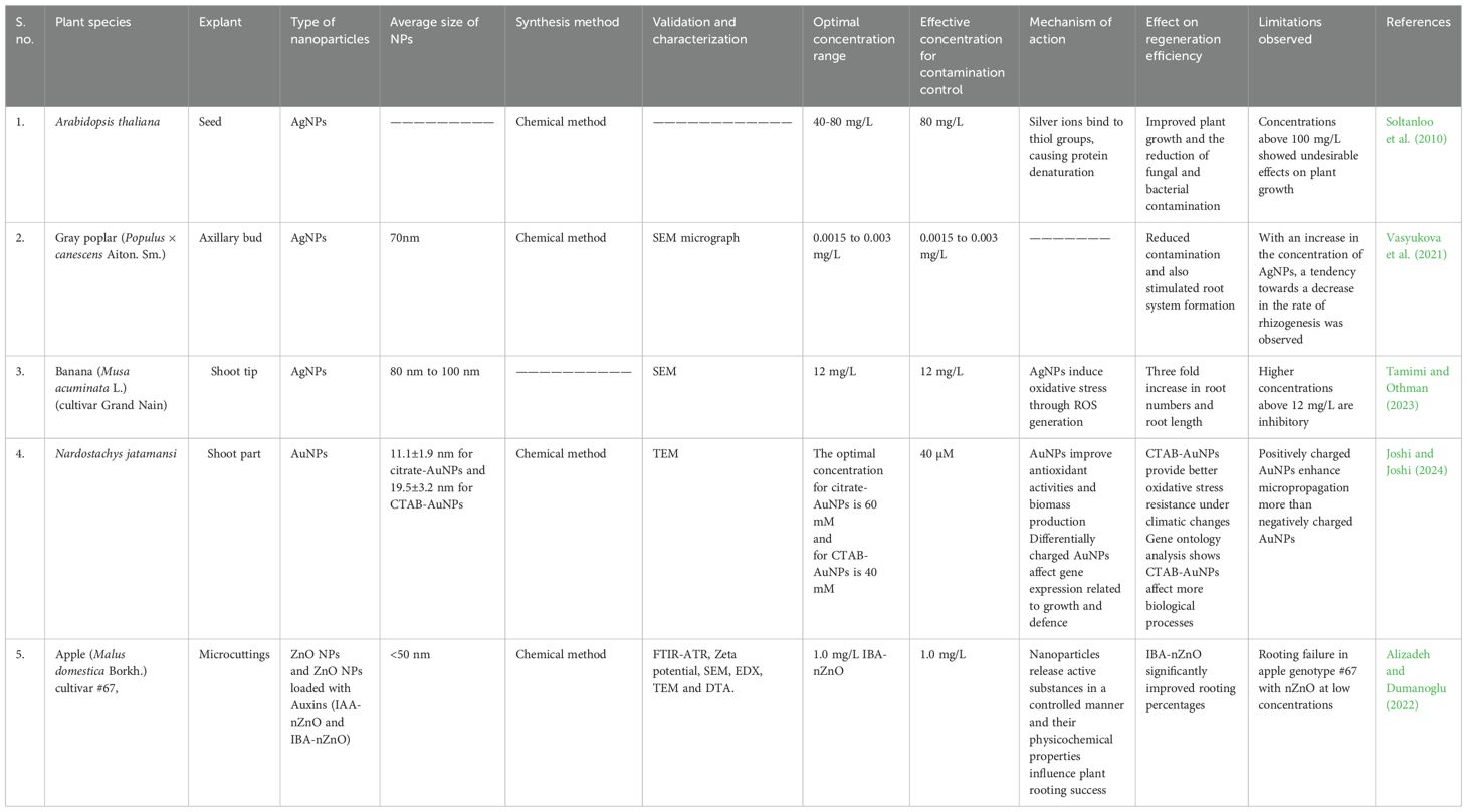
Table 3. Effect of nanoparticles on rooting stage of micropropagation.
Even though nanoparticles provide favorable advantages for micropropagation, it is important to assess the potential for toxicity and the environmental concerns associated with the use of materials at the molecular level. However, once again, given the potential outcomes of improved rooting, we need to consider any ecological concerns when utilizing this new form of plant biotechnology in the form of nanotechnology.
2.4 Acclimatization
The last micropropagation step is acclimatization, in which the plantlets are cultured in vitro and then moved to natural conditions. Acclimatization is a step where there is a risk to the plantlets for mortality due to desiccation, pathogen infection, and stressful environmental conditions. The acclimatization process, where a plant survives, can be improved with nanoparticles by increasing stress tolerance and antimicrobial protection from pathogens (El-Saadony et al., 2022). The use of nanoparticles influences the acclimatization and hardening steps in micropropagation (Table 4). The inclusion of AgNPs has been shown to improve the plant growth rate, increase the survival rate, and decrease microbial contamination, facilitating the transition from in vitro to ex vitro. These nanoparticles have been shown to improve somatic embryo formation and the overall number of somatic embryos in Panax vietnamensis (Cuong et al., 2021).
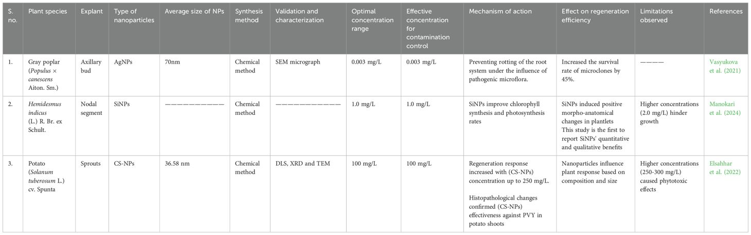
Table 4. Effect of nanoparticles on hardening/acclimatization stage of micropropagation.
For blueberry micropropagation, the addition of AgNPs improved shoot propagation rates, which suggests that there is potential for improving growth during acclimatization (Tejada-Alvarado et al., 2022). Compared with those of the control plants (44.44%), the survival of the plants exposed to silver nanoparticles was much greater (93.65%) (Cuong et al., 2021). AgNPs are functional sterilants that minimize microbial contamination of growth media, which is crucial for successful acclimatization. The use of these nanoparticles in chrysanthemum micropropagation resulted in better acclimatization under greenhouse conditions, leading to earlier developmental stages (Tung et al., 2021). More intensely, AgNPs act to protect plants during this acclimatization process from fungi and bacterial pathogens. These nanoparticles serve to limit microbial infections and diminish each plant’s risk of loss following transplantation (Gautam et al., 2020; Tariq et al., 2020).
In general, silicon nanoparticles enhance acclimatization by improving water use efficiency, the strength of plant cell wall composition and photosynthetic efficiency, allowing plants to acclimatize better post-transplantation to environmental stresses (Moraes and Lacava, 2022). The advantages of the use of nanoparticles in micropropagation are remarkable however, some studies have noted that excessive amounts of nanoparticles can also have negative effects on plant morphology and development therefore, the precise fine-tuning of nanoparticles throughout various acclimatization processes could be highly important (Evlakov et al., 2020).
3 Nanoparticles and their action mechanisms in micropropagation
Nanoparticles have emerged as promising tools for enhancing micropropagation efficiency. In plant tissue culture, their integration addresses persistent challenges such as microbial contamination, nutrient delivery, oxidative stress, and suboptimal morphogenesis (Kim et al., 2017). The mechanistic actions of nanoparticles in micropropagation can generally be classified into five primary functional areas (Figure 2): (i) antimicrobial activity, (ii) enhancement of nutrient delivery, (iii) modulation of hormonal signaling, (iv) alleviation of oxidative stress, and (v) activation of metabolic pathways. These mechanisms are extensive and overlap while cumulatively improving propagation efficiency, contamination resistance, acclimatization probability, and optimal growth (Mostafa et al., 2018; Rawat et al., 2018). The action mechanisms of important nanoparticles in major stages of micropropagation are shown in Figure 3.
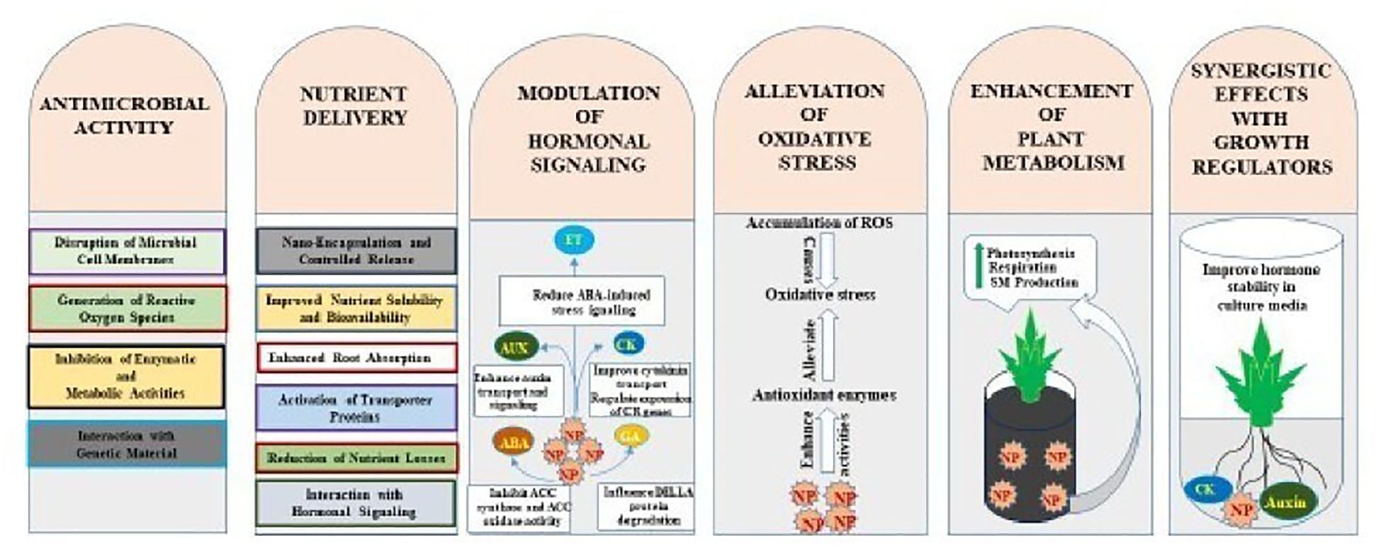
Figure 2. Action mechanisms of nanoparticles in micropropagantion.
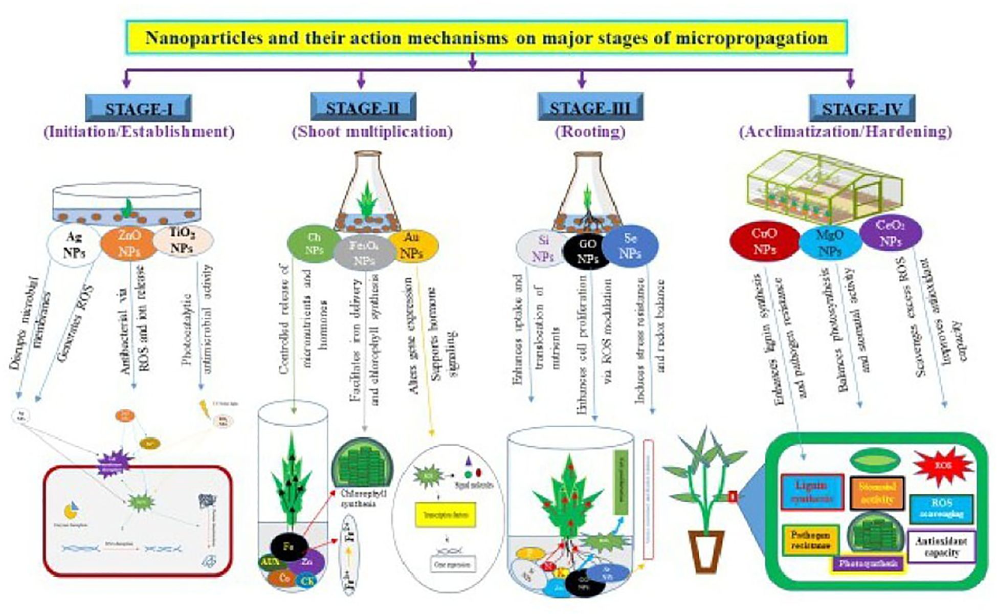
Figure 3. Nanoparticles and their action mechanisms on major stages of micropropaganation.
3.1 Antimicrobial activity
Microbial contamination, mostly by fungi and bacteria, is a continued barrier in plant tissue culture and results in large financial losses in the sector, while also ending with somatic origins that would later become genetically regulated plants. Traditional chemical disinfectants are normally effective, but they can cause phytotoxicity as well as residual toxicity. Hence, the use of inorganic nanoparticles with antimicrobial properties has presented itself as a superior alternative to chemical sterility agents. Because of their multidimensional characteristics, metals and metal oxides (AgNPs, ZnO NPs, CuO NPs, and TiO2 NPs) have inherently potent broad-spectrum antimicrobial activity via different mechanisms (Ranjan and Ramalingam, 2016; Kim et al., 2017; Fatima et al., 2021; Rahmawati et al., 2022; Kalra et al., 2022). Metallic nanoparticles, particularly silver and copper nanoparticles, have some effective antimicrobial properties that may reduce disease incidence by 50% (Francis et al., 2024).
The fundamental antimicrobial action of these nanoparticles is based on ROS (e.g., superoxide anions (O2-), hydroxyl radicals (•OH) and hydrogen peroxide (H2O2)). ROS can disturb microbial membranes, denature cellular proteins, and interfere with nucleic acids, leading to cell death (Kaur et al., 2023). Although silver nanoparticles are mostly composed of elemental silver (Ag), their surfaces may be functionalized with chemicals, which may also shed light on antimicrobial mechanisms. The interaction of AgNPs with thiol groups and phosphorus-containing compounds can lead to conformational changes and functional inactivity of these structural molecules. Importantly, these nanoparticles also permeabilize bacterial membranes, which also triggers cytoplasmic leakage and apoptosis (Bondarenko et al., 2018).
Both ZnO and TiO2 NPs have photocatalytic properties related to the induction of light-excited electrons into holes that can catalyze ROS. The antimicrobial capabilities of all types of nanoparticles, including ZnO and TiO2, are driven by their size at the nanoscale, surface area, and ability to interact intimately with microbial cells. The impressive capability of these phytocompatible drug nanotherapies for use in media sterilization means a stronger reliance on such methods than harsh disinfectants characterized by high toxicity (Zakharova et al., 2019).
3.2 Nutrient delivery
Tissue culture media contain a balanced mix of macronutrients and micronutrients; however, the nutrient bioavailability is often still limited by precipitation processes, pH swings, and chemical interactions. Nanoparticles offer a novel approach to nutrient delivery, with potential advantages in terms of greater solubility and bioavailability (Panpatte et al., 2016; Jampilek and Králová, 2017; Tariq et al., Singh et al., 2024c). Novel engineered nanofertilizers can entrap essential nutrients or chelate them and deliver them directly to plant cells, bypassing barriers and enhancing uptake efficiency (Gohar et al.; Ghazaryan et al., 2024, simultaneously improving nutrient uptake in both the apoplastic and symplastic pathways (Khlebnikova et al., 2023; Djanaguiraman et al., 2024).
In in vitro propagation, iron oxide nanoparticles (Fe34 NPs) have been successfully utilized to alleviate iron deficiency. Iron oxide nanoparticles can deliver Fe³+ ions in a controlled and sustained manner. This is important since Fe is critical for the biosynthesis of chlorophyll, electron transport, and a myriad of enzymatic functions (Feng et al., 2022). In addition, silicon nanoparticles positively contribute to the mechanical strength of cell walls, fortify cell walls, and increase stress tolerance, particularly under drought conditions, through regulating water retention and enzyme activities (Dhakte et al., 2022).
Researchers have also recently reported nanocarriers for macronutrients such as phosphorus (P) or potassium (K), which may be useful because of their limited ability in soil. Phosphorus in hydroxyapatite or other phosphate-bound nanocarriers assists with ATP synthesis and the metabolism of nucleic acids. Potassium-containing nanoparticles have been reported to regulate osmotic potential and activate enzymes. Overall, these advances improved nutrient uptake, promoting enhanced development of shoots and roots through various aspects of micropropagation (Ditta and Arshad, 2016).
3.3 Modulation of hormonal signaling pathways
Throughout micropropagation, phytohormones function to coordinate cellular differentiation, organogenesis, and plant morphogenesis. NPs alter phytohormone interactions by modifying biosynthesis, transport, sensitivity, and hormone-associated signaling pathways. Hormonal signaling modulation by NPs can take place either by direct contact with the hormone itself or indirectly through the transcriptional regulation of hormone-responsive genes.
In auxin (indole-3-acetic acid, IAA) signaling, which is critical for root induction, the addition of Fe34 NPs and graphene oxide NPs increases the level of auxin signaling itself. In both cases, it not only enables the polar transport of auxins but also increases the expression of auxin efflux carrier genes to improve meristematic activity and root organogenesis (Cheng et al., 2016; Yan et al., 2020, Yan et al., 2020; Yadav et al., 2022). As another example of an increase in hormone response and activity, AuNPs were found to have a synergistic effect on cytokinin, promote shoot proliferation, translocate cytokinins by stabilizing, and increase multiplication and nodal development via signals from cytokinins. The combination of nanoparticles and hormones in the micropropagation of plants is encouraging, especially because of the interaction with fewer hormones, where some nanoparticles can exert hormone-like effects, while some can work synergistically with exogenous hormone applications and generally have the prospect of better adjustment of hormone balance without the risk of adding more chemical burden to the media (Vinkovic et al., 2017).
Notably, NPs interact directly with abscisic acid (ABA) and gibberellins (GA) and can regulate responses to stress as well as elongation growth (Quesada, 2022). Other nanoparticles may even mimic hormonal action alone or be additive to exogenous hormone treatments and provide new methods to manipulate hormonal balance without increasing the chemical load (Kandhol et al., 2022).
3.4 Alleviation of oxidative stress
During the micropropagation process, oxidative stress commonly occurs due to high levels of ROS. The sources of oxidative stress include wounding, the application of exogenous hormones, and the artificial environmental components of in vitro cultivation (Hancock et al., 2001). In addition to acting as signaling molecules at lower concentrations, ROS can denature proteins, lipids, and nucleic acids at higher concentrations, leading to potentially nonviable plant tissue (Bhattacharjee, 2019). NPs can elicit these two types of ROS. They can produce ROS (especially at very high doses), but for the most part, they can increase the level of oxidative stress, which increases the activity of antioxidant defense systems (Kumar et al., 2018; Sachdev and Ahmad, 2021).
Silicon nanoparticles increase SOD, CAT, and POD enzyme activity to reduce oxidative damage and restore redox homeostasis (for example, during shoot elongation and root induction phases) (Mukarram et al., 2022). ZnO NPs and TiO2 NPs also effectively scavenge ROS, thereby protecting tissue as viable and reducing pentane (Singh and Chaudhary, 2020; Faizan et al., 2021).
When applied to stress-responsive genes, nanoparticles have been shown to provide upstream signaling for salicylic acid, jasmonate (JA), and ethylene for establishing systemic plant protection. This elicitor-like activity not only enhances the poor defense of plants but also further promotes their survival in stressful plant tissue cultures (Khlebnikova et al., 2023; Francis et al., 2024; Djanaguiraman et al., 2024).
3.5 Enhancements in plant metabolism and secondary metabolite production
Plant metabolism includes three common processes: photosynthesis, respiration and secondary metabolism; together, these processes serve as the basis for biomass accumulation, as well as adaptive responses to the environment. NPs affect these processes at the molecular and physiological levels, although the stage of the in vitro to ex vitro transition (i.e., the acclimation/hardening phase) is critical (Selvakesavan et al., 2023; Sharma and Chauhan, 2023). The application of Si NPs, TiO2 NPs and CeO2 NPs has been shown to improve photosynthetic efficiency, enhance chlorophyll contents and improve nutrient use efficiency (Gohar et al., 2024).
Photosynthetic systems are affected by TiO2 nanoparticles, as they increase photocatalytic activity in chloroplasts, promoting improved light absorption and better coupling to electron transport, which leads to an improved photosynthetic rate and increased stomatal conductance (Didaran et al., 2023). Similarly, Si NPs play seminal roles as osmotic buffers and induce structural changes in cell walls; these roles enhance stress resistance and ‘water retention’. These effects improve the overall ‘vigor’ and capacity for uniform hardening and acclimation of the plant (Siddiqui et al., 2020).
Furthermore, nanoparticles act as elicitors of secondary metabolite pathways, stimulating the production of phenolics, flavonoids, alkaloids and terpenoids, which are important for various facets of plant defenses and medicinal activities in plants (Sreelekshmi et al., 2022; Savanur, 2022; Inam et al., 2023).
While the use of nanoparticles as enhancers for micropropagation offers increased capacity to bioenhance multiple factors, including plant growth, re-establishment and rooting into natural growth media, it highlights the necessity for thorough attention to practical use in terms of nanoparticle dose-response relationships, phytotoxicity, and ecological cascading effects. Continued expansion of knowledge on plant–nanoparticle interactions are integral for understanding the molecular-level mechanisms that improve plant growth systems (Sharma et al., 2022; Khlebnikova et al., 2023).
4 Methodological framework with nanoparticle application
As the use of nanotechnology within the field of plant micropropagation continues to increase, the ability to improve propagation efficiency, disease resistance, nutrient transport, and overall plant performance continues to emerge. However, to ensure reproducibility, safety, and efficacy, an explicit methodological framework regarding the application of nanoparticles in plant tissue culture is needed. More specifically, the framework should include standardized characterization methods, methods of determining dose–responses, safety assessment techniques, and quality assessment protocols that minimize variability in outcomes, toxicity risks, and potential regulatory challenges when adopting/ascribing to nanobiotechnology in agricultural biotechnology and commercial plant propagation as a whole (Cinelli et al., 2016; Oomen et al., 2018; Mohamed and Kumar, 2019).
4.1 Standardization of nanomaterial characterization
An integral component of this framework is the standardization of nanomaterial characterization. Characterization ensures that the different physicochemical properties of NPs, which impact their biological performance, are relatively well defined and consistently reproduced. The key characterization parameters include the particle size distribution, morphology, surface area, zeta potential, chemical composition, and surface functionalization. Tools commonly employed for NP characterization include transmission electron microscopy (TEM), scanning electron microscopy (SEM), dynamic light scattering (DLS), X-ray diffraction (XRD), Fourier transform infrared spectroscopy (FTIR) and the Brunauer–Emmett–Teller (BET) surface area (Ramachandran et al., 2021).
A standardized characterization protocol should begin by measuring the particle size and distribution, which influences cellular uptake and reactivity. While DLS is a common technique because of its ability to analyze dispersed particles, it should always be followed up with TEM or SEM to obtain morphological confirmation. The next important measure is the zeta potential. The zeta potential helps characterize the coalescence of nanoparticles and their possible interactions with the plant cell membrane, providing further characterization of the nanoparticles. A relatively large absolute zeta potential (typically above ± 30 mV) reflects good coalescent particle stability, which is essential for determining the consistency of bioavailability in suspension culture media (Varenne et al., 2015a). The chemical composition and crystallinity of a nanoparticle is also an important part of its characterization because these parameters determine the rate of dissolution, the ion release profile and the redox potential of metal-based nanoparticles. These parameters may be characterized via XRD and energy-dispersive X-ray spectroscopy (EDX) (Reed-Gore et al., 2018; Deepty et al., 2019). The surface functional groups present on the nanoparticles matter because they contribute to the overall biocompatibility and plant hormone-mimicking capability of the nanoparticles to influence cellular signaling. The surface chemistry must also be characterized, which can be accomplished via FTIR or X-ray photoelectron spectroscopy (XPS). All these parameters must be reported by the researchers in detail. These guidelines must be followed when publishing and are recommended by the OECD Working Party on Manufactured Nanomaterials (WPMN), or ISO/TC 229 (Rasmussen et al., 2016).
4.2 Dose-response determination techniques
In addition to valuable characterization, dose–response determination approaches are integral to understanding the concentration-dependent responses exerted by NPs during micropropagation. Low dosages of NPs such as silver, zinc oxide, or graphene oxide may promote callus induction, root formation, or shoot elongation, but high dosages often react to oxidative stress, genotoxicity, and loss of growth. As a means to define practical application limits, reliable replications of dose-response curves must be developed. Thus, a tiered experimental design needs to be developed that includes several concentrations of the NPs (0.1 to 100 mg/L), multiple time points (24 h to weeks), and separation of the culture stages (initiation, proliferation, rooting, and acclimatization). The biological endpoints should consist of quantitative parameters such as callus mass, shoot number, root length, chlorophyll content, and antioxidant enzyme activities (SOD, CAT, POD) and stress markers (MDA, H2O2 levels). Additionally, transcriptomic and proteomic profiling can provide mechanistic details related to the dose-dependent modulation of signaling and metabolic pathways (Claeys et al., 2014; Kumar et al., 2016).
Aquiring clearance projections from the start, as well as acute and chronic exposure models, for both acute and chronic dose-response analyses. For example, some acute responses may only be seen as immediate toxicity, whereas chronic low doses could very easily lead to discrete epigenetic or physiological responses that occur cumulatively over successive subcultures. Advanced statistical models such as nonlinear regression, NOAEL (No Observed Adverse Effect Level) estimates, and benchmark dose (BMD) analysis can provide insight into safe and non-harmful nanoparticle concentrations, particularly when combined with in vitro–in vivo correlation studies, where plantlets initially treated in vitro are subsequently followed following acclimatization, and further bolster the ecological relevance of dose to indications (Nikinmaa, 2014; Savage et al, 2019).
4.3 Safety assessment protocols
Safety assessment protocols represent another critical element of the framework. It is much broader, as it demands attention beyond phytotoxicity to consider shifts in the microbial community, the expression of genes, and long-term ecology. For plant tissue culture, safety assessment should ultimately begin with cytotoxicity tests using a cell viability assay, such as 2,3,5-triphenyl tetrazolium chloride (TTC) or MTT, with suspension cultures or calli (Karakas et al., 2017). Genotoxicity tests, including the comet assay, micronucleus assay, and RAPD-PCR, may reveal DNA damage due to exposure to nanoparticles and, as such, can serve as a guide for the risk of chromosomal instability or unintended changes that can occur under somaclonal variation (Verdon et al., 2022). In addition, oxidative stress assays, which are used to determine the levels of ROS and lipid peroxidation, and assays of antioxidant enzymes produce a biochemical safety profile (Shukla et al., 2012; Gašparović et al., 2013; Agarwal et al., 2014).
At the microbial level, the unforeseen actions of various nanoparticles could also affect positive endophytes or rhizosphere consortia, either in the process or after hardening. Thus, culture-dependent and culture-independent techniques (e.g., qPCR and 16S rRNA sequencing) are useful for assessing microbial diversity in treated and control samples (Chhipa, 2023). Transgenerational studies, although infrequently published, are equally important for determining latent or heritable effects of NP exposure on seed germination, plant vigor, and reproductive outcomes. Future toxicology assessments will benefit from these multigenerational safety data to obtain regulatory clearance and general acceptance (Poma et al., 2014).
4.4 Quality control measures
QC measures must be taken during the entire process of NP production and applied to end-use studies. QC starts with the NP synthesis stage and must ensure consistency between different batches, and along a line, the particle size, charge and composition must be ensured. For green-synthesized NPs, extract variability from plants is a barrier to consistency; hence, standardization of the source material is needed (e.g., at the minimum age of the plant, the plant part used, or the solvent system). However, for all NPs, the sterility, endotoxin (most importantly for bioderived NPs) and stability must be assessed, including storage assessments. Where possible, certificates of analysis (CoAs) should be reported listing, at a minimum, all critical physicochemical properties (Maity et al., 2021; Saleh and Fadillah, 2023).
During application in micropropagation systems, media preparation must follow sterile, reproducible conditions, with uniform NP dispersion achieved via sonication or mechanical stirring. To avoid aggregation or precipitation, NP suspensions should be freshly prepared or stabilized via biocompatible agents (e.g., citrate and polyethylene glycol). The risk of cross-contamination between treatment groups should be minimized through spatial or temporal separation and rigorous tool sterilization. Regular checks for NP leaching, sedimentation, and degradation in culture media are essential to ensure consistent bioavailability (Kim et al., 2017; Gupta et al., 2020b).
Internal QC audits should involve routine calibration of instruments (e.g., pH meters, laminar flow cabinets), validation of assay protocols (e.g., controls in antioxidant enzyme tests), and replication of experiments across different labs or technicians (Kinns et al., 2013; Zhuravleva, 2014). Documentation standards, including detailed lab notebooks, digital records, and standardized reporting templates, support transparency and traceability. Furthermore, collaboration with accredited nanotechnology or toxicology laboratories for independent verification of findings adds robustness to the dataset and aids in regulatory compliance (Elberskirch et al., 2022).
As the field matures, there is a growing call for harmonized guidelines and databases for nanoparticle use in plant biotechnology. This includes establishing repositories for NP physicochemical and biological data, similar to the eNanoMapper or OECD NanoStat database. Such initiatives can help in the predictive modeling of NP behavior, reduce redundancy, and promote responsible innovation. The methodological framework described here aims to lay a foundation for such harmonization, ensuring that nanotechnology in plant tissue culture evolves along safe, effective, and scientifically rigorous lines (Kokina and Plaksenkova, 2022; Naidoo, 2022).
5 Standardization and protocol development
The reproducibility of nanoparticle assimilation into plant micropropagation systems is based on the elaboration of well-defined standardized protocols in terms of synthesis and characterization, use, and follow-up (Ankamwar et al., 2020). Standardization and protocol are key for the future of the translation of nanobiotechnology, not only to ensure scientific rigour but also to obtain regulatory acceptance, industrial scalability and environmental safety (Shen and Wang, 2011; Haydon, 2015) (Varenne et al., 2015b; Roubert et al., 2016). Without standardized protocols, fragmented results plague industry, resulting in nonreproducible results and inconsistent biological responses, which hinders both academic advancement and commercial opportunity (Boslough, 2013; Weller, 2021).
The intricate behavior of NPs, which is often influenced by variables such as size, shape, surface charge, propensity for aggregation, chemical composition, and their interaction with the biological matrix, requires detailed, stepwise, and context-specific standardization regimes. Such frameworks need to span disciplines, accommodate interspecies variation in plant response to 81 stresses, and ensure comparability between laboratory and field conditions (Ensor, 2011). Standardization must be performed via nanoparticle synthesis itself, at which point batch-to-batch reproducibility is still at the interface of what is feasible (Mülhopt et al., 2018). NPs may be produced via physical, chemical or biological (green) methods, and the resulting particles display different physicochemical characteristics (Triphati and Pirzadah, 2023).
Chemical synthesis methods usually produce well-defined particles; however, they can employ toxic precursors and hazardous solvents, whereas green synthesis using biological materials, such as plant extracts or microbial metabolites, is more ecofriendly but less controllable (Sharma and Sharma, 2021). The creation of standard syntheses involves the exact determination of precursor concentrations, reaction times, pH and temperature conditions and purification steps. These parameters together have to be tuned and synchronized to generate nanoparticles with a uniform size, size distribution, morphology, and surface functionalization. Otherwise, small differences during synthesis can have a profound effect on the reactivity, stability, and bioavailability of NPs, resulting in vastly different physiological effects when applied to tissue culture. Thus, it is essential to adhere to good manufacturing practice (GMP) principles for the production of nanoparticles for use in agriculture (Hühn et al., 2017; Sarlak and Abdi, 2022).
In the follow-up to particle synthesis, thorough characterization of the particles is essential and should be an integral aspect of the standardized procedure. Physicochemical characterization is essential for verifying the desired properties of nanoparticles and enables the correct interpretation of the biological effects of the system. Standard parameters that must be described include particle size (determined by dynamic light scattering or transmission electron microscopy), zeta potential (indicative of surface charge and colloidal stability), crystalline structure (e.g., by X-ray diffraction), functional groups (e.g., by means of FTIR spectroscopy) and surface area (by BET analysis) (Hodoroaba et al., 2020).
Furthermore, dispersibility in water and plant tissue culture media should be checked via UV–vis spectroscopy and dynamic light scattering. These properties determine the biological behavior of NPs, including their uptake efficiency by explants, their translocation through plant tissues or their interaction with components of media, such as sucrose or phytohormones (Moore et al., 2015; Rani et al., 2022). Characterization should be performed both before and after incubation in culture media to be aware of possible aggregation, dissolution or chemical modification. Without this, experimental interpretations are speculative, and dose-response relationships cannot be definitively quantified. Consequently, maintaining scientific transparency and reproducibility in any protocol of nanoparticle application in micropropagation, including characterization as a mandatory module, is crucial (Boverhof and David, 2010).
Following synthesis and characterization, the subsequent hurdle is developing protocols for the application of nanoparticles for the various stages of micropropagation. In developing protocols, standardization should consider plant species, genotype, developmental stage, and environmental conditions. Silver nanoparticles can again be employed to sterilize a culture vessel, which normally involves immersion for 5–10 min to ameliorate microbial contamination in explants (Hien et al., 2021), whereas zinc oxide nanoparticles can be added directly into the culture media to stimulate root elongation or mitigate oxidative stress (Molnár et al., 2020; Singh et al., 2022, Singh et al., 2023). Specific application methods (immersion, incorporation into culture media, and aerosols) and the frequency of application must be carefully defined, and exposure concentrations should be optimized with dose-response assays. Protocols should also contain control treatments that are subject to the same conditions as the treatment but lack the application of the nanoparticles to attribute any biological responses only to the nanoparticles. Another area of standardization is developing thresholds for usable and safe concentrations of nanoparticles. As a result, nanoparticles can produce biphasic (hormetic) effects by being beneficial at lower doses and toxic at higher doses; thus, cytotoxicity assays, oxidative stress markers such as malondialdehyde content or antioxidant enzyme activities, and viability assays such as TTC completely define the range of effective concentrations. For example, these concentration intervals should consider the plant species being tested, as well as the specific tissue type (e.g., leaf, node, meristem), since the biological responses of plants can vary tremendously (Tirumala et al., 2021).
Standardizing the composition of media containing nanoparticles is also important. Tissue culture media are complex mixtures of different sugars, salts, vitamins, hormones and gelling agents. These components can also react with nanoparticles, sometimes causing aggregation, chemical changes, or perhaps unintended bioactivity. Thus, standard protocols should indicate the compatibility of NPs with media components via stability studies that consider time, temperature and light exposure. The protocols should also describe how the media were prepared, including the order of addition of the NPs (before and/or after autoclaving), pH adjustments, and how mixing was performed, to avoid nanoparticle precipitation and/or degradation. Another consideration is NP-hormone interactions since nanoparticles could have synergistic or antagonistic effects with endogenous or exogenous plant growth regulators (PGRs), such as auxins and cytokinins. The standard protocols should also incorporate factorial experimental designs to evaluate the realized PGR and NP concentrations to optimize various morphogenetic responses, such as shoot proliferation or the promotion of a root system (Moore et al., 2015; Alagoz et al., 2022).
An important, although sometimes overlooked, consideration for standardization is quality control and reproducibility across laboratories. Validations of protocols in advance of publication should be institutionalized as inter-laboratory validations, comparing a common nanoparticle formulation and micropropagation protocol in different geographic or institutional settings. Ring trials in this way would allow for the identification of protocol-sensitive parameters and provide the framework for universally applicable SOPs. There is also a need for harmonization around documentation and reporting standards. Researchers should be expected to provide complete metadata, including but not limited to the source of the nanoparticles, route of synthesis, batch number, storage conditions, media composition, and biological data. Centralized databases or repositories of NP characterization and bioactivity in micropropagation would also facilitate meta-analyses and contributions to evidence synthesis efforts. They could also advance predictive modeling initiatives, such as machine learning efforts, to establish recommended nanoparticle types and concentrations for a given plant species and culture conditions (Roubert et al., 2016).
At the same time, there must be a robust biosafety element integrated into protocol development. Each standardized protocol will leave room for evaluations of both safety to the environment and safety to humans, typically ranging from ecotoxicity to the accumulation of residuals in regenerated plants. The proposed protocols will include long-term studies on the stability of nanoparticle-treated plantlets, for example, their soil stability (if returned to the environment) and leaching of NPs after being placed in the environment. These studies will have to be part of the protocol life cycles, as institutions will have to assess compliance with regulations but also the acceptance of nanotechnology by non-expert audiences and scientists alike, who need access to standardized protocols to study and regulatory experimental evidence as potential lifesaving technologies. By including safety endpoints such as assessment of DNA integrity, photosynthetic efficiency, and microbial community characterization in acclimatized plantlets, standardized NP protocols would have a greater degree of acceptability and robustness (Shende and Takke, 2021; Sharma et al., 2022).
Ultimately, protocol development has the possible future of creating integrated modular protocol packages such as diagnostic kits or vaccine platforms that are made of pre-validated modules (synthesis, characterization, application, safety, and data reporting modules) and could be tailored to the crop type and micropropagation phase. These kits are sellable by biotechnology companies and serve nurseries, agricultural research stations, and tissue culture laboratories. These kits increase the standardization of NP protocols, decrease the learning curve for non-expert researchers, and facilitate nanotoxic plant propagation protocols in agriculture (Mishra et al., 2022). Moreover, widely accepted protocols systematized by an internationally recognized body (e.g., FAO, ISO, BIS, and ICAR) with common characteristics will allow for community trust in the methods, along with assisting in the creation of policy and streamlining regulations (Bas et al., 2021).
6 Challenges and risks to nanoparticles in micropropagation
The use of nanoparticles in micropropagation has received considerable attention because of their unique physicochemical characteristics and ability to improve tissue culture results. Despite their advantages, there are numerous problems associated with implementing NPs in plant biotechnology systems. Given the nanoscale size and high level of reactivity of NPs, they can enable the precise delivery of nutrients, hormones, and antimicrobial agents within plant growth media. Conversely, nanoscales may also have unintended consequences for plant physiology and environmental and human health. This section reviews the key concerns raised when applying nanoparticles in micropropagation, including uncertainties in dose–response, environmental fate, regulatory deficiencies, economic limitations and standardization.
6.1 Toxicity thresholds and dose-response relationships
The biological efficacy and safety of nanoparticles in micropropagation are related to their dose-related response, which constitutes one of the greatest challenges in nanobiotechnology. NPs possess certain unique properties depending on their application in micropropagation, as they have a much higher surface area-to-volume ratio, are reactive and contain properties based on quantum mechanics, unlike traditional agrochemicals, which alter their association with plant tissues. These characteristics can promote or prevent the micropropagation process, as they have the potential to encourage growth in plants or raise concerns regarding phytotoxicity (Ramkumar et al., 2022). Whereas low concentrations of some NPs, such as AgNPs, ZnO NPs and Fe34 NPs, can stimulate growth or differentiation processes in the form of shoot proliferation, rooting and somatic embryogenesis, all of the above NPs select for cellular damage after certain thresholds involving oxidative stress, membrane leakage and genotoxicity (Irum et al., 2020; Tymoszuk et al., 2022).
A major complication in the analysis of dose-response relationships is related to the plant species and their developmental stages and the physical and chemical properties of the nanoparticles (Rawat et al., 2021). The size, shape, surface charge, and functionalization of nanoparticles can change their bioavailability and uptake (Ma et al., 2013). Smaller nanoparticles are more likely to simply traverse cell membranes and quickly be sequestered in organelles, whereas larger nanoparticles can end-up outside of the cell, with the potential to interfere with metabolic and signaling pathways (Mosquera et al., 2018). However, nanomaterials can generate ROS concentrations that are high-enough to potentially lead to oxidative stress, which may have a deleterious effect on cell viability, leading to lipid peroxidation, protein denaturation, and DNA damage (Manke et al., 2013).
The absence of a therapeutic index for the use of nanoparticles in plant systems has encouraged their use in micropropagation protocols. An index of the therapeutic window for dosage in pharmaceutical therapies has been established, but we do not have a similar range of dosages that are considered safe and efficacious from nanoparticles in plant systems (Kumari et al., 2022). Moreover, the interaction of nanoparticles with plant growth regulators can further complicate inference regarding optimal dosages. Therefore, specific treatments in a systematic manner for any one or a range of species are needed to assess potential concentrations that are intended to maximize efficacy with minimal cytotoxic effects. Longer-term studies may also be beneficial for identifying whether repeat doses through subcultures, or across generations, result in the collection of toxic effects or stress-related adaptations of the plantlets (Kandhol et al., 2022).
The inconsistency in experimental designs reported in the literature, including everything from the composition of culture media utilized to the environments imposed during our experiments, adds another layer of complexity to the establishment of dose-response curves (Cvrčková et al., 2015). Without standardized protocols, cross comparison of the impact of nanoparticles is an unreliable process. We need to standardize bioassay protocols that have measurable endpoints (i.e., chlorophyll concentration, root/shoot biomass, electrolyte leakage and enzyme activity). Advances in molecular diagnostics and imaging, such as transcriptomics and proteomics, could reveal valuable information on the sublethal effects of nanoparticles, which could help appropriately define toxicity thresholds and limits of exposure (Kang et al., 2023).
6.2 Environmental fate and bioaccumulation
The environmental impacts of the use of nanoparticles in plant micropropagation are becoming more significant, particularly with respect to their persistence, mobility and bioavailability through bioaccumulation in ecosystems. If nanoparticles are added to plantlets in the in vitro phase, once they are assembled into the ex vitro environment, they can be transferred to soils or aquatic environments, including the direct transfer of plantlets, disposal of culture media, or leaching from ex vitro additions. After being released into the environment, nanoparticles may undergo physical/chemical changes, such as aggregation, dissolution, biomolecular and macromolecular association and involvement, all of which have a compound effect on their fate and ecological toxicity (Batley et al., 2013; Wojcieszek and Ruzik, 2022).
Bioaccumulation in trophic chains may be one of the most serious hazards. For example, silver, copper, and zinc oxide nanoparticles, among others, may persist in soil matrices, become associated with, and be taken up by, plant roots and microbial communities and subsequently translocated into edible flora/plant tissues, thus entering the food chain. Numerous studies have shown the bioavailability of engineered nanoparticles with agricultural crops, which raises the possibility of engineered nanoparticles accumulating in higher organisms, potentially including humans. Many of the studies have focused primarily on those crops grown in soils, but the same hazards may also exist for crops derived from tissue cultures after acclimatization and following transplantation despite having undergone, in addition, planting medium recognition/realization (Murali et al., 2022).
The ecotoxicological effects of nanoparticles are not limited to only plants but also to soil microorganisms, fungi, nematodes, and aquatic organisms (Chhipa, 2021). For example, silver nanoparticles are used as in vitro antimicrobial agents but can inhibit nitrogen-fixing bacteria and interfere with the mycorrhizal relationships necessary for plant uptake of nutrients (Cao et al., 2017; Yu et al., 2021) in addition to causing unplanned collateral damage. These effects may negatively impact soil fertility and crop yield over time. Furthermore, nanoparticles that enter water can have a negative impact on the aquatic environment. Once in aquatic systems, they reduce photosynthetic productivity in algae, promote bioaccumulation in fish, and impair the coral cycle in invertebrates.
Methodologically, environmental monitoring of nanoparticles is an additive challenge because of their nanoscale size; they are uniquely variable in chemical composition and often interact with disparate environmental matrices. The environmental risk assessment tools are designed primarily for bulk chemicals. When attempting to add the exposure and effects of nanomaterials, this approach is not straightforward, and the use of complex compounds is unlikely to yield the same results as those of bulk chemical assessment methods. We need more advances in detection methodologies, such as isotopically labeled nanoparticles or synchrotron spectroscopy electronics, single particle inductively coupled plasma mass spectrometry (spICP-MS), and other more sophisticated methodologies to track the compartmental distribution and quantify the accumulation of nanoparticles within biological tissues and environmental compartments (Cornelis et al., 2021; Singh et al., 2024d).
There are at least two main methods for addressing environmental risks related to the use of nanoparticles in micropropagation. First, synthesis methods that are deemed more environmentally friendly and that use plant-based or microbial principles to make biodegradable or less persistent nanoparticles should be used whenever possible. Second, pre-acclimatization procedures should be able to degrade or eliminate any nanoparticles from plant tissues prior to field transplantation. Government regulatory agencies need to have some type of framework for the disposal of nanoparticles (more research related to this topic is needed). This is particularly important for laboratories and greenhouses where researchers use nanoparticles, and there are risks of unintended release into the environment (Kim et al., 2017).
6.3 Regulatory landscape and approval pathways
Governmental efforts to establish regulatory frameworks for the secure, uniform and ethical practice of nanoparticles in agriculture and biotechnology cannot seem to keep up with the acceptance of nanoparticles in micropropagation systems. The specific physicochemical traits that nanoparticles exhibit create complexities for regulatory frameworks that have relied on typical bulk assessment. Nanoparticles behave differently than typical agrochemicals because their size and reactivity are dependent, and the different interactions of nanoparticles depend on their performance, provider and biological system, so they do not assess nanoparticles in the same manner as traditional agrochemicals do. This gap in regulations has created a significant bottleneck to scaling up nanotechnology with plant tissue culture (Kumari et al., 2023).
Most regulatory authorities have not developed eligible and scientifically validated regulations specific to the use of nanoparticles in plant biotechnology. The few regulations that do exist, such as the EU REACH (Registration, Evaluation, Authorization and Restriction of Chemicals) regulations or U.S. EPA documents that discuss the usage of this nanomaterial, focus on its industrial usage rather than its applications in agricultural biotechnology. Consequently, researchers and commercial end-users practice in a regulated or informal regulated state that creates inconsistencies in applications, safety and quality control, including labeling. Furthermore, there are exposed liability concerns for institutions that use nanoparticle-based micropropagation methods without completed risk assessments (Ngarize et al., 2013; Bajpai et al., 2020).
The lack of standardized testing protocols only adds to the regulatory issue. Even if laboratories follow unique procedures to produce, characterize, and test nanoparticles, they compile different and heterogeneous datasets that are not representative, reliable or comparable, or aggregable in meta-analysis. The heterogeneity and endpoint variation make it much more difficult for regulators to determine the maximum acceptable exposure or the bioequivalence of different formulations of nanoparticles. Although international organizations responsible for developing protocols, such as the International Organization for Standardization (ISO) or the Organization for Economic Cooperation and Development (OECD), have developed high-throughput screening protocols for characterizing and testing nanoparticles as they relate to toxicology, many in plant sciences are just emerging (Gomes et al., 2021; Bleeker et al., 2023).
Risk assessment pathways for nanoparticles used in micropropagation should also include assessments of acute and chronic toxicity, in conjunction with ecological information, and the consequences of any genotoxicity and lifecycle assessment data. If regulatory pathways are developed to assess the safety of nanoparticles, they must also allow for any interactions with and how nanoparticles will function as plant growth regulators or other components of the culture media, as well as any potential synergies or antagonistic consequences. We need to be able to determine the direct or indirect effects (or absence) of nanoparticles on gene expression and epigenetic stability in regenerated plants, particularly when regenerated plants are consumed as food (Ranjan et al., 2021).
Tackling these issues involves active and cross-disciplinary collaboration. Researchers, regulators, and industry will have to combine their efforts to produce a comprehensive regulatory regime that will cater to the unique properties of nanoparticles. This needs to include premarket approval processes, required testing for safety, post-market surveillance of use and market disclosure requirements. An expanded role for regulatory agencies will be in producing guidelines for required documentation on nanoparticle-based culture media and formulations; labeling, disclosure and transparency within the value chain; and traceability.
6.4 Economic feasibility and scalability analysis
The economic viability and scale of nanoparticles for use in micropropagation are two of the most important but still underexplored dimensions for understanding why nanoparticles have not gained wider use and acceptance in commercial horticulture and plant biotechnologies (Wu and Li, 2022). The documented benefits of the use of nanoparticles, such as increased shoot multiplication, reduced contamination, increased rooting efficiency and stress tolerance, are well known under laboratory conditions (>500 papers), but determining how to obtain practical laboratory benefits or advantages for economically viable large-scale operations is challenging. This is primarily due, in part, to the costs incurred from producing, stabilizing, characterizing and integrating nanoparticles into tissue culture procedures (Abdelmajeed and Aboul-Nasr, 2013).
NPs are generally produced through energy-hungry physical or chemical processes that are generally costly due to expensive precursors, reducing agents and specialized instrumentation (ultrasonication, high-temperature furnaces, autoclaves, and reactors are just a few examples). While greener synthesis approaches using biological templates (such as plant extracts or microbes) are emerging, they have not achieved the consistency or scale of manufacture to be widely adopted in industrial applications. The functionalization of nanoparticles with appropriate ligands is fundamental to creating nanoparticles with stability, specificity, and controlled release, which also adds to production costs. In general, tissue culture laboratories (especially when they operate in low-resource situations or with non-commercial or low-margin crops such as trees) are unlikely to ever recoup the financial costs of adopting a consistently reliable nanoparticle-based protocol, regardless of any direct benefits such as production efficiency, improved quality (e.g., improved propagation accuracy), throughput or other secondary benefits from the normalization of nanoparticle use (Afonso et al., 2024).
When undertaking cost analyses, one must always consider how nanoparticle systems will fit with the current infrastructure. Most tissue culture laboratories have no existing experience with nanoparticle dispersion, characterization, or tracking, and the introduction of nanoparticles as a component in media preparation, sterilization and delivery will most likely require new methods, new equipment, new personnel training, and even significant changes to how waste is managed (e.g., disposal of residues of nanoparticles used in media preparation), so the associated costs to the operating budget, while likely not relevant when capital costs are considered, must be justified by justifiable measurable returns, such as propagation efficiencies, product quality (e.g., improved accuracy of propagation), throughput, or additional value for normalizing the use of nanoparticles (Tariq et al., 2020; Su et al., 2022).
In addition, the viability of using nanoparticles in micropropagation via economies of scale is also not understood. While many small-scale studies demonstrate the efficacy of a few milligrams or micrograms of nanoparticles, the process of scaling up to kilograms for commercial propagation, with millions of plantlets, creates many challenges with sourcing nanoparticles in the same form, preventing agglomeration of the nanoparticles, and obtaining batch consistency. These challenges could ultimately result in inconsistent responses of the nanoparticles and negatively impact product quality, as well as the trust of stakeholders and end-users alike (Kokina and Plaksenkova, 2022).
The cost-benefit ratio is also related to the type of crop being produced. For high-value crops, including orchids, elite fruit cultivars, and medicinal plants, any increase in propagation efficiency or improved plant resistance to disease increases the costs associated with nanoparticles. Low-margin crops, including leafy vegetables and fodder plants, may not be able to provide sustainable economic viability for practices that rely on costly nanoparticle protocols. Crop economic modeling provides a precise number that will equate break-even and profitability under different crop production scenarios (Zaman and Maitra, 2018).
Things that might help reduce economic feasibility include reusable nanoparticle matrices, time-released formulations to reduce waste, or the use of nanocomposites added to culture vessels or media gels to minimize the need for repeated applications. The use of various precision delivery systems, such as microfluidics, nanofiber scaffolds or encapsulated growth matrices, may lead to a gain in the delivery efficiency of these nanoparticles, indicating that the delivery of fewer nanoparticles will result in a problematic dose. Relationships with nanomaterial suppliers and some investments in local manufacturing will increase the cost of nanoparticle usage and access (Delgado, 2009).
With respect to some costs, if they are deemed in the best interest of governments or funding bodies, regulation, partnerships with nanotechnology regarding public and private relations, and international collaboration might also increase the likelihood of making lower cost barriers for adoption and the transfer of technologies. As an example, government and funding bodies explicitly subsidizing green nanoparticle synthesis as examples being used for agricultural uses or adding nanotechnology to the scopes of innovation-related schemes in agricultural use signifies alternatives to support the engagement of facilitating technologies for utilization. Generally, a multidisciplinary approach involving nanotechnology, economics, plant science, and industrial engineering means that knowledge of nanoparticle-assisted micropropagation should grow to be a meaningful, scalable, and economically useful technology in common culture.
6.5 Standardization challenges in synthesis and application
Importantly, standardization is a key component of scientific reproducibility and industrial scalability. Without standardization, we would be unable to scale up the production of nanoparticles or their integration into micropropagation. The inconsistencies in nanoparticle synthesis, characterization, and, ultimately, their application in horticulture are the primary impediments to integrating nanoparticles into the micropropagation process because they make it impossible to compare and establish best practices across studies. Furthermore, such inconsistencies mean that regulatory bodies are unable to regulate these substances or assess their risk and prevent the development of effective, crop specific, nanoparticle protocols (Zhang and Ge, 2018).
The range of nanoparticles used in plant tissue culture can vary significantly on the basis of core material (e.g., silver, gold, zinc oxide, iron oxide, silicon), morphology (e.g., spherical, rod, flower-like), size distributions, surface charges, and levels of functionalization. Small differences in synthesis conditions (e.g., pH, temperature, reactant concentration, reducing agents) can lead to large variations in the properties of nanoparticles and, consequently, in their biological effects. Small nanoparticles may penetrate cell walls more easily but could be more phytotoxic. However, without standardized protocols, researchers cannot reproduce research findings or transfer technology commercially (Mishra et al., 2022).
In addition to problems with synthesis variability, there is an absence of standard protocols for the characterization of nanoparticles. Techniques such as DLS, TEM, SEM, zeta potential measurements and UV-Vis spectroscopy have been used with varying degrees of application by researchers, including differing application parameters and reporting methods. These various methods lead to variable conclusions on the size, stability, and reactivity of nanoparticles, which makes it difficult to predict their biological outcomes. Most published studies report nominal concentrations (mg/L), without evaluation or reporting on the actual bioavailable fraction of nanoparticles in culture media. This would be informative in establishing their functional relevance (McNeil, 2011).
The application methods are also variable. Plant cultures can be exposed to nanoparticles in solid or liquid culture media via foliar spraying, seed priming, or coatings on the surfaces of culture vessels. The form of exposure, timing, time spent, frequency, etc., are also generally variable among the studies, leaving little to no common strategy for these variables across the literature. This inconsistency limits the generalizability of the results and complicates efforts to optimize nanoparticle use for specific micropropagation stages, such as callus induction, organogenesis, rooting, or acclimatization (Wohlmuth et al., 2022).
When these issues are considered, a priority is to create consensus standards regarding how particles are synthesized, characterized, and utilized in micropropagation. Creating a standard operating procedure (SOP) will require explicit descriptions of the physicochemical characteristics of the particles, protocols for the dispersion of particles, sterilization protocols and storage conditions. There is also potential for academic institutions, industry, and regulatory authorities in collaboration to develop certified reference materials (CRMs) and validated test methods related to agri-nanotechnology.
An additional important aspect of standardization includes incorporating nanotoxicology assessments into standard operating procedures. Considerations for standardized assays for cytotoxicity, genotoxicity and ecotoxicity should be included in the routine evaluation of nanoparticle formulations and could include high-throughput screening approaches and new omics-based technologies in search of elicited sub-lethal/long-lasting effects on plant physiological processes and genetic integrity for nanoparticles used in micropropagation (Koedrith et al., 2021; Pokhrel et al., 2021).
There should also be efforts toward digital standardization, and centralized databases or even metadata repositories where researchers can deposit and retrieve nanoparticle characterization, exposure conditions, and biological responses would be beneficial in considerably enabling meta-analyses, machine learning-based predictive modeling, and the evolution of evidence-based protocols (Elberskirch et al., 2022).
7 Future prospects: overcoming current barriers
Despite several challenges, the future of nanoparticles in micropropagation appears promising, especially with ongoing developments, innovations and interdisciplinary integration.
Emerging eco-synthesis methods, such as green synthesis using biological agents, are being developed as viable green alternatives to more conventional chemical approaches. These alternative syntheses are cost-effective and reduce hazardous byproducts while providing greater long-term environmental benefits. Functionalization approaches allow for the modification of the surface of nanoparticles, increasing their targeting efficacy, bioavailability and interaction with plant tissues, all of which may allow for the delivery of “smart” nanoparticles for the controlled release of nutrients, plant growth regulators, or antimicrobial agents while further improving efficient micropropagation outcomes.
The possibility of integrating nanoparticles with imaging methods (fluorescence microscopy or Raman spectroscopy) to study interactions in plant tissue in real-time is extremely exciting. It will be important to use these tools to deliver and implement nanoparticles properly, as they are related to performance. In addition, the combination of nanoparticles with genetic engineering tools such as CRISPR-Cas systems provides the opportunity to deliver genes, alter traits, and develop disease resistance. Additionally, nanoparticle-enabled sensors could enable precision agriculture, as we can start to monitor micropropagation environments to coordinate and optimize growth conditions, increase efficiencies, and reduce human error.
Nanotoxicity also needs to be reported in future research, as it is related to the biodegradability, mobility, and long-term impacts of nanoparticles in plant systems and ecosystems. It will be important to have biocompatible and degradable nanoparticles that do not bioaccumulate or persist. Multiple generations of studies need to be conducted to assess chronic exposure risk, especially with food crops. There is a need for nanotechnology researchers to be cautious with ecologists and plant biotechnologists to develop a safety profile for the use of nanomaterials in agriculture.
The cost associated with the production of nanoparticles decreases as production methods improve, and there is increasing industrial interest in biological-based processes. Recent developments, such as large-scale bioreactors and green synthesis methods, can facilitate the mass production of nanoparticles while reducing their impact on the environment. The more uniform and disease-free plant materials that are needed, particularly for high-value crops, there will continue to be commercially viable methods to utilize nanoparticles for micropropagation.
Nanoparticles are being investigated for new uses beyond conventional micropropagation, including somatic embryogenesis, cryopreservation and gene delivery. In some of these applications, particularly cryopreservation, nanoparticles can provide further cryoprotective functions via structural stabilization of the cell during freezing or enhance the formation of somatic embryos by enhancing the differentiation of the cells. Applications that use nanoparticles with these new applications offer tremendous possibilities and future perspectives for plant breeders and tissue culturists.
Nanomaterials have offered tools to increase nutrient uptake, increase disease resistance and thus overall performance, which decreases the dependency on synthetic agrochemicals. In addition, when used in micropropagation, they can be seen as sustainable agricultural practices since they improve propagation rates while reducing contamination and improving plantlet quality while providing a lower environmental footprint. With declining land availability and increasing food demands, nanomaterials or nanoparticles in conventional propagation schemes may contribute to resilient, resource-efficient crop production systems.
8 Conclusion
The incorporation of nanoparticles in micropropagation systems represents a breakthrough in plant tissue culture, providing new potential solutions for many traditional and existing stresses, including microbial contamination, nutrient restriction, hormonal imbalance, and environmental sensitivity during acclimatization. The characteristics of NPs, including their size, surface reactivity, and diverse physicochemical profiles, have the potential to improve nutrient uptake, increase phytohormone signaling capacity, alleviate oxidative stress, and increase shoot and root biomass proliferation, which contributes to the efficiency of plant regeneration. Nevertheless, several scientific, environmental, and regulatory questions remain. We still face many issues, not the least of which are the lack of optimal protocols for use, variability in species response to plant NPs, the potential for cytotoxicity at higher doses in micropropagation, and ecological issues such as bioaccumulation. Therefore, there is a need for a multidisciplinary systems approach to plant biology that combines nanotechnology, plant biology, toxicology, and environmental sciences to develop safe, scalable, and sustainable NP micropropagation systems for global food production. Future research should develop NP green synthesis, clearly defined NP assessment, and standardized regulatory frameworks. If proper due diligence between NP systems, global food production, and sustainable agriculture is attained, NPs can potentially transform plant biotechnology and provide real assets for sustainable agriculture, horticulture, and food security.
Author contributions
GG: Conceptualization, Visualization, Writing – original draft. PR: Writing – review & editing. CVá: Writing – review & editing. CVe: Writing – review & editing.
Funding
The author(s) declare financial support was received for the research and/or publication of this article. This research is connected to the Interreg VI-A IPA Hungary-Serbia Interreg Programme (Grant number: HUSRB/23S/11/027).
Conflict of interest
The authors declare that the research was conducted in the absence of any commercial or financial relationships that could be construed as a potential conflict of interest.
Generative AI statement
The author(s) declare that no Generative AI was used in the creation of this manuscript.
Any alternative text (alt text) provided alongside figures in this article has been generated by Frontiers with the support of artificial intelligence and reasonable efforts have been made to ensure accuracy, including review by the authors wherever possible. If you identify any issues, please contact us.
Publisher’s note
All claims expressed in this article are solely those of the authors and do not necessarily represent those of their affiliated organizations, or those of the publisher, the editors and the reviewers. Any product that may be evaluated in this article, or claim that may be made by its manufacturer, is not guaranteed or endorsed by the publisher.
References
Abdalla, N., El-Ramady, H., Kadry Seliem, M., El-Mahrouk, M. E., Taha, N., Bayoumi, Y., et al. (2022). An academic and technical overview on plant micropropagation challenges. Horticulturae 8, 677. doi: 10.3390/horticulturae8080677
Abdelmajeed, S. A. and Aboul-Nasr, M. H. (2013). Financial feasibility study of bananas tissue culture commercial production in Egypt. J. Financ. Account. Manag. 4, 87:96.
Abd-elsalam, K. A. (2013). Fungal genomics and biology nanoplatforms for plant pathogenic fungi management. Fungal Genomics Biol. 2, e, 107. doi: 10.4172/2165-8056.1000e107
Abdi, G., Salehi, H., and Khosh-Khui, M. (2008). Nano silver: A novel nanomaterial for removal of bacterial contaminants in valerian (Valeriana officinalis L.) tissue culture. Acta Physiol. Plant. 30, 709–714. doi: 10.1007/s11738-008-0169-z
Adebomojo, A. A. and Abdul Rahaman, A. A. (2020). Surface sterilization of Ocimum seeds and tissues with biosynthesized nanosilver and its effects on callus induction. IOP Conf. Series: Materials Sci. Eng. 805, 012024.
Afonso, I. S., Cardoso, B., Nobrega, G., Minas, G., Ribeiro, J. E., and Lima, R. A. (2024). Green synthesis of nanoparticles from olive oil waste for environmental and health applications: A review. J. Environ. Chem. Eng. 12, 114022. doi: 10.1016/j.jece.2024.114022
Agarwal, A., Cho, C.-L., and Sharma, R. (2014). Laboratory evaluation of reactive oxygen species. Encyclopedia Reprod. 5, 78–84. doi: 10.1016/B978-0-12-801238-3.64841-9
Aghdaei, M., Salehi, H., and Sarmast, M. K. (2012). Effects of silver nanoparticles on Tecomella undulate (Roxh.) Seem. micropropagation. Adv. Hortic. Sci 26, 21–24. doi: 10.13128/ahs-12748
Ahlawat, J., Sehrawat, A. R., Choudhary, R., and Yadav, S. K. (2020). Biologically synthesized silver nanoparticles eclipse fungal and bacterial contamination in micropropagation of Capparis decidua (Forsk.) Edgew: A substitute to toxic substances. Indian J. Exp. Biol. 58, 336–343.
Ahmadian, M., Babaei, A. R., Hessami, S. S. S., and Arab, M. M. (2015). Controlling the in vitro contamination of carnation (Dianthus caryophyllus L.) single nodes explant by nano-silver. Int. J. Agric. Biosci. 4, 167–170.
Alagoz, S. M., Arvanaghi, H. R., Dolatabadi, N., Khalaki, M. A., Moameri, M., Lajayer, B. A., et al. (2022). “Impact on nutritional status of plants treated with nanoparticles,” in Nanomaterial-Plant Interactions,Toxicity of Nanoparticles in Plants, vol. 5 . Eds. Rajput, V. D., Minkina, T., Sushkova, S., Mandzhieva, S. S., and Rensing, C. (San Diego, CA: Academic Press), 333–358. doi: 10.1016/B978-0-323-90774-3.00005-2
Alavi, M. and Ashengroph, M. (2023). Mycosynthesis of AgNPs: mechanisms of nanoparticle formation and antimicrobial activities. Expert Rev. Anti-Infective Ther. 21, 355–363. doi: 10.1080/14787210.2023.2179988
Alizadeh, S. and Dumanoglu, H. (2022). The effects of zinc oxide nanoparticles loaded with IAA and IBA on in vitro rooting of apple microcuttings. Turkish J. Agric. Forestry 46, 306–317. doi: 10.55730/1300-011X.3004
Ankamwar, B., Kirtiwar, S., and Shukla, A. C. (2020). “Plant-mediated green synthesis of nanoparticles,” in Advances in Pharmaceutical Biotechnology: Recent Progress and Future Applications. Eds. Patra, J., Shukla, A., and Das, G. (Springer, Singapore), 221–234. doi: 10.1007/978-981-15-2195-9_17
Arab, M. M., Yadollahi, A., Hosseini-Mazinani, M., and Bagheri, S. (2014). Effects of antimicrobial activity of silver nanoparticles on in vitro establishment of G×N15 (hybrid of almond × peach) rootstock. J. Genet. Eng. Biotechnol. 12, 103–110. doi: 10.1016/j.jgeb.2014.10.002
Athar, T., Khan, M. K., Pandey, A., Hamurcu, M., Saqib, Z. A., and Gezgin, S. (2022). “Current status and future directions for examining nanoparticles in plants,” in Nanomaterial-plant interactions, toxicity of nanoparticles in plants, vol. 5 . Eds. Rajput, V. D., Minkina, T., Sushkova, S., Mandzhieva, S. S., and Rensing, C. (San Diego, CA: Academic Press), 373–398. doi: 10.1016/B978-0-323-90774-3.00009-X
Bansod, S., Bawskar, M., and Rai, M. (2015). In vitro effect of biogenic silver nanoparticles on sterilisation of tobacco leaf explants and for higher yield of protoplasts. IET Nanobiotechnology 9, 239–245.
Bajpai, M., Shafi, H., and Kumari, S. (2020). “Nanoparticles: Importance and need for regulations,” in Nanoformulations in human health. Eds. Talegaonkar, S. and Rai, M. (Cham: Springer International Publishing). doi: 10.1007/978-3-030-41858-8_5
Bas, A. A., Burns, N. T., Gulotta, A. J., Junker, J. J., Drasler, B., Lehner, R., et al. (2021). Understanding the development, standardization, and validation process of alternative in vitro test methods for regulatory approval from a researcher perspective. Small 17, 2006027. doi: 10.1002/SMLL.202006027
Batley, G. E., Kirby, J. K., McLaughlin, M. J., and McLaughlin, M. J. (2013). Fate and risks of nanomaterials in aquatic and terrestrial environments. Accounts Chem. Res. 46, 854–862. doi: 10.1021/AR2003368
Bhandari, S., Sinha, S., Nailwal, T., and Thangadurai, D. (2022). “Nanotechnology: An approach for enhancement of plant system in terms of tissue culture,” in Biogenic nanomaterials: structural properties and functional applications. Eds. Thangadurai, D., Islam, S., Sangeetha, J., and Cruz-Martins, N. (New York: Apple Academic Press), 163–192. doi: 10.1201/9781003277149-9
Bhattacharjee, S. (2019). “ROS and oxidative modification of cellular components,” in Reactive Oxygen Species in Plant Biology. Ed. Bhattacharjee, S. (New Delhi: Springer), 81–105.
Bleeker, E. A. J., Swart, E., Braakhuis, H. M., Fernández-Cruz, M. L., Friedrichs, S., Gosens, I., et al. (2023). Towards harmonisation of testing of nanomaterials for EU regulatory requirements on chemical safety-A proposal for further actions. Regul. Toxicol. Pharmacol. 139, 105360. doi: 10.1016/j.yrtph.2023.105360
Bondarenko, O., Sihtmäe, M., Kuzmicˇiova, J., Rageliene˙, L., Kahru, A., Kahru, A., et al. (2018). Plasma membrane is the target of rapid antibacterial action of silver nanoparticles in Escherichia coli and Pseudomonas aeruginosa. Int. J. Nanomedicine 13, 6779–6790. doi: 10.2147/IJN.S177163
Boslough, M. (2013). Faulty protocols yield contaminated samples, unconfirmed results. Proc. Natl. Acad. Sci. United States America 110, E1651. doi: 10.1073/PNAS.1220567110
Boverhof, D. R. and David, R. M. (2010). Nanomaterial characterization: considerations and needs for hazard assessment and safety evaluation. Anal. Bioanal. Chem. 396, 953–961. doi: 10.1007/S00216-009-3103-3
Cao, J., Feng, Y., He, S., and Lin, X. (2017). Silver nanoparticles deteriorate the mutual interaction between maize (Zea mays L.) and arbuscular mycorrhizal fungi: a soil microcosm study. Appl. Soil Ecol. 119, 307–316. doi: 10.1016/J.APSOIL.2017.04.011
Cassells, A. C. (2002). “Tissue culture for ornamental breeding,” in Breeding for Ornamentals: Classical and Molecular Approaches. Ed. Vainstein, A. (Dordrecht: Springer), 139–153.
Cheng, F., Liu, Y. F., Lu, G. Y., Zhang., X. K., Xie, L. L., Yuan, C. F., et al. (2016). Graphene oxide modulates root growth of Brassica napus L. and regulates ABA and IAA concentration. J. Plant Physiol. 193, 57–63. doi: 10.1016/j.jplph.2016.02.011
Chhipa, H. (2021). “Nano-toxicity to microbes: potential implications of nanomaterials on microbial activity,” in Nanotoxicology and Nanoecotoxicology. Eds. Kumar, V., Guleria, P., Ranjan, S., Dasgupta, N., and Lichtfouse, E. (Cham: Springer, Cham), 99–123. doi: 10.1007/978-3-030-63241-0_4
Chhipa, H. (2023). “Influence of nanoparticles on the soil microbial flora and their rhizospheric activities,” in The impact of nanoparticles on agriculture and soil: Nanomaterial-plant interactions. Eds. Chauhan, N. S. and Gill, S. S. (San Diego, CA: Academic Press), 139–159. doi: 10.1016/b978-0-323-91703-2.00006-3
Cinelli, M., Coles, S. R., Sadik, O. A., Karn, B., and Kirwan, K. (2016). A framework of criteria for the sustainability assessment of nanoproducts. J. Cleaner Production 126, 277–287. doi: 10.1016/J.JCLEPRO.2016.02.118
Claeys, H., Van Landeghem, S., Dubois, M., Maleux, K., and Inzé, D. (2014). What is stress? Dose-response effects in commonly used in vitro stress assays. Plant Physiol. 165, 519–527. doi: 10.1104/PP.113.234641
Cornelis, G., Tuoriniemi, J., Montaño, M. D., Wagner, S., Gallego-Urrea, J. A., Mattsson, K., et al. (2021). “Challenges and current approaches toward environmental monitoring of nanomaterials,” in Monitoring environmental contaminants: Environmental contaminants. Ed. Johnson, S. (Quezon City, Philippines: Elsevier), 73–108. doi: 10.1016/B978-0-444-64335-3.00003-7
Cuong, D. M., Du, P. C., Tung, H. T., Ngan, H. T. M., Luan, V. Q., Phong, T. H., et al. (2021). Silver nanoparticles as an effective stimulant in micropropagation of Panax Vietnamensis -a valuable medicinal plant. Plant Cell Tissue Organ Culture 146, 577–588. doi: 10.1007/s11240-021-02095-2
Cvrčková, F., Luštinec, J., and Žárský, V. (2015). Complex, non-monotonic dose-response curves with multiple maxima: Do we (ever) sample densely enough? Plant Signaling Behav. 10, e1062198. doi: 10.1080/15592324.2015.1062198
Czyżowska, A. and Barbasz, A. (2019). Effect of ZnO, TiO2, Al2O3 and ZrO2 nanoparticles on wheat callus cells. Acta Biochim. Pol. 66, 365–370. doi: 10.18388/abp.2019_2775
Dakal, T. C., Kumar, A., Majumdar, R. S., and Yadav, V. (2016). Mechanistic basis of antimicrobial actions of silver nanoparticles. Front. Microbiol. 7. doi: 10.3389/FMICB.2016.01831
Dangariya, M., Khandhar, D., Monpara, J., Chudasama, K. S., and Thaker, V. S. (2020). Detection and identification of microbial contaminants from plant tissue culture. Trop. Plant Res. 7, 388–395. doi: 10.22271/TPR.2020.V7.I2.045
Darwesh, O. M., Hassan, S. A. M., and Abdallatif, A. M. (2023). Improve in vitro Multiplication of Olive shoots using environmental-safe Chitosan, Selenium, and Silver Nanostructures. Biointerface Res. Appl. Chem. 13, 419. doi: 10.33263/BRIAC135.419
Deepty, M., Srinivas, C., Ranjith Kumar, E., Krisha Mohan, N., Prajapat, C. L., Chandrasekhar Rao, T. V., et al. (2019). XRD, EDX, FTIR and ESR spectroscopic studies of co-precipitated Mn–substituted Zn–ferrite nanoparticles. Ceramics Int. 45, 8037–8044. doi: 10.1016/J.CERAMINT.2019.01.029
Delgado, G. C. (2009). Economical potential and safety of nanomaterials. Razón y Palabra 14, 16. Available online at: http://www.razonypalabra.org.mx/N/n68/16GianCarlo.pdf.
Dhakte, P., Kandhol, N., Raturi, G., Ray, P., Bhardwaj, A., Srivastava, A., et al. (2022). Silicon nano forms in crop improvement and stress management. Chemosphere 305, 135165.
Didaran, F., Ghasemi-Soloklui, A. A., Allakhverdiev, S. I., and Kordrostami, M. (2023). “Engineered nanoparticles enhance photosynthesis processes,” in Nanomaterial-Plant Interactions, Photosynthesis. Eds. Hou, H. J. M. and Allakhverdiev, S. I. (Amsterdam, Netherlands: Elsevier BV), 153–164.
Ditta, A. and Arshad, M. (2016). Applications and perspectives of using nanomaterials for sustainable plant nutrition. Nano Rev. 5, 209–229. doi: 10.1515/ntrev-2015-0060
Djanaguiraman, M., Anbazhagan, V., Dhankher, O. P., and Prasad, P. V. (2024). Uptake, translocation, toxicity, and impact of nanoparticles on plant physiological processes. Plants 13, 3137. doi: 10.3390/plants13223137
Elberskirch, L., Binder, K., Riefler, N., Sofranko, A., Liebing, J., Minella, C. B., et al. (2022). Digital research data: from analysis of existing standards to a scientific foundation for a modular metadata schema in nanosafety. Particle Fibre Toxicol. 19, 1. doi: 10.1186/s12989-021-00442-x
El-Esawi, M. A. (2016). “Micropropagation technology and its applications for crop improvement,” in Plant tissue culture: Propagation, conservation and crop improvement. Eds. Anis, M. and Ahmad, N. (Singapore: SpringerSingapore), 523–545. doi: 10.1007/978-981-10-1917-3_23
El-Kady, M. E., El-Boray, M. S., Shalan, A. M., and Mohamed, L. M. (2017). Effect of silicon dioxide nanoparticles on growth improvement of banana shoots in vitro within rooting stage. J. Plant Prod. 8, 913–916. doi: 10.21608/jpp.2017.40900
El-Kosary, S., Allatif, A. M. A., Stino, R. G., Hassan, M. M., and Kinawy, A. A. (2020). Effect of silver nanoparticles on micropropagation of date palm (Phoenix dactylifera L., cv. Sewi and Medjool). Plant Arch. 20, 9701–9706.
Elsahhar, S., Shahba, M., Elsayed, T., Mikhail, M., and Galal, A. (2022). Effect of chitosan nanoparticles (cs-nps) on in vitro regeneration response and production of potato virus Y (PVY)-free plants of potato. Agronomy 12, 2901. doi: 10.3390/agronomy12112901
El-Saadony, M. T., Saad, A. M., Soliman, M. S., Salem, H. M., Desoky, E. S. M., Babalghith, A. O., et al. (2022). Role of nanoparticles in enhancing crop tolerance to abiotic stress: A comprehensive review. Front. Plant Sci. 13. doi: 10.3389/fpls.2022.946717
El-Sharabasy, S. F., Ghazzawy, H. S., and Munir, M. (2017). In vitro application of silver nanoparticles as explant disinfectant for date palm cultivar Barhee. J. Appl. Horticulture 19, 106–112. doi: 10.37855/jah.2017.v19i02.19
Engelmann, F. and Dussert, S. (2000). Current development of cryopreservation for the conservation of plant genetic resources. Cahiers Agricultures 9, 237–244.
Ensor, D. S. (2011). “Implications of measurement standards for characterizing and minimizing risk of nanomaterials,” in Nanotechnology standards. Eds. Murashov, V. and Howard, J. (Springer, New York, NY), 165–177. doi: 10.1007/978-1-4419-7853-0_7
Evlakov, P. M., Fedorova, O. A., Grodetskaya, T. A., Zakharova, O. V., Zakharova, O. V., Zakharova, O. V., et al. (2020). Influence of copper oxide and silver nanoparticles on microclonal sprouts of downy birch. (Betula pubescens ehrh.). Nanotechnologies Russia. 15, 476–482. doi: 10.1134/S1995078020040035
Faizan, M., Yu, F., Chen, C., Faraz, A., and Hayat, S. (2021). Zinc oxide nanoparticles help to enhance plant growth and alleviate abiotic stress: A review. Curr. Protein Pept. Sci 22, 362–375. doi: 10.2174/1389203721666201016144848
Farrag, A., Taha, S. S., Darwish, O. S., Ashour, S., and Mahmoud, A. W. M. (2024). Improvement of potato (Solanum tuberosum L.) micro-tubers formation as effected by nano-particles in-vitro. Egyptian Pharm. J. 23, 507–517. doi: 10.4103/epj.epj_319_23
Fatima, K., Abbas, S. R., Zia, M., Sabir, S. M., Khan, R. T., Khan, A. A., et al. (2021). Induction of secondary metabolites on nanoparticles stress in callus culture of Artemisia annua L. Braz. J. Biol. 81, 474–483. doi: 10.1590/1519-6984.232937
Feng, Y., Kreslavski, V. D., Shmarev, A. N., Ivanov, A. A., Zharmukhamedov, S. K., Kosobryukhov, A., et al. (2022). Effects of iron oxide nanoparticles (Fe3O4) on growth, photosynthesis, antioxidant activity and distribution of mineral elements in wheat (Triticum aestivum) plants. Plants 11, 1894. doi: 10.3390/plants11141894
Francis, D. V., Abdalla, A., Mahakham, W., Sarmah, A. K., and Ahmed, Z. F. R. (2024). Interaction of plants and metal nanoparticles: Exploring its molecular mechanisms for sustainable agriculture and crop improvement. Environ. Int. 190, 108859. doi: 10.1016/j.envint.2024.108859
Gašparović, A.Č., Jaganjac, M., Mihaljević, B., Šunjić, S. B., and Zarkovic, N. (2013). “Assays for the Measurement of Lipid Peroxidation,” in Cell Senescence: Methods and Protocols, vol. 965 . Eds. Galluzzi, L., Vitale, I., Kepp, O., and Kroemer, G. (Humana Press, Totowa, NJ), 283–296. doi: 10.1007/978-1-62703-239-1_19
Gautam, N., Salaria, N., Thakur, K., Kukreja, S., Yadav, N., Yadav, R., et al. (2020). “Green Silver Nanoparticles for Phytopathogen Control,” in Proceedings of the National Academy of Sciences, India Section B: Biological Sciences, vol. 90 . Ed. Singh, Y. (New Delhi, India: Spriger Nature), 439–446. doi: 10.1007/S40011-019-01115-8
Ghazaryan, K., Pandey, D., Singh, S., Varagyan, V., Alexiou, A., Petropoulos, D., et al. (2024). Enhancing Crop Production: Unveiling the Role of Nanofertilizers in Sustainable Agriculture and Precision Nutrient Management. Egyptian J. Soil Sci 64 981–1007. doi: 10.21608/ejss.2024.282778.1748
Godel-Je˛drychowska, K., Milewska-Hendel, A., Sala, K., and Baranski, R. (2023). The impact of gold nanoparticles on somatic embryogenesis using the example of Arabidopsis thaliana. Int. J. Mol. Sci. 24, 10356. doi: 10.3390/ijms241210356
Gohar, F., Iqbal, U. Z., Khan, M. B., Rehman, F. U., Maryam, F. S. U., Azmat, M., et al. (2024). Impact of nanoparticles on plant growth, development and physiological processes: A comprehensive review. doi: 10.20944/preprints202410.0780.v1
Gomes, S. I. L., Scott-Fordsmand, J. J., and Amorim, M. J. B. (2021). Alternative test methods for (nano)materials hazards assessment: Challenges and recommendations for regulatory preparedness. Nano Today 40, 101242. doi: 10.1016/J.NANTOD.2021.101242
Gupta, A., Bano, A., Rai, S., Pathak, N., and Sharma, S. (2020b). “New Insights into Application of Nanoparticles for Plant Growth Promotion: Present and Future Prospects,” in Biogenic Nano-Particles and their Use in Agro-ecosystems. Eds. Ghorbanpour, M., Bhargava, P., Varma, A., and Choudhary, D. (Springer, Singapore), 259–279.
Gupta, N., Jain, V., Joseph, M. R., and Devi, S. (2020a). A review on micropropagation culture method. Asian J. Pharm. Res. Dev. 8, 86–93. doi: 10.22270/AJPRD.V8I1.653
Hancock, J. T., Desikan, R., and Neill, S. J. (2001). Role of reactive oxygen species in cell signalling pathways. Biochem. Soc. Trans. 29, 345–349. doi: 10.1042/bst0290345
Haydon, B. (2015). “Nanoengineering: A toolbox of standards for health and safety,” in Nanoengineering: Global approaches to health and safety issues. Ed. Dolez, P. I. (Quezon City, Philippines: Elsevier), 557–580. doi: 10.1016/B978-0-444-62747-6.00017-8
Hegazi, E. S., Yousef, A., Abd Allatif, A., Mahmoud, T., and Mostafa, M. (2021). Effect of silver nanoparticles, medium composition and growth regulators on in vitro propagation of Picual olive cultivar. Egyptian J. Chem. 64, 6961–6969. doi: 10.21608/ejchem.2021.78774.3853
Hegazi, E. S., Yousef, A. R. M., Abdallatif, A. M., Mahmoud, T. S. M., Mostafa, M. K. M., and Suliman, A. A. (2023). Effect of silver nanoparticles on microbial contaminants and growth of in vitro olive shoot cv. Sourani. Acad. J. Plant Sci. 16, 38–47. doi: 10.5829/idosi.ajps.2023.38.47
Hien, V. T., Tung, H. T., Khai, H. D., Luan, V. Q., Cuong, D. M., Lich, T. V., et al. (2021). Effect of silver nanoparticles on sterilization of different explant sources of Gerbera jamesonii cultured in vitro. Vietnam J. Biotechnol. 19, 705–715. doi: 10.15625/1811-4989/15672
Hodoroaba, V.-D., Unger, W. E. S., and Shard, A. G. (2020). “Characterization of nanoparticles: measurement processes for nanoparticles,” in Micro and Nano Technologies. Eds. Hodoroaba, V. D., Unger, W. E. S., and Shard, A. G. (Cham: St. Frisco, US: Elsevier Inc). doi: 10.1016/C2017-0-00312-9
Hühn, J., Carrillo-Carrión, C., Soliman, M., Pfeiffer, C., Valdeperez, D., Masood, A., et al. (2017). Selected standard protocols for the synthesis, phase transfer, and characterization of inorganic colloidal nanoparticles. Chem. Mater. 29, 399–461. doi: 10.1021/acs.chemmater.6b04738
Ibrahim, A., Fahmy, A. H., and Ahmed, S. S. (2019). Copper nanoparticles elevate regeneration capacity of (Ocimum basilicum L.) plant via somatic embryogenesis. Plant Cell Tissue Organ Culture 136, 41–50. doi: 10.1007/s11240-018-1489-3
Inam, M., Attique, I., Zahra, M., Khan, A. K., Hahim, M., Hano, C., et al. (2023). Metal oxide nanoparticles and plant secondary metabolism: unraveling the game-changer nano-elicitors. Plant Cell Tissue Organ Cul. 155, 1–18. doi: 10.1007/s11240-023-02587-3
Iqbal, Z., Javad, S., Naz, S., Shah, A. A., Shah, A. N., Paray, B. A., et al. (2022). Elicitation of the in vitro cultures of selected varieties of vigna radiata l. with zinc oxide and copper oxide nanoparticles for enhanced phytochemicals production. Front. Plant Sci. 13, 908532. doi: 10.3389/fpls.2022.908532
Irum, S., Jabeen, N., Ahmad, K. S., Shafique, S., Khan, T. F., Gul, H., et al. (2020). Biogenic iron oxide nanoparticles enhance callogenesis and regeneration pattern of recalcitrant Cicer arietinum L. PloS One 15, e0242829. doi: 10.1371/JOURNAL.PONE.0242829
Jalil, S. U. and Ansari, M. I. (2021). “Role of nanomaterials in regulating reactive species as a signaling molecule of abiotic stress in plants,” in Nanobiotechnology. Eds. Al-Khayri, J. M., Ansari, M. I., and Singh, A. K. (Springer, Cham), 291–304. doi: 10.1007/978-3-030-73606-4_12
Jadczak, P., Kulpa, D., Bihun, M., and Przewodowski, W. (2019). Positive effect of AgNPs and AuNPs in in vitro cultures of Lavandula angustifolia Mill. Plant Cell Tissue Organ Cult. 139, 191–197. doi: 10.1007/s11240-019-01656-w
Jampilek, J. and Králová, K. (2017). “Nanomaterials for delivery of nutrients and growth-promoting compounds to plants,” in Nanotechnology: An Agricultural Paradigm. Eds. Prasad, R., Kumar, M., and Kumar, V. (Springer, Singapore), 177–224.
Javed, R., Yucesan, B., Zia, M., and Gurel, E. (2022). “Nanoelicitation: A promising and emerging technology for triggering the sustainable in vitro production of secondary metabolites in medicinal plants,” in Plant and Nanoparticles. Ed. Chen, J. T. (Springer, Singapore), 265–280. doi: 10.1007/978-981-19-2503-0_10
Joshi, S., Dar, A. I., Acharya, A., and Joshi, R. (2022). Charged gold nanoparticles promote in vitro proliferation in Nardostachys jatamansi by differentially regulating chlorophyll content, hormone concentration, and antioxidant activity. Antioxidants 11, 1962. doi: 10.3390/antiox11101962
Joshi, S. and Joshi, R. (2024). Comparative transcriptome profiling unveils the role of differentially charged gold nanoparticle in growth, metabolic regulation and defence response of micro-propagated Nardostachys jatamansi Wall. ex DC. South Afr. J. Bot. 167, 585–596. doi: 10.1016/j.sajb.2024.02.052
Kalra, K., Chhabra, V., and Prasad, N. (2022). Antibacterial activities of zinc oxide nanoparticles: a mini review. J. Phys.: Conf. Ser. 2267, 012049. doi: 10.1088/1742-6596/2267/1/012049
Kandhol, N., Singh, V. P., White, J. C., Phan Trân, L. S., and Tripathi, D. K. (2022). Plant growth hormones and nanomaterial interface: Linking development to defence. Plant Cell Physiol. 63, 1840–1847. doi: 10.1093/pcp/pcac147
Kang, M., Weng, Y.-J., Liu, Y., Wang, H., Ye, L., Gu, Y., et al. (2023). A review on the toxicity mechanisms and potential risks of engineered nanoparticles to plants. Rev. Environ. Contam. Toxicol. 261, 1–33. doi: 10.1007/s44169-023-00029-x
Karakas, D., Ari, F., and Ulukaya, E. (2017). The MTT viability assay yields strikingly false-positive viabilities although the cells are killed by some plant extracts. Turkish J. Biol. 41, 919–925. doi: 10.3906/BIY-1703-104
Karimi, E. (2019). “Antimicrobial activities of nanoparticles,” in Nanotechnology for agriculture: Crop production and protection. Eds. Panpatte, D. and Jhala, Y. (Springer, , Singapore), 171–206. doi: 10.1007/978-981-32-9374-8_10
Kaur, H., Rauwel, P., and Rauwel, E. (2023). “Antimicrobial nanoparticles: Synthesis, mechanism of Actions,” in Antimicrobial Activity of Nanoparticles. Ed. Guisbiers, G. (Amsterdam: Elsevier), 155–202.
Khan, M. N., Khan, Z., Zhu, L., Li, Y., and Wu, H. (2022). “Impact of chitosan and chitosan-based metallic nanoparticles on the regulation of plant hormones,” in Nanomaterial-plant interactions, role of chitosan and chitosan-based nanomaterials in plant sciences. Eds. Kumar, S. and Madihally, S. V. (London, England: Academic Press), 343–356.
Khanna, K., Kohli, S. K., Bakshi, P., Sharma, P., Kour, J., Bhardwaj, T., et al. (2023). “Green biosynthesis of nanoparticles: mechanistic aspects and applications,” in Environmental Applications of Microbial Nanotechnology. Eds. Singh, P., Kumar, V., Bakshi, M., Hussain, C. M., and Sillanpää, M. (Netherlands: Elsevier), 99–126. doi: 10.1016/B978-0-323-91744-5.00020-5
Khlebnikova, D. A., Polivanova, O. B., Boytsova, M. V., Chepovoy, I. I., Munkhbaatar, N. O., and Cherednichenko, M. (2023). Physiological aspects of interaction of nanoparticles with plant and microorganism cells. Timiryazev Biol. J. 2, 77–93. doi: 10.26897/2949-4710-2023-2-77-93
Kim, D. H., Gopal, J., and Sivanesan, I. (2017). Nanomaterials in plant tissue culture: the disclosed and undisclosed. RSC Adv. 7, 36492–36505. doi: 10.1039/C7RA07025J
Kinns, H., Pitkin, S., Housley, D., and Freedman, D. B. (2013). Internal quality control: best practice. J. Clin. Pathol 66, 1027–1032. doi: 10.1136/JCLINPATH-2013-201661
Koedrith, P., Rahman, M., Jang, M., Shin, Y. J., Seo, D. Y., and Seo, Y. R. (2021). Nanoparticles: Weighing the pros and cons from an eco-genotoxicological perspective. J. Cancer Prev 26, 83–97. doi: 10.15430/JCP.2021.26.2.83
Kokina, I. and Plaksenkova, I. (2022). Nanoparticles in Plant Biotechnology: Achievements and Future Challenges. Proc. Latvian Acad. Sci. Section B Natural Exact Appl. Sci. 76, 204–210. doi: 10.2478/prolas-2022-0031
Krupa-Małkiewicz, M., Oszmiański, J., Lachowicz, S., Szczepanek, M., Jaśkiewicz, B., Pachnowska, K., et al. (2019). Effect of nanosilver (nAg) on disinfection, growth, and chemical composition of young barley leaves under in vitro conditions. J. Integr. Agric. 18, 1871–1881. doi: 10.1016/S2095-3119(18)62146-X
Krzepiłko, A. and Matyszczuk, K. (2024). Influence of zinc oxide nanoparticles in in vitro culture and bacteria Bacillus thuringiensis in ex vitro conditions on the growth and development of blackberry (Rubus fruticosus L.). Appl. Sci. 14, 3743. doi: 10.3390/app14093743
Kumar, D., Bansal, G., Narang, A., Basak, T., Basak, T., Abbas, T., et al. (2016). Integrating transcriptome and proteome profiling: Strategies and applications. Proteomics 16, 2533–2544. doi: 10.1002/PMIC.201600140
Kumar, V., Sharma, M., Khare, T., and Wani, S. H. (2018). “Impact of nanoparticles on oxidative stress and responsive antioxidative defense in plants,” in Nanomaterials in Plants, Algae, and Microorganisms. Eds. Tripathi, D. K., Ahmad, P., Sharma, S., Chauhan, D. K., and Dubey, N. K. (London, UK: Academic Press), 393–406.
Kumari, A., Rajput, S., Rajput, V. D., Nevidomskaya, D., Mandzhieva, S., Sushkova, S., et al. (2022). “Updated analysis of the exposure of plant to the nanomaterials,” in Nanomaterial-plant interactions, toxicity of nanoparticles in plants, vol. 5 . Eds. Rajput, V. D., Minkina, T., Sushkova, S., Mandzhieva, S. S., and Rensing, C. (London, UK: Academic Press), 25–45. doi: 10.1016/B978-0-323-90774-3.00011-8
Kumari, R., Suman, K., Karmakar, S., Mishra, V. K., Lakra, S. G., Saurav, G. K., et al. (2023). Regulation and safety measures for nanotechnology-based agri-products. Front. Genome Ed.. 5, 1200987. doi: 10.3389/fgeed.2023.1200987
Lee, S., Jiao, M., Zhang, Z., and Yu, Y. (2023). Nanoparticles for interrogation of cell signaling. Annu. Rev. Anal. Chem. 16, 333–351. doi: 10.1146/annurev-anchem-092822-085852
López-Valdez, F., Miranda-Arámbula, M., Ríos-Cortés, A. M., Fernández-Luqueño, F., and de-la-Luz, V. (2018). “Nanofertilizers and their controlled delivery of nutrients,” in Agricultural Nanobiotechnology. Eds. López-Valdez, F. and Fernández-Luqueño, F. (Springer, Cham), 35–48. doi: 10.1007/978-3-319-96719-6_3
Ma, N., Ma, C., Li, C., Wang, T., Tang, Y., Wang, H.-Y., et al. (2013). Influence of nanoparticle shape, size, and surface functionalization on cellular uptake. J. Nanosci. Nanotechnol. 13, 6485–6498. doi: 10.1166/JNN.2013.7525
Mahna, N., Vahed, S. Z., and Khani, S. (2013). Plant in vitro culture goes Nano: Nanosilver-mediated decontamination of ex vitro explants. J. Nanomed. Nanotechnol. 4, 1–5. doi: 10.4172/2157-7439.1000161
Maity, S., Adhikari, M., Banerjee, S., Guchhait, R., Chatterjee, A., and Pramanick, K. (2021). Critical analysis of biophysicochemical parameters for qualitative improvement of phytogenic nanoparticles. Biotechnol. Prog. 37, e3114. doi: 10.1002/BTPR.3114
Malik, W. A., Mahmood, I., Razzaq, A., Afzal, M., Shah, G. A., Iqbal, A., et al. (2021). Exploring potential of copper and silver nano particles to establish efficient callogenesis and regeneration system for wheat (Triticum aestivum L.). GM Crops Food 1–22, 564–585. doi: 10.1080/21645698.2021.1917975
Mandal, C. and Sahu, M. (2021). “Application of metal and metal oxide nanoparticles as potential antibacterial agents,” in Nanomaterials and Nanocomposites for Environmental Remediation. Energy, Environment, and Sustainability. Eds. Singh, S. P., Rathinam, K., Gupta, T., and Agarwal, A. K. (Springer, Singapore), 121–140. doi: 10.1007/978-981-16-3256-3_6
Manke, A., Wang, L., and Rojanasakul, Y. (2013). Mechanisms of nanoparticle-induced oxidative stress and toxicity. BioMed. Res. Int. 2013, 942916. doi: 10.1155/2013/942916
Manokari, M., Faisal, M., Alatar, A. A., and D. and Shekhawat, M. S. (2024). Silicon Nanoparticles on the Enhanced Micropropagation of Hemidesmus indicus (L.) R. Br. ex Schult. Silicon 16, 775–782. doi: 10.1007/s12633-023-02714-x
Mathew, S., Tiwari, D. K., and Tripathi, D. (2021). Interaction of carbon nanotubes with plant system: a review. Carbon Lett. 31, 167–176. doi: 10.1007/S42823-020-00195-1
McNeil, S. E. (2011). Challenges for nanoparticle characterization. Methods Mol. Biol. (Clifton, N.J) 697, 9–15. doi: 10.1007/978-1-60327-198-1_2
Mishra, D., Tripathi, S., Pant, S., and Chaturvedi, P. (2022). “Nanomaterials and Plant Tissue Culture: Developmental Path and Contradictory Facts,” in Commercial Scale Tissue Culture for Horticulture and Plantation Crops. Eds. Gupta, S. and Chaturvedi, P. (Springer, Singapore), 153–172. doi: 10.1007/978-981-19-0055-6_7
Mohamed, M. S. and Kumar, D. S. (2019). Application of Nanotechnology in Genetic Improvement in Crops (Cham: Springer), 3–24. doi: 10.1007/978-3-319-97852-9_1
Molnár, Á., Rónavári, A., Bélteky, P., Szőllősi, R., Valyon, E., Oláh, D., et al. (2020). ZnO nanoparticles induce cell wall remodeling and modify ROS/RNS signalling in roots of Brassica seedlings. Ecotoxicology Environ. Saf. 206, 111158. doi: 10.1016/J.ECOENV.2020.111158
Moore, T., Rodriguez-Lorenzo, L., Hirsch, V., Balog, S., Urban, D. A., Jud, C., et al. (2015). Nanoparticle colloidal stability in cell culture media and impact on cellular interactions. Chem. Soc. Rev. 44, 6287–6305. doi: 10.1039/C4CS00487F
Moraes, A. C. P. and Lacava, P. T. (2022). Use of silicon and nano-silicon in agrobiotechnologies. Silicon nano-silicon Environ. Stress Manage. Crop Qual. improvement (London, UK), 55–65. doi: 10.1016/B978-0-323-91225-9.00017-0
Mosquera, J., García, I., and Liz-Marzán, L. M. (2018). Cellular uptake of nanoparticles versus small molecules: a matter of size. Accounts Chem. Res. 51, 2305–2313. doi: 10.1021/ACS.ACCOUNTS.8B00292
Mostafa, M., Amal-Asran, H., and Abd-Elsalam, K. A. (2018). “Nanoantimicrobials mechanism of action,” in Nanobiotechnology Applications in Plant Protection. Nanotechnology in the Life Sciences. Eds. Abd-Elsalam, K. and Prasad, R. (Springer, Cham), 281–322. doi: 10.1007/978-3-319-91161-8_11
Mousavi Kouhi, S. M. and Lahouti, M. (2018). Application of ZnO nanoparticles for inducing callus in tissue culture of rapeseed. Int. J. NanoSci. Nanotechnol. 14, 133–141.
Mukarram, M., Petrík, P., Mushtaq, Z., Khan, M. M. A., Gulfishan, M., and Lux, A. (2022). Silicon nanoparticles in higher plants: Uptake, action, stress tolerance, and crosstalk with phytohormones, antioxidants, and other signalling molecules. Environ. Pollut. 310, 119855. doi: 10.1016/j.envpol.2022.119855
Mülhopt, S., Diabaté, S., Dilger, M., Adelhelm, C., Anderlohr, C., Bergfeldt, T., et al. (2018). Characterization of nanoparticle batch-to-batch variability. Nanomaterials 8, 311. doi: 10.3390/NANO8050311
Munkager, V., Vestergård, M., Priemé, A., Altenburger, A., Visser, E. D., Johansen, J. L., et al. (2020). AgNO3 sterilizes grains of barley (Hordeum vulgare) without inhibiting germination- A necessary tool for plant-microbiome research. Plants 9, 372. doi: 10.3390/plants9030372
Murali, M., Gowtham, H. G., Singh, S. B., Shilpa, N., Aiyaz, M., Alomary, M. N., et al. (2022). Fate, bioaccumulation and toxicity of engineered nanomaterials in plants: Current challenges and future prospects. Sci. Total Environ. 811, 152249. doi: 10.1016/j.scitotenv.2021.152249
Nagargade, M., Tyagi, V., Kumar, D., Shukla, S., and Pathak, A. (2022). “Nanofertilizers: Importance in Nutrient Management,” in Nanotechnology in Agriculture and Environmental ScienceEds. Deshmukh, S. K., Kochar, M., Kaur, P., and Singh, P. P. (Boca Raton: CRC Press), 69–80. doi: 10.1201/9781003323945-6
Naidoo, R. (2022). Development of a quality framework to monitor, evaluate and control broad-based engineered nanomaterials (Durban, South Africa: Doctor of Philosophy in Management Sciences Quality Management, Durban University of Technology). doi: 10.51415/10321/3336
Newsom, R. (2022). Influence of metallic, metallic oxide, and organic nanoparticles on plant physiology. Chemosphere 290, 133329. doi: 10.1016/j.chemosphere.2021.133329
Ngarize, S., Makuch, K. E., and Pereira, R. (2013). The case for regulating nanotechnologies: international, european and national perspectives. Rev. Eur. Comp. Int. Environ. Law 22, 131–145. doi: 10.1111/REEL.12029
Nikinmaa, M. (2014). “Acute and chronic toxicity,” in An Introduction to Aquatic Toxicology. Ed. Nikinmaa, M. (New York: Academic Press), 165–172. doi: 10.1016/B978-0-12-411574-3.00014-1
Oomen, A. G., Steinhäuser, K. G., Bleeker, E. A. J., van Broekhuizen, F., Sips, A. J. A. M., Dekkers, S., et al. (2018). Risk assessment frameworks for nanomaterials: Scope, link to regulations, applicability, and outline for future directions in view of needed increase in efficiency. NanoImpact 9, 1–13. doi: 10.1016/J.IMPACT.2017.09.001
Panpatte, D. G., Jhala, Y. K., Shelat, H. N., and Vyas, R. V. (2016). “Nanoparticles: The next generation technology for sustainable agriculture,” in microbial inoculants in sustainable agricultural productivity. Eds. Singh, D., Singh, H., and Prabha, R. (New Delhi: Springer), 289–300.
Parzymies, M. (2021). Nano-silver particles reduce contaminations in tissue culture but decrease regeneration rate and slows down growth and development of Aldrovanda vesiculosa explants. Appl. Sci. 11, 3653. doi: 10.3390/app11083653
Pathak, A., Haq, S., Meena, N., Dwivedi, P., Kothari, S. L., and Kachhwaha, S. (2023). Multifaceted role of nanomaterials in modulating in vitro seed germination, plant morphogenesis, metabolism and genetic engineering. Plants 12, 3126. doi: 10.3390/plants12173126
Piecková, E., Ahmed, F. K., Lehotská, R., and Globanová, M. (2021). “Novel silver-based nanomaterials for control of mycobiota and biocide analytical regulations in agri-food sector,” in Silver Nanomaterials for Agri-Food Applications. Ed. Abd-Elsalam, K. A. (Quezon City, Philippines: Elsevier), 187–216.
Pokhrel, L. R., Ubah, C. S., and Fallah, S. (2021). “Comprehensive Phytotoxicity Assessment Protocol for Engineered Nanomaterials,” in Methods in molecular biology. Environmental Toxicology and Toxicogenomics: Principles, Methods, and Applications, vol. 2326 . Eds. Pan, X. and Zhang, B. (New York: Humana), 251–266. doi: 10.1007/978-1-0716-1514-0_18
Poma, A., Colafarina, S., Fontecchio, G., and Chichiriccò, G. (2014). Transgenerational effects of NMs. Adv. Exp. Med. Biol. 811, 235–254. doi: 10.1007/978-94-017-8739-0_12
Quesada, V. (2022). Advances in the molecular mechanisms of abscisic acid and gibberellins functions in plants 2.0. Int. J. Mol. Sci. 23, 8524. doi: 10.3390/ijms23158524
Rahmawati, M., Mahfud, C., Risuleo, G., and Jadid, N. (2022). Nanotechnology in plant metabolite improvement and in animal welfare. Appl. Sci. 12, 838. doi: 10.3390/app12020838
Rai, P., Singh, V. P., Sharma, S., Tripathi, D. K., and Sharma, S. (2022). Iron oxide nanoparticles impart cross tolerance to arsenate stress in rice roots through involvement of nitric oxide. Environ. Pollut. 307, 119320. doi: 10.1016/j.envpol.2022.119320
Ramachandran, A. M., Ramachandran, A. M., Joseph, R., Asok, A., and Asok, A. (2021). “Characterization techniques for morphological and physicochemical evaluation of nanomaterials,” in Nanobiotechnology: Microbes and Plant Assisted Synthesis of Nanoparticles, Mechanisms and Applications. Eds. Ghosh, S. and Webster, T. J. (Quezon City, Philippines: Elsevier), 21–50. doi: 10.1016/B978-0-12-822878-4.00002-X
Ramírez-Mosqueda, M. A., Sánchez-Segura, L., Hernández-Valladolid, S. L., Bello-Bello, E., and Bello-Bello, J. J. (2019). Antifungal activity of silver nanoparticles during in-vitro culture of Stevia rebaudiana Bertoni. bioRxiv. doi: 10.1101/846733
Ramkumar, S., Thiruvengadam, M., Pooja, T., Thatchayani, G. S., Alwin, J., Harish, B., et al. (2022). “Effects of nanoparticles on phytotoxicity, cytotoxicity, and genotoxicity in agricultural crops,” in Nano-enabled Agrochemicals in Agriculture. Eds. Ghorbanpour, M. and Shahid, M. A. (San Diego, CA: Academic Press), 325–344.
Rani, S., Kumari, N., and Sharma, V. (2022). Uptake, translocation, transformation and physiological effects of nanoparticles in plants. Arch. Agron. Soil Sci. 69, 1579–1599. doi: 10.1080/03650340.2022.2103549
Ranjan, A., Rajput, V. D., Minkina, T., Bauer, T., Chauhan, A., and Jindal, T. (2021). Nanoparticles induced stress and toxicity in plants. Environ. Nanotechnol. Monit. Manag. 15, 100457. doi: 10.1016/J.ENMM.2021.100457
Ranjan, S. and Ramalingam, C. (2016). Titanium dioxide nanoparticles induce bacterial membrane rupture by reactive oxygen species generation. Environ. Chem. Lett. 14, 487–494. doi: 10.1007/s10311-016-0586-y
Rasmussen, K., González, M., Kearns, P., Riego Sintes, J., Rossi, F., and Sayre, P. (2016). Review of achievements of the OECD Working Party on Manufactured Nanomaterials’ Testing and Assessment Programme. From exploratory testing to test guidelines. Regul. Toxicol. Pharmacol. 74, 147–160. doi: 10.1016/J.YRTPH.2015.11.004
Rawat, S., Cantu, J., Apodaca, S. A., Wang, Y., Deng, C., López-Moreno, M. L., et al. (2021). Effects of Engineered Nanoparticles at Various Growth Stages of Crop Plants (Cham: Springer), 209–229. doi: 10.1007/978-3-030-65792-5_8
Rawat, M., Yadukrishnan, P., and Kumar, N. (2018). “Mechanisms of Action of Nanoparticles in Living Systems,” in Microbial Biotechnology in Environmental Monitoring and Cleanup. Eds. Pankaj and Sharma, A. (Hershey, PA: IGI Global Scientific Publishing), 220–236. doi: 10.4018/978-1-5225-3126-5.ch014
Reed-Gore, E. R., Moser, R. D., and Weiss, C. A. (2018). Characterization of nanosized crystallites using X-ray Diffraction (XRD): Standard operating procedure series: Characterization (C). U.S. Army Engineer Research and Development Center, Geotechnical and Structures Laboratory. Environ. Consequences Nanotechnologies. doi: 10.21079/11681/27802
Roubert, F., Beuzelin-Ollivier, M.-G., Hofmann-Amtenbrink, M., Hofmann, H., and Hool, A. (2016). Nanostandardization” in action: implementing standardization processes in a multidisciplinary nanoparticle-based research and development project. Nanoethics 10, 41–62. doi: 10.1007/S11569-015-0248-8
Sachdev, S. and Ahmad, S. (2021). “Role of nanomaterials in regulating oxidative stress in plants,” in Nanobiotechnology. Eds. Al-Khayri, J. M., Ansari, M. I., and Singh, A. K. (Springer, Cham), 305–326.
Saleh, T. A. and Fadillah, G. (2023). Green synthesis protocols, toxicity, and recent progress in nanomaterial-based for environmental chemical sensors applications. Trends Environ. Anal. Chem. 39, e00204. doi: 10.1016/j.teac.2023.e00204
Salisu, I. B., Abubakar, A. S., Sharma, M., and Pudake, R. N. (2014). Study of antimicrobial activity of nano silver (NS) in tissue culture media. Int. J. Curr. Res. Rev. 6, o1–o5.
Sarlak, N. and Abdi, M. R. (2022). “Biosynthesis and green synthesis of nanoparticles and their use in agriculture,” in Nano-enabled Agrochemicals in AgricultureEds. Ghorbanpour, M. and Shahid, M. A. (Quezon City, Philippines: Elsevier), 377–391. doi: 10.1016/b978-0-323-91009-5.00011-2
Savage, D. T., Hilt, J. Z., and Dziubla, T. D. (2019). In vitro methods for assessing nanoparticle toxicity. In. Methods Mol. Biol. 1894, 1–29. doi: 10.1007/978-1-4939-8916-4_1
Savanur, M. A. (2022). “Application of nanotechnology in plant secondary metabolites production,” in Phytonanotechnology. Eds. Shah, M. P. and Roy, A. (Singapore: Springer), 99–120.
Selvakesavan, R. K., Kruszka, D., Shakya, P., Mondal, D., and Franklin, G. (2023). Impact of nanomaterials on plant secondary metabolism. Nanomaterial Interact. Plant Cell. Mech. macromolecules Agric. implications (Cham), 133–170. doi: 10.1007/978-3-031-20878-2_6
Şener, S., Sayğı, H., and Duran, C. N. (2023). Responses of in vitro strawberry plants to drought stress under the influence of nano-silicon dioxide. Sustainability 15, 15569. doi: 10.3390/su152115569
Sharma, P. and Chauhan, N. S. (2023). “Effect on nanoparticles on plant cell morphology, physiology, and metabolism,” in Nanomaterial-Plant Interactions, The Impact of Nanoparticles on Agriculture and Soil. Eds. Chauhan, N. S. and Gill, S. S. (San Diego, CA: Academic Press), 95–113.
Sharma, M. and Sharma, M. K. (2021). “Fundamental and principles of green synthesis,” in Advances in Green Synthesis. Eds. Inamuddin, R., Boddula, M. I., and Ahamed, A. (Cham: Springer), 257–265. doi: 10.1007/978-3-030-67884-5_14
Sharma, P., Shukla, A., Yadav, M., Tiwari, A. K., Soni, R., Srivastava, S. K., et al. (2022). “Nanotechnology for plant growth promotion and stress management,” in Developments in Applied Microbiology and Biotechnology, Trends of Applied Microbiology for Sustainable Economy. Eds. Soni, R., Suyal, D. C., Yadav, A. N., and Goel, R. (San Diego, CA: Academic Press), 269–283.
Sharma, S., Vidya Shree, B., Sarmah, A. K., Irfan, M., and Kumar, P. (2022). Nanoparticle-based toxicity in perishable vegetable crops: Molecular insights, impact on human health and mitigation strategies for sustainable cultivation. Environ. Res. 212, 113168. doi: 10.1016/j.envres.2022.113168
Shen, D. H. and Wang, X. P. (2011). The Process of standardization of nanotechnology and related ethical issues (in Chinese). Chin. Sci. Bull. (Chinese Ver) 56, 131–134. doi: 10.1360/972010-1553
Shende, P. and Takke, A. (2021). “Phytotoxic impact of nanomaterials for nanosafety,” in Nanotoxicology and Nanoecotoxicology: Environmental Chemistry for a Sustainable World. Eds. Kumar, V., Guleria, P., Ranjan, S., Dasgupta, N., and Lichtfouse, E. (Springer, Cham). doi: 10.1007/978-3-030-69492-0_5
Shenoda, J. E., Emam, H. E., Abdel-Rahman, H. M., Ali, E. A. M., Hamid, E. A., and Alla, A. (2024). Different responses in micro-propagation of three avocado cultivars using silver and zinc nanoparticles. Egyptian J. Chem. 68 (11), 517–528. doi: 10.21608/ejchem.2024.334021.10745
Shokri, S., Babaei, A., Ahmadian, M., Arab, M. M., and Hessami, S. (2014). The effects of different concentrations of nano-silver on elimination of bacterial contaminations and phenolic exudation of rose (Rosa hybrida L.) in vitro culture. Int. J. Farm Allied Sci. 3, 50–54.
Shukla, S. J., Huang, R., Simmons, S. O., Tice, R. R., Witt, K. L., VanLeer, D., et al. (2012). Profiling environmental chemicals for activity in the antioxidant response element signaling pathway using a high throughput screening approach. Environ. Health Perspect. 120, 1150–1156. doi: 10.1289/EHP.1104709
Sichanova, M., Geneva, M., Petrova, M. N., Miladinova-Georgieva, K. D., Kirova, E., Nedev, T., et al. (2023). Influence of the abiotic elicitors ag salts of aspartic acid derivatives, self-organized in nanofibers with monomeric and dimeric molecular structures, on the antioxidant activity and stevioside content in micropropagated Stevia rebaudiana bert. Plants. Plants 12, 3574. doi: 10.3390/plants12203574
Siddiqui, H., Ahmed, K. B. M., Sami, F., and Hayat, S. (2020). “Silicon Nanoparticles and Plants: Current Knowledge and Future Perspectives,” in Sustainable Agriculture Reviews, vol. 41 . Eds. Hayat, S., Pichtel, J., Faizan, M., and Fariduddin, Q. (Springer, Cham), 129–142.
Singh, O. (2023). “Nanotechnology for sustainability and food security in agriculture: A nanopriming long story in short,” in Nanopriming Approach to Sustainable Agriculture. Eds. Singh, A., Rajput, V., Ghazaryan, K., Gupta, S., and Minkina, T. (IGI Global), 315–339. doi: 10.4018/978-1-6684-7232-3.ch014
Singh, V. and Chaudhary, I. J. (2020). Titanium Dioxide Nanoparticles and its Impact on Growth, Biomass and Yield of Agricultural Crops under Environmental Stress: A Review. Res. J. Nanosci. Nanotechnol. 10, 1–8. doi: 10.3923/rjnn.2020.1.8
Singh, D. and Gurjar, B. R. (2022). Nanotechnology for agricultural applications: Facts, issues, knowledge gaps, and challenges in environmental risk assessment. J. Environ. Manage. 322, 116033. doi: 10.1016/j.jenvman.2022.116033
Singh, A., Rawat, S., Rajput, V. D., Minkina, T., Mandzhieva, S., Eloyan, A. S., et al. (2024d). Nanotechnology products in agriculture and environmental protection: Advances and challenges. Egyptian J. Soil Sci 64, 1355–1378. doi: 10.21608/ejss.2024.300047.1802
Singh, A., Sengar, R. S., Shahi, U. P., Rajput, V. D., Minkina, T., and Ghazaryan, K. (2022). Prominent Effects of Zinc Oxide Nanoparticles on Roots of Rice (Oryza sativa L.) Grown under Salinity Stress. Stresses 3, 33–46. doi: 10.3390/stresses3010004
Singh, O., Shahi, U. P., Dhyani, B. P., Kumar, S., Vivek, S., and Sengar, R. S. (2023). Effect of zinc oxide nanoparticles application on growth and yield of basmati rice (Oryza sativa) in alkaline soil. Indian J. Agron. 68, 1–6. doi: 10.59797/ija.v68i3.2800
Singh, A., Sharma, A., Singh, O., Rajput, V. D., Movsesyan, H. S., Minkina, T., et al. (2024c). In-depth exploration of nanoparticles for enhanced nutrient use efficiency and abiotic stresses management: present insights and future horizons. Plant Stress 14, 100576. doi: 10.1016/j.stress.2024.100576
Singh, S., Singh, O., Singh, S., Singh, A., Rajput, V. D., Ghazaryan, K., et al. (2024a). “Impact of Nano-Enzyme and Nanomics for Sustainable Agriculture: Current Status and Future Prospective,” in Harnessing NanoOmics and Nanozymes for Sustainable Agricultur. Eds. Rajput, V., Singh, A., Ghazaryan, K., Alexiou, A., and Said Al-Tawaha, A. (Hershey, PA: IGI Global), 1–18. doi: 10.4018/979-8-3693-1890-4.ch001
Singh, O., Singh, S., Singh, P. K., Singh, V. K., Singh, A., Singh, S., et al. (2024b). “Nanoagriculture: Exploring Environmental Impacts and Sustainable Advantages,” in Harnessing NanoOmics and Nanozymes for Sustainable Agriculture. Eds. Rajput, V., Singh, A., Ghazaryan, K., Alexiou, A., and Said Al-Tawaha, A. (Hershey, PA: IGI Global), 275–296. doi: 10.4018/979-8-3693-1890-4.ch014
Soltanloo, H., Alimohammadi, M., Ramezanpour, S., and Bagherieh-Najjar, M. B. (2010). Nanosilver colloid: A novel antimicrobial candidate applicable in plant tissue culture medium. Aust. J. Basic Appl. Sci. 4, 5338–5345.
Sreelekshmi, R., Muthukrishnan, S., Bhagya, S., Parimelazhagan, T., and Siril, E. A. (2022). “Silver Nanoparticles for the in vitro Production of Phytochemicals,” in Nanophytomedicine. Eds. Thangaraj, P., Junior, L. J. Q., and Ponpandian, N. (Boca Raton: CRC Press).
Su, Y., Zhou, X., Meng, H., Xia, T., Liu, H., Rolshausen, P. E., et al. (2022). Cost-benefit analysis of nanofertilizers and nanopesticides emphasizes the need to improve the efficiency of nanoformulations for widescale adoption. Nat. Food 3, 1020–1030. doi: 10.1038/s43016-022-00647-z
Talebi, S. M. (2018). “Nanoparticle-induced morphological responses of roots and shoots of plants,” in Nanomaterials in Plants, Algae, and MicroorganismsEds. Tripathi, D. K., Ahmad, P., Sharma, S., Chauhan, D. K., and Dubey, N. K. (San Diego, CA: Academic Press).
Tamimi, S. and Othman, H. S. (2023). Silver nanoparticles for enhancing the efficiency of micropropagation of banana (Musa acuminata L.). Trop. Life Sci. Res. 34, 161–175. doi: 10.21315/tlsr2023.34.2.8
Tariq, A., Ilyas, S., and Naz, S. (2020). “Nanotechnology and Plant Tissue Culture,” in Nanoagronomy. Ed. Javad, S. (Cham: Springer), 23–35. doi: 10.1007/978-3-030-41275-3_2
Tawade, P. V. and Wasewar, K. L. (2023). “Nanotechnology in biological science and engineering,” in Environmental Applications of Microbial NanotechnologyEds. Singh, P., Kumar, V., Bakshi, M., Hussain, C. M., and Sillanpää, M. (Quezon City, Philippines: Elsevier), 43–64.
Tejada-Alvarado, J. J., Meléndez-Mori, J. B., Vilca-Valqui, N. C., Huaman-Huaman, E., Lapiz-Culqui, Y. K., Neri, J. C., et al. (2022). Optimizing factors influencing micropropagation of ‘Bluecrop’ and ‘Biloxi’ blueberries and evaluation of their morphophysiological characteristics during ex vitro acclimatization. J. Berry Res. 12, 347–364. doi: 10.3233/JBR-211565
Tekielska, D., Pečenka, J., Hakalová, E., Čechová, J., Bytešníková, Z., Richtera, L., et al. (2024). Elimination of Curtobacterium sp. strain A7_M15, a contaminant in Prunus rootstock tissue culture production, using reduced graphene oxide–silver–copper and silver–selenium nanocomposites. Chem. Biol. Technol. Agric. 11, 19. doi: 10.1186/s40538-024-00536-6
Tirumala, M. G., Anchi, P., Raja, S., Rachamalla, M., and Godugu, C. (2021). Novel methods and approaches for safety evaluation of nanoparticle formulations: A focus towards in vitro models and adverse outcome pathways. Front. Pharmacol. 12. doi: 10.3389/FPHAR.2021.612659
Tripathi, P., Subedi, S., Khan, A. L., Chung, Y.-S., and Kim, Y. (2021). Silicon Effects on the Root System of Diverse Crop Species Using Root Phenotyping Technology. Plants 10 (5), 885. doi: 10.3390/plants1005088
Triphati, A. and Pirzadah, T. B. (2023). “Synthesis methods of nanoparticles and their key applications,” in Synthesis of Bionanomaterials for Biomedical Applications. Eds. Ozturk, M., Roy, A., Bhat, R. A., Sukan, F. V., and Tonelli, F. M. P. (Quezon City, Philippines: Elsevier), 57–76. doi: 10.1016/b978-0-323-91195-5.00001-5
Tung, H. T., Bao, H. G., Cuong, D. M., Ngan, H. T. M., Hien, V. T., Luan, V. Q., et al. (2021). Silver nanoparticles as the sterilant in large-scale micropropagation of chrysanthemum. In Vitro Cell. Dev. Biol. - Plant 1–10, 897–906. doi: 10.1007/s11627-021-10163-7
Tymoszuk, A., Sławkowska, N., Szałaj, U., Kulus, D., Antkowiak, M., and Wojnarowicz, J. (2022). Synthesis, characteristics, and effect of zinc oxide and silver nanoparticles on the in vitro regeneration and biochemical profile of chrysanthemum adventitious shoots. Materials 15, 8192. doi: 10.3390/ma15228192
Tymoszuk, A., Wenda-Piesik, A., Szałaj, U., and Wojnarowicz, J. (2024). Analysis of architecture of chrysanthemum plantlets in response to zinc oxide, silver and auxin treatment in shoot-tip culture. Acta Soc. Bot. Pol. 93, 1–25. doi: 10.5586/asbp/183092
Varenne, F., Botton, J., Merlet, C., Beck-Broichsitter, M., Legrand, F.-X., and Vauthier, C. (2015a). Standardization and validation of a protocol of size measurements by dynamic light scattering for monodispersed stable nanomaterial characterization. Colloids Surfaces A: Physicochemical Eng. Aspects 486, 124–138. doi: 10.1016/J.COLSURFA.2015.08.043
Varenne, F., Botton, J., Merlet, C., Vachon, J.-J., Geiger, S., Geiger, S., et al. (2015b). Standardization and validation of a protocol of zeta potential measurements by electrophoretic light scattering for nanomaterial characterization. Colloids Surfaces A: Physicochemical Eng. Aspects 486, 218–231. doi: 10.1016/j.colsurfa.2015.08.044
Vasyukova, I., Gusev, A., Zakharova, O., Baranchikov, P., and Yevtushenko, N. (2021). Silver nanoparticles for enhancing the efficiency of micropropagation of gray poplar (Populus × canescens Aiton. Sm.). IOP Conf. Series: Earth Environ. Sci. 875, 012053. doi: 10.1088/1755-1315/875/1/012053
Verdon, R. F., Stone, V., Murphy, F., Christopher, E., Johnston, H. J., Doak, S. H., et al. (2022). The application of existing genotoxicity methodologies for grouping of nanomaterials: towards an integrated approach to testing and assessment. Particle Fibre Toxicol. 19, 32. doi: 10.1186/s12989-022-00476-9
Vinković, T., Novák, O., Strnad, M., Goessler, W., Jurašin, D., Parađiković, N., et al. (2017). Cytokinin response in pepper plants (Capsicum annuum L.) exposed to silver nanoparticles. Environ. Res. 156, 10–18. doi: 10.1016/j.envres.2017.03.015
Wen, H., Shi, H. B., Jiang, N., Qiu, J., Lin, F., and Kou, Y. (2022). Antifungal mechanisms of silver nanoparticles on mycotoxin-producing rice false smut fungus. Science 26, 105763–105763. doi: 10.1016/j.isci.2022.105763
Wohlmuth, J., Tekielska, D., Čechová, J., and Baránek, M. (2022). Interaction of the nanoparticles and plants in selective growth stages-usual effects and resulting impact on usage perspectives. Plants 11, 2405. doi: 10.3390/plants11182405
Wojcieszek, J. and Ruzik, L. (2022). Uptake, accumulation, and transformation of metal-based nanoparticles in plants: interaction of nanoparticles with environmental pollutants. Eds. J. Szpunar, J. Jiménez-Lamana. R. Soc. Chem., 260–284. doi: 10.1039/9781839166570-00260
Wu, H.-H. and Li, Z. (2022). Nano-enabled agriculture: How do nanoparticles cross barriers in plants? Plant Commun. 3, 100346. doi: 10.1016/j.xplc.2022.100346
Yadav, S., Yugandhar, P., Alavilli, H., Raliya, R., Singh, A., Sahi, S. V., et al. (2022). Potassium chloroaurate-mediated in vitro synthesis of gold nanoparticles improved root growth by crosstalk with sucrose and nutrient-dependent auxin homeostasis in. Arabidopsis thaliana. Nanomaterials (Basel) 12, 2099. doi: 10.3390/nano12122099
Yan, L., Li, P., Zhao, X., Ji, R., and Zhao, L. (2020). Physiological and metabolic responses of maize (Zea mays) plants to Fe3O4 nanoparticles. Sci Total Environ. 718, 137400. doi: 10.1016/j.scitotenv.2020.137400
Younis, A. B., Haddad, Y., Kosaristanova, L., and Šmerková, K. (2022). Titanium dioxide nanoparticles: Recent progress in antimicrobial applications. Wiley Interdiscip. Reviews-Nanomedicine Nanobiotechnology 15, e1860. doi: 10.1002/wnan.1860
Yu, Y., Cheng, H., Zhou, J., Zhou, J., Wang, S., Wei, M., et al. (2021). Particle size rather than concentration of silver nanoparticles mainly affects soil N2-fixing bacterial communities. Int. J. Environ. Sci Technol. 19, 5611–5622. doi: 10.1007/S13762-021-03509-6
Zakharova, O. V., Zakharova, O. V., Gusev, A. A., Gusev, A. A., and Gusev, A. A. (2019). Photocatalytically active zinc oxide and titanium dioxide nanoparticles in clonal micropropagation of plants: prospects. Nanotechnologies Russia 14, 311–324. doi: 10.1134/S1995078019040141
Zaman, A. and Maitra, S. (2018). Crop modeling: a tool for agricultural research. MOJ Food Process. Technol. 6, 350–353. doi: 10.15406/MOJFPT.2018.06.00186
Zhang, D. and Ge, G. (2018). Nanotechnology standardization: Challenges, current status and perspectives. Chin. Sci Bull. 63, 3697–3705. doi: 10.1360/N972018-00829
Zhao, S., Wang, W., Chen, X., Gao, Y., Wu, X. H., Ding, M., et al. (2022). Graphene oxide affected root growth, anatomy, and nutrient uptake in alfalfa. Ecotoxicol. Environ. Saf. 250, 114483. doi: 10.1016/j.ecoenv.2022.114483
Keywords: nanoparticles, micropropagation, nanotechnology, acclimatization, green synthesis, nanotoxicity, regulatory framework
Citation: Guru GR, Ramteke PW, Veres C and Vágvölgyi C (2025) Potential impacts of nanoparticles integration on micropropagation efficiency: current achievements and prospects. Front. Plant Sci. 16:1629548. doi: 10.3389/fpls.2025.1629548
Received: 16 May 2025; Accepted: 08 September 2025;
Published: 07 October 2025.
Edited by:
Abhishek Singh, Yerevan State University, ArmeniaReviewed by:
Hanuman Singh Jatav, Sri Karan Narendra Agriculture University, IndiaOmkar Singh, Sardar Vallabhbhai Patel University of Agriculture and Technology, India
Copyright © 2025 Guru, Ramteke, Veres and Vágvölgyi. This is an open-access article distributed under the terms of the Creative Commons Attribution License (CC BY). The use, distribution or reproduction in other forums is permitted, provided the original author(s) and the copyright owner(s) are credited and that the original publication in this journal is cited, in accordance with accepted academic practice. No use, distribution or reproduction is permitted which does not comply with these terms.
*Correspondence: Pramod W. Ramteke, cHdyYW10ZWtlQGdtYWlsLmNvbQ==; Csaba Vágvölgyi, Y3NhYmFAYmlvLnUtc3plZ2VkLmh1