 Ankush Prasad
 Claudio Rossi
 Pavel Pospíšil
- 1Department of Biophysics, Faculty of Science, Palacký University, Olomouc, Czechia
- 2Department of Biotechnology, Chemistry and Pharmacy, University of Siena, Siena, Italy
Editorial on the Research Topic
Visualizing plant-microbe dynamics: cutting-edge imaging methods in plant research
Understanding the complex interactions between plants and microbes is vital for advancing sustainable agriculture and improving plant health. Recent developments in technologies have revolutionized our ability to observe these interactions in real time and at high precision. Techniques such as stereoscopic and confocal microscope, in situ hybridization (FISH), and gene expression analysis allow researchers to understand microbial colonization, biofilm formation, and plant immune responses. Advanced OMICS and imaging methods are also being integrated with molecular tools and computational modelling to map spatial-temporal dynamics in the plant-microbe dynamics studies. These innovations are not only deepening our knowledge of plant-microbe relationships but also opening new avenues for targeted microbial interventions in crop systems. With the increasing urgency of climate change, food security, and sustainable farming practices, the need to unravel these interactions has never been more pressing. The Research Topic “Visualizing Plant-Microbe Dynamics: Cutting-Edge Imaging Methods in Plant Research” aims to spotlight the transformative potential of modern technologies in deepening our understanding of the complex interaction between plant roots and their microbial allies.
This Research Topic brings together innovative studies that apply microscopic imaging, in vitro symbiosis models, and other state-of-the-art methodologies to decipher plant-microbe associations. Among the notable contributions, the work by Liu et al. titled “Exogenous myristate promotes the colonization of arbuscular mycorrhizal fungi in tomato” stands out for its integration of symbiosis assessment with functional lipid supplementation experiments. Liu et al. explore the fascinating ability of arbuscular mycorrhizal fungi (AMF) to enhance root colonization under the influence of exogenously applied myristate. Utilizing dual in vitro culture systems and sand-based growth models, the authors provide a visually and quantitatively rich portrayal of AMF development, from hyphal branching and hyphopodia formation to intraradical structures. Their accurate observations show that myristate significantly boosts hyphal proliferation and the frequency of arbuscule and vesicle formation. This study highlights the pivotal role of environmental lipids in symbiotic signalling and colonization, made visible through precise measurements and root colonization metrics. Equally significant is the meta-analysis by Yuan et al., who evaluated the role of AMF in enhancing secondary metabolite production in medicinal plants. Though not imaging-focused per se, their findings emphasize the functional output of symbiosis and the enhanced level of active ingredients in medicinal plants.
In the study presented by Jiang et al., the authors shed light on the intricate interplay between soil microbiota and nutrient dynamics in iron-deficient citrus orchards on coastal saline-alkali lands. The authors demonstrate that citrus roots significantly influence the rhizosphere microenvironment by nurturing beneficial bacterial communities capable of enhancing iron availability and mitigating salt stress. By employing 16S rRNA amplicon sequencing and advanced ecological network analyses, they showed a distinct stratification in bacterial composition and function between rhizosphere and bulk soils, linked closely to variations in nutrient content and salinity with soil depth.
In the review by Xie et al., the authors highlight the importance of microbial stability in tea plantations for preserving tea quality and plant health. Microbial communities impact tea flavour and help plants resist pests and diseases. While biotic stress can harm plants, it also triggers defence mechanisms and microbial changes. Microbial biocontrol agents (MBCAs) offer a natural alternative to pesticides, controlling pests and shaping beneficial microbial communities. Although research on MBCAs is promising, their real-world impact in tea fields is poorly understood. The review explores plant-insect-microbe interactions to guide better pest and disease management in tea cultivation (Figure 1).
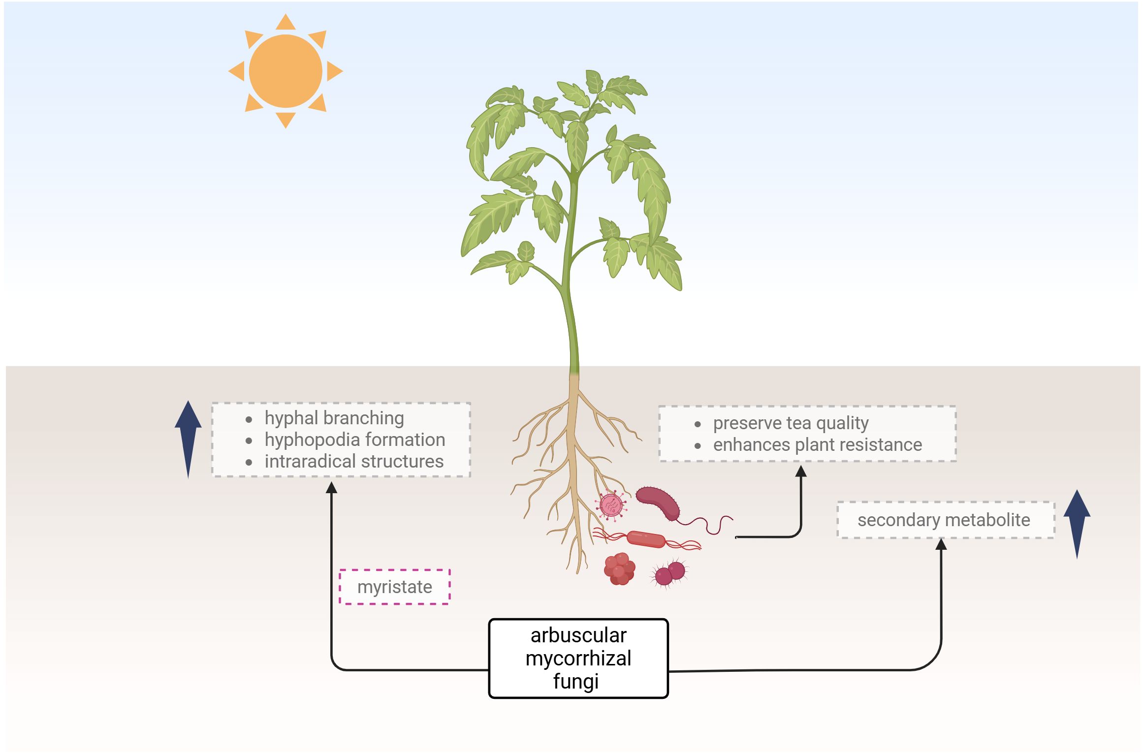
Figure 1. The illustration depicts the influence of arbuscular mycorrhizal fungi and microbial communities on plant development, highlighting their role in enhancing plant resistance and secondary metabolite production created using Biorender.com.
In an era of precision agriculture and ecological restoration, the ability to visualize these interactions in situ is more than a technical achievement; it is a pathway to more resilient and sustainable ecosystems. We invite readers to explore this Research Topic, which centres on the convergence of molecular biology and advanced imaging technologies.
Author contributions
AP: Conceptualization, Visualization, Writing – original draft, Writing – review & editing. CR: Writing – review & editing. PP: Writing – review & editing.
Conflict of interest
The authors declare that the research was conducted in the absence of any commercial or financial relationships that could be construed as a potential conflict of interest.
Generative AI statement
The author(s) declare that Generative AI was used in the creation of this manuscript. Language edits were performed with the help of generative AI.
Publisher’s note
All claims expressed in this article are solely those of the authors and do not necessarily represent those of their affiliated organizations, or those of the publisher, the editors and the reviewers. Any product that may be evaluated in this article, or claim that may be made by its manufacturer, is not guaranteed or endorsed by the publisher.
Keywords: plant-microbe interactions, imaging techniques, plant physiology, rhizosphere, arbuscular mycorrhizal fungi
Citation: Prasad A, Rossi C and Pospíšil P (2025) Editorial: Visualizing plant-microbe dynamics: cutting-edge imaging methods in plant research. Front. Plant Sci. 16:1653745. doi: 10.3389/fpls.2025.1653745
Received: 25 June 2025; Accepted: 27 June 2025;
Published: 08 July 2025.
Edited and Reviewed by:
Andrea Genre, University of Turin, ItalyCopyright © 2025 Prasad, Rossi and Pospíšil. This is an open-access article distributed under the terms of the Creative Commons Attribution License (CC BY). The use, distribution or reproduction in other forums is permitted, provided the original author(s) and the copyright owner(s) are credited and that the original publication in this journal is cited, in accordance with accepted academic practice. No use, distribution or reproduction is permitted which does not comply with these terms.
*Correspondence: Ankush Prasad, YW5rdXNoLnByYXNhZEB1cG9sLmN6
†ORCID: Ankush Prasad, orcid.org/0000-0002-2009-8987
Claudio Rossi, orcid.org/0000-0001-6032-1015
Pavel Pospíšil, orcid.org/0000-0001-9126-2011