- 1The Research Center for Ornamental Plants, College of Horticulture, South China Agricultural University, Guangzhou, China
- 2Guangdong Provincial Key Laboratory of Ornamental Plant Germplasm Innovation and Utilization, Environmental Horticulture Research Institute, Guangdong Academy of Agricultural Sciences, Guangzhou, China
- 3College of Life Sciences, South China Agricultural University, Guangzhou, China
The Phosphatidylethanolamine-binding protein (PEBP) gene family plays a crucial role in plant growth and development, particularly in regulating flowering time and morphogenesis. However, the diversity, expression patterns, and functions of PEBP genes in Hedychium coronarium remain largely unexplored. In this study, 14 HcPEBP genes were identified and classified into MFT, FT, and TFL1 subfamilies based on phylogenetic analysis. Intraspecific collinearity analysis revealed three collinear relationships within the HcPEBP gene family. Interspecific collinearity analysis across H. coronarium, rice, wild banana, and pineapple highlighted the evolutionary significance of specific PEBP genes. Motifs DPDxP and GxHR are conserved in HcPEBPs, which are essential for anion-binding activity. At the same position in the C-terminus, FT-likes contain the xGxGGR motif, while TFL1-likes possesses the TAARRR motif. 64.3% of HcPEBP genes consist of four exons and three introns. Promoter regions of HcPEBP genes are enriched with light-responsive elements, suggesting a primary response to light signals. Expression patterns analysis by qRT-PCR showed that seven FT-like genes are predominantly expressed in leaves, with increased expression during the transition from vegetative to reproductive growth. HcPEBP11, a FT-like gene, is highly expressed in inflorescence buds. Overexpression of HcPEBP11 in tobacco induced early flowering, confirming its role in promoting flowering. This study provides a foundation for further research on the biological functions of the PEBP gene family in H. coronarium and elucidates the role of HcPEBP11 in flowering regulation.
1 Introduction
Phosphatidylethanolamine-binding proteins (PEBPs) possess evolutionarily conserved phosphatidylethanolamine-binding domains and are prevalent in both prokaryotes and eukaryotes (Dong et al., 2020; Li et al., 2009; Rautureau et al., 2009). In plants, PEBPs are pivotal in regulating floral transition, determining architecture, and influencing seed dormancy and germination, as well as tuber formation and sink-source allocation (Abelenda et al., 2019, 2009; Bi et al., 2016; Chen et al., 2018; Goto, 2005; Lee et al., 2023; Navarro et al., 2015; Yamaguchi et al., 2005; Zhang S. H, et al., 2021). The PEBP gene family is categorized into three subfamilies: Flowering Locus T-like (FT-like), TERMINAL FLOWER 1-like (TFL1-like), and MOTHER OF FT AND TFL1-like (MFT-like) (Karlgren et al., 2011). MFT-like is considered the ancestral form of FT-like and TFL1-like, present in both basal land plants and seed plants. In contrast, FT-like and TFL1-like are exclusive to seed plants, suggesting their emergence as a result of seed plant evolution (Hedman et al., 2009; Karlgren et al., 2011; Liu et al., 2016; Xu et al., 2022). Despite extensive sequence similarity among PEBP genes, their functions diverge significantly (Wang et al., 2015).
MFT-like genes are implicated in seed development and germination through their involvement in abscisic acid (ABA) and gibberellin (GA) signaling pathways (Vaistij et al., 2018; Xi et al., 2010; Xi and Yu, 2010; Yu et al., 2019). FT-like and TFL1-like genes are essential in regulating the timing of flowering and morphogenesis. In Arabidopsis thaliana, FT and TFL1 proteins share 60% sequence identity but exhibit antagonistic functions (Ahn et al., 2006; Hanzawa et al., 2005). The FT protein, acting as florigen, plays a crucial role in promoting flowering (Corbesier et al., 2007). The FT gene integrates both external and internal signals to regulate flowering (Abe et al., 2005; Wigge et al., 2005a). In rice, Hd3 and RFT1, homologs of the A. thaliana FT gene, initiate flowering, also known as heading (Tamaki et al., 2007; Zhao et al., 2015). In maize, ZCN8, a FT-like gene, acts as a floral activator and contributes to photoperiod sensitivity. Its ectopic expression in vegetative shoot apices triggers early flowering in transgenic plants (Lazakis et al., 2011; Mach, 2011). FT-like genes, encoding florigens, have been identified in various plant species, including herbaceous plants, woody plants, and lianas (Fan et al., 2023; Igasaki et al., 2008; Kalia et al., 2023; Kim et al., 2022; Lembinen et al., 2023; Lin et al., 2019; J. Odipio et al., 2020; Patil et al., 2022). TFL1-like genes, comprising TFL1, CEN (CENTRORADIALIS), and BFT (BROTHER OF FT), act as floral repressors by delaying flowering and inhibiting flower primordia formation. TFL1 regulates shoot meristem identity and represses flowering by competitively binding to FD (a bZIP type transcription factor FLOWERING LOCUS D), thereby inhibiting FD-FT complex formation (Freytes et al., 2021; Kaneko-Suzuki et al., 2018; Zhu et al., 2020). In Arabidopsis, heterologous expression of apple (Malus × domestica Borkh) MdTFL1 or MdTFL2 significantly delays flowering, increases rosette leaf number, and plant height (Zuo et al., 2021). Mutations in the TFL1 homologs KSN of rose and strawberry result in a continuous flowering habit (Iwata et al., 2012; Soufflet-Freslon et al., 2021). Additionally, in Arabidopsis and pea, TFL1 modulates the length of the vegetative phase (Bradley et al., 1997; Foucher et al., 2003).
PEBP genes are integral to the growth, development, and reproduction of plants, making the analysis of their evolution and function are crucial for advancing plant cultivation and understanding. The PEBP gene family has been identified and researched in various plant species, including A. thaliana (Kardailsky et al., 1999), potato (Zhang et al., 2022), maize (Danilevskaya et al., 2008), rice (Zhao et al., 2022), Dendrobium huoshanense (Song et al., 2021), cotton (Wang et al., 2019; Zhang et al., 2016), Perilla frutescens (Xu et al., 2022), wheat (Dong et al., 2020), and tomato (Sun et al., 2023). However, data on the gene family’s member count, phylogeny, expression, and functions in H. coronarium remain unexplored. Known as “white ginger lily” or “butterfly ginger,” H. coronarium is a perennial herb and ornamental flowering plant native to the Eastern Himalayas and southern China (Baez et al., 2011). Its elegant floral morphology and refreshing scent have made it a popular choice for cultivation as a cut flower or garden plant in tropical and subtropical regions (Ke et al., 2019). Flowering, a critical agronomic trait for ornamental plants, affects their aesthetic and cultivation value. H. coronarium exhibits a continuous flowering habit, producing new aboveground stems and blooming from May to October.
This study conducted a genome-wide identification of the PEBP gene family in H. coronarium, revealing 14 HcPEBP genes. Using bioinformatics techniques, we analyzed sequence characteristics, phylogenetic relationships, chromosomal localization, conserved motifs, gene structures, collinearity, and cis-regulatory elements. qRT-PCR analysis provided insights into the expression patterns of HcPEBPs across various tissues. We specifically examined the expression level of HcPEBP11 in leaf buds and inflorescence bud’s three distinct developmental stages. Notably, HcPEBP11 exhibited higher expression in the inflorescence buds compared to other tissues. The role of HcPEBP11 in promoting flowering was further investigated by its heterologous expression in tobacco. In summary, this work offers a scientific reference for understanding the PEBP gene family of H. coronarium. Additionally, this research lays the groundwork for further detailed investigations into the molecular and biological functions of HcPEBP gene family members and enriches the study of PEBP gene family in plant species.
2 Materials and methods
2.1 Plant material
H. coronarium was cultivated in the open field at the College of Horticulture, South China Agricultural University (Guangzhou, China, 23.16°N, 113.36°E). The plant materials (root, rhizome, leaf, leaf buds, inflorescence buds) were collected from 19:00 on June 15, 2024. Root, rhizome and leaf were collected during reproductive growth. All tissues were immediately frozen in liquid N and stored at − 80 until use.
2.2 PEBP gene family elucidation in H. coronarium
Arabidopsis PEBP sequences were downloaded from TAIR (https://www.arabidopsis.org/). The H. coronarium genome (unpublished) was provided by Beijing Novogene Bioinformatics Technology Corporation (China). Arabidopsis PEBP sequences were used as queries to identify HcPEBP sequences from the H. coronarium genome using the BLAST module in TBtools (E-value ≤ 1.0 × 10⁻⁵) (Chen et al., 2020). Additionally, The HMM profile of the PEBP domain (PF01161) from the Pfam database (http://pfam.xfam.org/) (Finn et al., 2006) was used in the Simple HMM search module in TBtools. Sequences identified by both BLAST and HMM searches were merged, and duplicates were removed. All candidate sequences were analyzed for the PEBP domain using the NCBI-Conserved Domain Database (https://www.ncbi.nlm.nih.gov/Structure/cdd/wrpsb.cgi) (Morris et al., 2015). Only sequences containing the complete PEBP domain were retained for further analysis.
2.3 Chromosomal location and collinearity analysis
Chromosomal details (length, gene density, and gene positions) were extracted from the H. coronarium genome using TBtools (Chen et al., 2020). The chromosomal locations of HcPEBP genes were visualized using TBtools. Genome sequences and annotations for wild banana, pineapple, and rice were downloaded from NCBI (https://www.ncbi.nlm.nih.gov/) and Ensemble Plants (https://plants.ensembl.org). Gene duplication events and collinear relationships were analyzed using the One-step MCScanX-Super Fast module in TBtools with default settings. Collinear relationships were visualized using the Dual Synteny Plotter in TBtools.
2.4 Conserved motif and gene structure analysis
Conserved motifs within HcPEBP proteins were analyzed using the MEME suite (https://meme-suite.org/meme/tools/meme) (Bailey et al., 2015). The motif count was set to eight, with other parameters kept at default settings. The identified motifs and gene structures were visualized using the Gene Structure View (advanced) module in TBtools.
2.5 Investigation of physicochemical characteristics
Physicochemical properties of HcPEBP proteins, including molecular weight (MW), isoelectric point (pI), instability index, aliphatic index, and GRAVY (grand average of hydropathicity), were analyzed using the ProtParam tool on the ExPASy online platform (http://www.expasy.ch/tools-/pi_tool.html) (Artimo et al., 2012). Subcellular localization predictions for HcPEBP proteins were made using the WoLF PSORT tool (Horton et al., 2007).
2.6 Alignment of multiple sequences and evolutionary relationship study
PEBP protein sequences from Arabidopsis and rice were downloaded from TAIR and the Rice Genome Annotation Project (RGAP, http://rice.uga.edu/) databases, respectively. The GenBank accession numbers for PEBPs in Arabidopsis and rice are listed in Supplementary Table S1. Multiple sequence alignments of PEBP proteins from H. coronarium, Arabidopsis, and Oryza sativa were conducted using TBtools. A phylogenetic tree comprising 40 PEBP proteins from H. coronarium, Arabidopsis, and rice was constructed with TBtools using default parameters and visualized with Evolview (https://www.evolgenius.info/evolview-v2) (Zhang et al., 2012). Amino acid sequences of HcPEBPs were aligned using the ClustalW algorithm in MEGA11 and displayed using GeneDoc (Tamura et al., 2021).
2.7 Examination of cis-regulatory elements
Sequences 2000 bp upstream of the transcription start site (ATG) for HcPEBP genes were extracted from the genome sequence using TBtools. Cis-acting elements within the promoter sequences of HcPEBPs were predicted using the PlantCARE database (http://bioinformatics.psb.ugent.be/webtools/plantcare/html/) (Lescot et al., 2002), with results analyzed, classified, and visualized using TBtools.
2.8 Differential gene expression profiling across various tissues
The expression patterns of HcPEBP genes across various tissues of H. coronarium were investigated. Relative expression levels in five tissues (root, rhizome, leaf, inflorescence bud, leaf bud) were measured by qRT-PCR. Morphological characteristics of these tissues are depicted in Supplementary Figure S1. Primer sequences are provided in Supplementary Table S2.
2.9 Total RNA and DNA extraction, cDNA synthesis and qRT-PCR analysis
Total RNA was isolated from plant materials using the HiPure Plant RNA Mini Kit (Magen, Guangzhou, China), following the manufacturer’s protocol. DNA was extracted using the DNA Quick Plant System (Tian Gen, Beijing, China), as per the provided manual. For gene cloning, cDNA was synthesized using the PrimeScript™ RT Reagent Kit with gDNA Eraser (TaKaRa, Japan). For qRT-PCR, cDNA was reverse-transcribed using the Evo M-MLV RT Mix Kit with gDNA Clean for qPCR Ver.2 (Accurate Biology, Hunan, China). qRT-PCR was performed using Hieff® qPCR SYBR Green Master Mix (Yeasen Biotechnology, Shanghai, China) on an ABI 7500 Fast Real-Time PCR system (Applied Biosystems, USA). Reaction conditions followed a previously described protocol (Wang et al., 2021). The GAPDH gene was used as an internal reference for normalization. Relative gene expression levels were calculated using the 2−ΔΔCt method. Statistical analyses for Significant differences were determined using IBM SPSS Statistics and Origin 2021.
2.10 Molecular cloning and genetic engineering in plants
The coding sequence (CDS) of HcPEBP11 was amplified from H. coronarium cDNA using Phanta Max Super-Fidelity DNA Polymerase (Vazyme, Nanjing, China), following the manufacturer’s protocol. The CDS was cloned into the pOx vector (provided by the State Key Laboratory for Conservation and Utilization of Subtropical Agro-Bioresources, South China Agricultural University, China) using the ClonExpress II One Step Cloning Kit (Vazyme, Nanjing, China). The constructed plasmid was transformed into Agrobacterium tumefaciens GV3101 (WeDi, Shanghai, China) using the freeze-thaw method. Primers used are listed in Supplementary Table S2. Tobacco plants (Nicotiana tabacum cv. W38) were transformed using the leaf disc method (Horsch et al., 1986). Agrobacterium cultures containing pOx-HcPEBP11 were grown at 28°C in liquid medium with 50 µg/mL kanamycin and 25 µg/mL rifampicin until reaching an OD600 of 0.6–0.8. Bacteria were pelleted by centrifugation at 5000 rpm for 8 minutes and resuspended in MS liquid medium containing 100 µM acetosyringone to an OD600 of 0.7–0.8. Young leaf explants from sterile tobacco seedlings were pre-cultured for three days in darkness before transformation. Transformed plants were regenerated through a series of cultures: co-culture, bacteriostatic, differentiation, induction, rooting, and screening.
2.11 Selection and characterization of transgenic lines with phenotypic evaluation
Putative transgenic tobacco plants resistant to 20 mg/L hygromycin B in 1/2 MS medium were screened using PCR (Cordero Otero and Gaillardin, 1996; Thion et al., 2002; Walter et al., 1989). Universal primers pOx-F/R (flanking the multiple cloning sites of the pOx vector) and specific primers HPH-F/R (targeting the hygromycin B phosphotransferase gene) were used to confirm the integration of HcPEBP11 into the tobacco genome. Primer sequences are listed in Supplementary Table S2. Semi-quantitative RT-PCR (Semi-qRT-PCR) and qRT-PCR were performed to verify HcPEBP11 expression in transgenic lines (Tripathi et al., 2022). Total RNA was extracted from leaves at the flower bud stage. Semi-qRT-PCR was conducted using ABI VeritiPro PCR (Thermo Fisher) and Phanta Max Super-Fidelity DNA Polymerase, with specific primers for HcPEBP11. Transgenic and wild-type tobacco lines were grown under natural light conditions. The time to bolting (appearance of the first flower bud) and flowering (blooming of the first flower) was recorded to assess the role of HcPEBP11 in flowering regulation.
2.12 Subcellular localization of HcPEBP11 protein
The coding sequence (CDS) of the HcPEBP11 gene, lacking termination codons, was successfully cloned into the p35S-cGFP vector. Subsequently, the resultant recombinant vector underwent transformation into the A. tumefaciens strain GV3101 via the heat shock method. For the transformation assay, leaves of N. benthamiana at the five-leaf stage were utilized (Kokkirala et al., 2010). Post 72 hours of infiltration, the green fluorescent protein (GFP) signals were detected using a confocal laser scanning microscope.
3 Results
3.1 Identification and physicochemical characterization of HcPEBP gene family
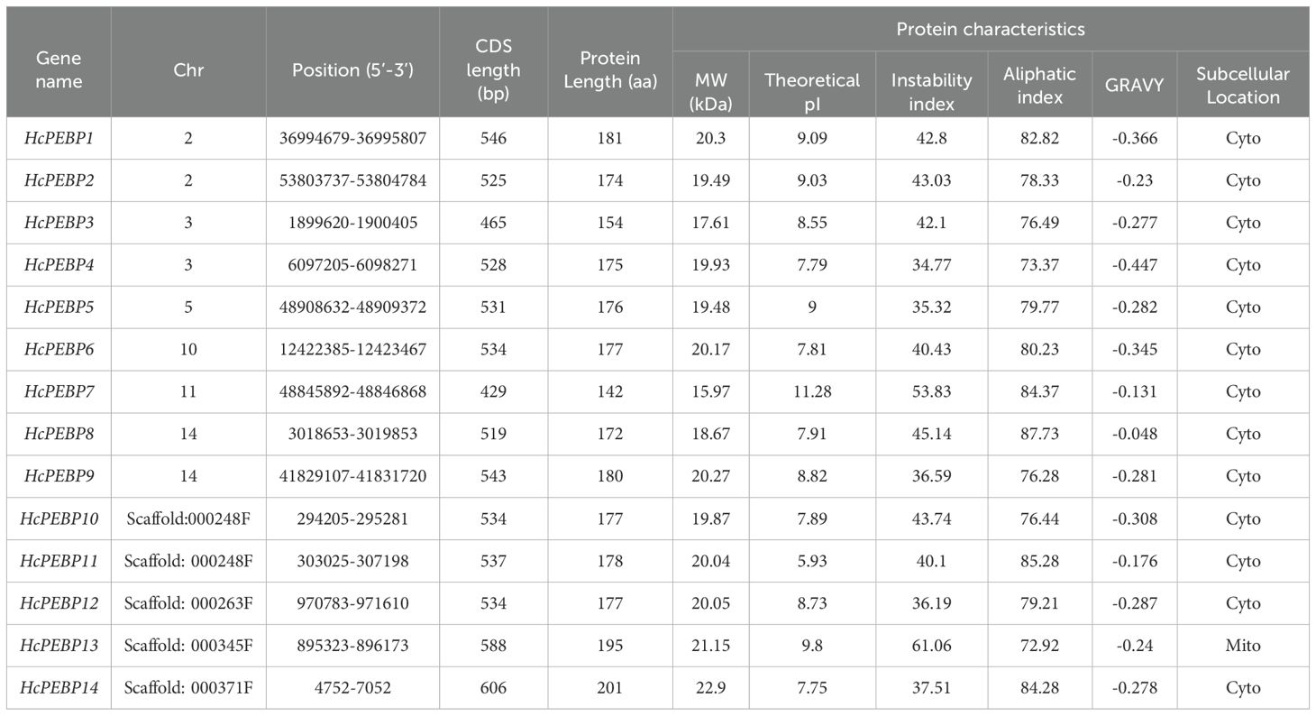
In H. coronarium, 14 PEBP genes were identified and named HcPEBP1–14 based on their chromosomal distribution. Key details about the HcPEBP family and the physicochemical properties of their encoded proteins are summarized in Table 1 and Supplementary Table S3. Protein length ranges from 142 aa (HcPEBP7) to 201 aa (HcPEBP14), with an average of 175 aa. Molecular weight averages 19.71 kDa, ranging from 15.97 kDa (HcPEBP7) to 22.90 kDa (HcPEBP14). Theoretical isoelectric points span from 5.93 (HcPEBP11) to 11.28 (HcPEBP7). Instability Index ranges from 34.77 (HcPEBP4) to 61.06 (HcPEBP13). Aliphatic index aries between 87.73 (HcPEBP8) and 72.92 (HcPEBP13). All 14 HcPEBP proteins are hydrophilic. Except for HcPEBP13 (mitochondrial), all other HcPEBPs are predicted to be cytoplasmic proteins.
3.2 Chromosomal localization and gene duplication analysis of HcPEBP genes
In H. coronarium, 14 HcPEBP genes were identified. 9 genes (HcPEBP1-9) mapped to six chromosomes (Hc-2, Hc-3, Hc-5, Hc-10, Hc-11, Hc-14), while the remaining five genes (HcPEBP10-14) are located on four genome scaffolds. Intraspecific collinearity analysis revealed three pairs of duplicated genes: HcPEBP2 and HcPEBP7, HcPEBP4 and HcPEBP14, HcPEBP6 and HcPEBP12 (Figure 1A). To assess the impact of selection pressure on the collinear HcPEBP gene pairs, the Ka/Ks ratios (non-synonymous to synonymous substitutions) were calculated for each duplicated pair. A Ka/Ks = 1 indicates neutral selection, Ka/Ks < 1 indicates purifying selection, and Ka/Ks > 1 suggests positive selection. The Ka/Ks ratios for the three collinear HcPEBP gene pairs ranged from 0.087 to 0.295 (Supplementary Table S4), indicating that these duplicated gene pairs have undergone purifying selection, which maintains functional stability by removing deleterious mutations.
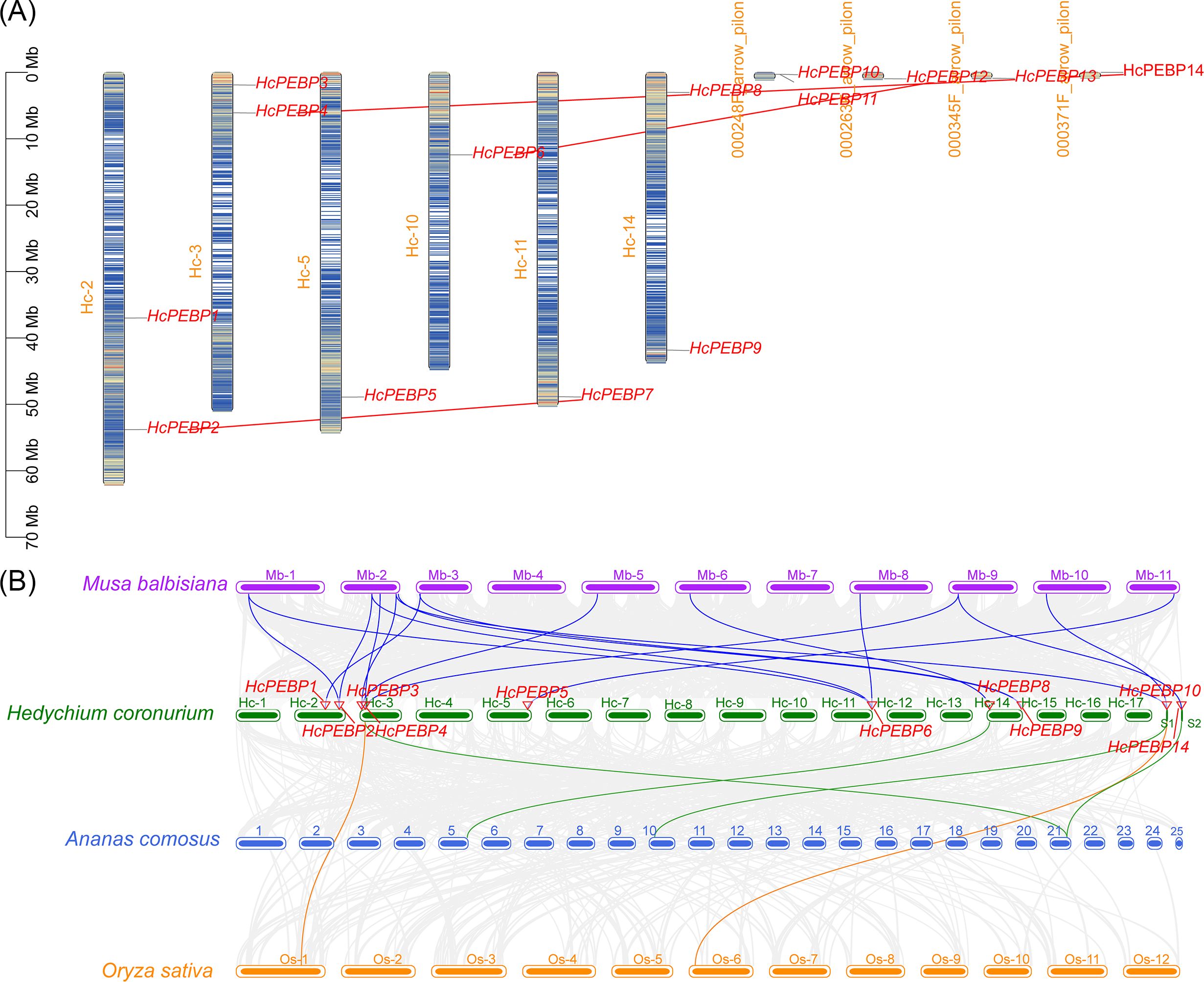
Figure 1. Chromosomal localization and collinearity analysis of HcPEBP genes. (A) Chromosome length is shown on the left scale. HcPEBP genes (1–14) are highlighted in red; chromosome numbers are in orange. The gene density is indicated by a blue-to-red gradient. Red lines show intraspecific collinearity among HcPEBP genes. (B) Interspecific collinearity analysis of HcPEBP genes with M. balbisiana, A. comosus, and O. sativa. Chromosome numbers are displayed above each chromosome. Colored lines indicate collinear relationships; red triangles mark HcPEBP gene locations.
To further explore the evolutionary relationships of HcPEBP genes with those of other species, a collinearity analysis was conducted using O. sativa, Musa balbisiana, and Ananas comosus alongside HcPEBP family members. The results revealed that the HcPEBP gene family exhibits 17 collinearities with wild banana, 2 with rice, and 4 with pineapple (Figure 1B, Supplementary Table S5). Among these species, HcPEBP genes show the closest evolutionary relationship with PEBP members from wild banana. Notably, HcPEBP4 and HcPEBP10 display collinearities with all three species, highlighting their conserved evolutionary roles.
3.3 Phylogenetic analysis of PEBP family members
To investigate the evolutionary relationships of HcPEBP genes with PEBP genes from other species, a phylogenetic tree was constructed using multiple sequence alignments of amino acids from 6 PEBP proteins of A. thaliana, 20 PEBP proteins of O. sativa, and 14 PEBP proteins of H. coronarium (Figure 2). The phylogenetic tree is divided into three subgroups: MFT, TFL1, and FT. HcPEBP8 clusters with AtMFT, OsMFT1, and OsMFT2, indicating it belongs to the MFT subgroup. Four genes (HcPEBP2/5/7/13) cluster with AtBFT, AtTFL1, AtATC, and OsRCEs, suggesting these four genes belong to the TFL1 subgroup. Nine genes (HcPEBP1/3/4/6/9/10/11/12/14) cluster with AtFT, AtTSF, Hd3a, and OsFTLs, indicating they belong to the FT subgroup.
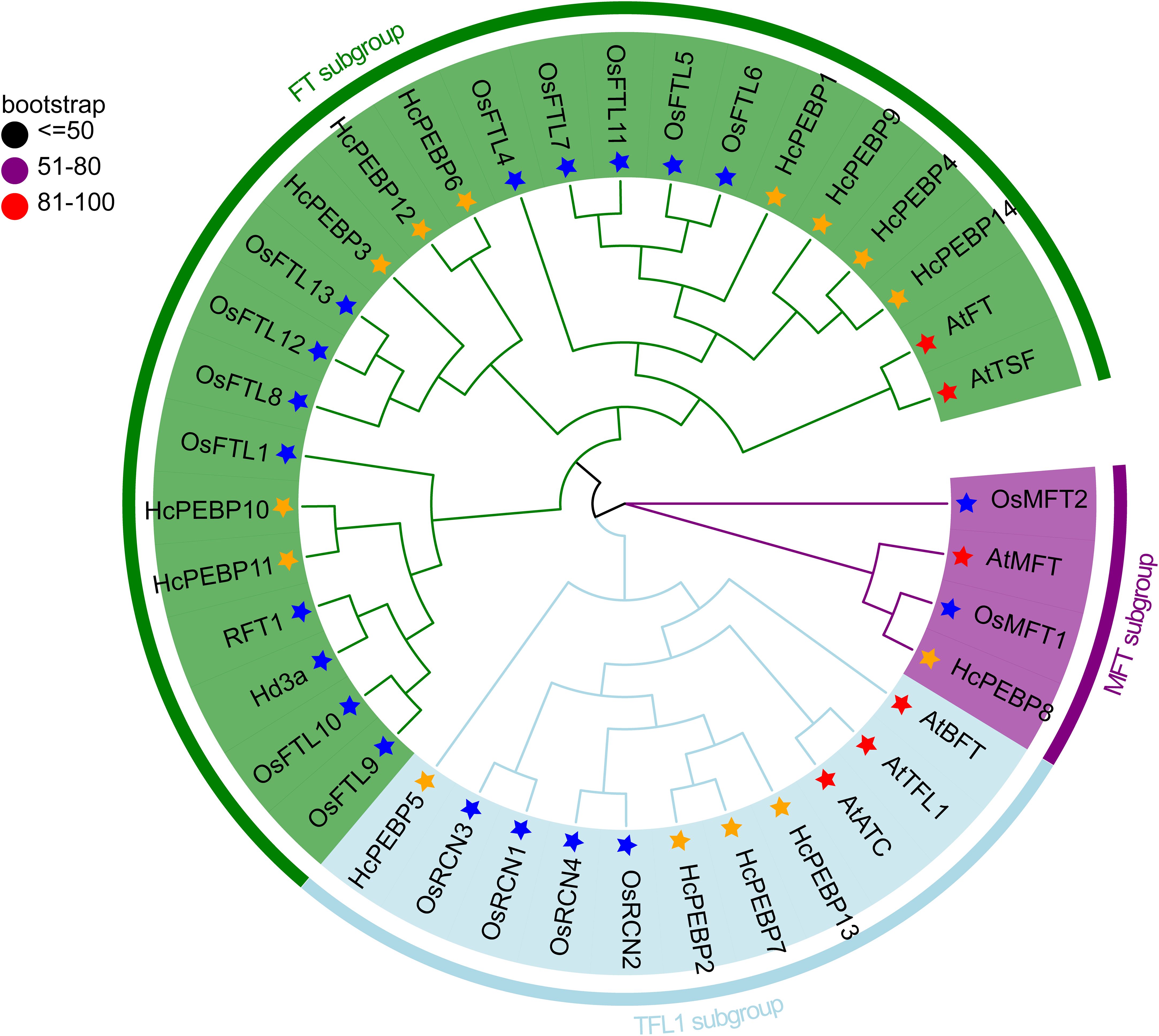
Figure 2. Phylogenetic tree analysis of 6 AtPEBP (A. thaliana), 20 OsPEBP (O. sativa), and 14 HcPEBP (H. coronarium) proteins. Species marked by colored stars: Red: A. thaliana, Blue: O. sativa, Orange: H. coronarium. Bootstrap percentage values (1,000 replications) at nodes: Black circles (0–0.5), Purple circles (0.51–0.8), Red circles (0.81–1.0). GenBank accession numbers for AtPEBPs and OsPEBPs are provided in Supplementary Table S1.
3.4 Alignment of amino acid sequences and analysis of conserved domains
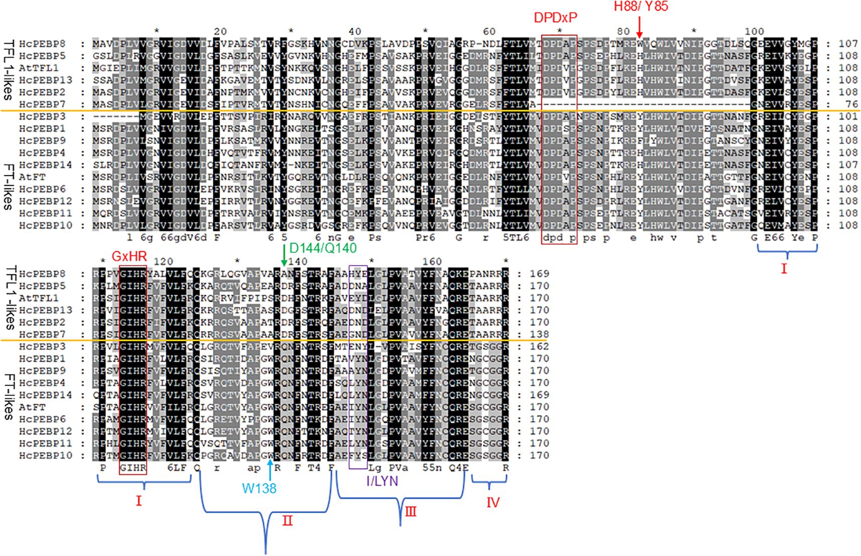
Phylogenetic tree analysis revealed that HcPEBP1/3/4/6/9/10/11/12/14 belong to the FT subfamily, while HcPEBP2/5/7/13 belong to the TFL1 subfamily. To further investigate the structural and functional conservation of these proteins, amino acid sequence alignment was performed for HcPEBPs, along with AtFT and AtTFL1 (Figure 3). All HcPEBP proteins contain the GxHR motif, a hallmark of the PEBP family. Except for HcPEBP7, all other HcPEBP proteins possess the DPDxP motif, another key functional domain. The C-terminal amino acid sequence can be divided into four segments (I–IV), each of which is essential for the FT protein’s function. The conserved motif LYN/IYN/in FT-like proteins is located in segment III, playing a critical role in enzymatic activity. In segment IV region, FT-like proteins contain the motif xGxGGR, whereas TFL1-like proteins contain a different motif (TAARRR) in the same region. FT-like proteins HcPEBP1/3/6/10/11/12 contain the key amino acid residues Tyr85, Trp138, and Gln140, which are critical for their function. In contrast, TFL1-like proteins HcPEBP2/5/13 possess the key residues His88 and Asp144, which are characteristic of their functional role. Interestingly, in FT-like proteins HcPEBP4/9/14, the residue Tyr85 is mutated to His, Phe, His, respectively.
3.5 Analysis of conserved motifs and gene structural features
A phylogenetic tree containing only HcPEBP genes was constructed, dividing them into three subfamilies (Figure 4A). To further investigate the characteristics of HcPEBP genes, we conducted conserved motif and gene structure analyses. In the conserved motif analysis (Figure 4B), 8 conserved motifs (named motifs 1–8) were identified, with detailed sequence information provided in Supplementary Table S6. The analysis revealed the following patterns: Motifs 3, 5, and 7 are shared by all HcPEBPs, indicating their fundamental role in the protein family. Motif 1 is present in all HcPEBPs except HcPEBP7. Motif 2 is found in all HcPEBPs except HcPEBP3. Motif 4 is absent in HcPEBP3 and HcPEBP14. Motif 6 is exclusively present in the FT subfamily, highlighting its specificity to this group. Motif 8 is unique to the TFL1 and MFT subfamilies, suggesting a functional role specific to these groups. Additionally, conserved domain analysis confirmed that all 14 HcPEBPs contain the PEBP domain (Figure 4C).
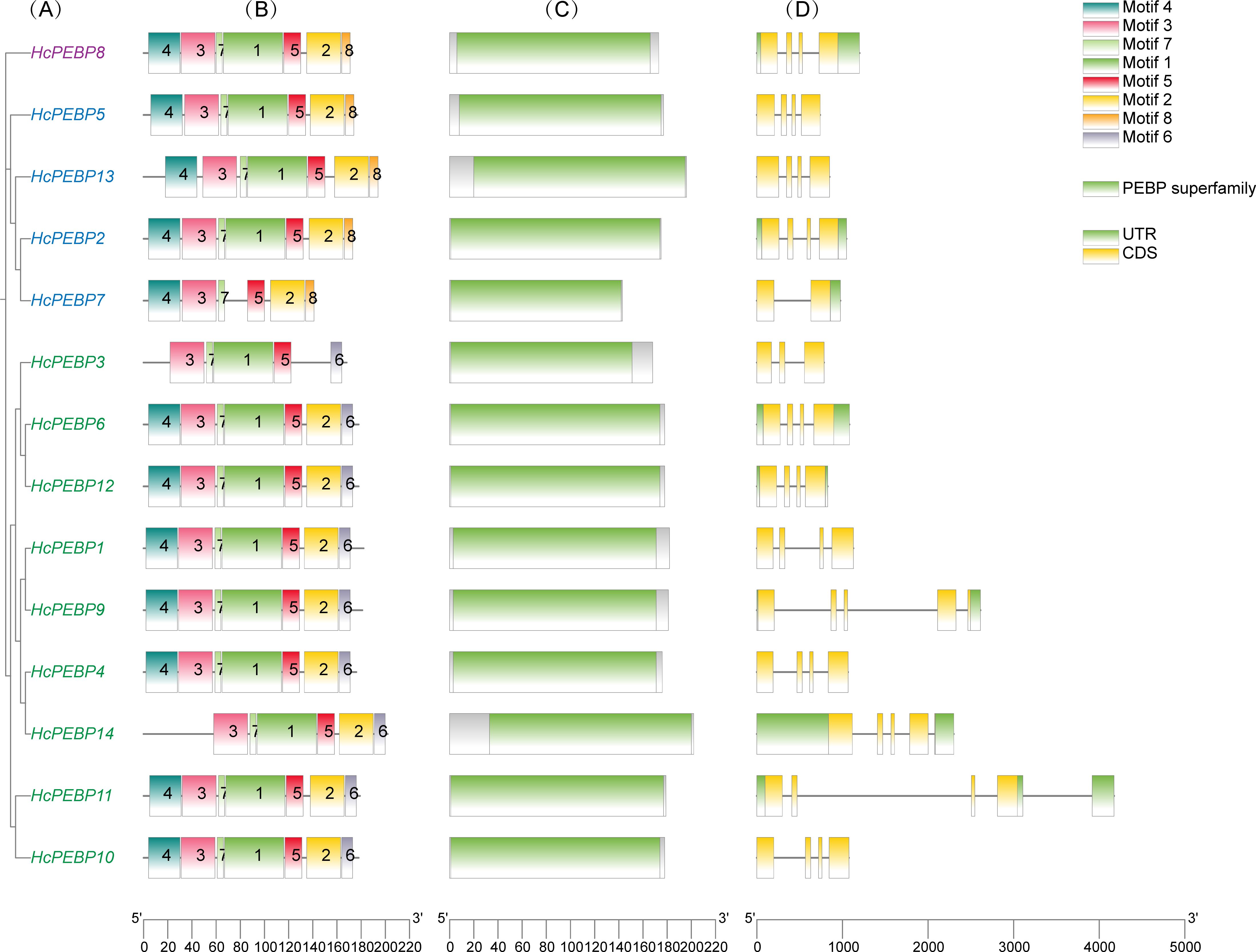
Figure 4. Phylogenetic tree, conserved motifs, domain, and gene structure analysis of HcPEBP gene family members. (A) Phylogenetic tree. (B) Distribution of conserved motifs in HcPEBP proteins, represented by colored boxes. (C) Conserved domains of HcPEBP proteins. (D) Gene structure of HcPEBP family members, showing UTRs (green rectangles), exons (yellow rectangles), and introns (grey lines). A scale at the bottom allows comparison of protein and gene lengths.
Gene structure analysis revealed that the 14 HcPEBP genes share a generally similar structural organization (Figure 4D). Among them: nine HcPEBP genes (64.3% of the total) contain four exons and three introns, representing the most common structural pattern in the HcPEBP gene family. Three HcPEBP genes (HcPEBP9/11/14) consist of five exons and four introns. HcPEBP3 with three exons and two introns. HcPEBP7 contain only two exons and one intron.
3.6 Prediction and characterization of cis-regulatory elements
Cis-acting elements are critical for regulating gene transcription. In this study, we analyzed the 2000 bp upstream sequences of the HcPEBP genes’ start codon to predict and characterize cis-acting elements (Figure 5A). A total of 45 distinct cis-acting elements were identified and classified into four functional categories (Figure 5B): hormone-responsive (98 elements), development-related (28 elements), light-responsive (161 elements), and defense and stress responsiveness-related (40 elements), summing up to 327 elements. Among these, light-responsive elements were the most abundant, while development-related element were the least frequent. All HcPEBP genes, except HcPEBP4 and HcPEBP14, contain the G-box element (light-Responsive Element). All genes, except HcPEBP10, contain the Box4 element. HcPEBP1/3/6/7 lack any development-related elements. Detailed information on the cis-acting elements in the promoter regions of HcPEBP genes is provided in Supplementary Table S7.
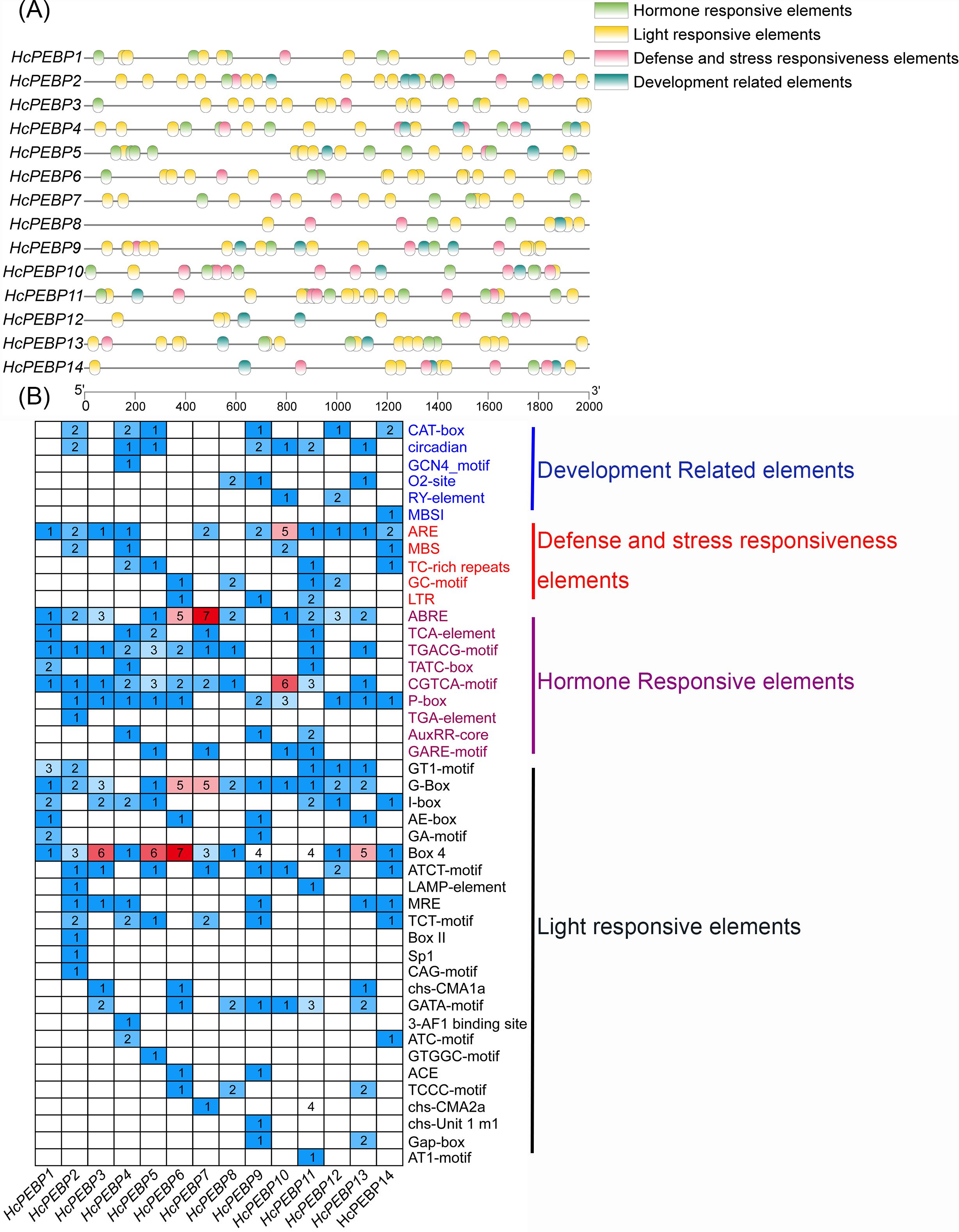
Figure 5. Cis-acting element analysis of HcPEBP gene promoter regions. (A) Distribution of cis-acting elements in the 2000 bp upstream promoter regions of HcPEBP genes. Different colors represent various element types. A ruler indicates sequence direction and length. (B) Classification and statistics of cis-acting elements. Numbers and colors indicate the count of specific elements per gene.
3.7 Tissue-specific expression profiling of HcPEBP genes
To investigate tissue-specific gene expression patterns, we analyzed the expression levels of HcPEBP genes across various tissues—root, rhizome, leaf, leaf bud, and inflorescence bud using qRT-PCR (Supplementary Figure S1). HcPEBP5 and HcPEBP8 showed the highest expression in roots, with HcPEBP8 significantly exceeding HcPEBP5 (Figures 6E, H, O). HcPEBP1/2/3/4/9/10/12 and HcPEBP14 exhibited significantly elevated expression in leaves compared to other tissues (Figures 6A–D, I, J, L, N). Among these, HcPEBP1/2/14 had notably higher expression levels (Figure 6Q). HcPEBP6/7/13 were most highly expressed in Rhizomes, with HcPEBP7 levels significantly surpassing those of HcPEBP6 and HcPEBP13 (Figures 6F–G, M, P). HcPEBP11 expression was significantly higher in inflorescence buds compared to other tissues (Figure 6K). Additionally, transcriptome data and qRT-PCR results indicate that the expression of HcPEBP11 progressively increases during the development of inflorescence buds. (Figures 7A, C, Supplementary Figure S2, Supplementary Table S9). Seven FT subfamily genes (HcPEBP1/3/4/9/10/12/14) exhibited high expression levels in leaves (Figures 6A, C, D, I, J, L, N). To further investigate their roles, we analyzed their expression patterns in leaves at four developmental stages of the apical meristem (Figures 7B, C). The results revealed that HcPEBP1 showed a continuous increase in expression from stage 1 to stage 4. HcPEBP3/4/9/10/12 displayed an initial increase followed by a decrease, with HcPEBP9/10/12 peaking at stage 2 and HcPEBP3/4 reaching their highest expression at stage 3. These findings suggest that HcPEBP1/9/10/12 may play roles in floral transition.
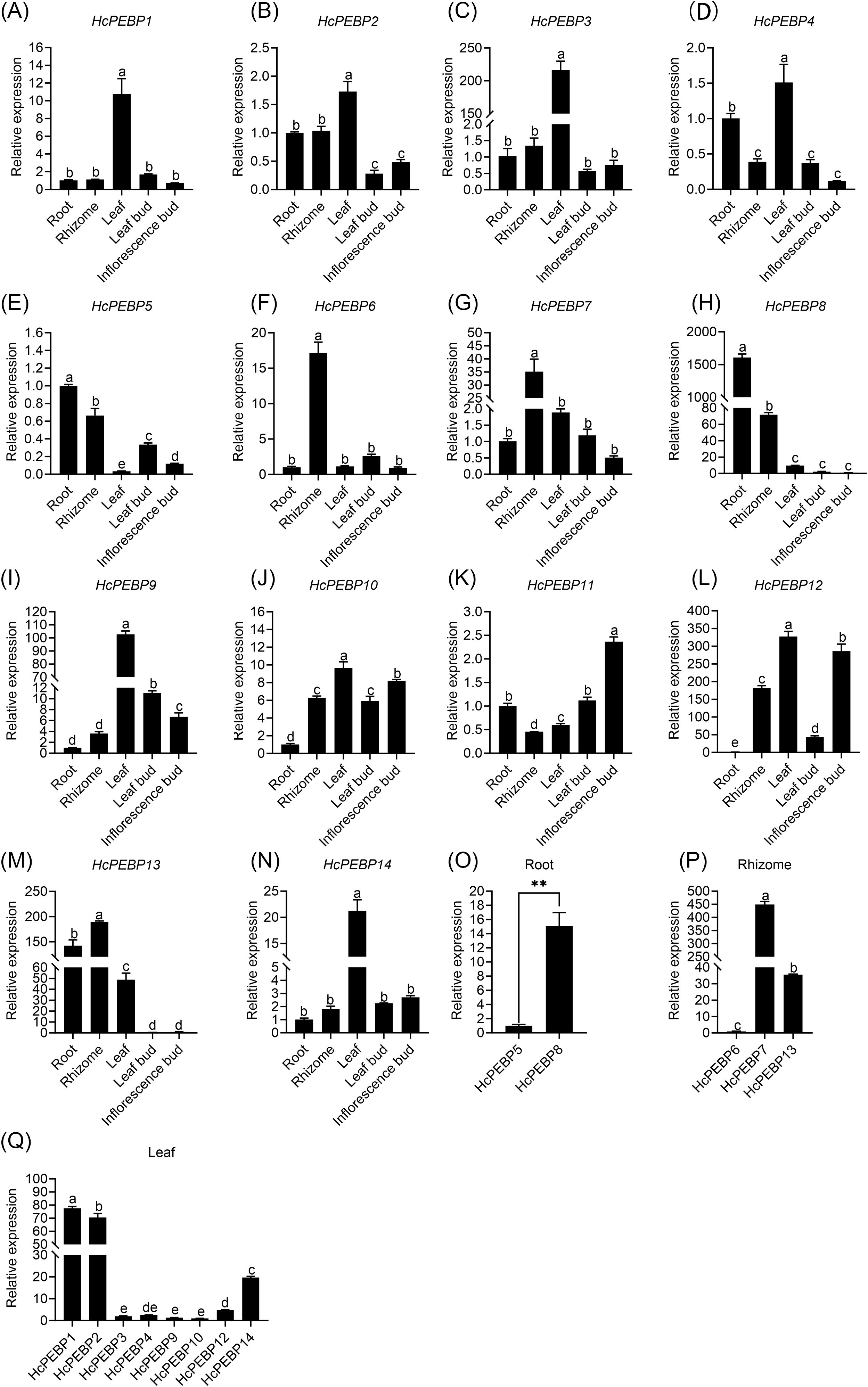
Figure 6. Tissue-specific expression analysis of HcPEBP genes. (A–N) Relative expression levels of HcPEBP genes in five tissues. (O) HcPEBP genes predominantly expressed in roots. (P) HcPEBP genes predominantly expressed in rhizomes. (Q) HcPEBP genes predominantly expressed in leaves. Error bars represent standard deviation (three biological replicates). Different lowercase letters indicate significant differences at P< 0.05 (after multiple comparison corrections). ** denotes P< 0.01.
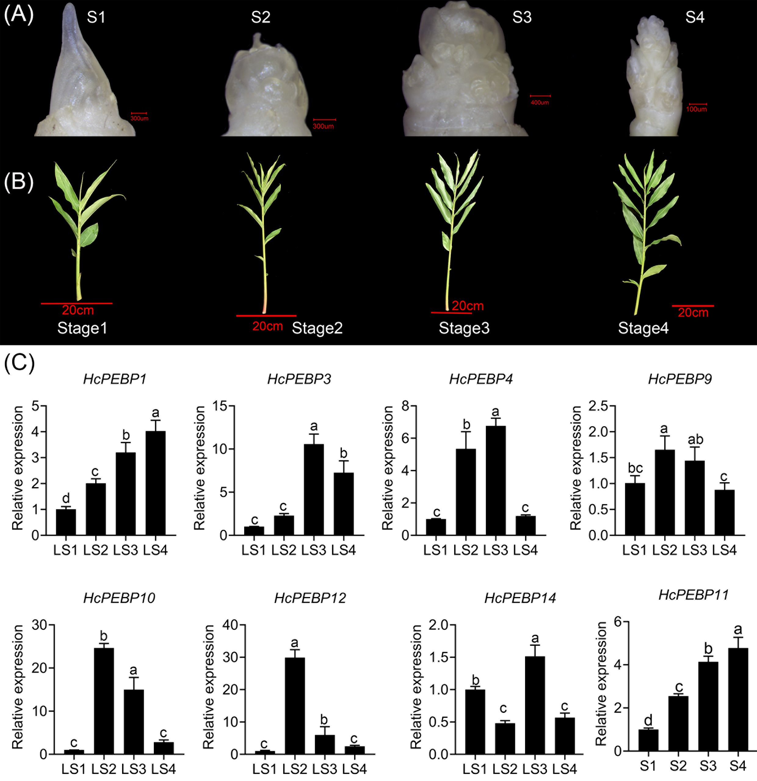
Figure 7. Quantitative expression analysis of eight FT subfamily HcPEBP genes. (A) Developmental stages of apical meristem in this study: S1, leaf bud. S2/3/4, early stage/middle stage/late stage of inflorescence bud differentiation. (B) Plants corresponding to apical meristem stages. (C) Expression levels of seven FT subfamily HcPEBP genes in leaves at four apical meristem stages and HcPEBP11 during apical meristem development. ‘LSs’ denotes leaves at different apical meristem stages.
3.8 Functional verification of heterologous overexpression of HcPEBP11 and subcellular localization of HcPEBP11
Expression analysis of the HcPEBP gene family suggests that HcPEBP11 may influence the timing of flowering. To elucidate HcPEBP11’s regulatory role in flowering, we overexpressed it in tobacco. PCR amplification confirmed the presence of HcPEBP11 in transgenic tobacco strains and the positive control, with consistent band positions, while the negative control displayed no bands (Supplementary Figures S3A, B). This indicates the successful integration of HcPEBP11 into the tobacco genome. Semi-quantitative PCR results revealed HcPEBP11 expression in all transgenic lines, absent in wild type (WT) (Supplementary Figure S3D), confirming its expression in the transgenic strains. Expression analysis showed that HcPEBP11 levels in the leaves and flower buds of transgenic strains L-1 and L-6 were significantly higher than in WT, with transgenic line L-8 exhibiting even higher levels in the flower bud (Figures 8B, C). Transgenic plants flowered earlier than WT, with bolting and flowering times occurring 9–11 and 8–12 days earlier, respectively (Figure 8A; Supplementary Table S8). Bioinformatic prediction and GFP assay validation indicated that HcPEBP11 proteins localize to the cytoplasm (Supplementary Table S4; Supplementary Figure S4). These findings suggest that HcPEBP11 promotes early flowering in tobacco and may function as a cytoplasmic regulator involved in flowering time control.
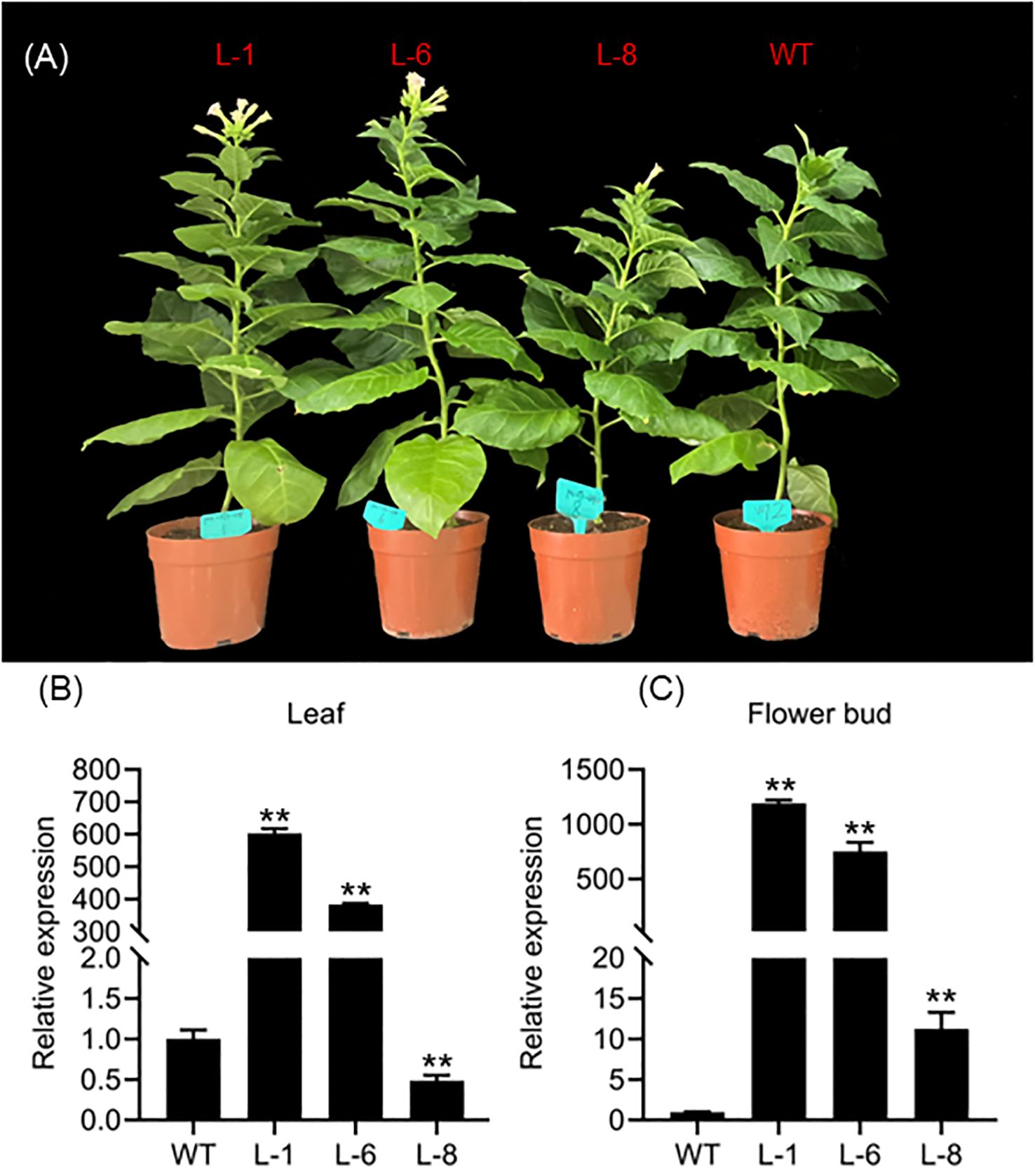
Figure 8. Flowering phenotype and HcPEBP11 expression analysis in transgenic tobacco. (A) Flowering traits comparison between HcPEBP11 transgenic tobacco and wild-type (WT) plants. ‘M’: Marker D2000, ‘WT’: Wild-type (negative control), ‘P’: pOx-PEBP11 plasmid (positive control). L-1, L-6, L-8: HcPEBP11 transgenic lines. (B) HcPEBP11 expression levels in leaves of transgenic tobacco. (C) HcPEBP11 expression levels in flower buds of transgenic tobacco. ** indicates P < 0.01.
4 Discussion
PEBP proteins constitute a class of proteins characterized by a conserved PEBP domain. Extensive research has demonstrated that PEBPs perform a conserved role in regulating plant growth and developmental processes (Chardon and Damerval, 2005; Danilevskaya et al., 2008; He et al., 2022). The PEBP gene family is categorized into three subfamilies—FT-like, TFL1-like, and MFT-like—based on phylogenetic relationships. The FT and TFL1 subfamilies are instrumental in regulating flowering time, morphogenesis, and plant architecture (Xi and Yu, 2009), while the MFT subfamily is involved in seed development, germination, and dormancy (Chen et al., 2018; Footitt et al., 2017). Although the identification and functional characterization of the PEBP gene family have been extensively studied in Arabidopsis and other angiosperms (He et al., 2022; Li et al., 2023; Song et al., 2021; Sun et al., 2023; Venail et al., 2022; Xu et al., 2022; Yang et al., 2023; Zhang et al., 2022; Zhang M, et al., 2021; Zhao et al., 2022; Zhong et al., 2022), genome-wide identification and functional analysis of PEBP genes in H. coronarium remain unexplored.
This study identified 14 PEBP genes in H. coronarium, designated HcPEBP1-14 (Supplementary Table S3). The number of PEBP genes in H. coronarium is fewer than in rice (20) and maize (25), similar to wheat (Triticum urartu) (16), but greater than in Arabidopsis (6). The variation in PEBP gene numbers across species suggests evolutionary differences, with monocotyledons generally having more members than dicotyledons, possibly due to the elimination of non-functional PEBP genes during evolution (Yang et al., 2019). Phylogenetic analysis classified 14 HcPEBP genes into three subfamilies: 1 MFT, 4 TFL1-likes, and 9 FT-likes (Figure 2), consistent with other plant species (Karlgren et al., 2011). Most HcPEBP genes clustered with PEBP genes from rice, reflecting their monocot status and aligning with previous studies (Yang et al., 2019). The number of FT genes and TFL1 genes is influenced by gene duplication, selective pressures, functional specialization, ecological adaptation, evolutionary trade-offs, and species-specific factors. These include gene duplication events, differential selective pressures, functional diversification, and the need for precise regulation of flowering time in response to environmental changes (Hedman et al., 2009; Pin and Nilsson, 2012; Wickland and Hanzawa, 2015; Andrés et al., 2012). In H. coronarium (ginger lily), the FT subfamily has more members than the TFL1 subfamily, which may reflect adaptations to environmental conditions and the need for reproductive success.
Chromosome localization analysis revealed that 9 HcPEBP genes are distributed across six chromosomes, while five are located on genomic scaffolds (Figure 1A). This distribution may reflect challenges in H. coronarium genome assembly. Gene duplication, a common evolutionary mechanism, enhances plant adaptability. Gene duplications include whole-genome and single-gene duplications, with the latter occurring through various mechanisms such as tandem, proximate, diffuse, and separated duplications (Lynch and Conery, 2000; Panchy et al., 2016; Qiao et al., 2019). Intraspecific collinearity analysis identified three pairs of duplicated HcPEBP genes (Figure 1), suggesting functional similarities. Interspecific collinearity analysis with rice, wild banana, and pineapple revealed homologous relationships among PEBP family members, with HcPEBP4 and HcPEBP10 showing collinearity with these species (Figure 1B; Supplementary Table S5), highlighting their potential significance in PEBP gene family evolution and function.
Conserved motif GxHR and DPDxP are critical for their biological function (Chen et al., 2022). Mutations in or near these regions may affect the interaction of FT protein with FD protein by altering the binding with phosphate ions (Si et al., 2018). All HcPEBPs possessed GxHR motif. Except HcPEBP7, all HcPEBPs contained the DPDxP motif, indicating evolutionary conservation. The absence of the DPDxP motif in HcPEBP7 may lead to the loss of its function. The xGxGGR motif in FT-like proteins and the TAARRR motif in TFL1-like proteins are key distinguishing features that determine their functional roles in flowering regulation. Specifically, FT-like proteins contain the xGxGGR motif, which is essential for their role in promoting flowering. TFL1-like proteins possess the TAARRR motif, which is critical for their function in repressing flowering. This difference in motifs underpins the antagonistic roles of FT and TFL1 proteins in controlling flowering time (Karlgren et al., 2011; Hanzawa et al., 2005; Wickland and Hanzawa, 2015). In Arabidopsis, PEBP homologues AtFT and AtTFL1 are key flowering regulators with opposing functions: AtFT promotes, while AtTFL1 represses flowering. Amino acid swaps from Tyr85 and Gln140 in AtFT to His88 and Asp144 in AtTFL1 reverse their regulatory roles in flowering (Ahn et al., 2006; Hanzawa et al., 2005). Tyr85 in HcPEBP4/9/14 (FT-likes) are mutated to His/Phe/His, this may cause them to lose their function in promoting flowering. The fourth exon’s amino acid residues, divided into four segments, determine gene function specificity, with segments II and III (LYN motif) being crucial for AtFT-induced flowering (Ahn et al., 2006). Among the nine FT-like HcPEBP genes, only HcPEBP4, HcPEBP11, and HcPEBP14 possess the LYN motif in segment III (Figure 3). Gene structure analysis indicates that the number of exons in HcPEBP genes ranges from two to five, with most having four exons (Figure 4D), a pattern observed in PEBP gene families of other species (Dong et al., 2020; Sun et al., 2023; Wang et al., 2019).
The core promoter and associated cis-acting elements are vital for gene transcription regulation, serving as specific protein binding sites (Burley and Roeder, 1996; Molina and Grotewold, 2005; Zou et al., 2011). Light, a key environmental stimulus, influences plant growth and development (Song et al., 2016), with the photoperiod pathway significantly affecting flowering by activating FT expression. Elements such as G-box, I-box, and GT1-motif, crucial for light response, are prevalent in the promoters of light-regulated genes (Giuliano et al., 1988; Menkens et al., 1995). The HcPEBP gene family is rich in light-responsive elements, suggesting their role in light-mediated functions. Additionally, HcPEBP genes contribute to plant growth, development, stress response, and hormone regulation. Yet, the specific functions of the PEBP gene family cis-acting elements in H. coronarium have not been studied.
To date, the tissue-specific expression of HcPEBP genes in H. coronarium remains unexamined. qRT-PCR analysis revealed distinct expression patterns (Figures 6, 7). In soybean and cotton, MFT genes, predominantly expressed in seeds, are implicated in oil content and germination (Cai et al., 2023; Yu et al., 2019). In Arabidopsis, TFL1 expression in shoot meristems prolonged vegetative and inflorescence phases when overexpressed (Fernandez-Nohales et al., 2014). Our study found that the MFT-like gene HcPEBP8 and the TFL1-like gene HcPEBP7 are highly expressed in roots and rhizomes, respectively (Figures 6E–H, M, O, P), indicating their potential roles in H. coronarium root development. Previous research has shown that the florigen encoded by the FT gene is produced in leaves and then transferred to the shoot apical meristem, initiating the transition to the reproductive phase (Abe et al., 2005; Susila et al., 2021; Tamaki et al., 2007; Wigge et al., 2005a). In this study, nine PEBP genes belong to the FT subfamily (Figure 2), with seven exhibiting the highest expression in leaves (Figures 6A, C–D, I, J, L, N). The FT-like gene HcPEBP11, highly expressed in the inflorescence bud, may influence flower differentiation and development (Figure 6K). OsFT-L1, expressed at the shoot apical meristem, encodes a florigen-like protein with strong florigenic activity, and its overexpression induces flowering (Giaume et al., 2023). Similarly, HcPEBP11 is highly expressed in the inflorescence bud, aligning with the expression pattern of OsFTL1. Phylogenetic analysis groups HcPEBP11 and OsFTL1 together (Figure 2). Overexpressing HcPEBP11 in tobacco confirms its role in promoting flowering, with transgenic lines flowering 9–11 days earlier than wild types. Overexpression of FT genes has been shown to accelerate floral organ development and flowering, as seen in cassava with the endogenous FT-like gene MeFT1 (Odipio et al., 2020). The potential regulatory mechanisms of HcPEBP11 require further investigation. We hypothesize that:1) The high expression of HcPEBP11 in floral buds suggests that it may directly function in the floral meristem, independent of long-distance transport from leaves to the shoot apex. 2) It may interact with specific transcription factors (Such as FD, MADS-box genes) in the floral meristem to directly activate the expression of downstream flowering-related genes. In this study’s findings clarify the number, bioinformatics features, and expression patterns of the PEBP gene family in H. coronarium, providing a basis for future research on the role of HcPEBP11 in regulating flowering.
5 Conclusion
In H. coronarium, 14 PEBP genes were identified and categorized into three subfamilies:9 FT-likes, 4 TFL1-likes, and 1 MFT. The study analyzed their physicochemical properties, phylogeny, gene structure, conserved motifs, and cis-acting elements. All HcPEBP proteins share the conserved GxHR motif, and all except HcPEBP7 contain the DPDxP motif. Promoter regions of HcPEBP genes are enriched with light-responsive elements. Organ-specific expression analysis via RT-qPCR revealed that HcPEBP1/2/3/4/9/10/12/14 are highly expressed in leaves, and with increased expression during the transition from vegetative to reproductive growth. HcPEBP11 shows the highest expression in inflorescence buds, increasing with bud development. Overexpression of HcPEBP11 in transgenic tobacco resulted in early flowering, suggesting its role in flowering regulation. This study provides a comprehensive overview of the PEBP gene family in H. coronarium and lays the groundwork for further research into the functional and regulatory mechanisms of HcPEBP genes in flowering.
Data availability statement
The original contributions presented in the study are included in the article/Supplementary Material. Further inquiries can be directed to the corresponding author/s.
Author contributions
QW: Writing – original draft, Data curation, Formal Analysis, Visualization, Validation. YZ: Writing – review & editing, Formal Analysis, Software, Data curation, Methodology. FW: Writing – review & editing, Data curation, Software. XL: Writing – review & editing, Resources, Supervision. YY: Writing – review & editing, Resources, Supervision. RY: Writing – review & editing. YF: Funding acquisition, Project administration, Supervision, Writing – review & editing, Conceptualization.
Funding
The author(s) declare that financial support was received for the research and/or publication of this article. This research was funded by the Provincial Rural Revitalization Strategy of Guangdong Province in 2024 (Grant no. 2024-NPY-00-038), Research Projects in Key Areas of Guangdong Province (Grant no. 2020B020220007), and National Agricultural Technology Modernization Pilot County Construction Project (Conghua District).
Conflict of interest
The authors declare that the research was conducted in the absence of any commercial or financial relationships that could be construed as a potential conflict of interest.
Publisher’s note
All claims expressed in this article are solely those of the authors and do not necessarily represent those of their affiliated organizations, or those of the publisher, the editors and the reviewers. Any product that may be evaluated in this article, or claim that may be made by its manufacturer, is not guaranteed or endorsed by the publisher.
Supplementary material
The Supplementary Material for this article can be found online at: https://www.frontiersin.org/articles/10.3389/fpls.2025.1482764/full#supplementary-material
References
Abe, M., Kobayashi, Y., Yamamoto, S., Daimon, Y., Yamaguchi, A., Ikeda, Y., et al. (2005). FD, a bZIP protein mediating signals from the floral pathway integrator FT at the shoot apex. Science 309, 1052–1056. doi: 10.1126/science.1115983
Abelenda, J. A., Bergonzi, S., Oortwijn, M., Sonnewald, S., Du, M. R., Visser, R. G. F., et al. (2019). Source-sink regulation is mediated by interaction of an FT homolog with a SWEET protein in potato. Curr. Biol. 29, 1178–1186.e6. doi: 10.1016/j.cub.2019.02.018
Abelenda, J. A., Prat, S., Navarro, C., and Silva, J. (2009). FT control of floral transition and tuber formation in potato. Comp. Biochem. Physiol. A Mol. Integr. Physiol. 153A, S196. doi: 10.1016/j.cbpa.2009.04.439
Ahn, J. H., Miller, D., Winter, V. J., Banfield, M. J., Lee, J. H., Yoo, S. Y., et al. (2006). A divergent external loop confers antagonistic activity on floral regulators FT and TFL1. EMBO J. 25, 605–614. doi: 10.1038/sj.emboj.7600950
Andrés, F. and Coupland, G. (2012). The genetic basis of flowering responses to seasonal cues. Nat. Rev. Genet. 13, 627–639. doi: 10.1038/nrg3291
Artimo, P., Jonnalagedda, M., Arnold, K., Baratin, D., Csardi, G., de Castro, E., et al. (2012). ExPASy: SIB bioinformatics resource portal. Nucleic Acids Res. 40, W597–W603. doi: 10.1093/nar/gks400
Baez, D., Pino, J. A., and Morales, D. (2011). Floral scent composition in hedychium coronarium J. Koenig analyzed by SPME. J. Essent. Oil Res. 23, 64–67. doi: 10.1080/10412905.2011.9700460
Bailey, T. L., Johnson, J., Grant, C. E., and Noble, W. S. (2015). The MEME suite. Nucleic Acids Res. 43, W39–W49. doi: 10.1093/nar/gkv416
Bi, Z. H., Li, X., Huang, H. S., and Hua, Y. W. (2016). Identification, functional study, and promoter analysis of hbMFT1, a homolog of MFT from rubber tree (Hevea brasiliensis). Int. J. Mol. Sci. 17, 247. doi: 10.3390/ijms17030247
Bradley, D., Ratcliffe, O., Vincent, C., Carpenter, R., and Coen, E. (1997). Inflorescence commitment and architecture in Arabidopsis. Science 275, 80–83. doi: 10.1126/science.275.5296.80
Burley, S. K. and Roeder, R. G. (1996). Biochemistry and structural biology of transcription factor IID (TFIID). Annu. Rev. Biochem. 65, 769–799. doi: 10.1146/annurev.bi.65.070196.004005
Cai, Z., Xian, P., Cheng, Y., Zhong, Y., Yang, Y., Zhou, Q., et al. (2023). MOTHER-OF-FT-AND-TFL1 regulates the seed oil and protein content in soybean. New Phytol. 239, 905–919. doi: 10.1111/nph.18792
Chardon, F. and Damerval, C. (2005). Phylogenomic analysis of the PEBP gene family in cereals. J. Mol. Evol. 61, 579–590. doi: 10.1007/s00239-004-0179-4
Chen, C., Chen, H., Zhang, Y., Thomas, H. R., Frank, M. H., He, Y., et al. (2020). TBtools: an integrative toolkit developed for interactive analyses of big biological data. Mol. Plant 13, 1194–1202. doi: 10.1016/j.molp.2020.06.009
Chen, W., Li, H., Zou, D., Yuan, Y., Li, C., Yang, A., et al. (2022). Expression profile of faFT1 and its ectopic expression in arabidopsis demonstrate its function in the reproductive development of fragaria ananassa. J. Plant Growth Regul. 41, 1687–1698. doi: 10.1007/s00344-021-10409-z
Chen, Y., Xu, X., Chen, X., Chen, Y., Zhang, Z., Xu, X., et al. (2018). Seed-specific gene MOTHER of FT and TFL1 (MFT) involved in embryogenesis, hormones and stress responses in dimocarpus longan lour. Int. J. Mol. Sci. 19, 2403. doi: 10.3390/ijms19082403
Corbesier, L., Vincent, C., Jang, S., Fornara, F., Fan, Q., Searle, I., et al. (2007). FT protein movement contributes to long-distance signaling in floral induction of Arabidopsis. Science 316, 1030–1033. doi: 10.1126/science.1141752
Cordero Otero, R. and Gaillardin, C. (1996). Efficient selection of hygromycin-B-resistant Yarrowia lipolytica transformants. Appl. Microbiol. Biotechnol. 46, 143–148. doi: 10.1007/s002530050796
Danilevskaya, O. N., Meng, X., Hou, Z., Ananiev, E. V., and Simmons, C. R. (2008). A genomic and expression compendium of the expanded PEBP gene family from maize. Plant Physiol. 146, 250–264. doi: 10.1104/pp.107.109538
Dong, L., Lu, Y., and Liu, S. (2020). Genome-wide member identification, phylogeny and expression analysis of PEBP gene family in wheat and its progenitors. PeerJ 8, e10483. doi: 10.7717/peerj.10483
Fan, Z., Gao, Y., Guan, C., Liu, R., Wang, S., and Zhang, Q. (2023). FLOWERING LOCUS T homologue in reblooming bearded iris (Iris spp.) plays a role in accelerating flowering and reblooming. S. Afr. J. Bot. 159, 10–16. doi: 10.1016/j.sajb.2023.04.009
Fernandez-Nohales, P., Domenech, M. J., Martinez de Alba, A. E., Micol, J. L., Ponce, M. R., and Madueno, F. (2014). AGO1 controls arabidopsis inflorescence architecture possibly by regulating TFL1 expression. Ann. Bot. 114, 1471–1481. doi: 10.1093/aob/mcu132
Finn, R. D., Mistry, J., Schuster-Bockler, B., Griffiths-Jones, S., Hollich, V., Lassmann, T., et al. (2006). Pfam: clans, web tools and services. Nucleic Acids Res. 34, D247–D251. doi: 10.1093/nar/gkj149
Footitt, S., Olcer-Footitt, H., Hambidge, A. J., and Finch-Savage, W. E. (2017). A laboratory simulation of Arabidopsis seed dormancy cycling provides new insight into its regulation by clock genes and the dormancy-related genes DOG1, MFT, CIPK23 and PHYA. Plant Cell Environ. 40, 1474–1486. doi: 10.1111/pce.1294023
Foucher, F., Morin, J., Courtiade, J., Cadioux, S., Ellis, N., Banfield, M. J., et al. (2003). DETERMINATE and LATE FLOWERING are two TERMINAL FLOWER1/CENTRORADIALIS homologs that control two distinct phases of flowering initiation and development in Pea. Plant Cell 15, 2742–2754. doi: 10.1105/tpc.015701
Freytes, S. N., Canelo, M., and Cerdán, P. D. (2021). Regulation of flowering time: when and where? Curr. Opin. Plant Biol. 63, 102049. doi: 10.1016/j.pbi.2021.102049
Giaume, F., Bono, G. A., Martignago, D., Miao, Y., Vicentini, G., Toriba, T., et al. (2023). Two florigens and a florigen-like protein form a triple regulatory module at the shoot apical meristem to promote reproductive transitions in rice. Nat. Plants 9, 525–533. doi: 10.1038/s41477-023-01383-3
Giuliano, G., Pichersky, E., Malik, V. S., Timko, M. P., Scolnik, P. A., and Cashmore, A. R. (1988). An evolutionarily conserved protein binding sequence upstream of a plant light-regulated gene. Proc. Natl. Acad. Sci. U.S.A. 85, 7089–7083. doi: 10.1073/pnas.85.19.7089
Goto, K. (2005). Intercellular protein trafficking of TFL1 and FT is necessary for the inflorescence meristem identity and floral transition in Arabidopsis. Plant Cell Physiol. 46, S11.
Hanzawa, Y., Money, T., and Bradley, D. (2005). A single amino acid converts a repressor to an activator of flowering. Proc. Natl. Acad. Sci. U.S.A. 102, 7748–7753. doi: 10.1073/pnas.0500932102
He, J., Gu, L., Tan, Q., Wang, Y., Hui, F., He, X., et al. (2022). Genome-wide analysis and identification of the PEBP genes of Brassica juncea var. Tumida. BMC Genomics 23, 535. doi: 10.1186/s12864-022-08767-3
Hedman, H., Kallman, T., and Lagercrantz, U. (2009). Early evolution of the MFT-like gene family in plants. Plant Mol. Biol. 70, 359–369. doi: 10.1007/s11103-009-9478-x
Horsch, R. B., Klee, H. J., Stachel, S., Winans, S. C., Nester, E. W., Rogers, S. G., et al. (1986). Analysis of Agrobacterium tumefaciens virulence mutants in leaf discs. Proc. Natl. Acad. Sci. U.S.A. 83, 2571–2575. doi: 10.1073/pnas.83.8.2571
Horton, P., Park, K.-J., Obayashi, T., Fujita, N., Harada, H., Adams-Collier, C. J., et al. (2007). WoLF PSORT: protein localization predictor. Nucleic Acids Res. 35, W585–W587. doi: 10.1093/nar/gkm259
Igasaki, T., Watanabe, Y., Nishiguchi, M., and Kotoda, N. (2008). The FLOWERING LOCUS T/TERMINAL FLOWER 1 family in Lombardy poplar. Plant Cell Physiol. 49, 291–300. doi: 10.1093/pcp/pcn010
Iwata, H., Gaston, A., Remay, A., Thouroude, T., Jeauffre, J., Kawamura, K., et al. (2012). The TFL1 homologue KSN is a regulator of continuous flowering in rose and strawberry. Plant J. 69, 116–125. doi: 10.1111/j.1365-313X.2011.04776.x
Kalia, D., Jose-Santhi, J., Kumar, R., and Singh, R. K. (2023). Analysis of PEBP genes in saffron identifies a flowering locus T homologue involved in flowering regulation. J. Plant Growth Regul. 42, 2486–2505. doi: 10.1007/s00344-022-10721-2
Kaneko-Suzuki, M., Kurihara-Ishikawa, R., Okushita-Terakawa, C., Kojima, C., Nagano-Fujiwara, M., Ohki, I., et al. (2018). TFL1-like proteins in rice antagonize rice FT-like protein in inflorescence development by competition for complex formation with 14-3–3 and FD. Plant Cell Physiol. 59, 458–468. doi: 10.1093/pcp/pcy021
Kardailsky, I., Shukla, V. K., Ahn, J. H., Dagenais, N., Christensen, S. K., Nguyen, J. T., et al. (1999). Activation tagging of the floral inducer FT. Science 286, 1962–1965. doi: 10.1126/science.286.5446.1962
Karlgren, A., Gyllenstrand, N., Kallman, T., Sundstrom, J. F., Moore, D., Lascoux, M., et al. (2011). Evolution of the PEBP gene family in plants: functional diversification in seed plant evolution. Plant Physiol. 156, 1967–1977. doi: 10.1104/pp.111.176206
Ke, Y., Abbas, F., Zhou, Y., Yu, R., Yue, Y., Li, X., et al. (2019). Genome-wide analysis and characterization of the Aux/IAA family genes related to floral scent formation in. Hedychium coronarium. Int. J. Mol. Sci. 20, 3235. doi: 10.3390/ijms20133235
Kim, G., Rim, Y., Cho, H., and Hyun, T. K. (2022). Identification and functional characterization of FLOWERING LOCUS T in Platycodon grandiflorus. Plants (Basel) 11, 325. doi: 10.3390/plants11030325
Kokkirala, V. R., Peng, Y., Abbagani, S., Zhu, Z., and Umate, P. (2010). Subcellular localization of proteins of Oryza sativa L. in the model tobacco and tomato plants. Plant Signal. Behav. 5, 1336–1341. doi: 10.4161/psb.5.11.13318
Lazakis, C. M., Coneva, V., and Colasanti, J. (2011). ZCN8 encodes a potential orthologue of Arabidopsis FT florigen that integrates both endogenous and photoperiod flowering signals in maize. J. Exp. Bot. 62, 4833–4842. doi: 10.1093/jxb/err129
Lee, A., Jung, H., Park, H. J., Jo, S. H., Jung, M., Kim, Y. S., et al. (2023). Their C-termini divide Brassica rapa FT-like proteins into FD-interacting and FD-independent proteins that have different effects on the floral transition. Front. Plant Sci. 13–2022. doi: 10.3389/fpls.2022.1091563
Lembinen, S., Cieslak, M., Zhang, T., Mackenzie, K., Elomaa, P., Prusinkiewicz, P., et al. (2023). Diversity of woodland strawberry inflorescences arises from heterochrony regulated by TERMINAL FLOWER 1 and FLOWERING LOCUS T. Plant Cell. 35 (6), 2079–2094. doi: 10.1093/plcell/koad086
Lescot, M., Dehais, P., Thijs, G., Marchal, K., Moreau, Y., Van de Peer, Y., et al. (2002). PlantCARE, a database of plant cis-acting regulatory elements and a portal to tools for in silico analysis of promoter sequences. Nucleic Acids Res. 30, 325–327. doi: 10.1093/nar/30.1.325
Li, F.-F., Song, S.-J., and Zhang, R.-N. (2009). Phosphatidylethanolamine-binding protein (PEBP) in basic and clinical study. Prog. Physiol. 40, 214–218.
Li, Y., Xiao, L., Zhao, Z., Zhao, H., and Du, D. (2023). Identification, evolution and expression analyses of the whole genome-wide PEBP gene family in Brassica napus L. BMC Genom Data 24, 27. doi: 10.1186/s12863-023-01127-4
Lin, T., Chen, Q., Wichenheiser, R. Z., and Song, G.-Q. (2019). Constitutive expression of a blueberry FLOWERING LOCUS T gene hastens petunia plant flowering. Sci. Hortic. 253, 376–381. doi: 10.1016/j.scienta.2019.04.051
Liu, Y. Y., Yang, K. Z., Wei, X. X., and Wang, X. Q. (2016). Revisiting the phosphatidylethanolamine-binding protein (PEBP) gene family reveals cryptic FLOWERING LOCUS T gene homologs in gymnosperms and sheds new light on functional evolution. New Phytol. 212, 730–744. doi: 10.1111/nph.14066
Lynch, M. and Conery, J. S. (2000). The evolutionary fate and consequences of duplicate genes. Science 290, 1151–1155. doi: 10.1126/science.290.5494.1151
Mach, J. (2011). Will the real florigen please stand up? Sorting FT homologs in maize. Plant Cell 23, 843. doi: 10.1105/tpc.111.230310
Menkens, A. E., Schindler, U., and Cashmore, A. R. (1995). The G-box: a ubiquitous regulatory DNA element in plants bound by the GBF family of bZIP proteins. Trends Biochem. Sci. 20, 506–510. doi: 10.1016/s0968-0004(00)89118-5
Molina, C. and Grotewold, E. (2005). Genome wide analysis of Arabidopsis core promoters. BMC Genomics 6, 25. doi: 10.1186/1471-2164-6-25
Morris, J. H., Wu, A., Yamashita, R. A., Marchler-Bauer, A., and Ferrin, T. E. (2015). cddApp: a Cytoscape app for accessing the NCBI conserved domain database. Bioinformatics 31, 134–136. doi: 10.1093/bioinformatics/btu605
Navarro, C., Cruz-Oro, E., and Prat, S. (2015). Conserved function of FLOWERING LOCUS T (FT) homologues as signals for storage organ differentiation. Curr. Opin. Plant Biol. 23, 45–53. doi: 10.1016/j.pbi.2014.10.008
Odipio, J., Getu, B., Chauhan, R. D., Alicai, T., Bart, R., Nusinow, D. A., et al. (2020). Transgenic overexpression of endogenous FLOWERING LOCUS T-like gene MeFT1 produces early flowering in cassava. PloS One 15, e0227199. doi: 10.1371/journal.pone.0227199
Panchy, N., Lehti-Shiu, M., and Shiu, S.-H. (2016). Evolution of gene duplication in plants. Plant Physiol. 171, 2294–2316. doi: 10.1104/pp.16.00523
Patil, H. B., Chaurasia, A. K., Kumar, S., Krishna, B., Subramaniam, V. R., Sane, A. P., et al. (2022). Synchronized flowering in pomegranate, following pruning, is associated with expression of the FLOWERING LOCUS T homolog. PgFT1. Physiol. Plant 174, e13620. doi: 10.1111/ppl.13620
Pin, P. A. and Nilsson, O. (2012). The multifaceted roles of FLOWERING LOCUS T in plant development. Plant Cell Environ. 35, 1742–1755. doi: 10.1111/j.1365-3040.2012.02558.x
Qiao, X., Li, Q., Yin, H., Qi, K., Li, L., Wang, R., et al. (2019). Gene duplication and evolution in recurring polyploidization-diploidization cycles in plants. Genome Biol. 20, 38. doi: 10.1186/s13059-019-1650-2
Rautureau, G., Jouvensal, L., Vovelle, F., Schoentgen, F., Locker, D., and Decoville, M. (2009). Expression and characterization of the PEBP homolog genes from. Drosophila. Arch. Insect Biochem. Physiol. 71, 55–69. doi: 10.1002/arch.20300
Si, Z., Liu, H., Zhu, J., Chen, J., Wang, Q., Fang, L., et al. (2018). Mutation of SELF-PRUNING homologs in cotton promotes short-branching plant architecture. J. Exp. Bot. 69, 2543–2553. doi: 10.1093/jxb/ery093
Song, C., Li, G., Dai, J., and Deng, H. (2021). Genome-wide analysis of PEBP genes in Dendrobium huoshanense: Unveiling the antagonistic functions of FT/TFL1 in flowering time. Front. Genet. 12. doi: 10.3389/fgene.2021.687689
Song, J., Liu, Q., Hu, B., and Wu, W. (2016). Comparative transcriptome profiling of Arabidopsis Col-0 in responses to heat stress under different light conditions. J. Plant Growth Regul. 79, 209–218. doi: 10.1007/s10725-015-0126-y
Soufflet-Freslon, V., Araou, E., Jeauffre, J., Thouroude, T., Chastellier, A., Michel, G., et al. (2021). Diversity and selection of the continuous-flowering gene, RoKSN, in rose. Hortic. Res. 8, 118. doi: 10.1038/s41438-021-00512-3
Sun, Y., Jia, X., Yang, Z., Fu, Q., Yang, H., and Xu, X. (2023). Genome-wide identification of PEBP gene family in Solanum lycopersicum. Int. J. Mol. Sci. 24, 9185. doi: 10.3390/ijms24119185
Susila, H., Juric, S., Liu, L., Gawarecka, K., Chung, K. S., Jin, S., et al. (2021). Florigen sequestration in cellular membranes modulates temperature-responsive flowering. Science 373, 1137–1141. doi: 10.1126/science.abh4054
Tamaki, S., Matsuo, S., Wong, H. L., Yokoi, S., and Shimamoto, K. (2007). Hd3a protein is a mobile flowering signal in rice. Science 316, 1033–1036. doi: 10.1126/science.1141753
Tamura, K., Stecher, G., and Kumar, S. (2021). MEGA11 molecular evolutionary genetics analysis version 11. Mol. Biol. Evol. 38, 3022–3027. doi: 10.1093/molbev/msab120
Thion, L., Vossen, C., Couderc, B., Erard, M., and Clemencon, B. (2002). Detection of genetically modified organisms in food by DNA extraction and PCR amplification. Biochem. Mol. Biol. Educ. 30, 51–55. doi: 10.1002/bmb.2002.494030010037
Tripathi, P., Chandra, A., and Prakash, J. (2022). Changes in physio-biochemical attributes and dry matter accumulation vis a vis analysis of genes during drought and stress recovery at tillering stage of sugarcane. Acta Physiol. Plant 44, 3. doi: 10.1007/s11738-021-03336-9
Vaistij, F. E., Barros-Galvão, T., Cole, A. F., Gilday, A. D., He, Z., Li, Y., et al. (2018). MOTHER OF FT AND TFL1 represses seed germination under far-red light by modulating phytohormone responses in Arabidopsis thaliana. Proc. Natl. Acad. Sci. U.S.A. 115, 8442–8447. doi: 10.1073/pnas.1806460115
Venail, J., da Silva Santos, P. H., Manechini, J. R., Alves, L. C., Scarpari, M., Falcão, T., et al. (2022). Analysis of the PEBP gene family and identification of a novel FLOWERING LOCUS T orthologue in sugarcane. J. Exp. Bot. 73, 2035–2049. doi: 10.1093/jxb/erab539
Walter, C. A., Nasr-Schirf, D., and Luna, V. J. (1989). Identification of transgenic mice carrying the CAT gene with PCR amplification. BioTechniques 7, 1065–1070.
Wang, C., Abbas, F., Zhou, Y., Ke, Y., Li, X., Yue, Y., et al. (2021). Genome-wide identification and expression pattern of SnRK gene family under several hormone treatments and its role in floral scent emission in. Hedychium coronarium. PeerJ 9, e10883. doi: 10.7717/peerj.10883
Wang, M., Tan, Y., Cai, C., and Zhang, B. (2019). Identification and expression analysis of phosphatidylethanolamine-binding protein (PEBP) gene family in cotton. Genomics 111, 1373–1380. doi: 10.1016/j.ygeno.2018.09.009
Wang, Z., Zhou, Z., Liu, Y., Liu, T., Li, Q., Ji, Y., et al. (2015). Functional evolution of phosphatidylethanolamine binding proteins in soybean and. Arabidopsis. Plant Cell 27, 323–336. doi: 10.1105/tpc.114.135103
Wickland, D. P. and Hanzawa, Y. (2015). The FLOWERING LOCUS T/TERMINAL FLOWER 1 gene family: Functional evolution and molecular mechanisms. Mol. Plant 8, 983–997. doi: 10.1016/j.molp.2015.01.007
Wigge, P. A., Kim, M. C., Jaeger, K. E., Busch, W., Schmid, M., Lohmann, J. U., et al. (2005a). Integration of spatial and temporal information during floral induction in. Arabidopsis. Sci. 309, 1056–1059. doi: 10.1126/science.1114358
Xi, W., Liu, C., Hou, X., and Yu, H. (2010). MOTHER OF FT AND TFL1 regulates seed germination through a negative feedback loop modulating ABA signaling in. Arabidopsis. Plant Cell 22, 1733–1748. doi: 10.1105/tpc.109.073072
Xi, W. and Yu, H. (2009). An expanding list: another flowering time gene, FLOWERING LOCUS T, regulates flower development. Plant Signal. Behav. 4, 1142–1144. doi: 10.4161/psb.4.12.9901
Xi, W. and Yu, H. (2010). MOTHER OF FT AND TFL1 regulates seed germination and fertility relevant to the brassinosteroid signaling pathway. Plant Signal. Behav. 5, 1315–1317.
Xu, H., Guo, X., Hao, Y., Lu, G., Li, D., Lu, J., et al. (2022). Genome-wide characterization of PEBP gene family in Perilla frutescens and PfFT1 promotes flowering time in. Arabidopsis thaliana. Front. Plant Sci. 13. doi: 10.3389/fpls.2022.1026696
Yamaguchi, A., Kobayashi, Y., Goto, K., Abe, M., and Araki, T. (2005). TWIN SISTER OF FT (TSF) acts as a floral pathway integrator redundantly with FT. Plant Cell Physiol. 46, 1175–1189. doi: 10.1093/pcp/pci151
Yang, J., Ning, C., Liu, Z., Zheng, C., Mao, Y., Wu, Q., et al. (2023). Genome-wide characterization of PEBP gene family and functional analysis of TERMINAL FLOWER 1 homologs in Macadamia integrifolia. Plants (Basel) 12, 2692. doi: 10.3390/plants12142692
Yang, Z., Chen, L., Kohnen, M. V., Xiong, B., Zhen, X., Liao, J., et al. (2019). Identification and characterization of the PEBP family genes in moso bamboo (Phyllostachys heterocycla). Sci. Rep. 9, 14998. doi: 10.1038/s41598-019-51278-7
Yu, X., Liu, H., Sang, N., Li, Y., Zhang, T., Sun, J., et al. (2019). Identification of cotton MOTHER OF FT AND TFL1 homologs, GhMFT1 and GhMFT2, involved in seed germination. PloS One 14, e0215771. doi: 10.1371/journal.pone.0215771
Zhang, H., Gao, S., Lercher, M. J., Hu, S., and Chen, W.-H. (2012). EvolView, an online tool for visualizing, annotating and managing phylogenetic trees. Nucleic Acids Res. 40, W569–W572. doi: 10.1093/nar/gks576
Zhang, G., Jin, X., Li, X., Zhang, N., Li, S., Si, H., et al. (2022). Genome-wide identification of PEBP gene family members in potato, their phylogenetic relationships, and expression patterns under heat stress. Mol. Biol. Rep. 49, 4683–4697. doi: 10.1007/s11033-022-07318-z
Zhang, M., Li, P., Yan, X., Wang, J., Cheng, T., and Zhang, Q. (2021). Genome-wide characterization of PEBP family genes in nine Rosaceae tree species and their expression analysis in P. mume. BMC Ecol. Evol. 21, 32. doi: 10.1186/s12862-021-01762-4
Zhang, S. H., Singh, M. B., and Bhalla, P. L. (2021). Molecular characterization of a soybean FT homologue, GmFT7. Sci. Rep. 11, 3651. doi: 10.1038/s41598-021-83305-x
Zhang, X., Wang, C., Pang, C., Wei, H., Wang, H., Song, M., et al. (2016). Characterization and functional analysis of PEBP family genes in upland cotton (Gossypium hirsutum L.). PloS One 11, e0161080. doi: 10.1371/journal.pone.0161080
Zhao, J., Chen, H., Ren, D., Tang, H., Qiu, R., Feng, J., et al. (2015). Genetic interactions between diverged alleles of Early heading date 1 (Ehd1) and Heading date 3a (Hd3a)/RICE FLOWERING LOCUS T1 (RFT1) control differential heading and contribute to regional adaptation in rice (Oryza sativa). New Phytol. 208, 936–948. doi: 10.1111/nph.13503
Zhao, C., Zhu, M., Guo, Y., Sun, J., Ma, W., and Wang, X. (2022). Genomic survey of PEBP gene family in rice: identification, phylogenetic analysis, and expression profiles in organs and under abiotic stresses. Plants (Basel) 11, 1576. doi: 10.3390/plants11121576
Zhong, C., Li, Z., Cheng, Y., Zhang, H., Liu, Y., Wang, X., et al. (2022). Comparative genomic and expression analysis insight into evolutionary characteristics of PEBP genes in cultivated peanuts and their roles in floral induction. Int. J. Mol. Sci. 23, 12429. doi: 10.3390/ijms232012429
Zhu, Y., Klasfeld, S., Jeong, C. W., Jin, R., Goto, K., Yamaguchi, N., et al. (2020). TERMINAL FLOWER 1-FD complex target genes and competition with FLOWERING LOCUS T. Nat. Commun. 11, 5118. doi: 10.1038/s41467-020-18782-1
Zou, C., Sun, K., Mackaluso, J. D., Seddon, A. E., Jin, R., Thomashow, M. F., et al. (2011). Cis-regulatory code of stress-responsive transcription in Arabidopsis thaliana. Proc. Natl. Acad. Sci. U.S.A. 108, 14992–14997. doi: 10.1073/pnas.1103202108
Keywords: Hedychium coronarium, PEBP gene family, flowering regulation, HcPEBP11, expression patterns
Citation: Wang Q, Zhou Y, Wang F, Li X, Yu Y, Yu R and Fan Y (2025) Genome-wide identification of PEBP gene family in Hedychium coronarium. Front. Plant Sci. 16:1482764. doi: 10.3389/fpls.2025.1482764
Received: 18 August 2024; Accepted: 19 June 2025;
Published: 09 July 2025.
Edited by:
Christos Noutsos, State University of New York at Old Westbury, United StatesReviewed by:
Zhongqi Fan, Fujian Agriculture and Forestry University, ChinaTeame Gereziher Mehari, Nantong University, China
Copyright © 2025 Wang, Zhou, Wang, Li, Yu, Yu and Fan. This is an open-access article distributed under the terms of the Creative Commons Attribution License (CC BY). The use, distribution or reproduction in other forums is permitted, provided the original author(s) and the copyright owner(s) are credited and that the original publication in this journal is cited, in accordance with accepted academic practice. No use, distribution or reproduction is permitted which does not comply with these terms.
*Correspondence: Yanping Fan, ZmFueWFucGluZ0BzY2F1LmVkdS5jbg==
 Qin Wang
 Yiwei Zhou
 Fang Wang1