- 1College of Biology Pharmacy and Food Engineering, Shangluo University, Shangluo, China
- 2Guangxi Key Laboratory of Plant Functional Phytochemicals and Sustainable Utilization, Guangxi Institute of Botany, Guangxi Zhuang Autonomous Region and Chinese Academy of Sciences, Guilin, China
- 3College of Biology and Environmental Sciences, Jishou University, Xiangxi, China
Long-chain acyl-CoA synthetases (LACSs) are involved in fatty acid metabolism and catabolism by converting free fatty acids to acyl-CoAs. They are essential for initiating β-oxidation of fatty acids and regulating lipid biosynthesis in plant growth and development, as well as in plant adaptation to various environmental stresses, including waterlogging stress. However, systematic identification and functional characterization of the LACS gene family have not been comprehensively studied in the waterlogging-tolerant kiwifruit germplasm Actinidia valvata Dunn. In this study, 22 AvLACS genes were identified within the A. valvata genome. The AvLACS genes were subsequently divided into five clusters on the basis of their phylogenetic relationships, and similar subcellular localizations, exon-intron structures, motif compositions, and protein tertiary structures were found within each cluster. Collinearity analysis identified 22 duplicated gene pairs in A. valvata, and these pairs have undergone purifying selection during evolution. Cis-acting element analysis revealed numerous hormone-responsive and stress-responsive elements in the promoter regions of the AvLACS genes. The expression levels of the AvLACS genes under waterlogging stress were determined using quantitative real-time PCR (qRT-PCR), and the results showed that the expression of the AvLACS1.1a/b, AvLACS1.2a, and AvLACS6a/b was significantly upregulated under waterlogging stress. Notably, AvLACS1.1a/b and AvLACS1.2a primarily facilitate short-term regulation of wax and triacylglycerol (TAG) synthesis, whereas AvLACS6a/b mediate TAG degradation through fatty acid β-oxidation during prolonged waterlogging. Transcriptome data revealed coordinated transcriptional regulation of TAG degradation pathway genes, which was supported by biochemical lipid profiling showing dynamic alterations in TAG content and degree of unsaturation correlated with waterlogging duration. These integrated molecular and biochemical data provide mechanistic insights highlighting distinct and coordinated roles of AvLACSs in lipid metabolic remodeling under waterlogging stress. These findings advance our understanding of the molecular mechanisms underlying waterlogging tolerance, and provide molecular targets and a theoretical basis for breeding waterlogging-tolerant kiwifruit and other crops.
1 Introduction
Lipids are structurally diverse biomolecules found in living organisms, performing essential biological functions, including energy storage, membrane formation, and signal transduction. As fundamental building blocks of lipids, fatty acids (FAs) are crucial for the formation of membrane glycerolipids, phospholipids, sphingolipids, and storage triacylglycerols (TAGs), and are indispensable for the synthesis of cell surface waxes, cutin, and suberin (Zhao et al., 2021). These molecules are vital for maintaining membrane integrity, supplying energy through metabolic pathways such as β-oxidation, and forming protective barriers against environmental stresses. In the metabolism of long-chain fatty acid-derived lipids, free FAs are activated by long-chain acyl-CoA synthetase (LACS) in a two-step enzymatic reaction to form acyl-CoA. First, free FAs react with ATP to produce an acyl-AMP intermediate, releasing pyrophosphate as a byproduct. In the second step, the enzyme-bound acyl-AMP intermediate couples with the thiol group of CoA to form acyl-CoA, releasing AMP (Groot et al., 1976; Grevengoed et al., 2014). Subsequently, the acyl-CoA produced by LACS participates in various metabolic pathways, including the synthesis of phospholipids, TAGs, cutin, cuticular wax, tryphine, and starch, as well as FA degradation via β-oxidation (Zhao et al., 2021; Li et al., 2016). Therefore, as key enzymes in FA metabolism, LACSs regulate lipid biosynthesis and energy homeostasis, playing critical roles in plant growth and adaptation to environmental stresses.
LACSs are a widespread family of enzymes crucial for lipid metabolism and are highly conserved across multicellular organisms. Since the identification of nine LACS family members in Arabidopsis (Shockey et al., 2002), numerous LACS members have been characterized in various species, including economically crops such as rice (Oryza sativa L.) (Kitajima-Koga et al., 2020), maize (Zea mays L.) (Wang et al., 2023; Yan et al., 2024) and wheat (Triticum aestivum L.) (Liu et al., 2022), oilseed crops such as soybean [Glycine max (L.) Merr.] (Wang et al., 2022), cotton (Gossypium spp.) (Zhong et al., 2023), rapeseed (Brassica napus L.) (Xiao et al., 2019), pecan [Carya illinoinensis (Wangenh.) K. Koch] (Ma et al., 2023), sunflower (Helianthus annuus L.) (Aznar-Moreno et al., 2014) and African oil palm (Elaeis guineensis Jacq.) (Wang et al., 2021), fruit crops such as apple [Malus domestica (Suckow) Borkh.] (Zhang et al., 2018) and ‘Dangshansuli’ pear (Pyrus bretschneideri rehd.) (Wang et al., 2025) and vegetable crop like tomato (Solanum lycopersicum L.) (Wu et al., 2024). In plants, although there are differences in the substrate specificity, subcellular distribution, and expression patterns of different LACS members, all LACS members possess a highly conserved AMP-binding domain, which is essential for the activation of long-chain fatty acids (LCFAs) to their respective long-chain-acyl-CoA thioesters (Zhao et al., 2021; Lewandowska et al., 2020). Additionally, a conserved acyl CoA synthetase (ACS) signature motif plays important roles in this activation process, facilitating the conversion of LCFAs to acyl-CoA thioesters (Ma et al., 2023; Zhou et al., 2024).
LACS enzymes are primarily localized in the endoplasmic reticulum (ER), peroxisome, plastid, and plasma membrane (PM), and the subcellular localization of LACS determines the subsequent fatty acid metabolic pathway it participates in. For example, LACSs localized to the plastid envelope, such as AtLACS9 in Arabidopsis, are involved in the activation of de novo synthesized LCFAs and facilitate lipid trafficking between plastids and the ER (Kitajima-Koga et al., 2020; Jessen et al., 2014). Peroxisome-localized LACSs, such as AtLACS6 and AtLACS7, play key roles in the degradation of TAGs to provide energy and maintain lipid homeostasis via FA β-oxidation (Yu et al., 2014; Wang et al., 2019). The majority of AtLACSs, including AtLACS1, AtLACS2, AtLACS4 and AtLACS8, are located in the ER, where FAs are metabolized to synthesize membrane lipids, cuticular lipids, and TAG (Zhao et al., 2021; Shockey et al., 2002; Kunst and Samuels, 2003; Fich et al., 2016). These generated lipids are also active components of plant adaptations to biotic and abiotic stresses. Membrane lipid remodeling is a key strategy for plants to adapt to environmental stresses (Zhang et al., 2016; Wang et al., 2014; Henschel et al., 2024). Additionally, the sequestration of acyl groups into TAGs stored in lipid droplets helps maintain cellular homeostasis under stress conditions (Wang et al., 2019; Yu et al., 2021). The cuticle on the cell surface, which is composed of cuticular wax and cutin, serves as a critical barrier against environmental stresses, including water stress (Bernard and Joubès, 2013). Thus, these LACS-mediated FA metabolic processes and their lipid products are crucial for plant growth and development, and are actively induced under various environmental stresses.
LACSs are involved in plant responses to various biotic factors, such as pathogen attacks, as well as abiotic stresses including drought, salinity, and hypoxia. In Arabidopsis, a reduction in wax and cutin, which function as a surface barrier against pathogens and water deprivation, was observed in the lacs1 and lacs2 mutant plants. As a result, the mutants exhibited increased susceptibility to the bacterial pathogen Pseudomonas syringae (Bernard and Joubès, 2013; Tang et al., 2007) as well as to drought stress (Weng et al., 2010). The expression of MdLACS1 was upregulated under drought stress, salt stress, and abscisic acid (ABA) treatment in apple (Zhang et al., 2018). Furthermore, MdLACS2 and MdLACS4 also contribute to enhanced drought and salt stress resistance in transgenic plants (Zhang et al., 2020a, Zhang et al., 2020b). Similarly, GmLACS2–3 in soybean (Glycine max L.), PoLACS4 in tree peony (Paeonia ostii ‘Feng Dan Bai’), and CiLACS9 and CiLACS9–1 in pecan (Carya illinoinensis L.) were highly expressed under drought and salt stresses (Ma et al., 2023; Zhang et al., 2022; Ayaz et al., 2021). Temperature fluctuations also affect the expression of LACS. For example, the expression of GmLACS9, GmLACS15, and GmLACS17 in soybean was signifiantly upregulated under low temperature (Wang et al., 2022), whereas ZmLACS9 was significantly induced by heat stress (Wang et al., 2023). Furthermore, hypoxia stress, induced by waterlogging, increased the expression of LACS6 in cucumber (Cucumis sativus L.) (Kęska et al., 2021) and four AvLACSs in kiwifruit (Actinidia valvata Dunn) (Li et al., 2022), suggesting that LACS plays a vital role in the plant response to waterlogging stress. Additionally, the response mechanism of AtLACS2 to hypoxia stress has been thoroughly investigated. AtLACS2 in Arabidopsis contributes to submergence tolerance by modulating the translocation of the ETHYLENE-RESPONSE FACTOR (ERF-VII) transcription factor from the membrane to the nucleus (Zhou et al., 2020) and by modulating cuticle permeability in plant cells (Xie et al., 2020).
Kiwifruit (Actinidia spp.) is a significant perennial fruit crop in the genus Actinidia and comprises more than 50 species (Huang, 2016). Kiwifruit is highly valued because of its high vitamin C content and essential minerals, which are beneficial for human health. However, most kiwifruit cultivars are highly sensitive to waterlogging stress due to their fleshy roots, resulting in reduced fruit yield under waterlogged conditions. Previous research has focused on screening for waterlogging-tolerant kiwifruit plants, and A. valvata has been identified as a highly waterlogging-tolerant kiwifruit variety and is now widely used as a rootstock in kiwifruit production (Li et al., 2021; Bai et al., 2022). To investigate the adaptive strategies employed by A. valvata under waterlogging stress, Li et al. (2022) conducted a transcriptome analysis on the roots of KR5 (A. valvata, a tolerant genotype) after 0, 12, 24, and 72 h of waterlogging stress, and the results showed that four differentially expressed genes (DEGs) encoding LACS were significantly upregulated under waterlogging stress. However, the systematic identification and characterization of the kiwifruit LACS gene family remain unstudied. In this study, genome-wide identification and investigation of the LACS gene family were conducted in A. valvata. The chromosomal locations, phylogenetic relationships, conserved domains, gene structures, and cis-acting elements were analyzed, along with gene synteny analysis and the expression patterns of AvLACSs at different fruit development stages and under salt stress. Additionally, the transcriptional level of AvLACSs under waterlogging stress was quantified by qRT-PCR analysis, and the functional mechanism of the upregulated genes was analyzed via the transcriptome analysis. Further lipid profiling was performed to validate the regulatory role of AvLACSs in wax synthesis and TAG metabolism under waterlogging stress. Our findings provide a scientific foundation for the functional validation of LACS genes in waterlogging stress adaptation, and offer valuable insights into breeding waterlogging-tolerant kiwifruit and variety identification.
2 Materials and methods
2.1 Identification of LACS Genes in A. valvata
We sequenced the entire genome of the A. valvata (BGI Inc., Shenzhen, China) and the genome sequencing data have been deposited at Sequence Read Archive database in NCBI under accession PRJNA1169670. A systematic annotation of genomic features, including protein-coding genes, was performed (unpublished), and the corresponding protein sequences were subsequently obtained. To identify the LACS gene family members from the A. valvata genome, we downloaded the HMM file of the AMP-binding domain (PF00501) using the Pfam database (https://pfam.xfam.org/) (accessed on 29 March 2024) (Mistry et al., 2020). The AMP domain was used to search the A. valvata protein database with HMMER 3.0 (https://www.ebi.ac.uk/Tools/hmmer/) (accessed on 29 March 2024) (Potter et al., 2018) with a threshold of E-value ≤ 1e−5 and other parameters set to defaults. Then, nine LACS protein sequences of A. thaliana were downloaded from The Arabidopsis Information Resource (TAIR, https://www.arabidopsis.org/) (accessed on 29 March 2024) (Lamesch et al., 2011) and used for the BLASTp analysis with the kiwifruit protein sequences with an E-value threshold of ≤1e−5. The merged results of these two methods were used to conduct a phylogenetic analysis with the 9 AtLACS proteins, 22 putative AvLACS proteins were finally identified. The putative AvLACS protein sequences were submitted to the NCBI-CDD website (https://www.ncbi.nlm.nih.gov/cdd) (accessed on 8 May 2024) and the SMART databases (http://smart.embl-heidelberg.de/) (accessed on 8 May 2024) to further confirm the existence of the AMP binding domain. These AvLACS members were named according to their affinities to AtLACS from A. thaliana.
2.2 Chromosomal location and physicochemical properties of the AvLACS genes
The chromosomal location of the AvLACSs was achieved from a gff3 file of the genome and mapped to different chromosomes using MG2C v2.1 online software (http://mg2c.iask.in/mg2c_v2.1/) (accessed on 26 May 2024) (Chao et al., 2021). The physicochemical properties of the AvLACS proteins, namely, the amino acid (A.A) length, molecular weight (M.W), isoelectric point (pI), instability index, the aliphatic index and grand average of hydropathicity (GRAVY) were evaluated by using the ExPASy website (https://www.expasy.org/) (accessed on 8 May 2024). Meanwhile, the subcellular localization of the AvLACS proteins was predicted using the PredictProtein web server(https://predictprotein.org/) (accessed on 9 May 2024) (Bernhofer et al., 2021).
2.3 Phylogenetic analysis, conserved motifs and gene structure analysis of the AvLACS genes
Multiple sequence alignment of 22 AvLACS proteins and 9 AtLACS proteins was performed using the Clustal W method in DNAMAN 8.0 software (Lynnon Biosoft, Vaudreuil, QC, Canada) with default parameters. To construct the phylogenetic tree, the LACS members from A. thaliana, A. valvata, Z. mays (Wang et al., 2023), and O. sativa (Ma et al., 2023) were renamed in accordance with their affinities to Arabidopsis (Supplementary Table 1). The evolutionary relationship of LACS family members was established by constructing a phylogenetic tree using MEGA 7.0 software (Mega Limited, Auckland, New Zealand) with the neighbor-joining (NJ) method following the default settings. A bootstrap analysis with 1000 replicates was performed, and the resulting tree were visualized via the online web tool iTOL (https://itol.embl.de/) (accessed on 16 May 2024) (Letunic and Bork, 2021). Gene structure information regarding the intron-exon distribution of AvLACS genes was retrieved from the General Feature Format (GFF) file. The conserved motifs of AvLACS protein sequences were predicted using the MEME (MEME 5.5.4) online tool (https://memesuite.org/meme/tools/meme) (accessed on 8 May 2024) (Bailey et al., 2015) and the maximum number of motifs was 10. The phylogenetic tree, gene structures and conserved motifs of AvLACS were visualized using the Gene Structure View (Advanced) within TBtools II software (Chen et al., 2023).
2.4 Conserved domains, secondary structure and 3D modeling of AvLACS proteins
The conserved domains in all AvLACS proteins were analyzed using the SMART database, and the domain structures were plotted using IBS 1.0.3 software (Liu et al., 2015). The secondary structures of the AvLACS proteins were predicted using SOPMA (https://npsa-prabi.ibcp.fr/cgi-bin/npsa_automat.pl?page=npsa_sopma.html) (accessed on 13 May 2024) (Geourjon and Deléage, 1995). Furthermore, we constructed 3D models of AvLACS proteins based on protein homology modeling using the online tool Phyre2 (http://www.sbg.bio.ic.ac.uk/phyre2/html/page.cgi?id=index) (accessed on 12 May 2024) (Kelley et al., 2015) with default parameters.
2.5 Gene duplication, collinearity analysis and Ka/Ks values calculation of AvLACS genes
Gene replication of AvLACS genes were identified by TBtools using the MCScanX toolkit package with default parameters (Chen et al., 2023). The collinearity of the LACS family members within A. valvata genome as well as between A. valvata and A. thaliana and A. chinensis, was determined using TBtools software (Chen et al., 2023). The Ka (non-synonymous) and Ks (synonymous) substitution rates and the Ka/Ks ratio of the duplicated AvLACS genes were calculated using TBtools software (Chen et al., 2023). Ka/Ks < 1 suggests the presence of purifying selection, whereas Ka/Ks > 1 implies positive selection and Ka/Ks = 1 is characteristic of neutral selection (Zhang, 2022). The divergence time (T, MYA; million years ago) was calculated by the following formula; T = Ks/r, (r = 6.78 × 10⁻⁹) (Luo et al., 2023; Tu et al., 2023).
2.6 Cis-regulatory element analysis of AvLACS genes
To predict the putative cis-regulatory elements in promoter regions of AvLACS genes, the 2000 bp upstream sequences of all the AvLACS genes were extracted from the genomic DNA sequences. The promoter sequences of each gene were submitted to PlantCARE (https://bioinformatics.psb.ugent.be/webtools/plantcare/html/) (accessed on 23 May 2024) (Lescot et al., 2002), and the distribution and the numbers of cis-regulatory elements were visualized in TBtools (Chen et al., 2023).
2.7 Gene expression analysis of AvLACS genes based on RNA-seq data
To investigate the expression pattern of AvLACS genes during plant development and under abiotic stresses, the publicly available A. valvata RNA-seq datasets deposited in the NCBI Sequence Read Archive (SRA) database were searched and downloaded (PRJNA984935 and PRJNA726156) (accessed on 1 November 2023). For plant development, the fruit flesh excluding seeds were collected at four different stages of fruit development (Green stage, Breaker stage, Color change stage and Mature ripe stage) from A. valvata (Bhargava et al., 2023). For salt stress, plants of A. valvata were subjected to 0.4% NaCl per net weight of the growing medium in the pot, and the roots of A. valvata were collected at 0 h (control), 12 h, 24 h and 72 h after the salt treatment (Abid et al., 2022). The RNA-seq data were converted intoFASTQ format using the Convert SRA to Fastq Files within TBtools II software (Chen et al., 2023). Subsequently, the transcriptional abundance of all transcripts was quantified using Kallisto Super GUI Wrapper of TBtools with default parameters through uploading the FASTQ files. The resulting expression values were normalized as Transcripts Per Million (TPM) and the normalized expression data of AvLACS genes were extracted using the Table Row Extractor and Filter tools within TBtools (Chen et al., 2023; Qiao et al., 2025). The Heatmap of TBtools was used to normalize data and draw heatmaps (Chen et al., 2023).
2.8 Plant materials and waterlogging treatments
Two-year-old A. valvata seedlings were cultivated in the greenhouse at Guangxi Key Laboratory of Plant Functional Phytochemicals and Sustainable Utilization and grown under normal conditions for three months. For the waterlogging experiment, each three potted seedings were placed in a plastic container (61 × 48 × 36 cm) filled with tap water. The seedlings were submerged to a final depth range of 3~5 cm beneath the water surface for 7 days under normal light/dark conditions (Zhang et al., 2024). Fresh root samples were collected at 0 h, 6 h, 24 h (1 d), 120 h and 7 d after the waterlogging treatment and immediately frozen in liquid nitrogen for further analyses. For each sample, a set of three replicates was established, with each replicate comprising three individual seedlings.
2.9 RNA extraction and qRT-PCR analysis
The total RNA of the root samples was extracted using the RNAprep Pure Plant Kit (TIANGEN, Beijing, China) according to the manufacturer’s instruction. The RNA integrity was checked using 1% agarose gel electrophoresis, and the RNA purity and concentration were measured using a spectrophotometer (UV-2550; Shimadzu, Co., Kyoto, Japan). The FastKing RT Kit With gDNase (TIANGEN, Beijing, China) was used to reverse the RNA into cDNA. Real-time PCRs were performed using the SuperReal PreMix Plus (SYBR Green) (TIANGEN, Beijing, China) following the manufacturer’s instructions. The relative expression levels were calculated using the 2−ΔΔCT method (Livak and Schmittgen, 2001) and three duplicates were performed. The primer pairs used for the qRT-PCR are provided in Supplementary Table 2.
2.10 Transcriptomic datasets analysis
The root samples were sent to Biomarker Technologies Co., Ltd. (Beijing, China) and performed transcriptome sequencing with three biological replicates. Total RNA was extracted from these samples using the RNAprep Pure Plant Kit (Tiangen, Beijing, China). RNA concentration and purity was measured using NanoDrop 2000 (Thermo Fisher Scientific, Wilmington, DE). RNA integrity was assessed using the RNA Nano 6000 Assay Kit of the Agilent Bioanalyzer 2100 system (Agilent Technologies, CA, USA). A total amount of 1 μg RNA per sample was used for library preparation with the Hieff NGS Ultima Dual-mode mRNA Library Prep Kit for Illumina (Yeasen Biotechnology, Co., Ltd. Shanghai, China) following the manufacturer’s instructions. The libraries were sequenced on an Illumina NovaSeq platform to generate 150 bp paired-end reads. Gene annotation was performed by comparing the DEGs with COG (Cluster of Orthologous Group of Proteins), GO (Gene Ontology), KEGG (Kyoto Encyclopedia of Genes) and Genomes, Swiss-prot, and NR (Non-redundant) databases. KEGG pathway enrichment analysis was conducted on the samples, and the DEGs were classified into regulatory pathways based on the annotation results. Based on RNA-seq data of A. valvata (Unpublished), the FPKM values mapped reads were used to calculate the expression levels of genes related to FAs metabolism under normal and waterlogging conditions, and the results were visualized by heat maps using TBtools (Chen et al., 2023).
2.11 Lipid analysis
For lipid analysis, the three biological samples were sent to BioMarker Technologies (Beijing, China). Root samples (100 mg) of 0 d, 1 d and 7 d after the waterlogging treatment were dissolved with 1.2 ml of 70% methanol, after vortexed for six times and then the samples were placed in the refrigerator at 4 °C overnight. After centrifugation, the supernatant was filtered and stored in an injection bottle for UPLC-MS/MS analysis using an Agilent SB-C18 column with a specific elution gradient of 0.1% formic acid aqueous solution and 0.1% formic acid acetonitrile. LC-MS/MS was performed on a triple quadrupole-linear ion trap mass spectrometer (QTRAP)in both positive and negative modes, with specific source parameters and mass calibration. The double bond index (DBI) was calculated from the mol % values derived from the LC-MS/MS data, according to the formula: DBI = [∑(number of double bonds × mol % of fatty acid)]/100 (Falcone et al., 2004).
2.12 Statistical analysis
Statistical analysis was performed using one-way ANOVA and Tukey’s test in SPSS (ANCOVA; SPSS26, SPSS Inc., Chicago, IL, USA), and * P < 0.05 and ** P < 0.01 indicated that the differences were significant and extremely significant, respectively. The data are presented as the mean ± standard error (± SE) of three biological replicates, with each treatment was repeated three times.
3 Results
3.1 Identification of LACS Gene Family Members in A. valvata
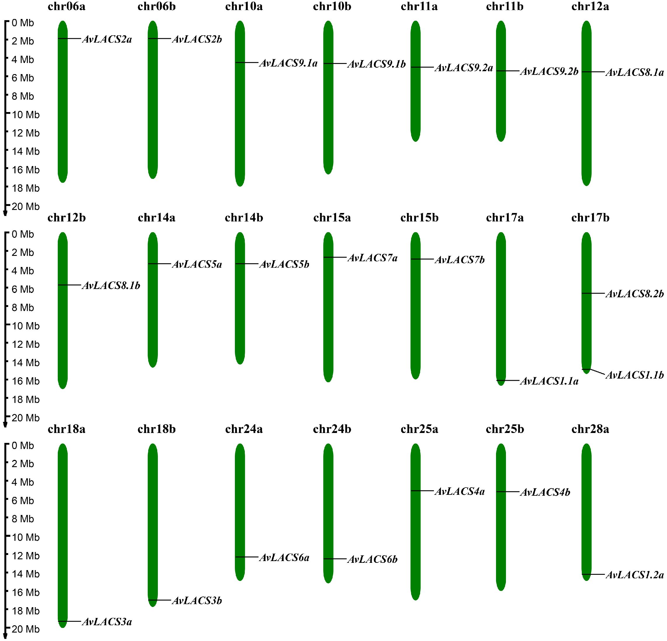
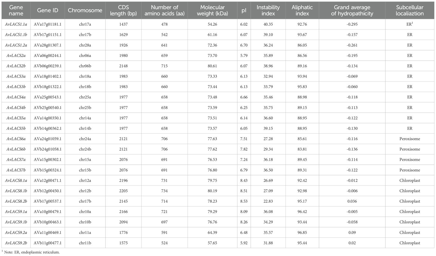
To identify LACS gene family members in A. valvata, we performed BLASTP searches and Hidden Markov Model (HMM), and the results were merged and filtered through phylogenetic analysis. NCBI-CDD and SMART domain searches were employed to verify the presence of AMP-binding domains in putative AvLACS candidate genes. Finally, 22 AvLACS family members were identified from the genome of A. valvata, and they were renamed according to their similarity to Arabidopsis (Supplementary Figure 1). All of the identified AvLACS genes were found to be located on 21 chromosomes, with 11 LACS genes on each subgenome a and subgenome b (Figure 1; Supplementary Table 3). The CDS lengths of the AvLACS genes ranged from 1437 to 2205 bp, with protein lengths varying between 478 and 734 amino acids (Table 1). Their molecular weights ranged from 54.26 to 80.61 kDa, and their pI values ranged from 5.79 to 8.53. The instability index of these AvLACS proteins ranged from 22.83 to 40.35, and their aliphatic index ranged from 83.81 to 96.85, and their grand average of hydropathicity (GRAVY) ranged from -0.295 to 0.09. Subcellular localization predictions indicated that 11 AvLACS proteins were located in the endoplasmic reticulum, 4 were located in the peroxisome, and 7 were located in the chloroplast (Table 1).
3.2 Phylogenetic analysis and characterization of conserved domains of LACSs in A. valvata
To explore the phylogenetic relationships and functional diversity of the AvLACSs, the LACS protein sequences from A. valvata, A. thaliana, Z. mays, and O. sativa were subjected to multiple sequence alignment, and a phylogenetic tree of these LACS proteins was constructed. The LACSs were divided into five clusters (Figure 2). Cluster I included AvLACS1.1a/b and AvLACS1.2b, cluster II contained the fewest genes, which were AvLACS2a/b; cluster III consisted of AvLACS3a/b, AvLACS4a/b, and AvLACS5a/b; cluster IV consisted of AvLACS6a/b, and AvLACS7a/b; and cluster V contained the highest number of genes, which were AvLACS8.1a/b, AvLACS8.2b, AvLACS9.1a/b, and AvLACS9.2a/b. The same AvLACS genes located on different subgenomes were phylogenetically closer, whereas LACSs from monocots on the evolutionary branch were relatively more distant from AvLACSs. The subcellular localization of LACS proteins clustered together on the phylogenetic tree is also consistent.
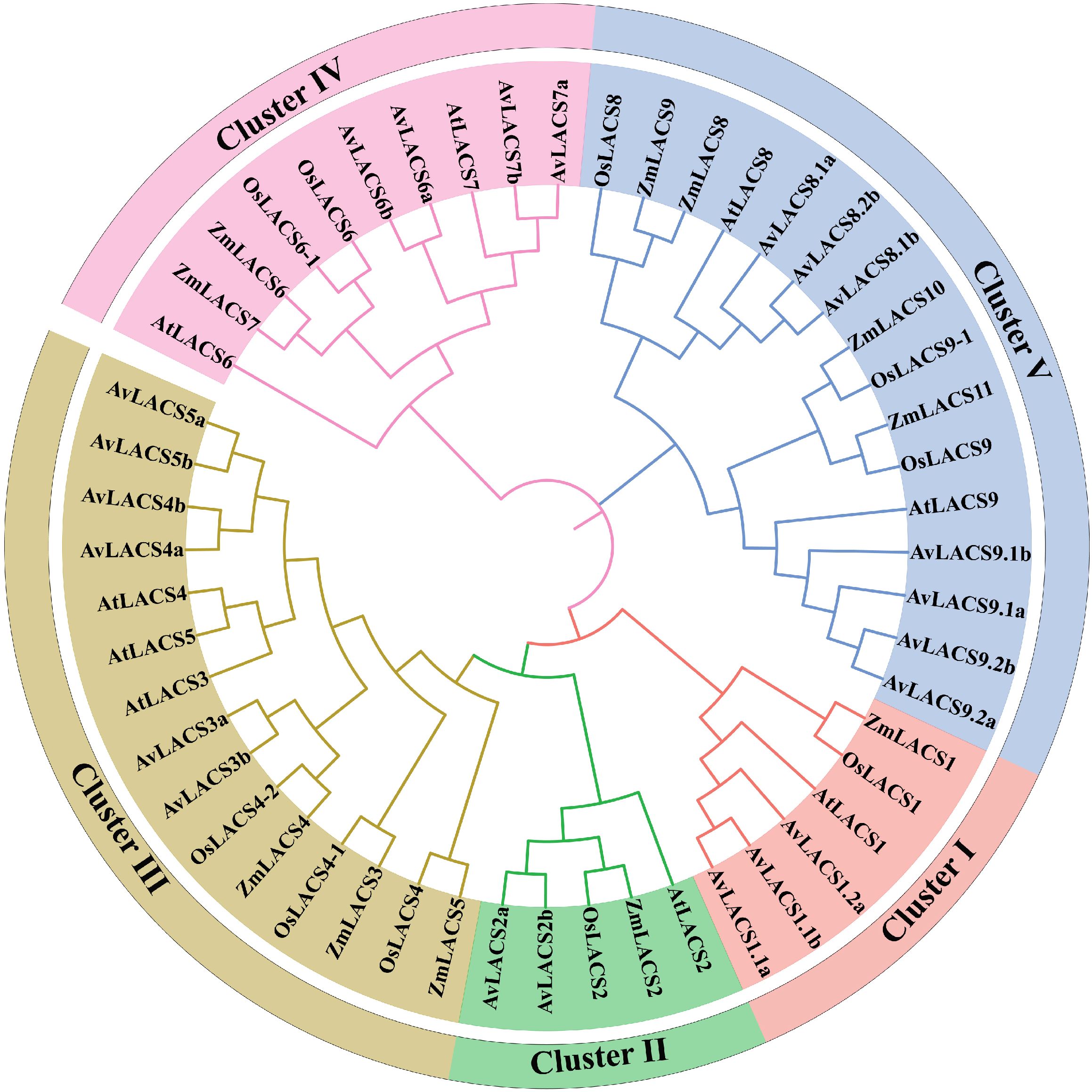
Figure 2. Phylogenetic analysis of the LACS proteins from A. valvata (Av), A. thaliana (At), O. sativa (Os), and Z. mays (Zm). The protein sequences were aligned with the Clustal W program using MEGA 7.0 and the phylogenetic tree was constructed using the neighbor-joining method with 1000 bootstrap replicates.
The conserved domains of the AvLACSs were analyzed using the SMART database and visualized with IBS software (version 1.0.3). As shown in Supplementary Figure 2, most AvLACSs contained an AMP-binding domain (PF00501). Compared with LACSs from other clusters, the AvLACSs from cluster II, namely, AvLACS2a/b, possessed an additional AMP-binding C domain. Similar domain positions and distributions were found within the same cluster.
3.3 protein sequence alignment and protein structure analysis of AvLACSs
To identify the conserved sequences in the AvLACSs, multiple sequence alignment of proteins was performed using DNAMAN software (Supplementary Figure 3). According to the alignment, each AvLACS protein contained two conserved motif domains (AMP-binding domain signature and ACS signature motif). In addition, the amino acid sequences of the conserved motif domain in AvLACS located within the same cluster were highly similar.
To further understand the AvLACS protein structure, the secondary and tertiary structures of AvLACS were predicted. The protein secondary structure of the AvLACS family members was predominantly composed of α-helices (35.70%-42.23%) and random coils (33.89%-38.00%), followed by extended strands (16.21%-20.64%), whereas β-turns (6.11%-8.64%) accounted for the smallest proportion (Supplementary Figure 4; Supplementary Table 4). Additionally, three-dimensional models of all AvLACSs proteins were built. As shown in Figure 3, similar 3D structures were found in the AvLACSs from the same cluster, and the composition and position of the secondary structures of the AvLACS proteins could be clearly observed.
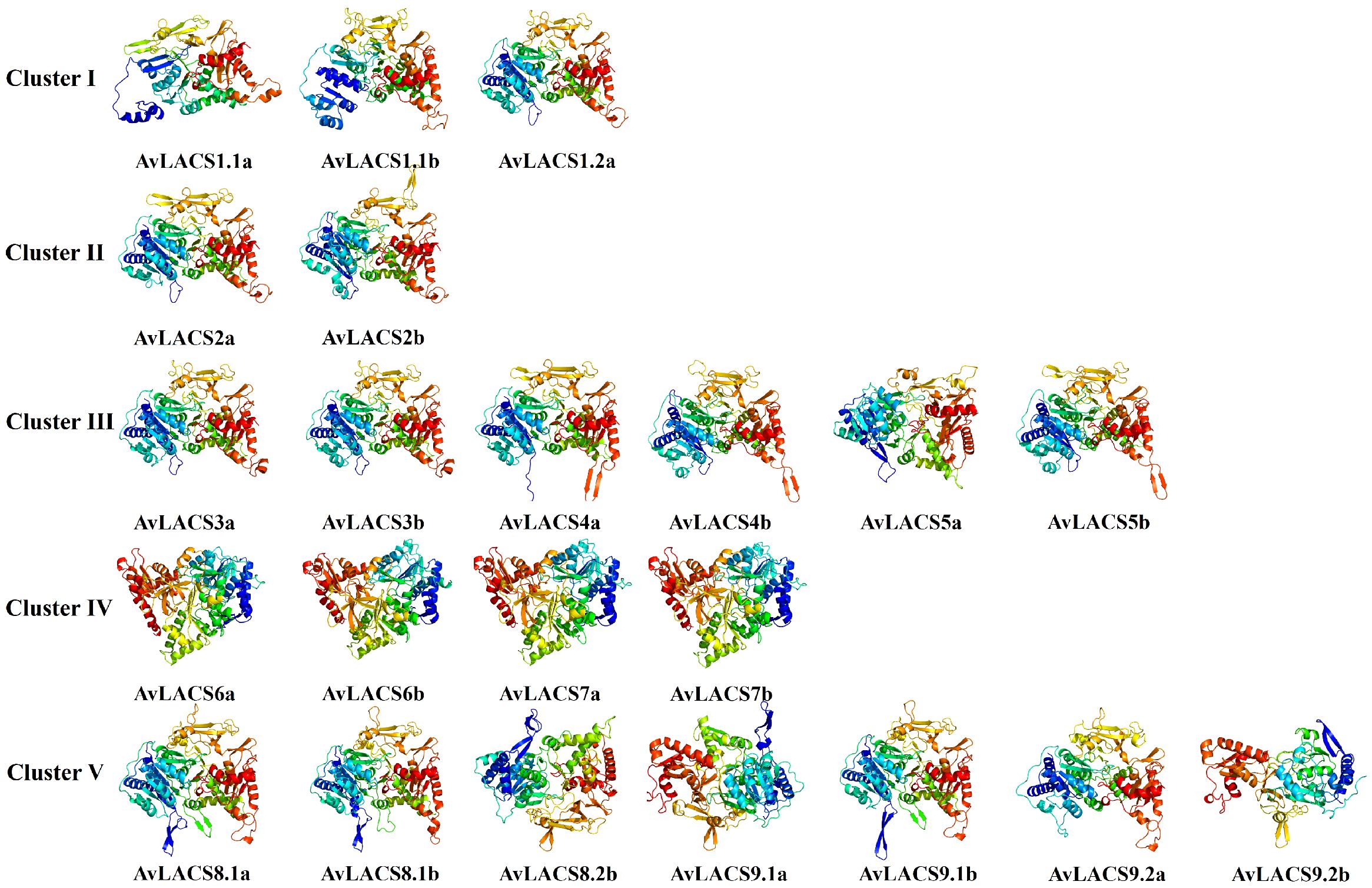
Figure 3. Three-dimensional (3D) structure of AvLACS proteins. The 3D models were constructed using the online Phyre2 server in default mode.
3.4 Gene structure and conserved motif analysis of AvLACS
To further investigate the evolutionary conservation of the AvLACS family genes, the conserved motifs and exon-intron organization of the AvLACS gene structure were analyzed (Figure 4). We predicted 10 conserved motifs in all the AvLACS proteins using the MEME software (Figure 4B; Supplementary Figure 5). The results revealed that a similar motif distribution was observed within each cluster. For instance, the AvLACS proteins within cluster III presented all 10 motifs, all of which were arranged in the same sequence. Interestingly, they contained two motif 9. Cluster IV also contained all 10 motifs and all motifs were arranged in the same order. However, unlike cluster III, all the AvLACS from cluster IV contained two motif 3. Gene structure analysis revealed that the AvLACS genes within the same cluster presented similar exon-intron gene structures (Figure 4C; Supplementary Figure 6). For example, the cluster II and III members all contained 19 exons and 18 introns, while the cluster IV members all contained 23 exons and 22 introns. Most members of Cluster V had 11 exons and 10 introns, with the exception of AvLACS9.2a/b.
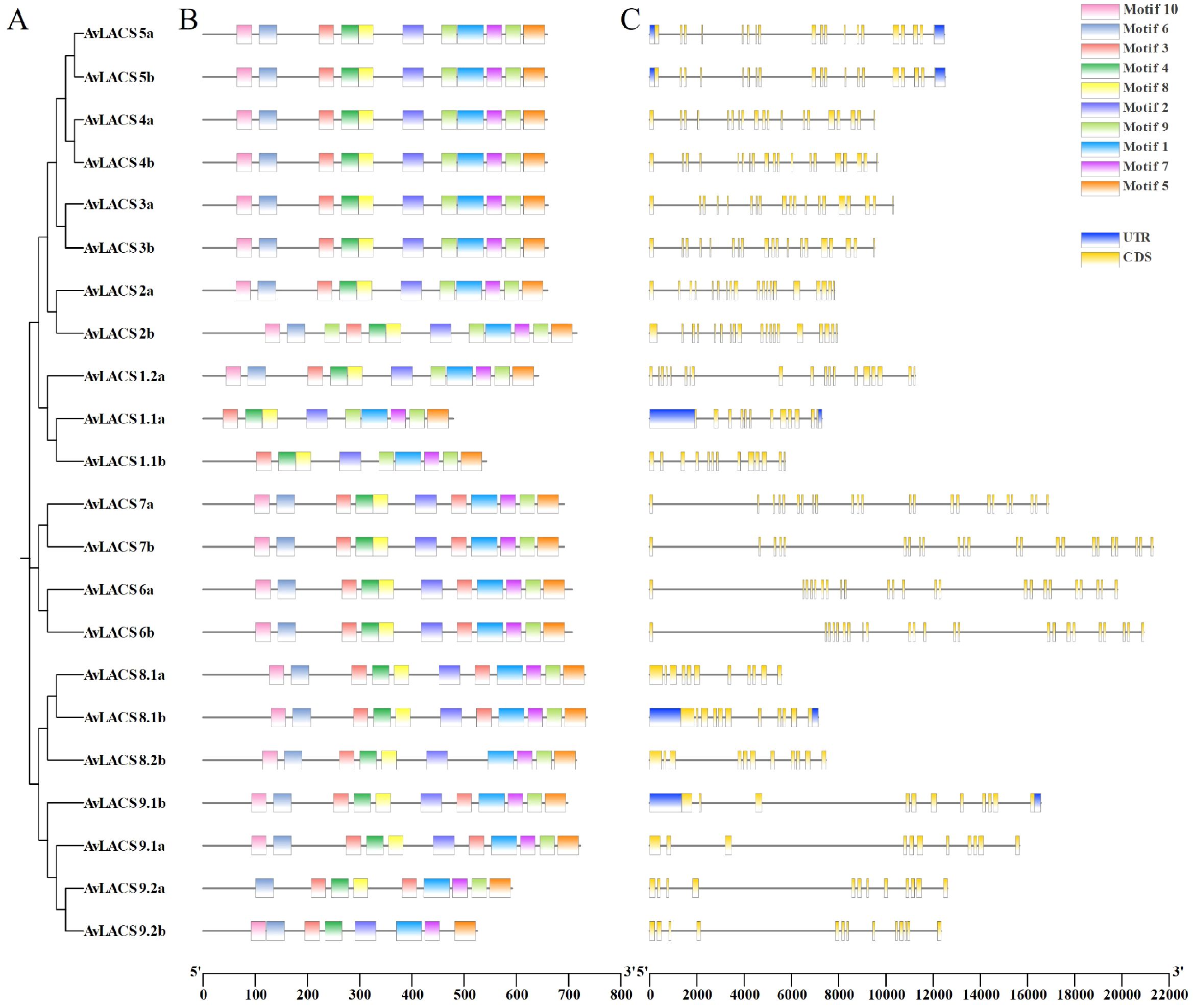
Figure 4. The phylogenetic tree, conserved motif, and gene structure of the AvLACS genes. (A) Phylogenetic tree of AvLACS proteins. (B) Conserved motif distribution of AvLACS proteins. (C) Exon-intron structure of AvLACS genes.
3.5 Gene duplication and collinearity analysis of AvLACS genes
To elucidate the synteny relationships among homologous LACS genes and infer gene duplication events, we conducted a collinearity analysis by using MCScanX. A total of 22 duplicated gene pairs were found in the A. valvata genome (Figure 5; Supplementary Table 5). The kiwifruit A. valvata is a tetraploid, and its genome can be divided into subgenome a and subgenome b. Each subgenome a or subgenome b has 3 duplicated gene pairs. However, there were 16 duplicated gene pairs between subgenome a and subgenome b. Then, we analyzed the selection pressure of replicated gene pairs by calculating the nonsynonymous (Ka) and synonymous (Ks) substitution rates. In A. valvata, all duplicated AvLACS gene pairs showed a Ka/Ks ratio of less than 1, indicating that they have undergone purifying selection during evolution (Supplementary Table 5). Additionally, the results showed that the gene duplication events occurred approximately 4.37 to 67.68 million years ago (MYA). In subgenome a, the gene duplication events occurred at 24.27, 25.90 and 67.12 MYA, while in subgenome b, the gene duplication events took place at 19.35, 25.98 and 67.45 MYA.
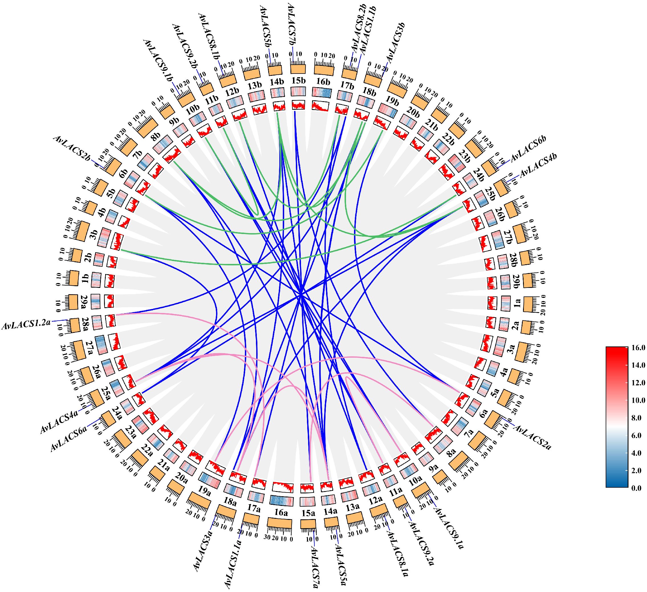
Figure 5. A collinearity analysis of AvLACSs. The grey lines indicate all duplicate genes, while the blue lines indicate the duplicated LACS gene pairs between A. valvata subgenome a and subgenome b. The pink lines in the circle indicate the duplicated LACS gene pairs within the subgenome a, and the green lines indicate the duplicated LACS gene pairs within the subgenome b. The heatmap and line graph indicate gene density.
To further elucidate the orthologous relationships of the AvLACS gene family, a multicollinearity analysis was conducted between A. valvata, A. thaliana and Actinidia chinensis ‘Hongyang’ (Supplementary Figure 7). Within A. valvata, there were 16 AvLACS gene pairs between subgenome a and subgenome b (Supplementary Figure 7A). The result showed that 14 and 15 gene pairs of LACS collinearity were found between A. chinensis ‘Hongyang’ and A. valvata subgenome a and subgenome b, respectively (Supplementary Figure 7B). In addition, there were each 14 gene pairs of LACS collinearity between A. thaliana and the A. valvata subgenome a or subgenome b (Supplementary Figure 7C).
3.6 Cis-acting elements analysis in the promoter regions of the AvLACS genes
To predict the potential molecular functions of the AvLACS genes, the cis-acting elements located in the upstream 2,000 bp promoter regions of all the AvLACS genes were analyzed using PlantCARE. The distribution and numbers of these cis-acting elements on the promoters of the AvLACS genes are shown in Figure 6. All of the AvLACS gene promoter regions contained numerous light response cis-elements (Figure 6A), such as GT1-motif, AE-box, and G-box (Figure 6B). The phytohormone-responsive cis-acting elements, especially the methyl jasmonate (MeJA) and abscisic acid (ABA) responsive elements, are widely distributed. The phytohormone responsive category further included salicylic acid (SA) responsive element (TCA-element), gibberellin (GA) responsive elements (GARE-motif, P-box and TATC-box), and auxin responsiveness elements (AuxRR-core and TGA-element) (Figure 6B). Among them, the least number of cis-element from the phytohormone category was AuxRR-core, which was found in only three AvLACS genes. Moreover, the stress response category included cis-elements related to biotic and abiotic stress, such as anaerobic induction, wound responsiveness, drought responsiveness, low-temperature responsiveness and defense and stress responsiveness (Figure 6A). Notably, almost all the AvLACS genes contained anaerobic responsive cis-elements, such as ARE and GC-motif, which could respond to hypoxia stress. The expression of AtLACS2 and LACS enzyme activity are affected by hypoxia, which restricts energy production via aerobic respiration (Zhao et al., 2021; Wundersitz et al., 2025). In contrast, there is only one gene involved in the response to defense and stress, namely, AvLACS9.2b.
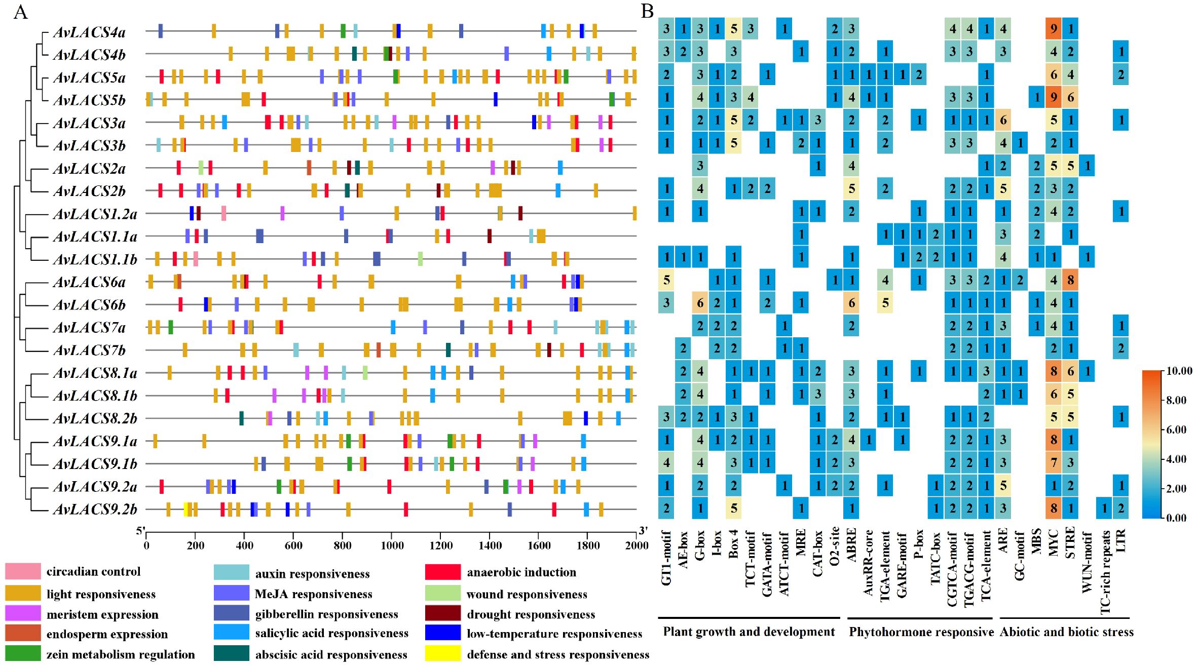
Figure 6. The cis-acting elements in the AvLACS gene promoter. (A) Cis-acting elements distribution in the promoter regions, with each colored rectangle representing a distinct type of cis-element; (B) Statistics on the number of cis-acting elements associated with plant growth and development and phytohormone and stress responses in the promoter region of AvLACS genes.
3.7 Expression Analysis of AvLACS genes in A. valvata
To understand the expression patterns of the AvLACS genes in A. valvata, the RNA-seq data (PRJNA984935) of kiwifruit were extracted and the FPKM (fragments per million mapped readings per thousand base transcripts) were used to evaluate their expression levels in the fruit flesh at different developmental stages. As shown in Figure 7A, AvLACS5a, AvLACS6a/b, AvLACS8.1a/b and AvLACS9.1a/b were highly expressed in the fruit flesh across four developmental stages – the stage 1 (mature green fruit stage), the stage 2 (breaker fruit stage), the stage 3 (color change fruit stage), and the stage 4 (ripe fruit stage). In contrast, genes like AvLACS2a/b, AvLACS3a/b and AvLACS9.2a/b showed relatively lower expression levels. Additionally, the expression patterns of AvLACS family members exhibited differences at different stages of fruit development (Figure 7A). The expression levels of AvLACS1.1a and AvLACS9.2a/b decreased during fruit development, while nearly half of the AvLACS family members showed an increase in expression during the breaker fruit stage (stage 2), such as AvLACS4a/b, AvLACS5a/b, AvLACS6a/b, and AvLACS8.1a/b, indicating AvLACSs may play a key role at this stage.
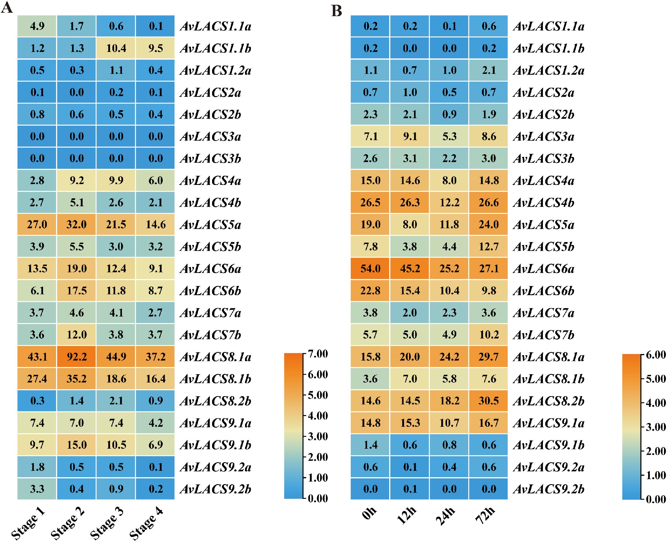
Figure 7. Expression profiles of AvLACSs in different fruit stages and under salt stress: (A) expression profiles of AvLACSs in fruit flesh at stage 1 (mature green fruit stage), stage 2 (breaker fruit stage), stage 3 (color change fruit stage), and stage 4 (ripe fruit stage); (B) expression profiles of AvLACSs in the roots under salt stress at 0 h, 12 h, 24 h, and 72 h.
We further analyzed the expression patterns of AvLACS genes in the roots of A. valvata under salt stress by utilizing another RNA-seq dataset (PRJNA726156) of kiwifruit. As shown in Figure 7B, the AvLACS4b and AvLACS6a/b were highly expressed in the root under normal conditions. However, the expression levels of AvLACS6a/b gradually decreased after the salt stress treatment. In contrast, several genes like AvLACS7b, AvLACS8.1a/b and AvLACS8.2b showed an increase expression after salt treatment for 72 h, suggesting that these genes were involved in regulating response to salt stress, particularly long-time salt stress.
3.8 RT-qPCR analysis of AvLACSs under waterlogging stress at different times
To further assess the role of the AvLACSs in the response to waterlogging stress, the expression of all of the AvLACS genes was measured by real-time quantitative polymerase chain reaction (RT-qPCR). As shown in Figure 8, the relative expression levels of nearly all AvLACS genes were significantly altered after the waterlogging treatment. Among them, most AvLACS genes were downregulated after the waterlogging treatment, such as AvLACS2a/b, AvLACS3a/b, AvLACS4a/b, and AvLACS5a/b. The reduction of LACS activity resulted the shift of the composition of LCFAs which triggers the activation of anaerobic metabolism genes (Wundersitz et al., 2025). In contrast, the expression of AvLACS1.1a/b, AvLACS1.2a and AvLACS6a/b increased under the waterlogging stress. Interestingly, the expression pattern of these five genes was different at different treatment time. Of these, AvLACS1.1a/b and AvLACS1.2a were rapidly induced by the submergence treatment at 6 h and displayed a trend of decline following the waterlogging stress. However, the expression of AvLACS6a/b increased significantly until the waterlogging treatment at 120 h. The results suggested that AvLACS1.1a/b and AvLACS1.2a were crucial for the short-term response, whereas AvLACS6a/b might be involved in regulating the long-term response to waterlogging stress.
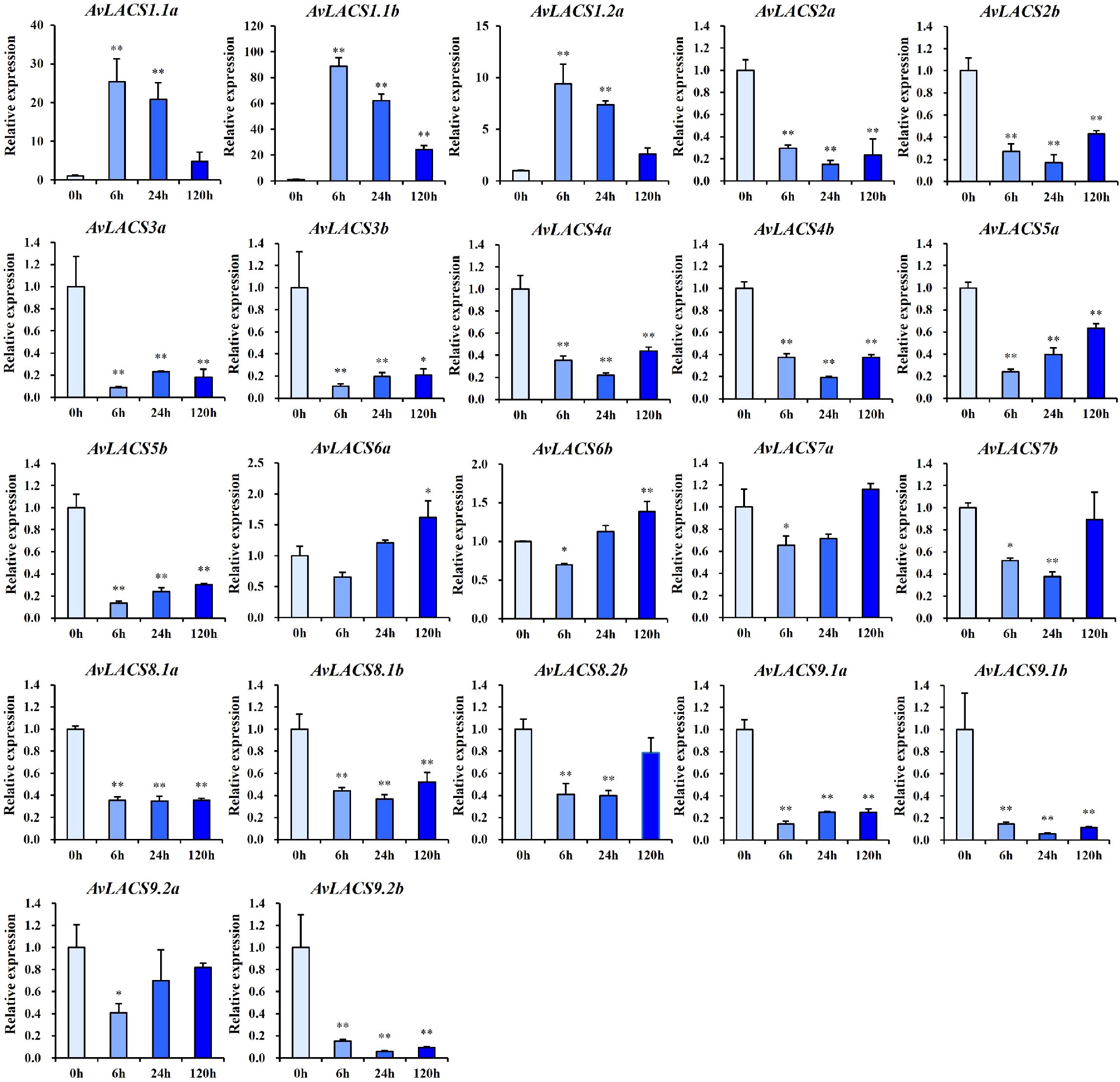
Figure 8. The relative expression profiles of AvLACSs in the roots under waterlogging stress at 0 h, 6 h, 24 h, and 120 h. Data are presented as means ± SE (n = 3), with statistical significance denoted by *(p < 0.05) and **(p < 0.01).
3.9 Expression of cuticular cutin, wax, and TAG biosynthesis genes under waterlogging stress
To understand the metabolic pathway that AvLACS1 (AvLACS1.1a/b and AvLACS1.2a) will subsequently participate in, the expression patterns of related genes involved in these pathways under waterlogging stress were analyzed based on the transcriptomic data (Figure 9). In Arabidopsis, AtLACS1 located in ER is prominently involved in several fatty acid-derived metabolic pathways, including the synthesis of wax, cutin, and TAG. Given that AvLACS1.1a/b and AvLACS1.2a are predicted to be located in the ER, and they exhibit a close evolutionary relationship with AtLACS1, it is hypothesized that AvLACS1 (AvLACS1.1a/b and AvLACS1.2a) may be involved in the synthesis of wax, cutin, and TAG. As shown in Figure 9, LCFAs were transferred to the ER and converted to acyl-CoA by AvLACS1. Under waterlogging stress, the expression levels of AvLACS1.1a/b and AvLACS1.2a rapidly increased at 6 h of treatment, and then gradually decreased, which is consistent with our qRT-PCR results. Subsequently, the synthesized acyl-CoA can be directed into three metabolic pathways: cutin biosynthesis, cuticular wax biosynthesis and TAG biosynthesis. In the cutin biosynthesis pathway, the expression of most genes was downregulated after waterlogging treatment (Figure 9; Supplementary Table 6). In the cuticular wax biosynthesis pathway, four FAR genes, six WSD1 genes, four CER1/3 genes and four MAH1 genes were identified (Figure 9; Supplementary Table 6). Among them, three FAR genes were downregulated after waterlogging treatment, while one FAR gene was upregulated until 120 h of treatment. In the WSD1 gene family, the expression of a WSD1 gene gradually increased after waterlogging treatment, another WSD1 gene showed an initial decrease followed by an increase in expression, and the expression of one WSD1 gene was downregulated after treatment. In another pathway involved in cuticular wax precursor synthesis, the expression of two CER1/3 genes was downregulated after treatment, while one MAH1 gene exhibited an initial decrease followed by an increase in expression after treatment, and one gene showed a gradual increase in expression in response to waterlogging stress. In the TAG biosynthesis pathway, one GPAT gene, one PAP gene and one DGAT gene rapidly increased at 6 h of treatment, and then gradually decreased, which is consistent with the expression patterns of AvLACS1.1a/b and AvLACS1.2a (Figure 9; Supplementary Table 7). Based on the transcription levels of related biosynthetic genes in these three metabolic pathways, it is inferred that acyl-CoA synthesized by AvLACS1 is primarily utilized in the synthesis of cuticular wax and TAG.
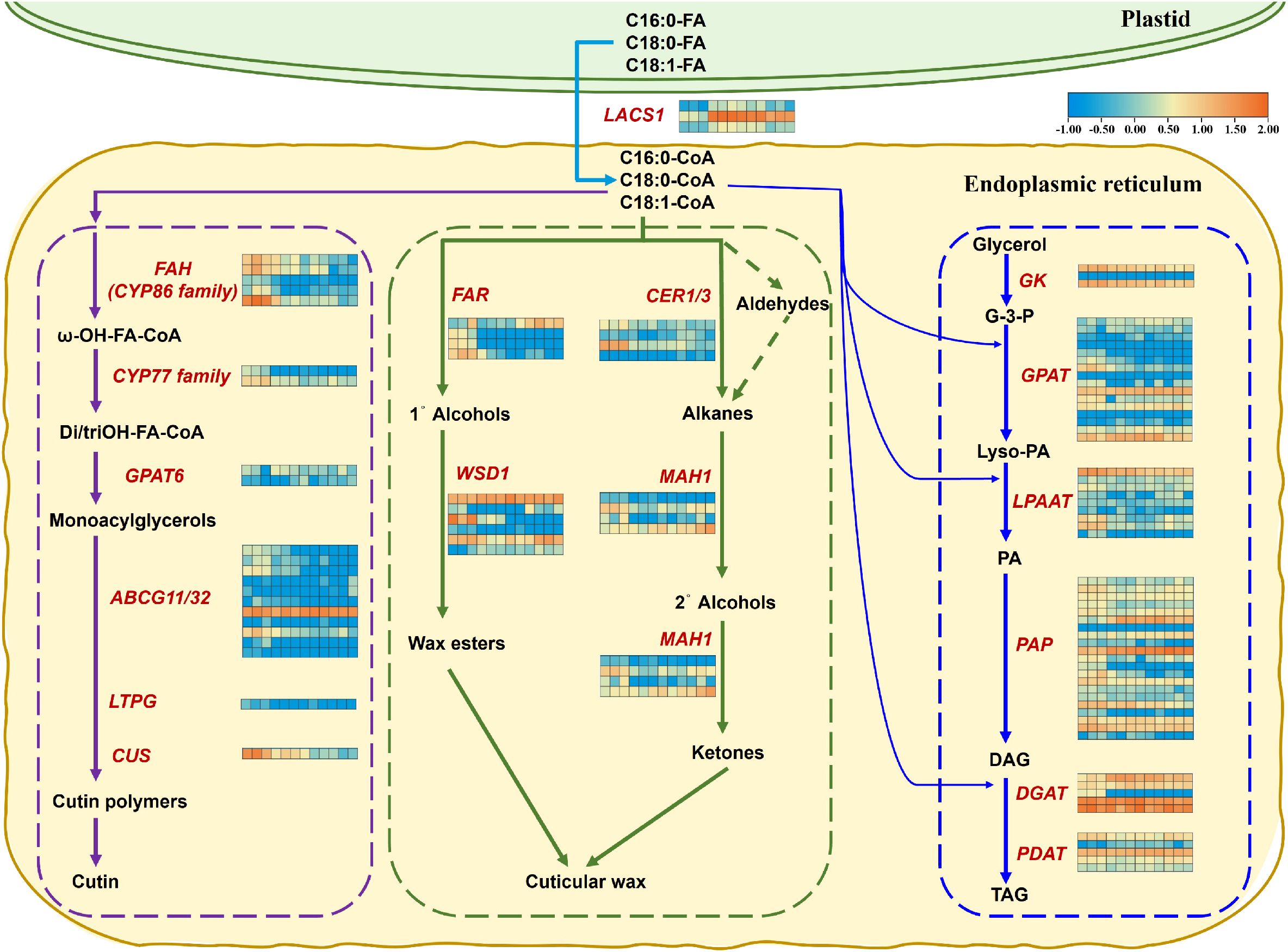
Figure 9. Expression analysis of genes responsible for cuticular cutin, wax, and TAG biosynthesis in the roots of A. valvata under waterlogging stress at 0 h, 6 h, 24 h, and 120 h. Purple dotted box indicates genes involved in cuticular cutin biosynthesis, green dotted box indicates genes involved in cuticular wax biosynthesis, and blue dotted box indicates genes involved in TAG biosynthesis. Transcript IDs for the heatmap are listed in the corresponding Supplementary materials files. Heatmaps were generated using the transcriptome data using the TBtools, and orange and blue colors in the heatmaps indicate higher and lower abundances, respectively. The twelve squares in each horizontal row represent the twelve samples (0 h-1, 0 h-2, 0 h-3, 6 h-1, 6 h-2, 6 h-3, 24 h-1, 24 h-2, 24 h-3, 120 h-1, 120 h-2 and 120 h-3). Abbreviations used in the figure: LACS1, long chain acyl-CoA synthetase 1; FAH, fatty acyl ω-hydroxylase; CYP86 family, cytochrome P450-CYP86 family; CYP77 family,cytochrome P450-CYP77 family; GPAT6, glycerol-3-phosphate acyl-transferase 6; ABCG11/32, ABC transporter G family member 11 and 32; LTPG, GPI-anchored lipid transfer protein; CUS, cutin synthase; FAR, fatty acyl-CoA reductase; WSD1, bifunctional wax ester synthase/acyl-CoA: diacylglycerol acyltransferase 1; CER1/3, very-long-chain aldehyde decarbonylase 1 and 3; MAH1, midchain alkane hydroxylase 1; GK, glycerol kinase; LPAAT, lysophosphatidic acid acyltransferase; PAP, phosphatidate phosphatase; DGAT, diacylglycerol acyl-transferase; PDAT, phospholipid: diacylglycerol acyltransferase; FA, fatty acid; CoA, coenzyme A; G-3-P, glycerol 3-phosphate; Lyso-PA, lysophosphatidic acid; PA, phosphatidic acid; DAG, diacylglycerol; TAG, triacylglycerol.
3.10 Expression of TAG degradation pathway genes under waterlogging stress
Considering that AvLACS6a/b are located in the peroxisome and that phylogenetic analysis reveals a close evolutionary relationship to AtLACS6, it is hypothesized that AvLACS6a/b may mediate TAG degradation through fatty acid β-oxidation like AtLACS6. The fatty acid β-oxidation pathway is summarized in Figure 10, and the expression patterns of genes under waterlogging stress associated to this pathway were presented as heatmap. During β-oxidation, twenty-two AvTAGL genes were identified to be differentially expressed under waterlogging stress at different times, and two of them were relatively upregulated with the extension of treatment time (Figure 10; Supplementary Table 8). Five AvMAGL genes were identified and one of them was downregulated rapidly after the waterlogging treatment at 6 h and then gradually upregulated at 120 h. Subsequently, the FFAs are then transported to peroxisomes. In peroxisomes, acyl-CoA is synthesized from the FFAs by AvLACS6. Similar to our qRT-PCR results, AvLACS6a and AvLACS6b were both upregulated under waterlogging stress at 120 h. In contrast, ten AvACT genes were identified and two of them were downregulated under waterlogging stress. The acyl-CoAs are first converted to 2-trans-enoyl-CoAs by ACX, which are subsequently processed through 3-hydroxyacyl-CoAs by MFP to form 3-ketoacyl-CoAs (Yoshitake et al., 2019). A total of eight AvACX genes were identified, three of which were downregulated at the initial stage of waterlogging treatment and subsequently upregulated at 120 h. Two AvMFP genes exhibited similar trends in expression with the aforementioned three AvACX genes. At the last step, the 3-ketoacyl-CoAs are then hydrolyzed to produce acyl-CoAs by KAT, and the hydrolyzed acyl-CoA is used as substrates for acyl-CoA oxidases. Three AvKAT genes were identified and all of them downregulated after waterlogging treatment. Based on the transcription levels of related biosynthetic genes in the TAG degradation pathway, at the long-term waterlogging stress, the esterification of fatty acids to acyl-CoAs by AvLACS6a/b results in their activation for oxidative attack at the β-carbon position until the complete degradation of long-chain acyl-CoAs to C2 acetyl units.
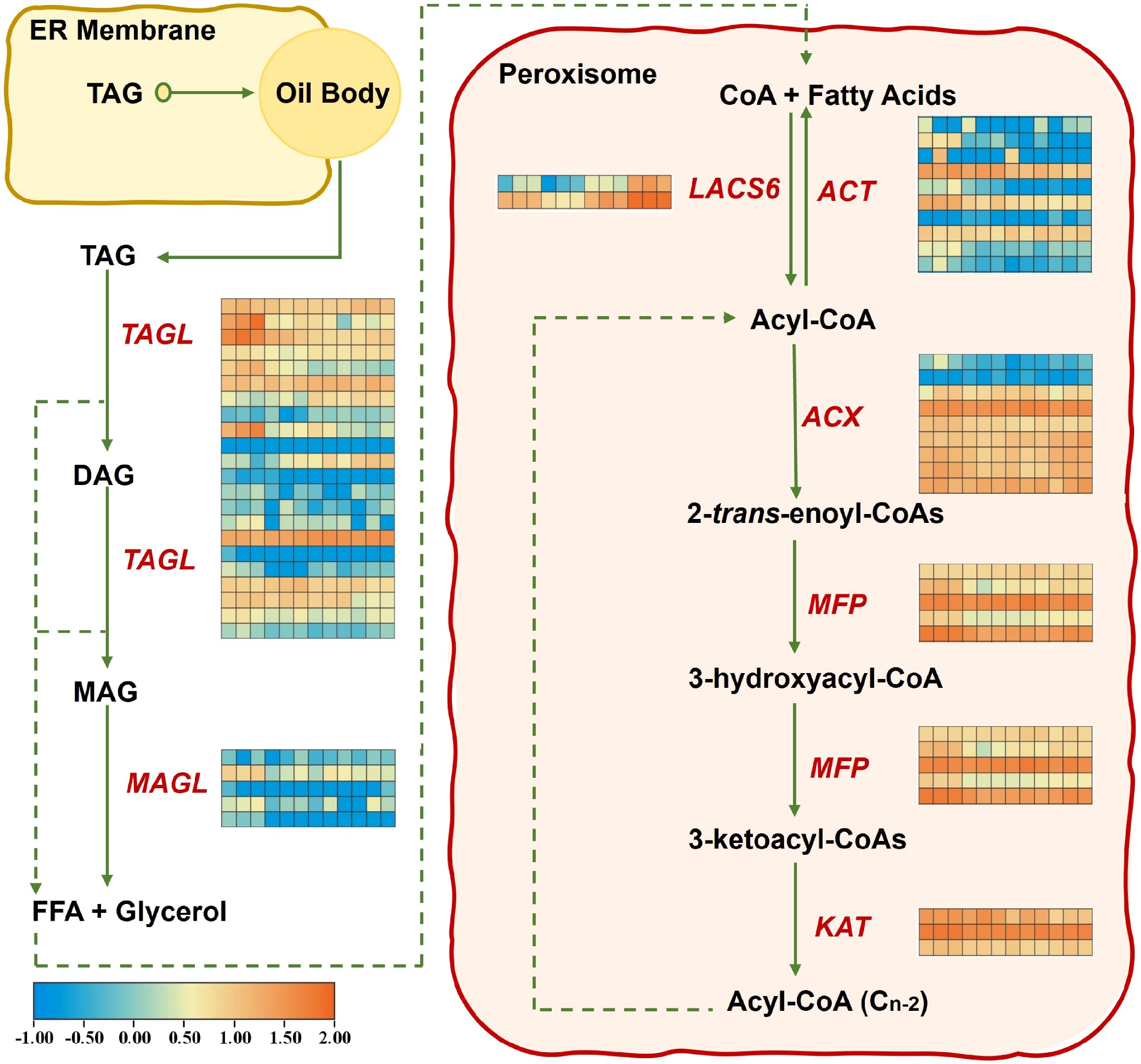
Figure 10. Expression analysis of genes related to TAG degradation in the roots of A. valvata under waterlogging stress at 0 h, 6 h, 24 h, and 120 h. Transcript IDs for the heatmap are listed in the corresponding Supplementary Materials file. Heatmaps were generated using the transcriptome data using TBtools, and orange and blue colors in the heatmaps indicate higher and lower abundances, respectively. The twelve squares in each horizontal row represent the twelve samples (0 h-1, 0 h-2, 0 h-3, 6 h-1, 6 h-2, 6 h-3, 24 h-1, 24 h-2, 24 h-3, 120 h-1, 120 h-2 and 120 h-3). Abbreviations used in the figure: TAGL, triacylglycerol lipase; MAGL, monoacylglycerol lipase; LACS6, long chain acyl-CoA synthetase 6; ACT, acyl-CoA thioesterase; ACX, acyl-CoA oxidase; MFP, multifunctional protein; KAT, 3-ketoacyl-CoA thiolase; ER, endoplasmic reticulum; TAG, triacylglycerol; DAG, diacylglycerol; MAG, monoacylglycerol; FFA, free fatty acid; CoA, coenzyme A.
3.11 Lipids profiling in the roots of A. valvata under waterlogging stress
To further validate our hypothesis regarding the metabolic pathways involving AvLACS1 and AvLACS6, the FFA, DAG and TAG from the roots of A. valvata at 0, 1and 7 days of waterlogging treatment were measured and identified by LC-MS/MS. As shown in Figure 11A, the content of FFA and DAG decreased significantly after submergence treatment, and although FFA and DAG showed some recovery after seven days of waterlogging, their levels remained significantly lower than those of the control. By contrast, the content of TAG accumulated significantly at 1 day of submergence treatment, while it returned to the normal level at 7 days. Meanwhile, the TAG/DAG ratio increased sharply at 1 day of submergence treatment and then decreased slightly at 7 days, but their levels were significantly higher than those of the control group (Figure 11B). The changes in the contents of these three lipids and the TAG/DAG ratio are consistent with our previous hypothesis that short-term waterlogging stress mediates TAG synthesis through AvLACS1, while long-term waterlogging stress mediates TAG degradation through AvLACS6. In addition, the double bond index (DBI), which indicates the degree of unsaturation, of FFA, DAG and TAG was calculated (Figure 11C), and the DBI of TAG was relatively large compared to that of FFA and DAG. Under waterlogging stress, the DBI of FFA decreased with the prolongation of submergence treatment, while the DBI of DAG and TAG increased significantly during submergence treatment. Significant changes in the lipid composition were observed in the roots of A. valvata with prolonged waterlogging stress. Waterlogging treatment induced a significant decrease in the content of various fatty acids in FFA and DAG, however, the content of the saturated FFA (16:0-FFA and 18:0-FFA) and DAG (32:0-DAG, 34:2-DAG and 34:3-DAG) gradually increased at 7 days of waterlogging stress (Figures 11D, E). In contrast, the content of the polyunsaturated TAG increased under waterlogging stress in parallel with the decreasing of less unsaturated TAG (Figure 11F), indicating that TAG serves as a pool for polyunsaturated fatty acids (Lu et al., 2020), and the incorporation of polyunsaturated fatty acids into TAG during waterlogging stress could be an important mechanism of plant adaptation to waterlogging stress.
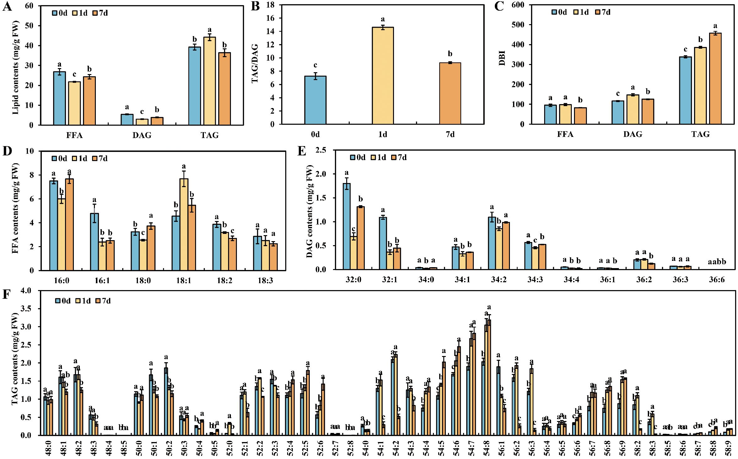
Figure 11. The composition and content of lipids in roots of A. valvata under waterlogging stress: (A) the lipid content of FFA, DAG and TAG in the roots under waterlogging stress at 0 d, 1d and 7 d; (B) the TAG/DAG ratio in the roots under waterlogging stress at 0 d, 1d and 7 d; (C) the DBI of FFA, DAG and TAG in the roots under waterlogging stress at 0 d, 1d and 7 d; (D–F) fatty acid composition of FFA (D), DAG (E) and TAG (F) in the roots under waterlogging stress at 0 d, 1d and 7 d. FFA, free fatty acids; DAG, diacylglycerol; TAG, triacylglycerol; DBI, double bond index. Values are presented as means ± standard deviation, different letters indicate significant differences at the p < 0.05 level.
4 Discussion
Long-chain acyl-CoA synthetase (LACS) plays a pivotal role in fatty acid metabolism and catabolism by converting free fatty acids to acyl-CoAs (Zhao et al., 2021). The generated acyl-CoAs serve as substrates for the synthesis of various lipids, including membrane lipids, cuticular lipids and TAG, which are not only essential for normal plant growth, but also are crucial for plant adaption to environmental stresses, such as waterlogging stress (Kęska et al., 2021; Zhou et al., 2020; Xie et al., 2020). As a waterlogging-tolerant kiwifruit, an increasing number of recent studies on A. valvata have focused on the adaptive strategies employed by A. valvata under waterlogging stress. Previous studies reported that four DEGs encoding LACS were significantly upregulated under waterlogging stress in A. valvata (Li et al., 2022). However, systematic identification and characterization of the LACS gene family in A. valvata is lacking to date. In this study, we identified 22 AvLACS members from the genome of A. valvata, which were located on 21 chromosomes. The number of AvLACS genes identified in the A. valvata genome was higher than the numbers found in A. thaliana (11 AtLACSs) (Shockey et al., 2002), Z. mays (11 ZmLACSs) (Wang et al., 2023), M. domestica (11 MdLACSs) (Zhang et al., 2018), G. max (17 GmLACSs) (Wang et al., 2022) and C. illinoinensis (11 CiLACSs) (Ma et al., 2023), but less than those in T. aestivum (30 TaLACSs) (Liu et al., 2022) and B. napus (34 BnaLACSs) (Xiao et al., 2019), which may be due to the differences in the sizes of their genomes. The AvLACSs encoded proteins ranging from 478 to 734 amino acids, and these proteins were located in the endoplasmic reticulum, peroxisome, and chloroplast. The subcellular localization of the AvLACSs was similar to the LACSs from other plants (Shockey et al., 2002; Liu et al., 2022; Zhou et al., 2024).
The multiple sequence alignment of AvLACS and AtLACS confirmed that AvLACS protein sequences contained two conserved motif domains (AMP-binding domain signature and ACS signature motif), which is consistent with CiLACS (Ma et al., 2023). Moreover, the AvLACS proteins were classified into five clusters based on phylogenetic tree analysis, and similar subcellular localization, exon-intron numbers, motif compositions, and protein structure were found within each cluster. The gene structure of AvLACS members in cluster II and III contained 19 exons and 18 introns, which is consistent with the number of exons found in the same clusters in C. illinoinensis (Ma et al., 2023). The conserved exon-intron structure in cluster II and III indicates that the LACS genes may have originated from a common ancestor, and have been significantly influenced by recurrent gene duplication events throughout their evolutionary history (Lynch et al., 2001). The greatest number of exons were found in AvLACS6a/b and AvLACS7a/b in cluster IV, which contained 23 exons. It is consistent with those of AtLACS6 and AtLACS7 in Arabidopsis (Shockey et al., 2002), MdLACS6.1 and MdLACS6.2 in M. domestica (Zhang et al., 2018), BnaLACS6-1/2/3/4 and BnaLACS7-1/2 in B. napus (Xiao et al., 2019), CiLACS6, CiLACS6–1 and CiLACS7 in C. illinoinensis (Ma et al., 2023). These variant exon-intron structures of AvLACS genes in different clusters suggested that AvLACS gene family may possess diverse functions, as it has been reported that the divergences in exon-intron structures can lead to changes of gene function (Xu et al., 2012).
The functional diversification of AvLACS genes is also reflected in the diversity of cis-acting elements in their promoter regions. In our study, cis-acting elements in the promoter regions of 22 AvLACS genes were predicted and classified into three subfamilies, including plant growth and development, phytohormone responsive, and stress-responsive subfamilies. Among them, the phytohormone-responsive cis-acting elements that are widely distributed include MeJA and ABA responsive elements, as well as elements responsive to SA, GA, and auxin. Some of the predictions were confirmed by the expression analysis, for instance, the expression level of PoLACS4 were significantly downregulated in mature leaves treated with 100 μmol/L ABA (Zhang et al., 2022), while the expression of MdLACS genes was upregulated with ABA treatment (Zhang et al., 2018). In addition, the stress response category contained cis-elements related to biotic stress such as defense and stress responsiveness, and to abiotic stress, such as anaerobic induction, wound responsiveness, drought responsiveness, and low-temperature responsiveness. Under biotic stress, loss of function of AtLACS2 could increase resistance to Botrytis cinerea (Bessire et al., 2007) and susceptibility to avirulent Pseudomonas syringae (Tang et al., 2007). Under abiotic stress, the expression of GmLACS9/15/17 was significantly upregulated under alkali, low temperature and drought stress (Wang et al., 2022). Similarly, several MdLACS genes were significantly upregulated under drought stress (Zhang et al., 2018), as the LACSs are crucial in the synthesis of cuticular lipids, and the formed cuticular can serve as surface barriers to prevent further water loss under drought stress (Zhao et al., 2021; Weng et al., 2010). The accumulation of cuticular wax enhanced the resistance of MdLACS2 or MdLACS4 transgenic plants to drought and salt stress (Zhang et al., 2020a, Zhang et al., 2020b). Furthermore, the expression of LACS6 in cucumber and four LACSs in kiwifruit (A. valvata) were significantly upregulated under waterlogging stress (Li et al., 2022; Kęska et al., 2021). AvLACSs might play an important role in A. valvata under waterlogging stress, as the anaerobic responsiveness cis-element was widely distributed in the promoter regions of AvLACSs.
To further elucidate the function of the AvLACS genes, we analyzed the expression patterns of AvLACSs during different stages of fruit development and under salt stress using transcriptome data available from NCBI. Based on the transcriptome data, nearly half of the AvLACS family members showed an increase expression at the breaker fruit stage, such as AvLACS4a/b, AvLACS5a/b, AvLACS6a/b, and AvLACS8.1a/b. Fleshy fruits are covered by the cuticle, which has an important protective role during fruit development and ripening (Trivedi et al., 2019). To prevent fruit softening and enhance the resistance to pathogens or water loss, the cuticular wax load increases during fruit development leading to a thick cuticle at maturity (Trivedi et al., 2019). The upregulated expression of these AvLACS genes at the breaker fruit stage might play a role in the synthesis of fruit cuticular wax to protect the fruit against the pathogens and water loss. Moreover, the expression levels of LACS genes under salt stress were different at various treatment times. The expression of AvLACS6a/b gradually decreased after salt stress treatment. Similarly, the expression levels of most of the MdLACS genes decreased under salt treatment in apple (Zhang et al., 2018). In pecan, the expression levels of CiLACS1, CiLACS1-1, and CiLACS4–1 were down-regulated under salt stress (Ma et al., 2023). However, AvLACS7b, AvLACS8.1a/b and AvLACS8.2b showed an increased expression after salt treatment for 72 h. The expression levels of CiLACS6, CiLACS7, CiLACS9, and Ci-LACS9–1 were upregulated after salt treatment at 8 and 16 days (Ma et al., 2023). These results indicate that LACSs play a crucial role in regulating response to salt stress, particular long-time salt stress.
In addition to their roles in drought and salt stress, increasing evidence suggests that LACSs are involved in plant response to waterlogging stress. In Arabidopsis, AtLACS2 is a key enzyme for wax and cutin biosynthesis, and the transgenic lines overexpressing LACS2 displayed enhanced resistance to submergence by modulating the cuticle integrity and permeability (Xie et al., 2020), as well as modulating the translocation of the ERF-VII transcription factor from the membrane to the nucleus (Zhou et al., 2020). In our study, the expression of all AvLACS genes was measured by RT-qPCR, and the results showed that the expression of AvLACS1.1a/b, AvLACS1.2a and AvLACS6a/b increased significantly after waterlogging treatment. Similarly, the expression level of LACS6 in cucumber exhibited a significant increase in response to waterlogging treatment (Kęska et al., 2021). Moreover, the expression of AvLACS1.1a/b and AvLACS1.2a was rapidly upregulated at 6 h, suggesting their involvement in the immediate response to short-term waterlogging stress. In contrast, AvLACS6a/b likely regulates the long-term adaptation to waterlogging, as its expression progressively increased up to 120 h of submergence treatment.
To elucidate the distinct mechanisms of different AvLACS genes in short-term or long-term waterlogging responses, transcriptomic analysis was performed to examine the expression profiles of associated genes within relevant metabolic pathways. During the short-term waterlogging response, the data suggest that acyl-CoA synthesized by AvLACS1 is primarily utilized in the synthesis of wax and TAG. Immediately after waterlogging, the rapid synthesis of wax might help plants regulate cuticle permeability, thereby regulating water transmission, improving gas exchange, and enhancing root resistance to pathogens caused by hypoxia (Xie et al., 2021). Homologs of AvLACS1, such as SlLACS1 in tomato has been demonstrated to play a crucial role in wax biosynthesis (Wu et al., 2024). Simultaneously, TAG serves as a safe transient reservoir of acyl groups, and its rapid accumulation plays a pivotal role in buffering lipid homeostasis and protecting cells against lipotoxic death by FA overload, which often results from extensive membrane remodeling during the stress response (Wang et al., 2019; He and Ding, 2020). Our lipid analysis of A. valvata roots under waterlogging stress confirmed the hypothesis that TAG plays a critical role in stress adaptation, demonstrating that FFA and DAG levels significantly decreased, likely due to their rapid conversion into TAG, while both the content and the unsaturated level of TAG significantly increased during short-term waterlogging stress. The increase of TAG content and the degree of polyunsaturation were also observed in tomato roots under hypoxia stress by waterlogging for 48 h (Striesow et al., 2024). Similarly, lipid droplets in the leaves of ramie (Boehmeria nivea L.) increased and aggregated in the cytoplasmic in response to submergence stress (Shao et al., 2024). The incorporation of polyunsaturated fatty acids into TAG, leading to increased TAG content and unsaturation, might be a key mechanism of plant adaptation to waterlogging stress.
At the long-term waterlogging response, AvLACS6a/b may mediate TAG degradation through fatty acid β-oxidation in peroxisome, similar to AtLACS6. The analysis of lipid composition in roots under long-term waterlogging has provided valuable insights into lipid metabolism during waterlogging stress conditions. The gradual decrease in TAG content is coupled with significant increases in FFA and DAG levels, which strongly supports the hypothesis that TAG undergoes degradation under prolonged waterlogging stress. During the long-term waterlogging stress, the liberation of fatty acids stored in cytoplasmic lipid droplets by TAG degradation can provide fatty acids to facilitate membrane reconstruction (Yu et al., 2021), and then maintain intracellular lipid homeostasis (Wang et al., 2019). Notably, the unsaturation degree of TAG remained unchanged, indicating that TAG continues to serve as a reservoir for polyunsaturated fatty acids even under prolonged waterlogging conditions. Thus, we proposed and have verified that the AvLACS gene family in the tetraploid A. valvata genome enhances the tolerance of kiwifruit to waterlogging stress through multiple pathways, including the short-term regulation of wax and TAG synthesis, as well as the long-term regulation of TAG degradation during prolonged waterlogging stress. This differential temporal stress-response strategy through regulation of LACS activity affecting TAG metabolism and wax synthesis in A. valvata improves survival rate under both short- and long-term waterlogging stress.
5 Conclusion
In this study, a total of 22 AvLACS genes were identified and systematically analyzed in A. valvata. AvLACSs were divided into five clusters based on a phylogenetic tree, and similar subcellular localization, exon-intron structures, motif compositions and protein structures were found within each phylogenetic cluster. Collinearity analysis identified 22 duplicated gene pairs in the A. valvata genome, which have undergone purifying selection during evolution. Expression profiling revealed that AvLACS1.1a/b, AvLACS1.2a, and AvLACS6a/b were significantly upregulated at specific time points after waterlogging treatment, indicating that the AvLACS gene family in the tetraploid A. valvata’ genome contributes to enhancing kiwifruit tolerance to waterlogging stress through multiple regulatory pathways. In the initial phase of waterlogging, AvLACS1.1a/b and AvLACS1.2a are involved in regulating wax and TAG synthesis, which is vital for maintaining cuticle permeability, preserving lipid homeostasis, and enhancing pathogen resistance. Conversely, AvLACS6a/b are pivotal during prolonged waterlogging, facilitating TAG degradation through fatty acid β-oxidation to maintain membrane integrity and intracellular lipid stability under extended stress. This coordinated regulation between AvLACS1(AvLACS1.1a/b and AvLACS1.2a) and AvLACS6a/b facilitates dynamic lipid metabolic adaptation, enabling A. valvata to adapt to both early and sustained phases of waterlogging stress. Integrated lipid profiling and transcriptomic analyses validate that AvLACS1.1a/b and AvLACS1.2a mediate wax and TAG synthesis, whereas AvLACS6a/b are involved in TAG degradation via β-oxidation. These findings, supported by combined lipidomic and transcriptomic data, enhance our understanding of lipid metabolism in A. valvata and identify potential molecular targets for breeding waterlogging-tolerant kiwifruit and other crops. Overall, this study elucidates the distinct functional roles of LACS family members in kiwifruit, providing a foundation for future studies on the molecular mechanisms of AvLACS genes.
Data availability statement
The original contributions presented in the study are included in the article/Supplementary Material. Further inquiries can be directed to the corresponding author.
Author contributions
MZ: Conceptualization, Formal Analysis, Investigation, Writing – original draft, Writing – review & editing. KY: Investigation, Writing – original draft. JG: Investigation, Writing – original draft. SL: Resources, Writing – original draft. RL: Investigation, Methodology, Writing – original draft. YC: Formal Analysis, Writing – original draft. QZ: Formal Analysis, Investigation, Writing – original draft. YL: Writing – review & editing. JL: Supervision, Writing – review & editing. CL: Conceptualization, Funding acquisition, Supervision, Writing – review & editing. FW: Conceptualization, Funding acquisition, Supervision, Writing – review & editing.
Funding
The author(s) declare that financial support was received for the research and/or publication of this article. This study was supported by the National Natural Science Foundation of China (32160084), Guangxi Science and Technology Program (Guike AD23026228), Natural Science Foundation of Hunan Province (2023JJ30488), Science and Technology Department of Shaanxi Province (2024JC-YBMS-187), Guangxi Science and Technology Major Project (Guike AA23023008), Earmarked Fund for China Agriculture Research System (nycytxgxcxtd-2023-13-01), Doctoral Research Funding from Shangluo University (17SKY016), Science and Technology Research Project of Shangluo University (23KYPY07), Guangxi Key Laboratory of Plant Functional Phytochemicals and Sustainable Utilization (ZRJJ2024-19), and Guilin Innovation Platform and Talent Plan (20220125-7).
Conflict of interest
The authors declare that the research was conducted in the absence of any commercial or financial relationships that could be construed as a potential conflict of interest.
Generative AI statement
The author(s) declare that no Generative AI was used in the creation of this manuscript.
Publisher’s note
All claims expressed in this article are solely those of the authors and do not necessarily represent those of their affiliated organizations, or those of the publisher, the editors and the reviewers. Any product that may be evaluated in this article, or claim that may be made by its manufacturer, is not guaranteed or endorsed by the publisher.
Supplementary material
The Supplementary Material for this article can be found online at: https://www.frontiersin.org/articles/10.3389/fpls.2025.1580003/full#supplementary-material
References
Abid, M., Gu, S., Zhang, Y. J., Sun, S., Li, Z., Bai, D. F., et al. (2022). Comparative transcriptome and metabolome analysis reveal key regulatory defense networks and genes involved in enhanced salt tolerance of Actinidia (kiwifruit). Hortic. Res. 9, uhac189. doi: 10.1093/hr/uhac189
Ayaz, A., Huang, H., Zheng, M., Zaman, W., Li, D., Saqib, S., et al. (2021). Molecular cloning and functional analysis of GmLACS2–3 reveals its involvement in cutin and suberin biosynthesis along with abiotic stress tolerance. Int. J. Mol. Sci. 22, 9175. doi: 10.3390/ijms22179175
Aznar-Moreno, J. A., Venegas Calerón, M., Martínez-Force, E., Garcés, R., Mullen, R., Gidda, S. K., et al. (2014). Sunflower (Helianthus annuus) long-chain acyl-coenzyme A synthetases expressed at high levels in developing seeds. Physiol. Plant 150, 363–373. doi: 10.1111/ppl.12107
Bai, D., Li, Z., Gu, S., Li, Q., Sun, L., Qi, X., et al. (2022). Effects of kiwifruit rootstocks with opposite tolerance on physiological responses of grafting combinations under waterlogging stress. Plants 11, 2098. doi: 10.3390/plants11162098
Bailey, T. L., Johnson, J., Grant, C. E., and Noble, W. S. (2015). The MEME suite. Nucleic Acids Res. 43, W39–W49. doi: 10.1093/nar/gkv416
Bernard, A. and Joubès, J. (2013). Arabidopsis cuticular waxes: advances in synthesis, export and regulation. Prog. Lipid Res. 52, 110–129. doi: 10.1016/j.plipres.2012.10.002
Bernhofer, M., Dallago, C., Karl, T., Satagopam, V., Heinzinger, M., Littmann, M., et al. (2021). PredictProtein - predicting protein structure and function for 29 years. Nucleic Acids Res. 49, W535–W540. doi: 10.1093/nar/gkab354
Bessire, M., Chassot, C., Jacquat, A. C., Humphry, M., Borel, S., Petétot, J. M. C., et al. (2007). A permeable cuticle in Arabidopsis leads to a strong resistance to Botrytis cinerea. EMBO J. 26, 2158–2168. doi: 10.1038/sj.emboj.7601658
Bhargava, N., Ampomah-Dwamena, C., Voogd, C., and Allan, A. C. (2023). Comparative transcriptomic and plastid development analysis sheds light on the differential carotenoid accumulation in kiwifruit flesh. Front. Plant Sci. 14. doi: 10.3389/fpls.2023.1213086
Chao, J., Li, Z., Sun, Y., Aluko, O. O., Wu, X., Wang, Q., et al. (2021). MG2C: a user-friendly online tool for drawing genetic maps. Mol. Hortic. 1, 16. doi: 10.1186/s43897-021-00020-x
Chen, C., Wu, Y., Li, J., Wang, X., Zeng, Z., Xu, J., et al. (2023). TBtools-II: A “one for all, all for one” bioinformatics platform for biological big-data mining. Mol. Plant 16, 1733–1742. doi: 10.1016/j.molp.2023.09.010
Falcone, D. L., Ogas, J. P., and Somerville, C. R. (2004). Regulation of membrane fatty acid composition by temperature in mutants of Arabidopsis with alterations in membrane lipid composition. BMC Plant Biol. 4, 17. doi: 10.1186/1471-2229-4-17
Fich, E. A., Segerson, N. A., and Rose, J. K. C. (2016). The plant polyester cutin: biosynthesis, structure, and biological roles. Annu. Rev. Plant Biol. 67, 207–233. doi: 10.1146/annurev-arplant-043015-111929
Geourjon, C. and Deléage, G. (1995). SOPMA: significant improvements in protein secondary structure prediction by consensus prediction from multiple alignments. Bioinformatics 11, 681–684. doi: 10.1093/bioinformatics/11.6.681
Grevengoed, T. J., Klett, E. L., and Coleman, R. A. (2014). Acyl-CoA metabolism and partitioning. Annu. Rev. Nutr. 34, 1–30. doi: 10.1146/annurev-nutr-071813-105541
Groot, P. H. E., Scholte, H. R., and Hülsmann, W. C. (1976). “Fatty acid activation: specificity, localization, and function,” in Advances in Lipid Research, vol. 14 . Eds. Paoletti, R. and Kritchevsky, D. (Amsterdam, Netherlands: Elsevier), 75–126. doi: 10.1016/B978-0-12-024914-5.50009-7
He, M. and Ding, N. Z. (2020). Plant unsaturated fatty acids: multiple roles in stress response. Front. Plant Sci. 11. doi: 10.3389/fpls.2020.562785
Henschel, J. M., Andrade, A. N., dos Santos, J. B. L., da Silva, R. R., da Mata, D. A., Souza, T., et al. (2024). Lipidomics in plants under abiotic stress conditions: an overview. Agronomy 14, 1670–1670. doi: 10.3390/agronomy14081670
Huang, H. (2016). “Chapter 1 - Systematics and genetic variation of Actinidia,” in Kiwifruit. Ed. Huang, H. (Academic Press, San Diego), 9–44. doi: 10.1016/B978-0-12-803066-0.00001-0
Jessen, D., Roth, C., Wiermer, M., and Fulda, M. (2014). Two activities of long-chain acyl-coenzyme a synthetase are involved in lipid trafficking between the endoplasmic reticulum and the plastid in Arabidopsis. Plant Physiol. 167, 351–366. doi: 10.1104/pp.114.250365
Kelley, L. A., Mezulis, S., Yates, C. M., Wass, M. N., and Sternberg, M. J. E. (2015). The Phyre2 web portal for protein modeling, prediction and analysis. Nat. Protoc. 10, 845–858. doi: 10.1038/nprot.2015.053
Kęska, K., Szcześniak, M. W., Makałowska, I., and Czernicka, M. (2021). Long-term waterlogging as factor contributing to hypoxia stress tolerance enhancement in cucumber: comparative transcriptome analysis of waterlogging sensitive and tolerant accessions. Genes 12, 189. doi: 10.3390/genes12020189
Kitajima-Koga, A., Baslam, M., Hamada, Y., Ito, N., Taniuchi, T., Takamatsu, T., et al. (2020). Functional analysis of rice long-chain acyl-CoA synthetase 9 (OsLACS9) in the chloroplast envelope membrane. Int. J. Mol. Sci. 21, 2223. doi: 10.3390/ijms21062223
Kunst, L. and Samuels, A. L. (2003). Biosynthesis and secretion of plant cuticular wax. Prog. Lipid Res. 42, 51–80. doi: 10.1016/S0163-7827(02)00045-0
Lamesch, P., Berardini, T. Z., Li, D., Swarbreck, D., Wilks, C., Sasidharan, R., et al. (2011). The Arabidopsis Information Resource (TAIR): Improved gene annotation and new tools. Nucleic Acids Res. 40, D1202–D1210. doi: 10.1093/nar/gkr1090
Lescot, M., Déhais, P., Thijs, G., Marchal, K., Moreau, Y., Van de Peer, Y., et al. (2002). PlantCARE, a database of plant cis-acting regulatory elements and a portal to tools for in silico analysis of promoter sequences. Nucleic Acids Res. 30, 325–327. doi: 10.1093/nar/30.1.325
Letunic, I. and Bork, P. (2021). Interactive Tree Of Life (iTOL) v5: An online tool for phylogenetic tree display and annotation. Nucleic Acids Res. 49, W293–W296. doi: 10.1093/nar/gkab301
Lewandowska, M., Keyl, A., and Feussner, I. (2020). Wax biosynthesis in response to danger: Its regulation upon abiotic and biotic stress. New Phytol. 227, 698–713. doi: 10.1111/nph.16571
Li, Z., Bai, D., Zhong, Y., Abid, M., Qi, X., Hu, C., et al. (2021). Physiological responses of two contrasting kiwifruit (Actinidia spp.) rootstocks against waterlogging stress. Plants 10, 2586. doi: 10.3390/plants10122586
Li, Z., Bai, D., Zhong, Y., Lin, M., Sun, L., Qi, X., et al. (2022). Full-length transcriptome and RNA-Seq analyses reveal the mechanisms underlying waterlogging tolerance in kiwifruit (Actinidia valvata). Int. J. Mol. Sci. 23, 3237. doi: 10.3390/ijms23063237
Li, N., Xu, C., Li-Beisson, Y., and Philippar, K. (2016). Fatty acid and lipid transport in plant cells. Trends Plant Sci. 21, 145–158. doi: 10.1016/j.tplants.2015.10.011
Liu, Y., Liu, Z., Zhang, H., Yuan, S., Li, Y., Zhang, T., et al. (2022). Genome-wide identification and expression profiling analysis of the long-chain acyl-CoA synthetases reveal their potential roles in wheat male fertility. Int. J. Mol. Sci. 23, 11942. doi: 10.3390/ijms231911942
Liu, W., Xie, Y., Ma, J., Luo, X., Nie, P., Zuo, Z., et al. (2015). IBS: An illustrator for the presentation and visualization of biological sequences. Bioinformatics 31, 3359–3361. doi: 10.1093/bioinformatics/btv362
Livak, K. J. and Schmittgen, T. D. (2001). Analysis of relative gene expression data using real-time quantitative PCR and the 2(-Delta Delta C(T)) method. Methods 25, 402–408. doi: 10.1006/meth.2001.1262
Lu, J., Xu, Y., Wang, J., Singer, S. D., and Chen, G. (2020). The role of triacylglycerol in plant stress response. Plants 9, 472. doi: 10.3390/plants9040472
Luo, J., Abid, M., Zhang, Y., Cai, X., Tu, J., Gao, P., et al. (2023). Genome-wide identification of kiwifruit SGR family members and functional characterization of SGR2 protein for chlorophyll degradation. Int. J. Mol. Sci. 24, 1993. doi: 10.3390/ijms24031993
Lynch, M., O’Hely, M., Walsh, B., and Force, A. (2001). The probability of preservation of a newly arisen gene duplicate. Genetics 159, 1789–1804. doi: 10.1093/genetics/159.4.1789
Ma, W., Zhu, K., Zhao, J., Chen, M., Wei, L., Qiao, Z., et al. (2023). Genome-wide identification, characterization, and expression analysis of long-chain acyl-CoA synthetases in Carya illinoinensis under different treatments. Int. J. Mol. Sci. 24, 11558. doi: 10.3390/ijms241411558
Mistry, J., Chuguransky, S., Williams, L., Qureshi, M., Salazar, G. A., Sonnhammer, E. L. L., et al. (2020). Pfam: The protein families database in 2021. Nucleic Acids Res. 49, D412–D419. doi: 10.1093/nar/gkaa913
Potter, S. C., Luciani, A., Eddy, S. R., Park, Y., Lopez, R., and Finn, R. D. (2018). HMMER web server: 2018 update. Nucleic Acids Res. 46, W200–W204. doi: 10.1093/nar/gky448
Qiao, Q., Ren, Z., Fu, X., Qiao, W., Sheng, F., Li, S., et al. (2025). Genome-wide exploration and characterization of the RALFs and analysis of its role in peanut (Arachis hypogaea L.). BMC Plant Biol. 25, 337. doi: 10.1186/s12870-025-06356-6
Shao, D., Yu, C., Chen, Y., Qiu, X., Chen, J., Zhao, H., et al. (2024). Lipids signaling and unsaturation of fatty acids participate in ramie response to submergence stress and hypoxia-responsive gene regulation. Int. J. Biol. Macromol. 263, 130104. doi: 10.1016/j.ijbiomac.2024.130104
Shockey, J. M., Fulda, M. S., and Browse, J. A. (2002). Arabidopsis contains nine long-chain acyl-coenzyme A synthetase genes that participate in fatty acid and glycerolipid metabolism. Plant Physiol. 129, 1710–1722. doi: 10.1104/pp.003269
Striesow, J., Welle, M., Busch, L. M., Bekeschus, S., Wende, K., and Stöhr, C. (2024). Hypoxia increases triacylglycerol levels and unsaturation in tomato roots. BMC Plant Biol. 24, 909. doi: 10.1186/s12870-024-05578-4
Tang, D., Simonich, M. T., and Innes, R. W. (2007). Mutations in LACS2, a long-chain acyl-coenzyme A synthetase, enhance susceptibility to avirulent Pseudomonas syringae but confer resistance to Botrytis cinerea in Arabidopsis. Plant Physiol. 144, 1093–1103. doi: 10.1104/pp.106.094318
Trivedi, P., Nguyen, N., Hykkerud, A. L., Häggman, H., Martinussen, I., Jaakola, L., et al. (2019). Developmental and environmental regulation of cuticular wax biosynthesis in fleshy fruits. Front. Plant Sci. 10. doi: 10.3389/fpls.2019.00431
Tu, J., Abid, M., Luo, J., Zhang, Y., Yang, E., Cai, X., et al. (2023). Genome-wide identification of the heat shock transcription factor gene family in two kiwifruit species. Front. Plant Sci. 14. doi: 10.3389/fpls.2023.1075013
Wang, K., Durrett, T. P., and Benning, C. (2019). Functional diversity of glycerolipid acylhydrolases in plant metabolism and physiology. Prog. Lipid Res. 75, 100987. doi: 10.1016/j.plipres.2019.100987
Wang, J., Li, X., Zhao, X., Na, C., Liu, H., Miao, H., et al. (2022). Genome-wide identification and characterization of the abiotic-stress-responsive LACS gene family in soybean (Glycine max). Agronomy 12, 1496. doi: 10.3390/agronomy12071496
Wang, X., Tian, X., Zhang, H., Li, H., Zhang, S., Li, H., et al. (2023). Genome-wide analysis of the maize LACS gene family and functional characterization of the ZmLACS9 responses to heat stress. Plant Stress 10, 100271. doi: 10.1016/j.stress.2023.100271
Wang, S., Uddin, M. I., Tanaka, K., Yin, L., Shi, Z., Qi, Y., et al. (2014). Maintenance of chloroplast structure and function by overexpression of the rice MONOGALACTOSYLDIACYLGLYCEROL SYNTHASE gene leads to enhanced salt tolerance in tobacco. Plant Physiol. 165, 1144–1155. doi: 10.1104/pp.114.238899
Wang, X., Wang, X., Chen, X., Chen, L., Dou, L., Yu, J., et al. (2025). Characterization of the PbrLACS2 in pear and role in wax biosynthesis in Arabidopsis. Tree Genet. Genomes 21, 3. doi: 10.1007/s11295-024-01685-3
Wang, Y., Zhao, J., Chen, H., Zhang, Q., Zheng, Y., Li, D., et al. (2021). Molecular cloning and characterization of long-chain acyl-CoA synthetase 9 from the mesocarp of African oil palm (Elaeis guineensis Jacq.). Sci. Hortic. 276, 109751. doi: 10.1016/j.scienta.2020.109751
Weng, H., Molina, I., Shockey, J., and Browse, J. (2010). Organ fusion and defective cuticle function in a lacs1lacs2 double mutant of Arabidopsis. Planta 231, 1089–1100. doi: 10.1007/s00425-010-1110-4
Wu, P., Li, S., Yu, X., Guo, S., and Gao, L. (2024). Identification of long-chain acyl-CoA synthetase gene family reveals that SlLACS1 is essential for cuticular wax biosynthesis in tomato. Int. J. Biol. Macromol. 277, 134438. doi: 10.1016/j.ijbiomac.2024.134438
Wundersitz, A., Hoffmann, K. M. V., and van Dongen, J. T. (2025). Acyl-CoA-binding proteins: bridging long-chain acyl-CoA metabolism to gene regulation. New Phytol. 246, 1960–1966. doi: 10.1111/nph.70142
Xiao, Z., Li, N., Wang, S., Sun, J., Zhang, L., Zhang, C., et al. (2019). Genome-wide identification and comparative expression profile analysis of the long-chain acyl-CoA synthetase (LACS) gene family in two different oil content cultivars of Brassica napus. Biochem. Genet. 57, 781–800. doi: 10.1007/s10528-019-09921-5
Xie, L., Tan, W., Yang, Y., Tan, Y., Zhou, Y., Zhou, D., et al. (2020). Long-chain acyl-CoA synthetase LACS2 contributes to submergence tolerance by modulating cuticle permeability in Arabidopsis. Plants 9, 262. doi: 10.3390/plants9020262
Xie, L., Zhou, Y., Chen, Q., and Xiao, S. (2021). New insights into the role of lipids in plant hypoxia responses. Prog. Lipid Res. 81, 101072. doi: 10.1016/j.plipres.2020.101072
Xu, G., Guo, C., Shan, H., and Kong, H. (2012). Divergence of duplicate genes in exon–intron structure. Proc. Natl. Acad. Sci. U.S.A. 109, 1187–1192. doi: 10.1073/pnas.1109047109
Yan, Z., Hou, J., Leng, B., Yao, G., Ma, C., Sun, Y., et al. (2024). Genome-wide identification and characterization of maize long-chain acyl-CoA synthetases and their expression profiles in different tissues and in response to multiple abiotic stresses. Genes 15, 983. doi: 10.3390/genes15080983
Yoshitake, Y., Ohta, H., and Shimojima, M. (2019). Autophagy-mediated regulation of lipid metabolism and its impact on the growth in algae and seed plants. Front. Plant Sci. 10. doi: 10.3389/fpls.2019.00709
Yu, L., Tan, X., Jiang, B., Sun, X., Gu, S., Han, T., et al. (2014). A peroxisomal long-chain Acyl-CoA synthetase from Glycine max involved in lipid degradation. PLoS One 9, e100144. doi: 10.1371/journal.pone.0100144
Yu, L., Zhou, C., Fan, J., Shanklin, J., and Xu, C. (2021). Mechanisms and functions of membrane lipid remodeling in plants. Plant J. 107, 37–53. doi: 10.1111/tpj.15273
Zhang, Z. (2022). KaKs_calculator 3.0: calculating selective pressure on coding and non-coding sequences. Genom. Proteom. Bioinform. 20, 536–540. doi: 10.1016/j.gpb.2021.12.002
Zhang, M., Deng, X., Yin, L., Qi, L., Wang, X., Wang, S., et al. (2016). Regulation of galactolipid biosynthesis by overexpression of the rice MGD gene contributes to enhanced aluminum tolerance in tobacco. Front. Plant Sci. 7. doi: 10.3389/fpls.2016.00337
Zhang, C., Hu, X., Zhang, Y., Liu, Y., Wang, G., You, C., et al. (2020a). An apple long-chain acyl-CoA synthetase 2 gene enhances plant resistance to abiotic stress by regulating the accumulation of cuticular wax. Tree Physiol. 40, 1450–1465. doi: 10.1093/treephys/tpaa079
Zhang, M., Liu, C., Wang, F., Liu, S., Gao, J., Li, J., et al. (2024). Genome-wide identification of the DGK gene family in kiwifruit (Actinidia valvata Dunn) and an expression analysis of their responses to waterlogging stress. Horticulturae 10, 310. doi: 10.3390/horticulturae10040310
Zhang, C., Mao, K., Zhou, L., Wang, G., Zhang, Y., Li, Y., et al. (2018). Genome-wide identification and characterization of apple long-chain Acyl-CoA synthetases and expression analysis under different stresses. Plant Physiol. Biochem. 132, 320–332. doi: 10.1016/j.plaphy.2018.09.004
Zhang, C., Zhang, Y., Hu, X., Xiao, X., Wang, G., You, C., et al. (2020b). An apple long-chain acyl-CoA synthetase, MdLACS4, induces early flowering and enhances abiotic stress resistance in Arabidopsis. Plant Sci. 297, 110529. doi: 10.1016/j.plantsci.2020.110529
Zhang, H., Zhang, S., Li, M., Wang, J., and Wu, T. (2022). The PoLACS4 gene may participate in drought stress resistance in tree peony (Paeonia ostii ‘Feng dan bai’). Genes 13, 1591. doi: 10.3390/genes13091591
Zhao, H., Kosma, D. K., and Lü, S. (2021). Functional role of long-chain acyl-CoA synthetases in plant development and stress responses. Front. Plant Sci. 12. doi: 10.3389/fpls.2021.640996
Zhong, Y., Wang, Y., Li, P., Gong, W., Wang, X., Yan, H., et al. (2023). Genome-wide analysis and functional characterization of LACS gene family associated with lipid synthesis in cotton (Gossypium spp.). Int. J. Mol. Sci. 24, 8530. doi: 10.3390/ijms24108530
Zhou, Y., Tan, W., Xie, L., Qi, H., Yang, Y., Huang, L., et al. (2020). Polyunsaturated linolenoyl-CoA modulates ERF-VII-mediated hypoxia signaling in Arabidopsis. J. Integr. Plant Biol. 62, 330–348. doi: 10.1111/jipb.12875
Keywords: long-chain acyl-CoA synthetases (LACSs), Actinidia valvata, genome-wide characterization, expression analysis, lipid analysis, waterlogging stress
Citation: Zhang M, Ye K, Gao J, Liu S, Liu R, Chen Y, Zhao Q, Lei Y, Li J, Liu C and Wang F (2025) Identification and functional analysis of AvLACS genes unveils their role in lipid homeostasis and waterlogging tolerance in kiwifruit (Actinidia valvata Dunn). Front. Plant Sci. 16:1580003. doi: 10.3389/fpls.2025.1580003
Received: 19 February 2025; Accepted: 05 June 2025;
Published: 30 June 2025.
Edited by:
Yogeshwar Vikram Dhar, Ruhr University Bochum, GermanyReviewed by:
Humberto Prieto, Agricultural Research Institute (Chile), ChileDaniel Bimpong, Yangtze University, China
Copyright © 2025 Zhang, Ye, Gao, Liu, Liu, Chen, Zhao, Lei, Li, Liu and Wang. This is an open-access article distributed under the terms of the Creative Commons Attribution License (CC BY). The use, distribution or reproduction in other forums is permitted, provided the original author(s) and the copyright owner(s) are credited and that the original publication in this journal is cited, in accordance with accepted academic practice. No use, distribution or reproduction is permitted which does not comply with these terms.
*Correspondence: Cuixia Liu, bGl1Y3VpeGlhQGd4aWIuY24=
†These authors have contributed equally to this work
 Meijuan Zhang
 Kaiyu Ye2†