- 1The Engineering Research Institute of Agriculture and Forestry, Ludong University, Yantai, Shandong, China
- 2College of Horticulture, Ludong University, Yantai, Shandong, China
- 3Yantai Technology Center of Characteristic Plant Gene Editing and Germplasm Innovation, Ludong University, Yantai, Shandong, China
- 4Research and Development Department, Bestplant (Shandong) Stem Cell Engineering Co., Ltd., Yantai, Shandong, China
- 5Zhaoyuan Shenghui Agricultural Technology Development Co., Ltd, Zhaoyuan, Shandong, China
Introduction: The sucrose synthase (SUS), a crucial enzyme in the sucrose metabolism, is encoded by a multigene family in plant kingdom.
Methods: In our study, we utilized bioinformatics tools to identify and characterize the members of the SUS gene family within the blueberry genome. Our analysis encompassed the physicochemical properties, gene structures, conserved motifs, promoter cis-acting elements, chromosomal locations, evolutionary relationships and expression profiles of these family members, allowing us to predict their potential functions.
Results: We identified seven distinct SUS genes, mapped across six chromosomes, showcasing the complexity of this gene family in blueberries. Phylogenetic analysis, constructed through a multi-species phylogenetic tree, revealed that the SUS gene family can be categorized into three subfamilies: SUS I, SUS II and SUS III. Notable variations were observed among the VdSUS gene family members, particularly in the number of amino acids, molecular weight, isoelectric point, and hydrophobicity of the encoded proteins. Intriguingly, our predictive analysis of the promoter regions of VdSUS genes uncovered a wealth of cis-acting elements linked to light response, hormonal regulation, and stress responses, suggesting a role in adaptive mechanisms. Expression studies indicated that VdSUS genes were highly expressed in fruit tissues, with the application of exogenous sucrose leading to significant downregulation of VdSUS2, VdSUS3 and VdSUS6. Furthermore, the expression of VdSUS genes was found to be responsive to abiotic stresses, such as salt, drought, and low temperatures, with varying degrees of upregulation or downregulation observed. Most notably, the overexpression of VdSUS4 in Arabidopsis thaliana resulted in enhanced tolerance to salt stress.
Discussion: These findings have shed new light on the multifaceted roles of VdSUS gene family members in the complex physiological processes of blueberries, highlighting their potential in the context of stress adaptation and fruit development.
1 Introduction
In the plant kingdom, sucrose serves as a vital source of carbon and energy, underpinning cellular life cycles (Huang et al., 2021). It is the predominant carbohydrate produced during photosynthesis and is subsequently transported to non-photosynthetic organs via the phloem (Liu and Zheng, 2022). Sucrose is implicated in a myriad of metabolic pathways that are central to plant growth and development. These include critical processes such as cell division, tissue differentiation, seed germination, flowering initiation, fruit maturation, and the accumulation of metabolic products. Sucrose also plays a pivotal role in responding to both biotic and abiotic stresses, as well as facilitating recovery from damage, as highlighted in various studies (Huang et al., 2021; Gaudin et al., 2000; Iraqi and Tremblay, 2001; Rook et al., 2001; Yang et al., 2001).
In higher plants, sucrose synthesis occurs via two pathways: (1) Sucrose-phosphate synthase (SPS) catalyzes the formation of sucrose-6F-phosphate (Suc6P), which is then hydrolyzed to sucrose by sucrose-phosphatase (SPP); (2) Sucrose synthase (SUS) catalyzes the conversion of UDP-glucose and fructose to sucrose and UDP, though this process is reversible (Huber and Huber, 1996; Lunn and MacRae, 2003). Research has shown that Trehalose 6-phosphate (Tre6P) functions as a specific signal for sucrose, with Tre6P levels regulating sucrose concentration. When sucrose levels increase or decrease, corresponding changes in Tre6P levels occur due to the relative activities of trehalose 6-phosphate synthases (TPS) and trehalose 6-phosphate phosphatases (TPP). In source leaves, Tre6P modulates sucrose synthesis by influencing sucrose levels, while in sink organs it regulates sucrose consumption (Figueroa and Lunn, 2016; Yadav et al., 2014). Furthermore, sucrose has been established as a signaling molecule that modulates gene expression and shapes enzyme metabolic pathways (Ciereszko et al., 2001). Sucrose specifically promotes the expression of the Ugp gene, which encodes UDP-glucose pyrophosphorylase. This enzyme converts UDP-glucose, produced via sucrose cleavage by SUS, into glucose-1-phosphate (Yoon et al., 2021). For instance, when detached Arabidopsis leaves are fed with 50 mM sucrose, Ugp gene expression is upregulated (Ciereszko et al., 2001). In isolated barley leaves, exogenous sucrose application increases fructan concentration by inducing the expression of the fructan 6-fructosyltransferase (6-SFT) gene (Nagaraj et al., 2001).
Prior to its transport to recipient tissues within the plant, sucrose is subjected to hydrolysis by two key enzymes. Invertase (INV) is responsible for breaking down sucrose into glucose and fructose, while SUS facilitates the transformation of sucrose and uridine diphosphate (UDP) into UDP-glucose and fructose (Chourey et al., 1998; Yoon et al., 2021). These enzymes are crucial for sucrose unloading in the phloem, with SUS playing a key role in carbon partitioning, biomass accumulation, and sink tissue strength (Stein and Granot, 2019). SUS has been shown to regulate the distribution of sucrose between source and sink tissues (Haigler et al., 2001), and to influence starch synthesis (Barratt et al., 2001). It also contributes to cellulose synthesis in secondary cell walls (Albrecht and Mustroph, 2003), impacts nitrogen fixation (Baier et al., 2010), and is associated with survival rates following exposure to stress (Harada et al., 2005). These functions underscore the multifaceted importance of SUS in plant physiology and adaptation.
SUS, a key player in carbohydrate metabolism, is encoded by a compact yet diverse multigene family that can be found across both monocotyledonous and dicotyledonous plants. The SUS gene family size differs among species: it comprises six members in Arabidopsis (Baud et al., 2004), rice (Hiros et al., 2008), cocoa (Li et al., 2015), tomato (Duan et al., 2021), and citrus (Islam et al., 2014), five in grape (Zhu et al., 2017), and three in maize (Duncan et al., 2006). Notably, cotton has seven SUS genes (Chen et al., 2012), apple boasts eleven (Tong et al., 2018), and poplar exhibits an expansive family of fourteen (An et al., 2014). Remarkably, the largest SUS gene family, with an impressive thirty members, has been identified in pear (Harada et al., 2005). Variation is a hallmark of the SUS gene family across species, with each member exhibiting unique functional roles and structural characteristics. Their expression patterns also diverge, highlighting distinct stages of plant growth and development. For example, in pea, the SUS gene family is divided into three clear subfamilies SUS I, SUS II, and SUS III each with its own distinct expression profile during organogenesis. SUS I members demonstrate broad tissue expression and are particularly abundant in developing seeds. In contrast, SUS II members are predominantly expressed in mature ovaries and leaves, while SUS III members exhibit limited expression, primarily in flowers and young ovaries (Barratt et al., 2001). This tissue-specific expression pattern underscores the evolution of SUS genes towards specialized physiological functions, reflecting their intricate involvement in the developmental and adaptive processes of plants.
In maize, SUS genes associated with cell wall synthesis are abundantly expressed in developing endosperm, while ZmSUS1 is widely expressed and plays a central role in starch synthesis (Duncan et al., 2006). Moreover, the overexpression of ZmSUS1 significantly improves maize seed traits, increasing starch content (Li et al., 2023). In Arabidopsis, the six AtSUS genes also exhibit differential expression, and extensive research has been conducted on their specific functions in studies involving knockout mutants (Baud et al., 2004; Bieniawska et al., 2007). Mutants of pea SUS (rug4) exhibit reduced seed starch content, while the overexpression of SUS in potato stems leads to starch accumulation (Barratt et al., 2001; Zrenner et al., 1995; Baroja-Fernández et al., 2009). The wheat SUS gene TaSUS1 is a determinant of grain number per spike (Shen et al., 2023). In hybrid aspen (Populus tremula × tremuloides), specific reduction of SUS (PtrSUS1 and PtrSUS2) expression levels in wood through RNAi technology, leads to changes in cell wall structure and significant reduction in wood density in the transgenic lines (Gerber et al., 2014). Similarly, overexpression of PsnSUSy1 and PsnSUSy2 genes in tobacco thickens the secondary cell wall, enhancing nutritional growth and mechanical strength (Li et al., 2019). In stress studies, the Arabidopsis sus1/sus4 double mutant exhibits normal growth but shows significant growth retardation when the roots are subjected to hypoxic conditions (Bieniawska et al., 2007). In cucumber, the expression and activity of CsSUS3 increase when subjected to flooding low-oxygen stress, especially in lateral roots (Wang et al., 2014).
Blueberry, recognized as one of the five major health foods for humans and hailed as the “king of fruits worldwide”, is known for its delicious taste and rich content of functional components such as organic acids, phenolics, minerals and vitamins. It possesses medicinal value with antioxidant, anti-inflammatory, anti-cancer, neuroprotective and vision-improving properties (Duan et al., 2022). The storage of sugars in blueberry fruits begins with SUS, which converts fructose and UDP-glucose into sucrose and UDP. Understanding the function of SUS in sucrose synthesis and cleavage is crucial for addressing fruit over-acidification and enhancing the quality of blueberry fruits.
In this study, using blueberry (O’ Neal) as the material, we identified members of the SUS gene family from the genome database of evergreen blueberry (Vaccinium darrowii). We conducted bioinformatic analysis, including chromosome localization, gene structure, conserved motifs and evolutionary relationships. Additionally, we investigated the expression patterns of VdSUS genes in different tissues and developmental stages. By externally applying sucrose to blueberry fruits, we analyzed the role of VdSUS genes in fruit ripening. We also investigated the response of the VdSUS genes to abiotic stress. Moreover, overexpression of the VdSUS4 gene enhanced salt tolerance in transgenic Arabidopsis. In summary, our results contributed to a comprehensive understanding of physiological functions of the blueberry SUS gene family in abiotic stress tolerance.
2 Materials and methods
2.1 Identification of VdSUS gene family members in blueberry
We obtained the blueberry protein sequences from the NCBI database (https://www.ncbi.nlm.nih.gov/). The SUS family protein domain feature files (PF00534 and PF00862) were obtained from the Pfam website (https://pfam.xfam.org/). A Hidden Markov Model (HMM) was built using HMMER 3.0, and the hmmsearch program from HMMER 3.0 was employed to search for protein sequences containing the SUS family protein domain features in the blueberry protein database. Redundant protein sequences were manually removed. A total of 7 protein sequences were identified as candidate members of the blueberry SUS family. The identified protein sequences were validated for conserved domains using NCBI-CDD (https://www.ncbi.nlm.nih.gov/cdd/) and SMART (http://smart.embl-heidelberg.de/). The physicochemical properties of the VdSUS gene family proteins, including the number of amino acids, molecular weight, theoretical isoelectric point and hydropathicity, were predicted using the ExPASy (https://web.expasy.org/protparam/) online website. The subcellular localization of VdSUS proteins were predicted using the website (http://www.genscript.com/tools/wolf-psort).
2.2 Chromosome localization, sequence alignment, gene structure, conserved motifs and three-dimensional structural domain analysis
According to information from the GFF annotation file (NCBI), the TBtools software was employed to visualize the chromosome positions and gene structures of VdSUS gene family (Chen et al., 2020). Sequence alignment of SUS protein sequences was conducted using Jalview software. The analysis of conserved motifs was performed using the online tool MEME (https://meme-suite.org/meme/). Three-dimensional structural analysis of VdSUS protein sequences was carried out using the online tool SWISS-MODEL (https://swissmodel.expasy.org/). To ensure the accuracy of the model, AtSUS1, which shares high sequence similarity with VdSUS, was used as a template.
2.3 Phylogenetic tree analysis of the SUS gene family across multiple species
To construct a systematic phylogenetic tree of SUS, protein sequences of SUS from Arabidopsis, maize, rice, tomato, wheat, and sugar beet were extracted from previous studies. Multiple sequence alignment was performed using MEGA X software, and a neighbor-joining (NJ) method was employed to build the phylogenetic tree with a bootstrap value of 1000 (Kumar et al., 2018). Subsequently, the tree was visualized and enhanced using the online tool Evolgenius (https://evolgenius.info//evolview-v2).
2.4 Collinearity analysis
Downloaded the genome files and GFF files for Arabidopsis, rice, and grape from the NCBI database. Utilized the multiple collinearity scanning toolkit in MCScanX within TBtools for the analysis of cross-species collinearity relationships, followed by visualization using TBtools (Chen et al., 2020).
2.5 The analysis of the promoter cis-elements of VdSUS
A 2 kb DNA sequence upstream of the start codon (ATG) was extracted from the VdSUS gene family. Predictions for cis-acting elements were performed using the Plant-CARE (http://bioinformatics.psb.ugent.be/webtools/plantcare/html/) website (Higo et al., 1999). Subsequently, a classification analysis was conducted using Excel, and visualization was performed using TBtools.
2.6 Plant materials, growth conditions, sucrose and stress treatments
This study utilized blueberry (O’Neal) and Arabidopsis thaliana (Columbia-0) as experimental materials. Blueberry and Arabidopsis plants were cultivated in plant growth chambers under 16 h of light and 8 h of darkness, with a light intensity of 100 μmolm−2s−1, and temperatures at 23°C (light) and 20°C (dark). Various tissues, representing different developmental stages and tissue types, were collected from soil-grown blueberry plants, including roots, young stems, mature stems, young leaves, mature leaves, young flowers, mature flowers, early green fruits, late green fruits and mature fruits.
Sucrose treatment involved the external application of sucrose to late green fruits. Uniform-sized fruits were selected, and 100 μL 50 mM sucrose was injected into the fruits using 1 mL syringe. And 100 μL 50 mM sorbitol served as an osmotic potential control. Samples were collected at 0 h, 6 h, 12 h and 24 h post-treatment. Additionally, blueberry seedlings were subjected to drought, salt and low-temperature (4°C) treatments. For drought treatment, seedlings were exposed to drought for 0–15 d. Salt stress was treated using 200 mM NaCl for 0–11 d. Low-temperature stress involved placing the seedlings in a 4°C incubator for 0–24 h after pre-cooling the incubator to 4°C the day before. After treatment, plant leaves were immediately frozen in liquid nitrogen and stored at -80°C for further analysis.
For salt stress treatment, wild-type and transgenic Arabidopsis seeds were surface-sterilized and sown on Murashige and Skoog medium (pH 5.9) supplemented with 100 mM NaCl. The plates were placed at 4°C for 2 days, and then at 22°C for vertical growth. Phenotypic images were captured, and root length and fresh weight were recorded. In addition, wild-type and transgenic Arabidopsis seeds were surface-sterilized and sown on 1/2 MS medium. After 7 days of cultivation, seedlings were transplanted into pots (10 × 10 cm) filled with a 1:1 mixture of nutrient soil and vermiculite, and then cultured in a plant growth chamber. Three weeks post-transplantation, healthy wild-type and transgenic Arabidopsis plants were irrigated with equivalent volumes of 150 mM NaCl solution. Growth performance was subsequently monitored and recorded. Regarding the measurement of seed germination rate, surface-sterilized seeds were sown on 1/2 MS medium, and germination rates were assessed after 4 d of incubation based on the emergence of radicles and cotyledons.
2.7 Physiological analysis
Following salt stress treatment, collected leaf samples (0.2 g) were homogenized in 5 mL of ice-cold 25 mM phosphate-buffered saline (PBS, pH 7.8) containing 0.2 mM EDTA. POD (peroxidase), SOD (superoxide dismutase), CAT (Catalase) activities and MDA contents were determined using commercial assay kits according to the manufacturer’s instructions (Nanjing Jiancheng Bioengineering Institute, China).
2.8 Vector construction and plant transformation
To construct the plant expression vector pCAMBIA1300-VdSUS4 under the control of the Cauliflower Mosaic Virus (CaMV) 35S promoter, the complete coding sequence of VdSUS4 was inserted into the pCAMBIA1300 vector using the ClonExpress II One Step Cloning Kit (Vazyme, China). The recombinant vector was transformed into Agrobacterium tumefaciens strain GV3101, and the transformation of Arabidopsis plants was performed as previously described (Clough and Bent, 1998). Homozygous transgenic plants were selected on 1/2 MS medium containing 50 mg/L kanamycin for subsequent experiments.
2.9 RNA extraction and quantitative real-time PCR analysis
Total RNA extraction was performed using the FastPure Universal Plant Total RNA Isolation Kit (Vazyme, China), and cDNA synthesis was conducted using the HiScript III 1st Strand cDNA Synthesis Kit (Vazyme, China). The reference gene VdTub2 (Vda09G008900.1) was utilized, and quantitative PCR was carried out using the CFX Connect Real-Time System (Bio-Rad, America) with ChamQ Universal SYBR qPCR Master Mix (Vazyme, China). Gene-specific primers for VdSUS were designed using Premier 5.0 software, and the sequences are listed in Supplementary Table S3. The reaction conditions were as follows: pre-denaturation at 95°C for 1 min, denaturation at 95°C for 15 s, annealing/extension at 60°C for 15 s, and a total of 40 cycles. The comparative CT method was employed to assess the relative expression levels of qRT-PCR products (Schmittgen and Livak, 2008).
2.10 Data analysis
Data were organized and categorized using Excel (Version 2019), visualized with TBtools (Version 2.042), and refined in Adobe Illustrator 2020. Statistical analyses were carried out using Prism 8 and SPSS 20. All results are presented as means ± standard deviation (SD) from three biological replicates. Significant differences were analyzed by Student’s t-test.
3 Results
3.1 Identification and characterization of the blueberry SUS gene family members
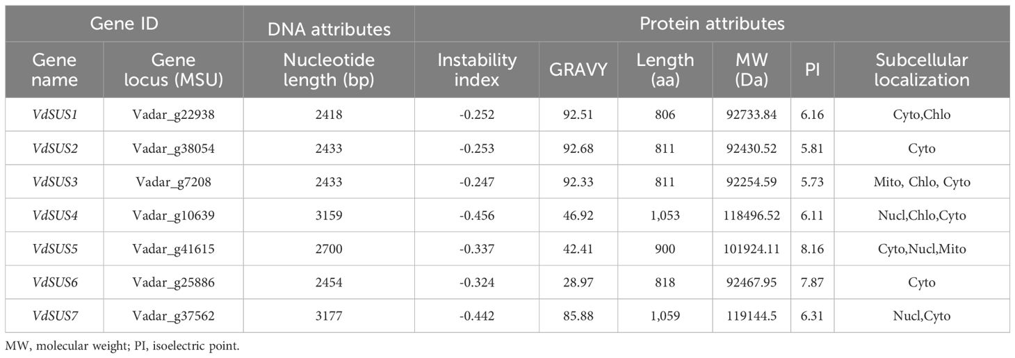
To elucidate the biological functions of the SUS gene family in blueberries, we constructed a Hidden Markov Model. Seven protein sequences containing the Sucrose_synth and Glyco_trans_1_4 domains were identified in the blueberry whole genome. Based on their phylogenetic relationship with Arabidopsis, they were named VdSUS1-VdSUS7 (Supplementary Figure S1). The amino acid sequences of the blueberry SUS gene family ranged from 811 to 1059. The molecular weights ranged from 92254.59 (VdSUS3) to 119144.5 (VdSUS7), with isoelectric points between 5.73 (VdSUS3) and 8.16 (VdSUS5). The instability index ranged from 28.97 (VdSUS6) to 92.68 (VdSUS2), and all had negative average hydrophobicity coefficients, indicating hydrophilic proteins. Subcellular localization prediction indicates that VdSUS may be located in the cytoplasm, nucleus, and mitochondria (Table 1).
The conserved domains and phylogenetic relationships of VdSUS proteins were explored through a multiple sequence alignment of the VdSUS gene family. The results of the multiple sequence alignment showed that all seven VdSUS proteins possessed two domains, Sucrose_synth and Glyco_trans_1_4 (Supplementary Figure S2A). Analysis of the position of these two domains in the protein sequences revealed that the domain sequences of VdSUS4 and VdSUS7 were slightly shorter than those of the other members (Supplementary Figure S2B). The amino acid sequences of the SUS gene family proteins exhibited a predominance of α-helix secondary structures, each exceeding 50%, with the least proportion being β-turn, which was around 6% (Supplementary Figure S2C). The tertiary structure of VdSUS proteins consisted of two symmetric tetramers (Supplementary Figure S2D, Supplementary Figure S3), forming a three-lobed structure, with four distinct domains (Supplementary Figure S2E). The first two domains were designated as the Cellulose Targeting Domain (CTD, residues 1–121) and the Early Nodulin 40 Peptide Binding Domain (EPBD, residues 161–271). The last two domains included the GT-B glycosyltransferase with its Rossmann fold domain (Supplementary Figure S2E). These folds form an active site suited for substrate binding, responsible for recognizing and binding sucrose and UDP, and participating in the catalytic reaction of glycosyl transfer. The N-terminal and C-terminal domains of GT-B glycosyltransferase are referred to as GT-BN and GT-BC, respectively (Zheng et al., 2011). The GT-BN domain extended from residues 275 to 528, and the GT-BC domain extended from residues 529 to 760.
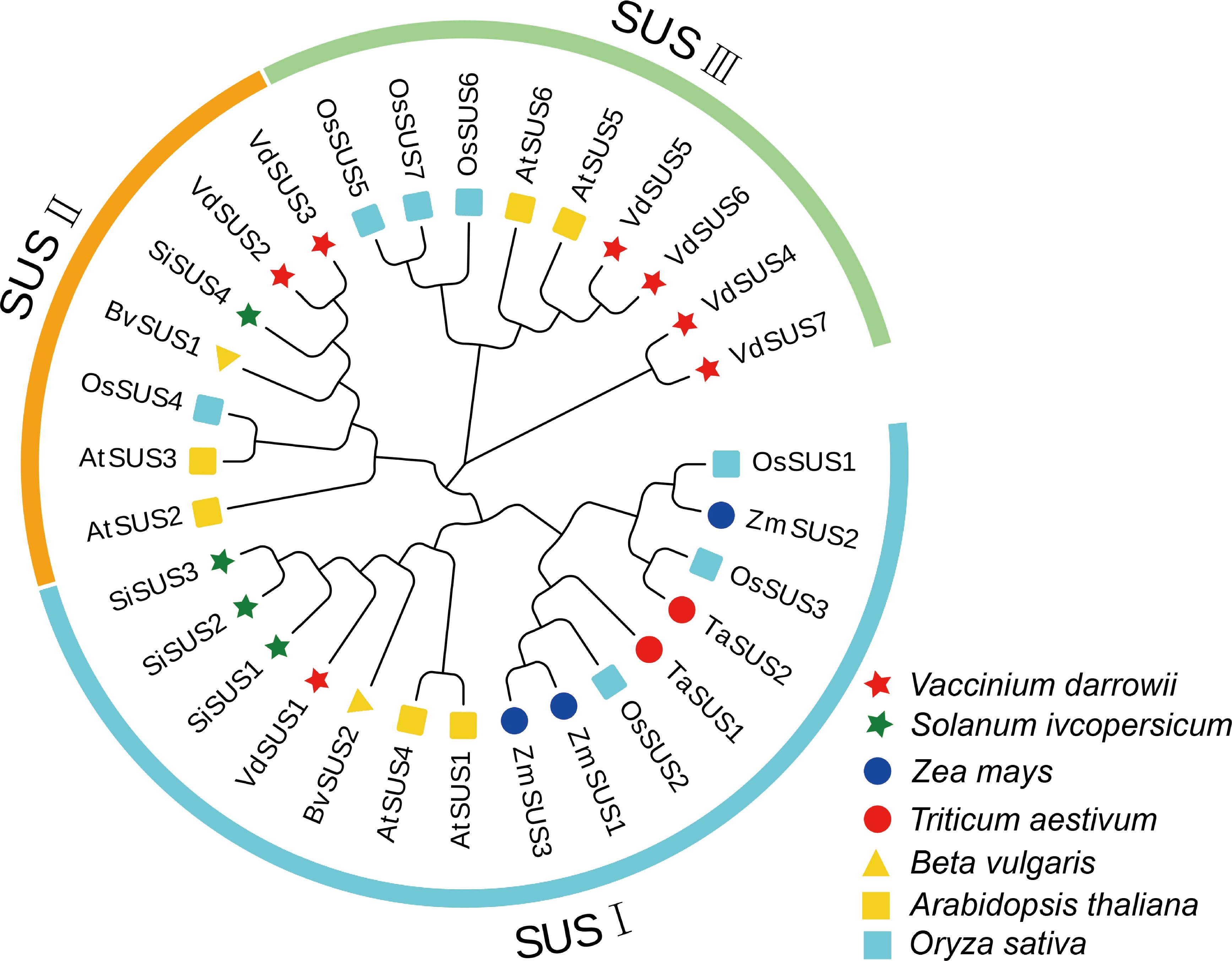
To investigate the evolution of the SUS gene family across different plant species, we collated the amino acid sequences of seven VdSUS proteins from blueberry, six AtSUS proteins from Arabidopsis (Bieniawska et al., 2007), seven OsSUS proteins from rice, four SiSUS proteins from tomato, three ZmSUS proteins from maize, two TaSUS proteins from wheat, and two BvSUS proteins from sugar beet. We constructed a neighbor-joining (NJ) phylogenetic tree (Figure 1; Supplementary Table S1) to analyze the evolutionary relationships. The results of the phylogenetic tree analysis revealed that the 31 SUS proteins from the seven species could be divided into three groups: SUS I, SUS II and SUS III. Blueberry VdSUS proteins were distributed in each subgroup, with only one protein (VdSUS1) in the SUS I group. Notably, these three groups were present in both monocotyledonous and dicotyledonous plants, suggesting a common ancestor. Additionally, VdSUS exhibited a unique clustering pattern, but most collinear orthologous gene pairs were distributed in AtSUS and OsSUS, indicating a shared evolutionary history among these genes across different species (Figure 1). This observation may be attributed to evolutionary variations between monocotyledonous and dicotyledonous plants, influencing the distribution of these gene pairs in their respective species.
3.2 Chromosomal localization and collinearity of the blueberry SUS gene family
To investigate the chromosomal localization of SUS gene in the blueberry genome, we downloaded the blueberry GFF file from NCBI and visualized it using TBtools. The results showed that the seven SUS genes were distributed on 6 chromosomes of blueberry (Figure 2A). The vd-10 chromosome has 2 SUS genes, while the other chromosomes have one gene each (Figure 2A).
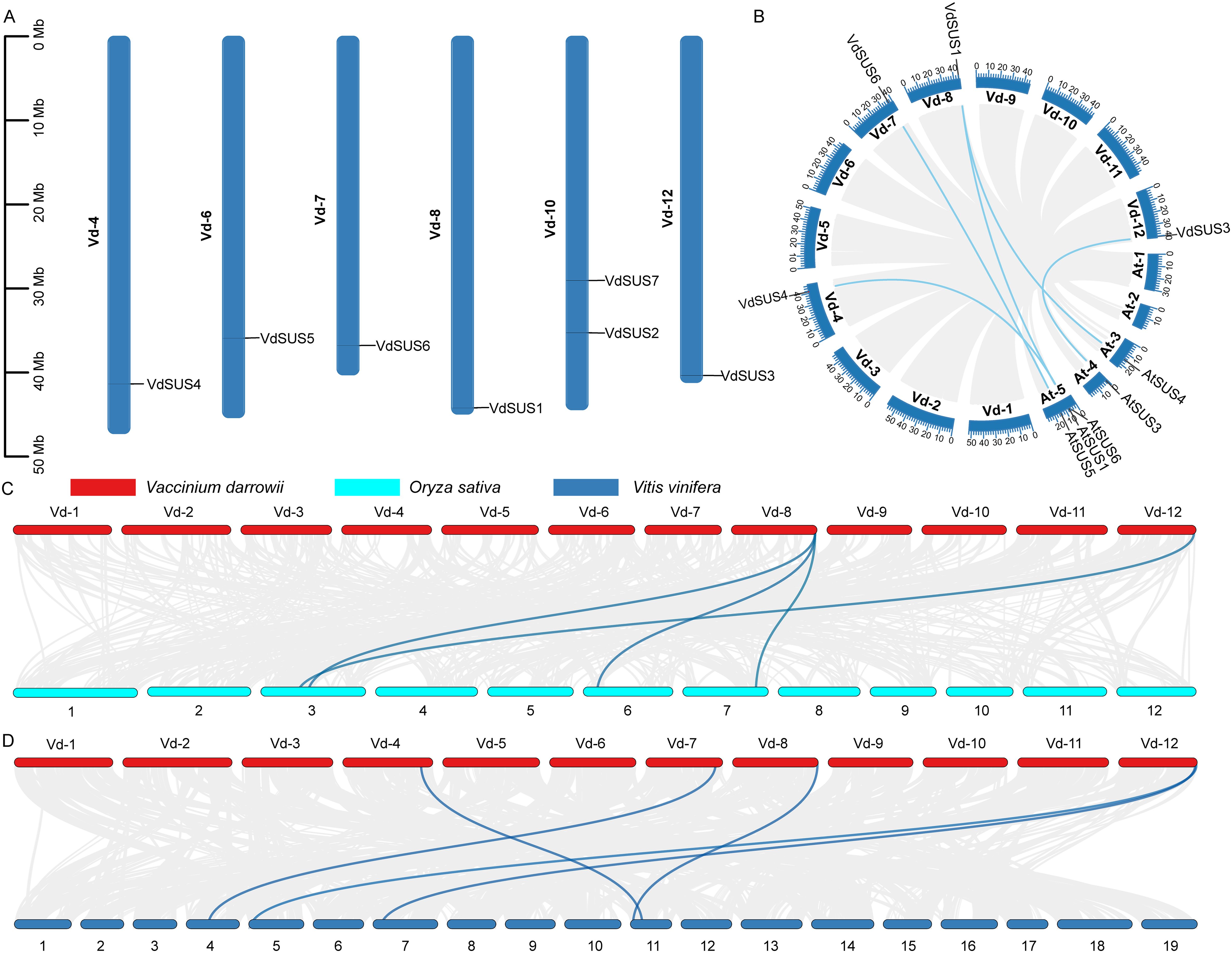
Figure 2. Characteristics of VdSUS proteins. (A) Chromosomal localization. (B) Collinearity relationship between blueberry and Arabidopsis SUS genes. (C) Collinearity relationship between blueberry and rice SUS genes. (D) Collinearity relationship between blueberry and grape.
Gene duplication was a common occurrence in plant evolution, including whole-genome duplication, tandem duplication and segmental duplication, which could generate homologous genes with similar sequences. We performed intraspecific collinearity analysis of the VdSUS gene family and found no collinear gene pairs (Figure 2B). To further investigate the phylogenetic and evolutionary relationships of SUS genes between species, and to reveal the collinearity relationships of SUS genes in different species, we selected three representative model species, including two dicotyledonous plants (Arabidopsis and grape) and one monocotyledonous plant (rice), for collinearity analysis with blueberry. These results showed that there were 5 pairs of collinear SUS gene pairs between blueberry and Arabidopsis, including VdSUS4 and AtSUS6, VdSUS6 and AtSUS5, VdSUS1 and AtSUS3, VdSUS1 and AtSUS1, and VdSUS3 and AtSUS3 (Figure 2B). In comparison with rice genome (7 genes), four collinear gene pairs were identified between VdSUS and OsSUS (Figure 2C; Supplementary Table S2). The VdSUS genes exhibited higher homology with the VvSUS genes of grape (5 genes) (Figure 2D; Supplementary Table S2), indicating a close relationship between them. These results also suggested that, compared to monocotyledonous plant genomes, blueberry exhibited more significant collinearity with dicotyledonous plant genomes, and individual homologous genes showed one-to-many or many-to-one homology. These genes had undergone multiple genes duplication events, indicating a close phylogenetic relationship between the studied species. Their evolutionary functions might be conserved, and their ancestral functions had not been lost or altered during the duplication process, playing an important role in the evolution of the SUS gene family.
3.3 Blueberry SUS gene family structure and conserved motif analysis
To further investigate the structural features and evolutionary mechanisms of the SUS gene family, we conducted phylogenetic tree analysis of the SUS proteins in the blueberry genome, as well as a comparative analysis of the distribution of conserved motifs and intron-exon structures. Predicted gene structures revealed that SUS gene sequences contained 12 or more introns (Figure 3B). The sequences of all VdSUS proteins contained Motif 1, Motif 2, Motif 4, Motif 5 and Motif 9 (Figure 3C), with most motifs consisting of 50 amino acids (except for Motif 4 and Motif 9) (Figure 3D). VdSUS5 and VdSUS6 belonged to the SUS III group (Figure 3A), but they exhibited differences in gene structure while sharing the same motif distribution. This divergence in gene structure may contribute to functional differences among VdSUS genes. In contrast, the gene structures and conserved motifs of VdSUS2/VdSUS3 and VdSUS4/VdSUS7 were consistent, indicating a strong correlation between phylogenetic relationships among gene family members and their gene structures. This might suggest functional redundancy among these genes in blueberries.
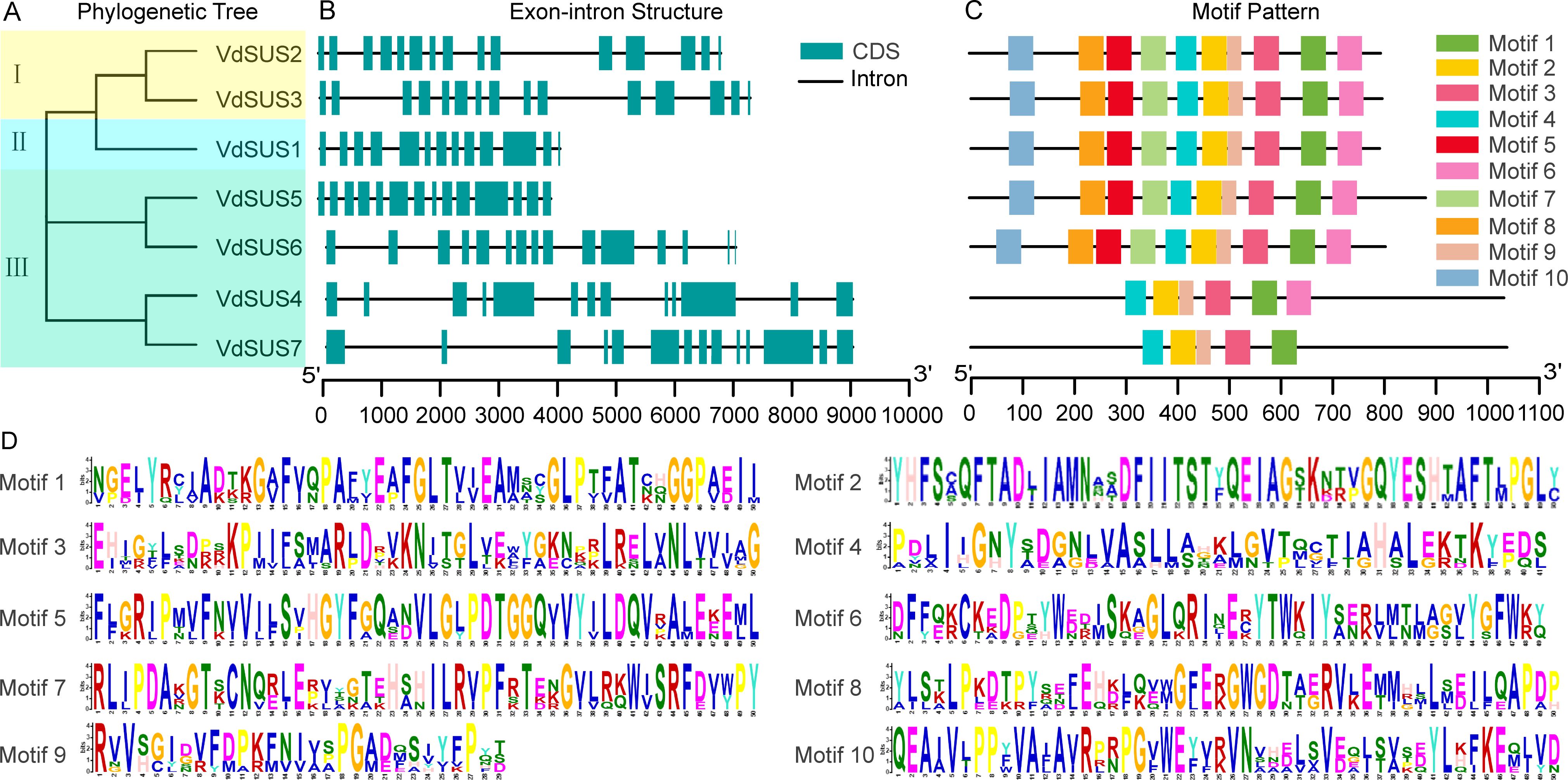
Figure 3. Structural features of VdSUS sequences. (A) Phylogenetic tree of the VdSUS protein family, where I represents SUS I, II represents SUS II, and III represents SUS III. (B) Gene structure features of the VdSUS genes family. (C) Motif analysis of the VdSUS proteins. (D) Conserved motif sequences.
3.4 Analysis of promoter cis-acting elements in the blueberry SUS gene family
Cis-regulatory elements (CREs) are non-coding DNA sequences in the gene promoter region that play a crucial role in gene expression, widely participating in the regulation of plant growth, development and stress responses (Zhao et al., 2018). By analyzing the CREs of VdSUS, we aimed to further understand the potential roles of VdSUS in plant growth and development, as well as in response to plant hormones and abiotic stress. To identify genes functions and regulatory patterns, we investigated the CREs in the promoter regions of each gene by searching the 2000 bp region around each transcription activation site in the Plant CARE database. The analysis of VdSUS promoter regions revealed various CREs. We selected 20 representative CREs for visualization of their distribution (Figure 4B). Among them, VdSUS3 had the highest number of CREs, totaling 33, while VdSUS7 had 21 CREs. All CREs could be broadly classified into three categories (Figure 4C). The first category was hormone response, with a total of 51 CREs in the VdSUS gene family. Some genes, such as VdSUS4 and VdSUS6, contained multiple hormone response elements in their promoter regions, suggesting a rapid and intense response to specific hormones. The second category was abiotic stress, with a total of 120 CREs in the VdSUS gene family. All VdSUS genes contained abiotic stress-related elements such as ARE (anaerobic stress-related) and MYC (salt stress-related), indicating their potential role in regulating anaerobic stress and salt stress responses. The third category was growth and development, with a total of 31 CREs related to growth and development. Among them, VdSUS1 and VdSUS6 contained the highest number of AAGAA-motif (auxin response element), suggesting their involvement in blueberry growth and development regulation.

Figure 4. Analysis of cis-regulatory elements in the promoter regions of SUS genes in blueberry. (A) Phylogenetic tree of the VdSUS protein family in blueberry. (B) The positions of cis-regulatory elements in the promoter regions of VdSUS genes in blueberry. (C) Statistical analysis of the number of cis-regulatory elements in the promoter regions of VdSUS genes in blueberry.
In summary, these results indicated that the composition and quantity of CREs in different VdSUS promoter regions vary among subfamilies (Figure 4A). This suggested that the functional expression of SUS genes in blueberries was regulated by various CREs associated with hormones, abiotic stress, and plant growth and development processes.
3.5 Expression analysis of blueberry SUS genes in different organs, tissues and developmental stages
To better elucidate the physiological functions of VdSUS gene family members, we employed qRT-PCR to examine the spatiotemporal gene expression patterns of VdSUS genes in different organs, tissues and developmental stages of blueberry. Fluorescence quantitative PCR primers were designed based on the CDS region of the 7 members of the VdSUS gene family, VdTub2 serving as the internal reference control. The expression levels of the 7 VdSUS genes were detected by qRT-PCR in various tissues, including young flowers (YFl), mature flowers (MFl), early green fruits (EGF), late green fruits (LGF), mature fruits (MF), young stems (YS), mature stems (MS), young leaves (YL), mature leaves (ML) and roots (R) (Figure 5A). In flowers, the expression levels of most VdSUS genes were higher in mature flowers than young flowers. In fruits, except for VdSUS4, which showed higher expression in mature fruits than in early and late green fruits, other VdSUS members exhibited the highest expression in early green fruits, followed by a decline in late green fruits (Figure 5B). Interestingly, their expression levels increased again in mature fruits. In stems, the expression levels of VdSUS1, VdSUS3 and VdSUS7 were relatively high, while the expression levels of other members were generally low (Figure 5B). In leaves, except for VdSUS2, the expression levels of other members were higher in young leaves than in mature leaves (Figure 5B). In roots, the expression levels of VdSUS3, and VdSUS5 were relatively high, while expression levels of other members were low (Figure 5B). Overall, VdSUS5 and VdSUS6 showed lower expression levels in various tissues, indicating their contribution to the growth, sucrose metabolism and fruit development in blueberries (Figure 5B).
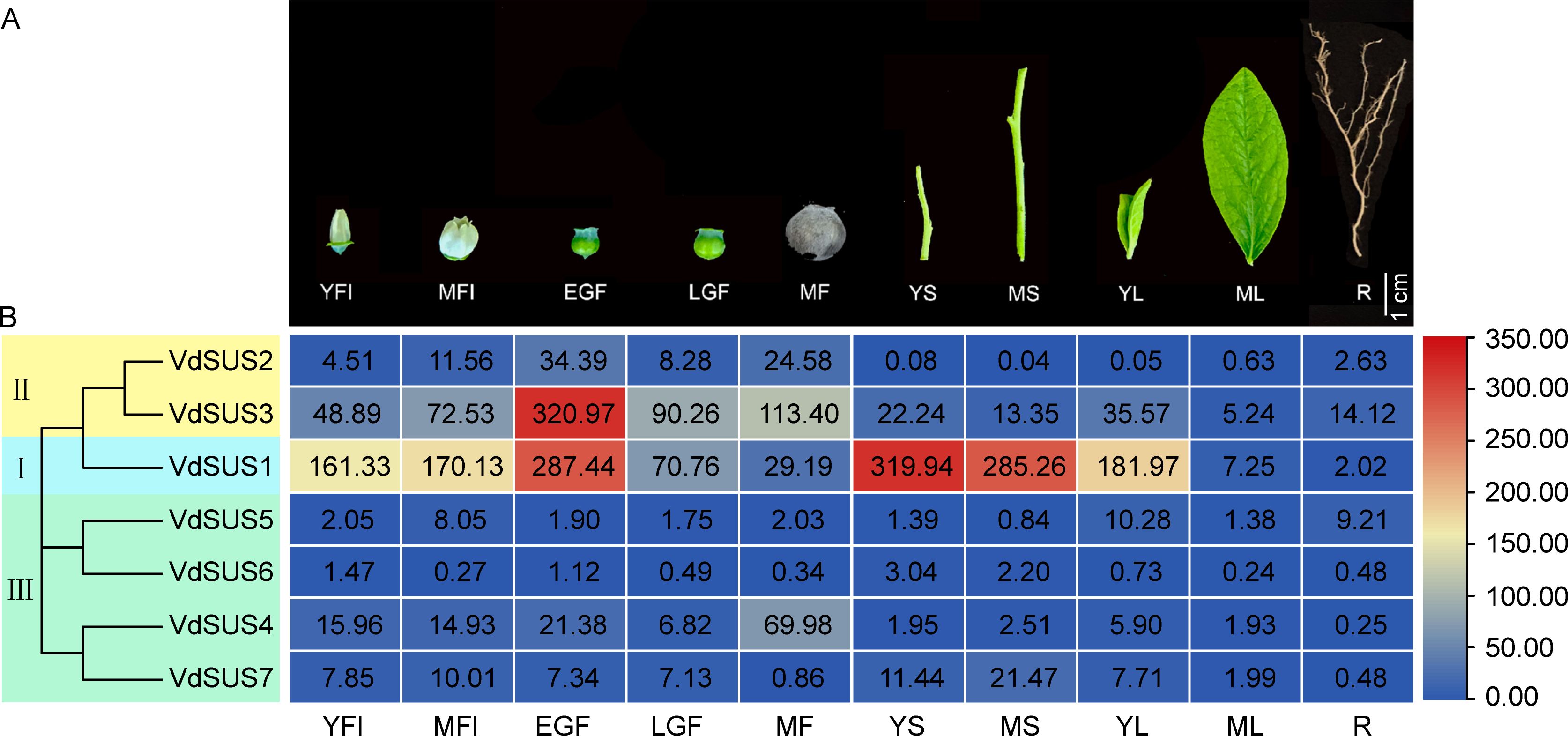
Figure 5. Spatial expression patterns analysis of VdSUS genes. (A) Phenotypes of extracted blueberry samples in different organs, tissues and developmental stages. YFl, young flowers; MFl, mature flowers; EGF, early green fruits; LGF, late green fruits; MF, mature fruits; YS, young stems; MS, mature stems; YL, young leaves; ML, mature leaves; R, roots. (B) Expression levels of VdSUS family genes.
3.6 Expression patterns of VdSUS genes in blueberry fruits under sucrose treatment and abiotic stress conditions
Exogenous sucrose had been demonstrated to act as a signaling molecule, promoting the ripening of tomato and strawberry fruits (Jia et al., 2013, 2016). In order to further identify which VdSUS genes influence the ripening and softening of blueberry fruits, the expression of VdSUS genes in blueberry fruits was modulated by exogenous sucrose application. The results revealed that, compared to blueberry fruits treated with sorbitol (negative control), the expression of VdSUS4 in blueberry fruits was upregulated after 6 hours of sucrose treatment, reaching its peak after 12 h with a threefold increase compared to the negative control. VdSUS1 and VdSUS7 showed an upregulation in expression after 24 h of sucrose treatment, while expression of VdSUS2, VdSUS3, and VdSUS6 exhibited a slight decrease after sucrose treatment. VdSUS5 showed no significant change compared to the negative control (Supplementary Figure S4).
In order to further explore the potential role of VdSUS in abiotic stress, we investigated the expression patterns of VdSUS under drought, salt and low temperature stress conditions. The results revealed differential expression patterns of VdSUS under drought (Figure 6A), salt (Figure 6C), and low temperature stress (Figure 6E). The expression levels of VdSUS1, VdSUS6 and VdSUS7 were downregulated, while the expression levels of VdSUS2-VdSUS5 were upregulated after drought treatment (Figure 6B). After NaCl treatment, the general trend of VdSUS1-VdSUS5 gene expression was upregulated, and VdSUS7 showed initial upregulation followed by downregulation at 5 d, while VdSUS6 was almost unaffected (Figure 6D). Under low temperature treatment, except for VdSUS5 was induced upregulation, all other genes were significantly downregulated (Figure 6F).
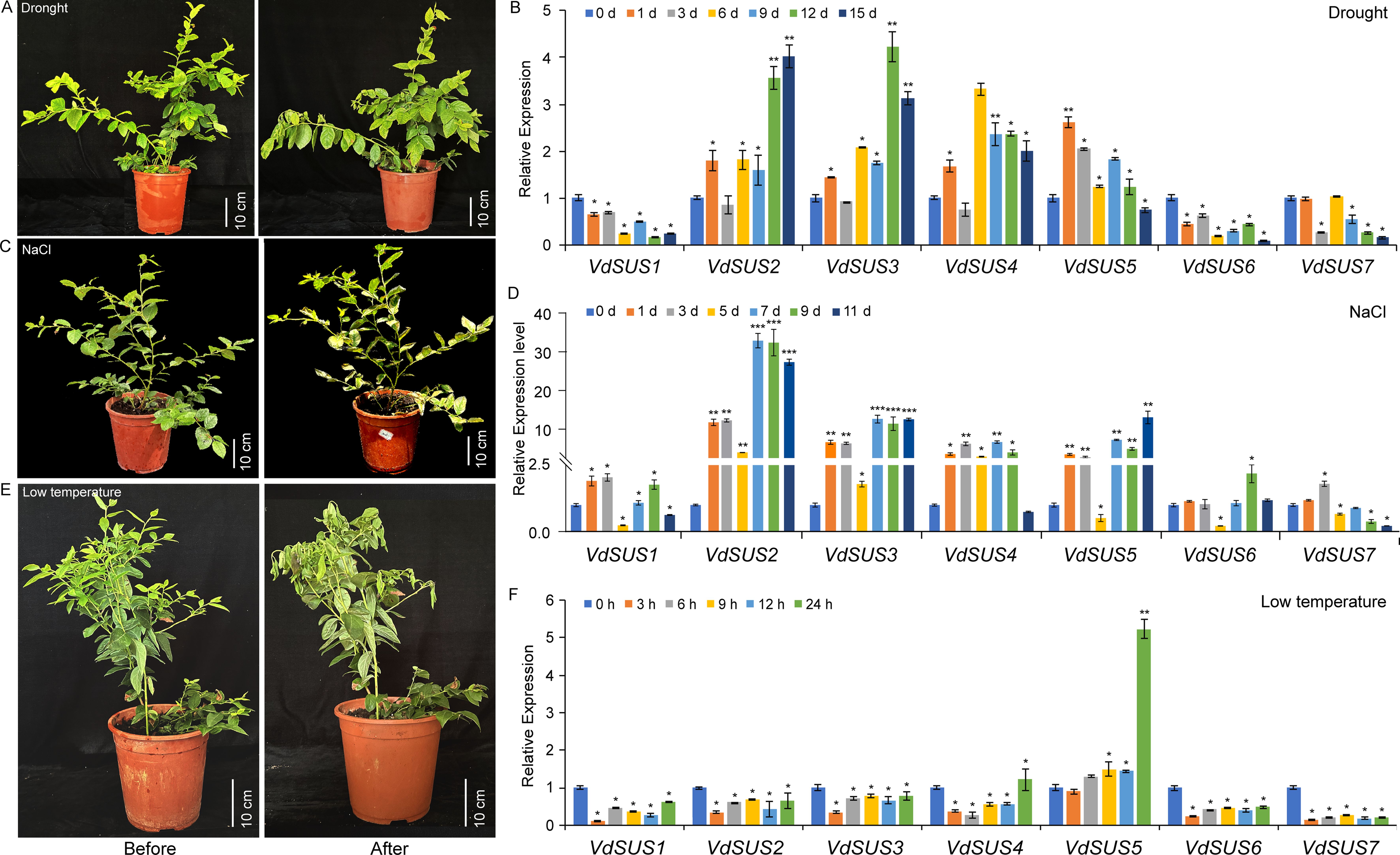
Figure 6. Expression patterns of VdSUS genes in response to abiotic stress treatment. (A) Phenotypic charts before and after drought treatment. (B) Expression levels of VdSUS genes under drought treatment conditions. (C) Phenotypic charts before and after NaCl treatment. (D) Expression levels of VdSUS genes under NaCl treatment conditions. (E) Phenotypic charts before and after low temperature treatment. (F) Expression levels of VdSUS genes under low temperature treatment conditions. Values are the average ± standard deviation of three biological replicates. The transcription levels of VdSUS genes at 0 days and 0 hours were set as “1”. P-values < 0.05, 0.01 and 0.001 are denoted by “*”, “**” and “***” respectively (Student’s t-test).
3.7 Overexpression of VdSUS4 confers salt stress tolerance
To explore the potential functions of VdSUS4 in enhancing plant salt tolerance, we first generated transgenic Arabidopsis overexpressing VdSUS4 (Supplementary Figure S5) and assessed their seedling growth under salt stress conditions. Under normal conditions, there were no significant differences in germination and root elongation between wild-type and transgenic seedlings. However, compared with the wild-type, transgenic plants exhibited a nearly 100% increase in root elongation on media containing NaCl (Figures 7A, B). And, the fresh weight of transgenic plants demonstrated a 40% increase compared to the wild-type plants (Figures 7A, C). Further analysis revealed an exceeding 40% increase in germination rate of transgenic seeds relative to wild-type under 150 mM NaCl treatment (Figures 8A, B).
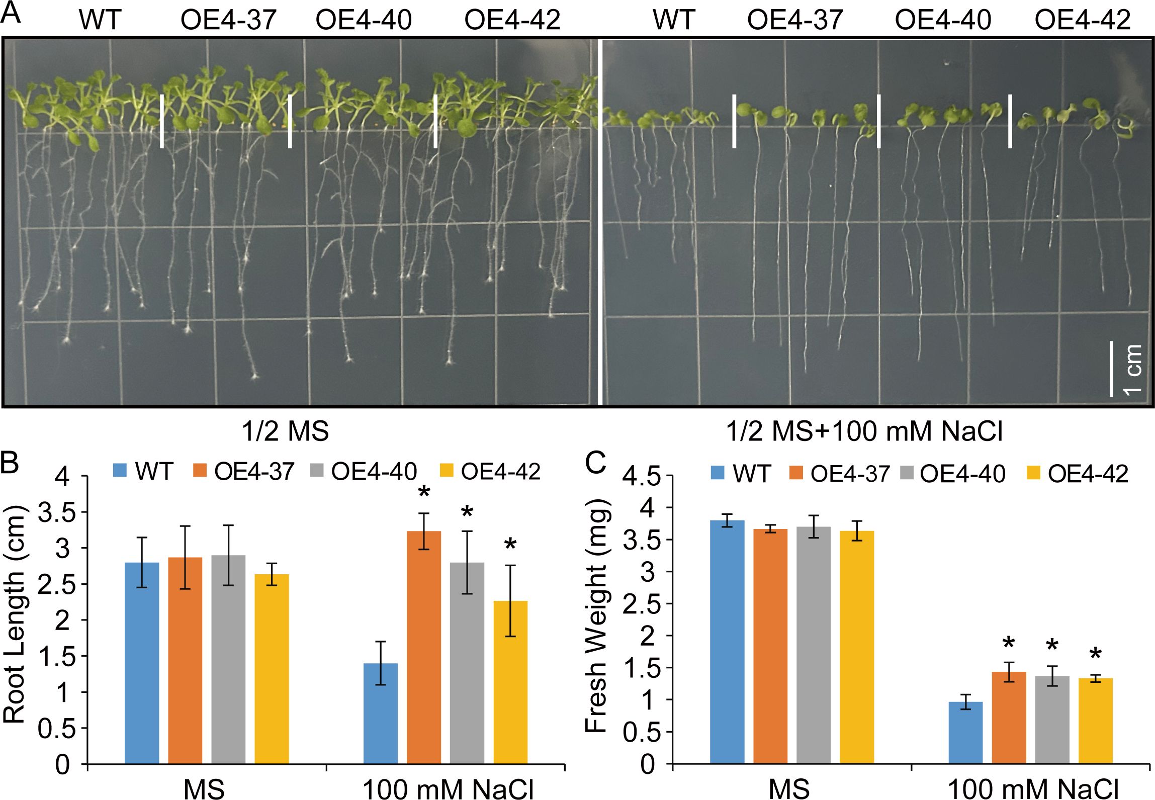
Figure 7. Overexpression of VdSUS4 enhanced salt stress tolerance in Arabidopsis. (A) Germination of WT and VdSUS4 overexpressing lines on media with or without 100 mM NaCl, photographed after 15 d. (B) Root length measurement of 15-day-old WT and VdSUS4 overexpressing lines. (C) Fresh weight measurement of 15-day-old WT and VdSUS4 overexpressing lines. Values are the mean ± standard deviation of three biological replicates. p-values < 0.05 are represented by “*” (Student’s t-test).
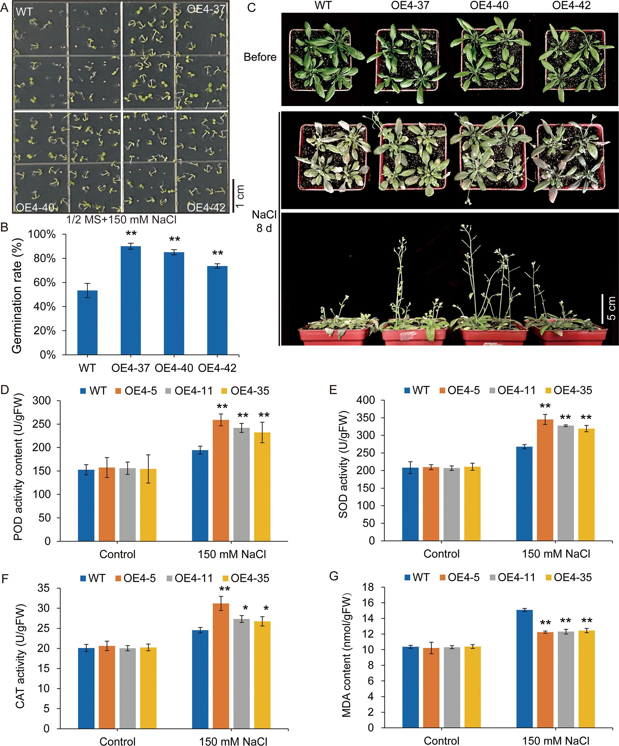
Figure 8. VdSUS4 enhances salt tolerance by strengthening the plant antioxidant system. (A, B) Germination rate of WT and transgenic seeds. (C) Phenotypic comparison of WT and transgenic lines after treatment with 150 mM NaCl for 8 d. (D) POD activity. (E) SOD activity. (F) CAT activity. (G) MDA content. Values are the mean ± standard deviation of three biological replicates. p-values < 0.01 are represented by “**” (Student’s t-test). p-values < 0.05 are represented by "*".
We also performed salt tolerance experiments with wild-type and transgenic plants in soil. The plants were watered with or without 150 mM NaCl solution for 8 days, clearly difference was observed in the leaves and boltings of wild-type and transgenic plants. Both plant groups completed flowering under salt stress, but wild-type plants displayed significantly chlorosis leaves, shorter stems and fewer pods compared to the transgenic lines (Figure 8C). All these results indicated that overexpression of VdSUS4 enhanced salt stress tolerance in Arabidopsis.
Plants adapt to saline-alkaline environments through specific physiological and biochemical regulatory mechanisms. The level of plants salt tolerance can be evaluated by measuring key physiological and biochemical parameters. Our experimental results indicated that, under normal growth conditions, the activities of peroxidase (POD), superoxide dismutase (SOD), and catalase (CAT) showed no significant differences between wild-type and transgenic lines. However, under salt stress conditions, the activities of POD, SOD, and CAT increased in both wild-type and transgenic plants, with significantly higher enzyme activities observed in the transgenic lines. This suggested that plants activated their antioxidant defense systems to mitigate salt-induced oxidative damage, and the transgenic lines possessed a stronger capacity to scavenge reactive oxygen species (ROS), thereby reducing the harmful effects of superoxide radicals and enhancing salt tolerance (Figures 8D–F).
Lipid hydroperoxidation serves as an effective indicator of cellular oxidative damage (Yoshimura et al., 2004). Oxidative stress-induced alterations in lipid hydroperoxide accumulation kinetics were assessed through quantitative determination of malondialdehyde (MDA) in foliar disk samples. Upon NaCl stress, malondialdehyde (MDA) concentration markedly increased in wild-type plants, whereas the transgenic lines exhibited only a marginal increase (Figure 8G). These findings suggested that VdSUS4 enhanced the plant’s antioxidant defense system, reduced ROS accumulation, and helped maintain cellular homeostasis, thereby mitigating salt stress-induced damage.
4 Discussion
As one of the most common berries, blueberry was domesticated during the 20th century (Miller et al., 2019). Due to its flavor and health-promoting properties, blueberries are now cultivated and consumed worldwide. However, blueberry cultivation still faces various challenges, including the demand for varieties with higher yield and better fruit quality. Moreover, compared to other high-value crops, molecular tools for blueberry breeding are relatively limited, making breeding more challenging (Yocca et al., 2023). In this context, the identification of valuable genetic resources becomes a crucial step in promoting genetic improvement through the application of genetic engineering technologies.
Sucrose synthase (SUS) is widely recognized as a key enzyme involved in sucrose metabolism in higher plants and is considered a biochemical marker for crop strength (Xu et al., 2019). Therefore, conducting systematic study on SUS and identifying candidate genes involved in blueberry fruit ripening and response to abiotic stress is of great significance. In our study, seven members of the VdSUS gene family were identified from the blueberry genome (Table 1). SUS is a member of the conserved GT-4 glycosyltransferase subfamily, belonging to the larger metal-independent GT-B glycosyltransferase superfamily. In Arabidopsis, AtSUS1 possesses a typical sequence structure of SUS. Similarly, the SUS proteins in blueberries form a symmetrical tetramer (Supplementary Figure S1D), with the polypeptide chains folding into four distinct domains. This structural arrangement is well-suited for catalyzing and cleaving sucrose and regulating SUS. Additionally, the secondary structure is predominantly composed of α-helices (Supplementary Figure S1C).
Differences in the number of SUS genes across species are primarily due to gene duplication and chromosomal segmental or whole-genome duplication events, which lead to varying rates of SUS gene birth and loss. This process is a major driving force in species evolution, involving the functional innovation of new genes and the evolution of their expression patterns (Agarwal et al., 2017). Chromosome localization and collinearity analysis suggest that the SUS gene family in blueberries may primarily undergo segmental duplication (Figure 2). Although genes within a gene family evolve through various mechanisms, comprehensive phylogenetic and structural analysis can provide insights into the evolutionary origin and relationships of different isozymes. Based on their phylogeny and molecular structure, plant SUS proteins have been classified into three major groups (Zou et al., 2013). Our phylogenetic analysis of VdSUS genes in blueberry, Arabidopsis, rice, tomato, corn, wheat, and sugar beet confirmed this classification (Supplementary Figure S1F).
As observed in other plants, such as Arabidopsis (Bieniawska et al., 2007) and rice (Hiros et al., 2008), closely related VdSUS members generally exhibit similar motifs and exon/intron structures (Figures 3A, B), indicating that different SUS proteins may function in a similar manner. However, we also observed differences in genes structure between VdSUS5 and VdSUS6, despite their close proximity in the phylogenetic tree (Figure 3B), suggesting that structural divergence among gene family members is caused by mechanisms such as exon/intron loss or gain, insertion/deletion and exonization. Therefore, the analysis of exon/intron structure is crucial for revealing the evolutionary footprint of gene families (Xu et al., 2012). Among the identified 10 motifs, motif 1, 2, 4, 5 and 9 are present in all VdSUS proteins (Figure 3C), indicating their high conservation. These motifs constitute the conserved SUS domains essential for the specificity of SUS functions. Cis-regulatory elements (CREs) are closely related to gene function and play a crucial role in the transduction of biological signals. In this study, we found that the promoter region of VdSUS contains numerous CREs related to hormone regulation, abiotic stress and growth and development (Figure 4B), such as response elements related to AAGAA-motif (auxin response), ARE (anaerobic stress-related) and MYC (salt stress-related) (Figure 4C), indicating a close correlation between VdSUS and the potential regulatory effects on growth and development under different environmental changes.
Functional diversity resulting from gene duplication leads to changes in protein properties and differential expression. This is one of the major evolutionary driving forces for plants to adapt to new environments (Flagel and Wendel, 2009). The expression levels of VdSUS1 and VdSUS3 were higher in flowers (YFI and MFI) than other VdSUS genes, and both expression levels in MFI were higher than those in YFI (Figure 5B), suggesting their potential role in providing energy during the maturation process of flowers. Similarly, VvSUS4 in grapes is also highly expressed in flowers (Zhu et al., 2017). In fruits, VdSUS3 shows more expression in MF than those in LGF, while the expression of VdSUS1 decreases accompanied by the fruit ripening (Figure 5B). This phenomenon had been observed in apples as well, where SUS transcription levels were higher in the early stages of fruit development, but decrease as the fruit continues to grow (Li et al., 2012). In the stem, the expression level of VdSUS1 was significantly higher than that of other members, and its expression in YS was higher than in MS (Figure 5B).
In sweet potatoes, IbSUS3, IbSUS4 and IbSUS8 were highly expressed in the stem (Jiang et al., 2023). These SUS gene families were likely involved in carbohydrate transport and assimilation. Additionally, in transgenic poplars, enhanced cellulose deposition leaded to thicker secondary cell walls in the xylem, thereby increasing wood density (Coleman et al., 2009). Sucrose, as the main form of transport for photosynthetic assimilates, was produced in mature leaves, which served as source organs for sucrose synthesis and acted as centers for sucrose export. On the other hand, young leaves, flowers and fruits acted as sink organs, receiving sucrose for development or storing sucrose (Nebauer et al., 2011). In tobacco, SUS2 and SUS3 were highly expressed in leaves (Wang et al., 2015). The expression of VdSUS1 reached peak in young leaves and decreased with leaf development, reaching its lowest level in mature leaves (Figure 5B). Meanwhile, BjSUS5, 6, and 7 are significantly overexpressed in young leaves (Koramutla et al., 2019). Similarly, we observed that except for VdSUS2, the expression levels of other members in young leaves were higher than those in mature leaves, indicating their unique roles in leaf development. Moreover, in roots, VdSUS3 exhibits the highest expression level among different VdSUS genes, indicating its important role in the development of roots (Figure 5B).
An increasing body of research had indicated that the SUS gene families were associated with plant responses to environmental stress. Under abiotic stresses such as cold, drought, salinity, and hypoxia, sucrose biosynthesis can protect cell membrane integrity, stabilize proteins, and accelerate metabolism (Strand et al., 2003). During plant recovery from abiotic stress, sucrose serves as an energy source to promote metabolic activity (Strand et al., 2003). Soluble sugars regulate various functions, acting as fuel for growth and development, precursors in metabolism, short- and long-distance signaling molecules, and as components of osmotic protection and reactive oxygen species scavenging systems under stress conditions (Hennion et al., 2019; Salmon et al., 2020). The expression of AtSUS1 could be induced by cold or drought treatments, and AtSUS3 could serve as a molecular marker for dehydration (Baud et al., 2004). HbSUS5 responded to low-temperature and drought stress (Xiao et al., 2014), while HvSUS1 and HvSUS3 also responded to low-temperature and drought stress (Barrero-Sicilia et al., 2011). VvSUS5 was induced by high temperature, cold, salt, darkness and drought conditions (Zhu et al., 2017). ItbSUS2, ItbSUS5 and ItbSUS6 responded to salt stress (Jiang et al., 2023). In our study, after drought treatment, VdSUS2, VdSUS3, VdSUS4 and VdSUS5 were highly expressed with increasing treatment time, while VdSUS1, VdSUS6 and VdSUS7 gradually decreased (Figure 6B). After salt treatment, the expression levels of VdSUS1-VdSUS5 enhanced with increasing treatment time (Figure 6D). Under low-temperature treatment, except for VdSUS5 was induced upregulation, all other genes were significantly downregulated (Figure 6F). The expression levels of SUS genes might be due to increased demand for glycolysis under abiotic stress conditions (Kleines et al., 1999). The observed downregulation of some VdSUS genes in blueberry fruits after sucrose application may be attributed to high concentrations of sucrose acting through energy sensors, such as SnRK1 kinase, to suppress SUS gene expression. This suppression likely serves to prevent further sucrose breakdown, thereby avoiding carbon metabolic imbalance.
In our study, we observed that transgenic Arabidopsis overexpressing VdSUS4 did not exhibit growth and developmental defects, while it enhanced plant tolerance to salt stress (Figure 7). The overexpression of VdSUS4 enhanced salt stress tolerance in Arabidopsis, indicating that VdSUS4 was a valuable candidate gene for improving plant tolerance to abiotic stress through genetic engineering. However, the molecular mechanisms behind this phenomenon are not yet clear, posing an interesting question for future research. In conclusion, our study provides important insights into the SUS gene family of blueberry, validates their functions, and lays a theoretical foundation for further functional studies of SUS genes in blueberry. This research holds potential applications in future genetic engineering projects.
5 Conclusion
In conclusion, our study identified seven VdSUS genes within the blueberry genome. Analyses were conducted on their physicochemical properties, phylogenetic relationships, conserved motifs, gene structure, cis-acting elements in promoters and expression patterns to elucidate their potential functions in blueberries. Functional studies in transgenic Arabidopsis suggested that VdSUS genes may play a role in response to salt stress. Our research findings will provide valuable insights into the response of VdSUS genes to various abiotic stresses in blueberry.
Data availability statement
The original contributions presented in the study are included in the article/supplementary material. Further inquiries can be directed to the corresponding author.
Author contributions
LY: Funding acquisition, Project administration, Supervision, Writing – original draft, Writing – review & editing. YW: Data curation, Formal Analysis, Writing – original draft. WG: Data curation, Writing – original draft. HXZ: Funding acquisition, Project administration, Supervision, Writing – review & editing. HJZ: Funding acquisition, Project administration, Supervision, Writing – review & editing.
Funding
The author(s) declare that financial support was received for the research and/or publication of this article. This research has been jointed supported by the following grants: the Natural Science Foundation of Shandong Province, China (ZR2020MC160), the National Natural Science Foundation of China (Grant No. 32001330), the Cooperation Project of University and Local Enterprise in Yantai of Shandong Province (2021XDRHXMPT09) and the Double-Hundred Talents Project of Yantai City.
Acknowledgments
This research has been jointed supported by the following grants: the Natural Science Foundation of Shandong Province, China (ZR2020MC160), the National Natural Science Foundation of China (Grant No. 32001330), the Cooperation Project of University and Local Enterprise in Yantai of Shandong Province (2021XDRHXMPT09) and the Double-Hundred Talents Project of Yantai City.
Conflict of interest
Authors LY, HXZ, and HJZ were employed by Bestplant Shandong Stem Cell Engineering Co., Ltd. Authors LY, HXZ, and HJZ were employed by Zhaoyuan Shenghui Agricultural Technology Development Co., Ltd.
The remaining authors declare that the research was conducted in the absence of any commercial or financial relationships that could be construed as a potential conflict of interest.
Generative AI statement
The author(s) declare that no Generative AI was used in the creation of this manuscript.
Publisher’s note
All claims expressed in this article are solely those of the authors and do not necessarily represent those of their affiliated organizations, or those of the publisher, the editors and the reviewers. Any product that may be evaluated in this article, or claim that may be made by its manufacturer, is not guaranteed or endorsed by the publisher.
Supplementary material
The Supplementary Material for this article can be found online at: https://www.frontiersin.org/articles/10.3389/fpls.2025.1581182/full#supplementary-material
Supplementary Figure 1 | Phylogenetic tree of the SUS genes family in blueberry and Arabidopsis.
Supplementary Figure 2 | Characteristics and evolutionary relationships of VdSUS proteins. (A) Multiple sequence alignment of VdSUS family proteins. (B) Visualization of the Sucrose_synthesis and Glyco_trans_1_4 domains of VdSUS proteins. (C) Statistic analysis of the secondary structure of VdSUS amino acid sequences. (D) Front view of the overall tertiary structure of VdSUS amino acid sequences. (E) Peptide chain structure of VdSUS amino acids.
Supplementary Figure 3 | Three-dimensional structures of VdSUS proteins.
Supplementary Figure 4 | Expression levels of VdSUS genes family after treated with sucrose. Values are the average ± standard deviation of three biological replicates. The transcription levels of VdSUSs at 0 h were set as “1”. P-values < 0.05 is denoted by “*” respectively (Student’s t-test).
Supplementary Figure 5 | Identification of VdSUS4 transgenic Arabidopsis. (A) Construct used for Arabidopsis transformation. (B) PCR verification of different transgenic Arabidopsis lines. M: marker, P: VdSUS4 plasmid, WT: Wild-type. (C) RT-PCR confirmed the expression of VdSUS4 in different transgenic lines. (D) qRT-PCR analysed the expression of VdSUS4 in different transgenic lines.
References
Agarwal, P. K., Gupta, K., Lopato, S., and Agarwal, P. (2017). Dehydration responsive element binding transcription factors and their applications for the engineering of stress tolerance. J. Exp. Bot. 68, 2135–2148. doi: 10.1093/jxb/erx118
Albrecht, G. and Mustroph, A. (2003). Localization of sucrose synthase in wheat roots: increased in situ activity of sucrose synthase correlates with cell wall thickening by cellulose deposition under hypoxia. Planta 217, 252–260. doi: 10.1007/s00425-003-0995-6
An, X. M., Chen, Z., Wang, J. C., Ye, M. X., Ji, L. X., Wang, J., et al. (2014). Identification and characterization of the Populus sucrose synthase gene family. Gene 539, 58–67. doi: 10.1016/j.gene.2014.01.062
Baier, M. C., Keck, M., Gödde, V., Niehaus, K., Küster, H., and Hohnjec, N. (2010). Knockdown of the symbiotic sucrose synthase MtSucS1 affects arbuscule maturation and maintenance in mycorrhizal roots of Medicago truncatula. Plant Physiol. 152, 1000–1014. doi: 10.1104/pp.109.149898
Baroja-Fernández, E., Muñoz, F. J., Montero, M., Etxeberria, E., Sesma, M. T., Ovecka, M., et al. (2009). Enhancing sucrose synthase activity in transgenic potato (Solanum tuberosum L.). tubers results in increased levels of starch., ADPglucose and UDPglucose and total yield. Plant Cell Physiol. 50, 1651–1662. doi: 10.1093/pcp/pcp108
Barratt, D. H., Barber, L., Kruger, N. J., Smith, A. M., Wang, T. L., and Martin, C. (2001). Multiple, distinct isoforms of sucrose synthase in pea. Plant Physiol. 127, 655–664. doi: 10.1104/pp.127.2.655
Barrero-Sicilia, C., Hernando-Amado, S., González-Melendi, P., and Carbonero, P. (2011). Structure, expression profile and subcellular localisation of four different sucrose synthase genes from barley. Planta 234, 391–403. doi: 10.1007/s00425-011-1408-x
Baud, S., Vaultier, M. N., and Rochat, C. (2004). Structure and expression profile of the sucrose synthase multigene family in Arabidopsis. J. Exp. Bot. 55, 397–409. doi: 10.1093/jxb/erh047
Bieniawska, Z., Paul Barratt, D. H., Garlick, A. P., Thole, V., Kruger, N. J., Martin, C., et al. (2007). Analysis of the sucrose synthase gene family in Arabidopsis. Plant J. 49, 810–828. doi: 10.1111/j.1365-313X.2006.03011.x
Chen, C., Chen, H., Zhang, Y., Thomas, H. R., Frank, M. H., He, Y., et al. (2020). TBtools: An integrative toolkit developed for interactive analyses of big biological data. Mol. Plant 13, 1194–1202. doi: 10.1016/j.molp.2020.06.009
Chen, A., He, S., Li, F., Li, Z., Ding, M., Liu, Q., et al. (2012). Analyses of the sucrose synthase gene family in cotton: structure, phylogeny and expression patterns. BMC Plant Biol. 12, 85. doi: 10.1186/1471-2229-12-85
Chourey, P. S., Taliercio, E. W., Carlson, S. J., and Ruan, Y. L. (1998). Genetic evidence that the two isozymes of sucrose synthase present in developing maize endosperm are critical, one for cell wall integrity and the other for starch biosynthesis. Mol. Gen. Genet. 259, 88–96. doi: 10.1007/s004380050792
Ciereszko, I., Johansson, H., and Kleczkowski, L. A. (2001). Sucrose and light regulation of a cold-inducible UDP-glucose pyrophosphorylase gene via a hexokinase-independent and abscisic acid-insensitive pathway in Arabidopsis. Biochem. J. 354, 67–72. doi: 10.1042/0264-6021:3540067
Clough, S. J. and Bent, A. F. (1998). Floral dip: a simplified method for Agrobacterium-mediated transformation of Arabidopsis thaliana. Plant J. 16, 735–743. doi: 10.1046/j.1365-313x.1998.00343.x
Coleman, H. D., Yan, J., and Mansfield, S. D. (2009). Sucrose synthase affects carbon partitioning to increase cellulose production and altered cell wall ultrastructure. Proc. Natl. Acad. Sci. U. S. A 106, 13118–13123. doi: 10.1073/pnas.0900188106
Duan, Y., Tarafdar, A., Chaurasia, D., Singh, A., Bhargava, P. C., Yang, J. F., et al. (2022). Blueberry fruit valorization and valuable constituents: A review. Int. J. Food Microbiol. 381, 109890. doi: 10.1016/j.ijfoodmicro.2022.109890
Duan, Y., Yang, L., Zhu, H., Zhou, J., Sun, H., and Gong, H. (2021). Structure and expression analysis of sucrose phosphate synthase, sucrose synthase and invertase gene families in Solanum lycopersicum. Int. J. Mol. Sci. 22, 4698. doi: 10.3390/ijms22094698
Duncan, K. A., Hardin, S. C., and Huber, S. C. (2006). The three maize sucrose synthase isoforms differ in distribution, localization, and phosphorylation. Plant Cell Physiol. 47, 959–971. doi: 10.1093/pcp/pcj068
Figueroa, C. M. and Lunn, J. E. (2016). A tale of two sugars: trehalose 6-phosphate and sucrose. Plant Physiol. 172, 7–27. doi: 10.1104/pp.16.00417
Flagel, L. E. and Wendel, J. F. (2009). Gene duplication and evolutionary novelty in plants. New Phytol. 183, 557–564. doi: 10.1111/j.1469-8137.2009.02923.x
Gaudin, V., Lunness, P. A., Fobert, P. R., Towers, M., Riou-Khamlichi, C., Murray, J. A., et al. (2000). The expression of D-cyclin genes defines distinct developmental zones in snapdragon apical meristems and is locally regulated by the Cycloidea gene. Plant Physiol. 122, 1137–1148. doi: 10.1104/pp.122.4.1137
Gerber, L., Zhang, B., Roach, M., Rende, U., Gorzsás, A., Kumar, M., et al. (2014). Deficient sucrose synthase activity in developing wood does not specifically affect cellulose biosynthesis but causes an overall decrease in cell wall polymers. New Phytol. 203, 1220–1230. doi: 10.1111/nph.12888
Haigler, C. H., Ivanova-Datcheva, M., Hogan, P. S., Salnikov, V. V., Hwang, S., Martin, K., et al. (2001). Carbon partitioning to cellulose synthesis. Plant Mol. Biol. 47, 29–51. doi: 10.1023/A:1010615027986
Harada, T., Satoh, S., Yoshioka, T., and Ishizawa, K. (2005). Expression of sucrose synthase genes involved in enhanced elongation of pondweed (Potamogeton distinctus). turions under anoxia. Ann. Bot. 96, 683–692. doi: 10.1093/aob/mci220
Hennion, N., Durand, M., Vriet, C., Doidy, J., Maurousset, L., Lemoine, R., et al. (2019). Sugars en route to the roots. Transport, metabolism and storage within plant roots and towards microorganisms of the rhizosphere. Physiol. Plant 165, 44–57. doi: 10.1111/ppl.12751
Higo, K., Ugawa, Y., Iwamoto, M., and Korenaga, T. (1999). Plant cis-acting regulatory DNA elements (PLACE). database: 1999. Nucleic Acids Res. 27, 297–300. doi: 10.1093/nar/27.1.297
Hiros, T., Scofield, G. N., and Terao, T. (2008). An expression analysis profile for the entire sucrose synthase gene family in rice. Plant Sci. 174, 534–543. doi: 10.1016/j.plantsci.2008.02.009
Huang, T., Luo, X., Fan, Z., Yang, Y., and Wan, W. (2021). Genome-wide identification and analysis of the sucrose synthase gene family in cassava (Manihot esculenta Crantz). Gene 769, 145191. doi: 10.1016/j.gene.2020.145191
Huber, S. C. and Huber, J. L. (1996). Role and regulation of sucrose-phosphate synthase in higher plants. Annu. Rev. Plant Physiol. Plant Mol. Biol. 47, 431–444. doi: 10.1146/annurev.arplant.47.1.431
Iraqi, D. and Tremblay, F. M. (2001). Analysis of carbohydrate metabolism enzymes and cellular contents of sugars and proteins during spruce somatic embryogenesis suggests a regulatory role of exogenous sucrose in embryo development. J. Exp. Bot. 52, 2301–2311. doi: 10.1093/jexbot/52.365.2301
Islam, M. Z., Hu, X. M., Jin, L. F., Liu, Y. Z., and Peng, S. A. (2014). Genome-wide identification and expression profile analysis of citrus sucrose synthase genes: investigation of possible roles in the regulation of sugar accumulation. PloS One 9, e113623. doi: 10.1371/journal.,pone.0113623
Jia, H., Jiu, S., Zhang, C., Wang, C., Tariq, P., Liu, Z., et al. (2016). Abscisic acid and sucrose regulate tomato and strawberry fruit ripening through the abscisic acid-stress-ripening transcription factor. Plant Biotechnol. J. 14, 2045–2065. doi: 10.1111/pbi.12563
Jia, H., Wang, Y., Sun, M., Li, B., Han, Y., Zhao, Y., et al. (2013). Sucrose functions as a signal involved in the regulation of strawberry fruit development and ripening. New Phytol. 198, 453–465. doi: 10.1111/nph.12176
Jiang, Z., Zhang, H., Gao, S., Zhai, H., He, S., Zhao, N., et al. (2023). Genome-Wide identification and expression analysis of the sucrose synthase gene family in sweet potato and its two diploid relatives. Int. J. Mol. Sci. 24, 12493. doi: 10.3390/ijms241512493
Kleines, M., Elster, R. C., Rodrigo, M. J., Blervacq, A. S., Salamini, F., and Bartels, D. (1999). Isolation and expression analysis of two stress-responsive sucrose-synthase genes from the resurrection plant Craterostigma plantagineum (Hochst.). Planta 209, 13–24. doi: 10.1007/s004250050602
Koramutla, M. K., Ram, C., Bhatt, D., Annamalai, M., and Bhattacharya, R. (2019). Genome-wide identification and expression analysis of sucrose synthase genes in allotetraploid Brassica juncea. Gene 707, 126–135. doi: 10.1016/j.gene.2019.04.059
Kumar, S., Stecher, G., Li, M., Knyaz, C., and Tamura, K. (2018). MEGA X: Molecular evolutionary genetics analysis across computing platforms. Mol. Biol. Evol. 35, 1547–1549. doi: 10.1093/molbev/msy096
Li, M., Feng, F., and Cheng, L. (2012). Expression patterns of genes involved in sugar metabolism and accumulation during apple fruit development. PloS One 7, e33055. doi: 10.1371/journal.,pone.0033055
Li, F., Hao, C., Yan, L., Wu, B., Qin, X., Lai, J., et al. (2015). Gene structure, phylogeny and expression profile of the sucrose synthase gene family in cacao (Theobroma cacao L.). J. Genet. 94, 461–472. doi: 10.1007/s12041-015-0558-1
Li, P., Ma, H., Xiao, N., Zhang, Y., Xu, T., and Xia, T. (2023). Overexpression of the ZmSUS1 gene alters the content and composition of endosperm starch in maize (Zea mays L.). Planta 257, 97. doi: 10.1007/s00425-023-04133-z
Li, M., Wang, S., Liu, Y., Zhang, Y., Ren, M., Liu, L., et al. (2019). Overexpression of PsnSuSy1, 2 genes enhances secondary cell wall thickening, vegetative growth, and mechanical strength in transgenic tobacco. Plant Mol. Biol. 100, 215–230. doi: 10.1007/s11103-023-01353-5
Liu, L. and Zheng, J. (2022). Identification and expression analysis of the sucrose synthase gene family in pomegranate (Punica granatum L.). PeerJ 10, e12814. doi: 10.7717/peerj.12814
Lunn, J. E. and MacRae, E. (2003). New complexities in the synthesis of sucrose. Curr. Opin. Plant Biol. 6, 208–214. doi: 10.1016/s1369-5266(03)00033-5
Miller, K., Feucht, W., and Schmid, M. (2019). Bioactive compounds of strawberry and blueberry and their potential health effects based on human intervention studies: A brief overview. Nutrients 11, 1510. doi: 10.3390/nu11071510
Nagaraj, V. J., Riedl, R., Boller, T., Wiemken, A., and Meyer, A. D. (2001). Light and sugar regulation of the barley sucrose: fructan-6-fructosyltransferase promoter. J. Plant Physiol. 158, 1601–1607. doi: 10.1078/0176-1617-00592
Nebauer, S. G., Renau-Morata, B., Guardiola, J. L., and Molina, R. V. (2011). Photosynthesis down-regulation precedes carbohydrate accumulation under sink limitation in Citrus. Tree Physiol. 31, 169–177. doi: 10.1093/treephys/tpq103
Rook, F., Corke, F., Card, R., Munz, G., Smith, C., and Bevan, M. W. (2001). Impaired sucrose-induction mutants reveal the modulation of sugar-induced starch biosynthetic gene expression by abscisic acid signalling. Plant J. 26, 421–433. doi: 10.1046/j.1365-313x
Salmon, Y., Lintunen, A., Dayet, A., Chan, T., Dewar, R., Vesala, T., et al. (2020). Leaf carbon and water status control stomatal and nonstomatal limitations of photosynthesis in trees. New Phytol. 226, 690–703. doi: 10.1111/nph.16436
Schmittgen, T. D. and Livak, K. J. (2008). Analyzing real-time PCR data by the comparative C(T). method. Nat. Protoc. 3, 1101–1108. doi: 10.1038/nprot.2008.73
Shen, L., Zhang, L., Yin, C., Xu, X., Liu, Y., Shen, K., et al. (2023). The wheat sucrose synthase gene TaSus1 is a determinant of grain number per spike. Crop J. 12, 295–300. doi: 10.1016/j.cj.2023.11.007
Stein, O. and Granot, D. (2019). An overview of sucrose synthases in plants. Front. Plant Sci. 10. doi: 10.3389/fpls.2019.00095
Strand, Å., Foyer, C. H., Gustafsson, P., GardestrÖM, P., and Hurry, V. (2003). Altering flux through the sucrose biosynthesis pathway in transgenic Arabidopsis thaliana modifies photosynthetic acclimation at low temperatures and the development of freezing tolerance. Plant Cell Environ. 26, 523–535. doi: 10.1046/j.1365-3040.2003.00983.x
Tong, X., Wang, Z., Ma, B., Zhang, C., Zhu, L., Ma, F., et al. (2018). Structure and expression analysis of the sucrose synthase gene family in apple. J. Integr. Agr. 17, 847–856. doi: 10.1016/S2095-3119(17)61755-6
Wang, Z., Wei, P., Wu, M., Xu, Y., Li, F., Luo, Z., et al. (2015). Analysis of the sucrose synthase gene family in tobacco: structure, phylogeny, and expression patterns. Planta 242, 153–166. doi: 10.1007/s00425-015-2297-1
Wang, H., Sui, X., Guo, J., Wang, Z., Cheng, J., Ma, S., et al. (2014). Antisense suppression of cucumber (Cucumis sativus L.). sucrose synthase 3 (CsSUS3). reduces hypoxic stress tolerance. Plant Cell Environ. 37, 795–810. doi: 10.1111/pce.12200
Xiao, X., Tang, C., Fang, Y., Yang, M., Zhou, B., Qi, J., et al. (2014). Structure and expression profile of the sucrose synthase gene family in the rubber tree: indicative of roles in stress response and sucrose utilization in the laticifers. FEBS. J. 281, 291–305. doi: 10.1111/febs.12595
Xu, S. M., Brill, E., Llewellyn, D. J., Furbank, R. T., and Ruan, Y. L. (2012). Overexpression of a potato sucrose synthase gene in cotton accelerates leaf expansion, reduces seed abortion, and enhances fiber production. Mol. Plant 5, 430–441. doi: 10.1093/mp/ssr090
Xu, X., Yang, Y., Liu, C., Sun, Y., Zhang, T., Hou, M., et al. (2019). The evolutionary history of the sucrose synthase gene family in higher plants. BMC Plant Biol. 19, 566. doi: 10.1186/s12870-019-2181-4
Yadav, U. P., Ivakov, A., Feil, R., Duan, G. Y., Walther, D., Giavalisco, P., et al. (2014). The sucrose-trehalose-6-phosphate (Tre6P). nexus: specificity and mechanisms of sucrose signalling by Tre6P. J. Exp. Bot. 65, 1051–1068. doi: 10.1093/jxb/ert457
Yang, J., Zhang, J., Wang, Z., and Zhu, Q. (2001). Activities of starch hydrolytic enzymes and sucrose-phosphate synthase in the stems of rice subjected to water stress during grain filling. J. Exp. Bot. 52, 2169–2179. doi: 10.1093/jexbot/52.364.2169
Yocca, A. E., Platts., A., Alger, E., Teresi, S., Mengist, M. F., Benevenuto, J., et al. (2023). Blueberry and cranberry pangenomes as a resource for future genetic studies and breeding efforts. Hortic. Res. 10, 202. doi: 10.1093/hr/uhad202
Yoon, J., Cho, L. H., Tun, W., Jeon, J. S., and An, G. (2021). Sucrose signaling in higher plants. Plant Sci. 302, 110703. doi: 10.1016/j.plantsci.2020.110703
Yoshimura, K., Miyao, K., Gaber, A., Takeda, T., and Shigeoka, S. (2004). Enhancement of stress tolerance in transgenic tobacco plants overexpressing Chlamydomonas glutathione peroxidase in chloroplasts or cytosol. Plant J. 37, 21–33. doi: 10.1046/j.1365-313X.2003.01930.x
\Zhao, H., Gao, Z., Wang, L., Wang, J., Wang, S., Fei, B., et al. (2018). Chromosome-level reference genome and alternative splicing atlas of moso bamboo. Gigascience 7, 115. doi: 10.1093/gigascience/giy115
Zheng, Y., Anderson, S., Zhang, Y., and Garavito, R. M. (2011). The structure of sucrose synthase-1 from Arabidopsis thaliana and its functional implications. J. Biol. Chem. 286, 36108–36118. doi: 10.1074/jbc.M111.275974
Zhu, X., Wang, M., Li, X., Jiu, S., Wang, C., and Fang, J. (2017). Genome-wide analysis of the sucrose synthase gene family in grape (Vitis vinifera). Structure, evolution, and expression profiles. Genes 8, 111. doi: 10.3390/genes8040111
Zou, C., Lu, C., Shang, H., Jing, X., Cheng, H., Zhang, Y., et al. (2013). Genome-wide analysis of the Sus gene family in cotton. J. Integr. Plant Biol. 55, 643–653. doi: 10.1111/jipb.12068
Keywords: blueberry, sucrose synthase, bioinformatics, tissue-specific expression, salt stress
Citation: Wang Y, Yang L, Geng W, Zhang H and Zhou H (2025) The sucrose synthase gene family in blueberry (Vaccinium darrowii): functional insights into the role of VdSUS4 in salt stress tolerance. Front. Plant Sci. 16:1581182. doi: 10.3389/fpls.2025.1581182
Received: 21 February 2025; Accepted: 12 May 2025;
Published: 02 June 2025.
Edited by:
Boping Wu, Zhejiang Agriculture and Forestry University, ChinaReviewed by:
Luis Morales-Quintana, Autonomous University of Chile, ChileJiyi G. E. Zhang, Harpe Bioherbicide Solutions Inc., United States
Copyright © 2025 Wang, Yang, Geng, Zhang and Zhou. This is an open-access article distributed under the terms of the Creative Commons Attribution License (CC BY). The use, distribution or reproduction in other forums is permitted, provided the original author(s) and the copyright owner(s) are credited and that the original publication in this journal is cited, in accordance with accepted academic practice. No use, distribution or reproduction is permitted which does not comply with these terms.
*Correspondence: Lei Yang, bGVpeWFuZ0BuanUuZWR1LmNu; Hongxia Zhang, aHh6aGFuZ0BzaWJzLmFjLmNu; Houjun Zhou, aGp6aG91QGxkdS5lZHUuY24=
†These authors have contributed equally to this work
 Yanwen Wang
 Lei Yang
 Wenzhu Geng1,2,3†
 Hongxia Zhang
 Houjun Zhou